vanin-1 mice show decreased nsaid- and · mg/kg in 5% sodium carbonate to vanin-1 –/– (n =5)...
TRANSCRIPT
Vanin-1–/– mice show decreased NSAID- andSchistosoma-induced intestinal inflammationassociated with higher glutathione stores
Florent Martin, … , Philippe Naquet, Bouchra Gharib
J Clin Invest. 2004;113(4):591-597. https://doi.org/10.1172/JCI19557.
Vanin-1 is a membrane-anchored pantetheinase highly expressed in the gut and liver. Ithydrolyzes pantetheine to pantothenic acid (vitamin B5) and the low-molecular-weight thiolcysteamine. The latter is believed to be a key regulating factor of several essentialmetabolic pathways, acting through sulfhydryl-disulfide exchange reactions betweensulfhydryl groups of the enzymes and the oxidized form, cystamine. Its physiologicalimportance remains to be elucidated, however. To explore this point, we developed Vanin-1–deficient mice that lack free cysteamine. We examined the susceptibility of deficient miceto intestinal inflammation, either acute (NSAID administration) or chronic (Schistosomainfection). We found that Vanin-1–/– mice better controlled inflammatory reaction andintestinal injury in both experiments. This protection was associated with increased g-glutamylcysteine synthetase activity and increased stores of reduced glutathione, as well asreduced inflammatory cell activation in inflamed tissues. Oral administration of cystaminereversed all aspects of the deficient phenotype. These findings suggest that one cysteaminefunction is to upregulate inflammation. Consequently, the pantetheinase activity of Vanin-1molecule could be a target for a new anti-inflammatory strategy.
Article Immunology
Find the latest version:
http://jci.me/19557-pdf
The Journal of Clinical Investigation | February 2004 | Volume 113 | Number 4 591
IntroductionVanin genes were discovered recently following mousethymus reconstitution experiments after irradiation(1). The mouse gene cluster includes Vanin-1 andVanin-3 (2, 3) preferentially expressed by epithelial andmyeloid cells, respectively (4). Vanin-1 and Vanin-3transcripts show ubiquitous but heterogeneous tissuedistribution in vivo, with Vanin-1 being the predomi-nantly expressed isoform in the gut. Vanin-1 is a glyco-sylphosphatidyl inositol (GPI) anchored to cell mem-branes, whereas Vanin-3 is secreted. Vanin genes encodea pantetheinase (EC 3.5.1.) (5, 6) that hydrolyzes pan-tetheine to pantothenic acid (vitamin B5) and cys-
teamine (7). Cysteamine is a low-molecular thiol broad-ly distributed in organisms, from Drosophila to humans.This wide distribution and the mechanisms involvinglow-molecular thiols are likely to reflect the early stepsof molecular evolution (8). In vivo, cysteamine is inequilibrium with its oxidized form cystamine at con-centrations of approximately 20 µM (9–12). Cystamineis believed to be a key regulator of essential metabolicpathways. It acts by sulfhydryl-disulfide exchange reac-tions between sulfhydryl groups at or near the activesite of the enzymes and the oxidized form, cystamine(13–16). The enzymes putatively inhibited by cys-teamine include γ-glutamylcysteine synthetase (γ-GCS),which catalyzes the key step in glutathione (GSH) syn-thesis (17–19). This enzyme activity is of central impor-tance because GSH fulfills many important and chem-ically complex roles in protecting cellular componentsfrom the deleterious effects of toxic species (20). Cys-teamine can have dramatic effects, since its adminis-tration is widely used as an experimental model of duo-denal ulcers (21). Nevertheless, the physiologicalimportance of cysteamine remains elusive.
To explore this point, we developed Vanin-1–deficient(Vanin-1–/–) mice. Deficient mice lack free cysteamine (6).Because of the high Vanin-1 gene expression in the gutand cysteamine-induced intestinal injury, we examinedthe susceptibility of Vanin-1–/– mice to NSAID intestinalinjury. In addition, we used a model of chronic intestinalinflammation, Schistosoma infection. The Schistosoma
Vanin-1–/– mice show decreased NSAID- and Schistosoma-induced intestinal inflammation associated with higherglutathione stores
Florent Martin,1 Marie-France Penet,2 Fabrice Malergue,1 Hubert Lepidi,3 Alain Dessein,2
Franck Galland,1 Max de Reggi,2 Philippe Naquet,1 and Bouchra Gharib2
1Centre d’Immunologie de Marseille-Luminy, Centre National de la Recherche Scientifique–Institut National de la Santé et de la Recherche Médicale,
2Institut National de la Santé et de la Recherche Médicale U 399, and 3Unité des Rickettsies–Centre National de la Recherche Scientifique, UMR 6020, Université de la Méditerranée, Marseille, France
Vanin-1 is a membrane-anchored pantetheinase highly expressed in the gut and liver. It hydrolyzespantetheine to pantothenic acid (vitamin B5) and the low-molecular-weight thiol cysteamine. The lat-ter is believed to be a key regulating factor of several essential metabolic pathways, acting throughsulfhydryl-disulfide exchange reactions between sulfhydryl groups of the enzymes and the oxidizedform, cystamine. Its physiological importance remains to be elucidated, however. To explore this point,we developed Vanin-1–deficient mice that lack free cysteamine. We examined the susceptibility of defi-cient mice to intestinal inflammation, either acute (NSAID administration) or chronic (Schistosomainfection). We found that Vanin-1–/– mice better controlled inflammatory reaction and intestinal injuryin both experiments. This protection was associated with increased γ-glutamylcysteine synthetase activ-ity and increased stores of reduced glutathione, as well as reduced inflammatory cell activation ininflamed tissues. Oral administration of cystamine reversed all aspects of the deficient phenotype.These findings suggest that one cysteamine function is to upregulate inflammation. Consequently,the pantetheinase activity of Vanin-1 molecule could be a target for a new anti-inflammatory strategy.
J. Clin. Invest. 113:591–597 (2004). doi:10.1172/JCI200419557.
Received for publication July 22, 2003, and accepted in revised formDecember 16, 2003.
Address correspondence to: Bouchra Gharib, INSERM U 399,Faculté de médecine, 27, Boulevard Jean Moulin, 13385, MarseilleCedex 5, France. Phone: 33-491-32-44-54; Fax: 33-491-79-60-63; E-mail: [email protected] Martin and Marie-France Penet contributed equally tothis work.Philippe Naquet and Bouchra Gharib are co-senior authors.Conflict of interest: The authors have declared that no conflict ofinterest exists.Nonstandard abbreviations used: glycosylphosphatidyl inositol (GPI); γ-glutamylcysteine synthetase (γ-GCS);glutathione (GSH); hemoglobin (Hb); cyanin-3 (Cy3); epithelialcell adhesion molecule (Ep-CAM); myeloperoxidase (MPO);cyclooxygenase-1 (COX-1); macrophage inhibitory protein-2(MIP-2); hypoxanthine phosphoribosyl transferase (HPRT); GSH reductase (GR).

592 The Journal of Clinical Investigation | February 2004 | Volume 113 | Number 4
model is extensively used to investigate liver delayedhypersensitivity response. In humans, the severity ofhepatic injury associated with schistosomiasis is underthe control of the 6q22-q23 locus, referred to as SM2 (22).This region is highly syntenic with mouse 10A2B1, thelocation of Vanin gene cluster. In this study, we investi-gated intestine injury following Schistosoma infection,because the intestine is the second main site of egg depo-sition injury (23). In both models, tissue damage is asso-ciated with oxidative stress. Mice that lack NADPH oxi-dase activity (gp91phox–/–) are less susceptible to NSAIDinjury than WT mice (24). Similarly, schistosomiasis ischaracterized by an altered balance between pro-oxidantand antioxidant process inflammation (25–28).
We showed that Vanin-1–/– mice better controlledinflammatory reaction and intestinal injury in bothexperiments. This protection was associated withincreased γ-GCS activity and GSH stores, as well asreduced inflammatory cell activation in inflamed tis-sues. Oral administration of cysteamine reversed allaspects of the deficient phenotype. These findings sug-gest that one of cysteamine’s function is to upregulateinflammation. Consequently, the pantetheinase activ-ity of the Vanin-1 molecule could be a target for a newanti-inflammatory strategy.
MethodsMice. Vanin-1–/– mice backcrossed on a BALB/c back-ground (nine generations) were kept in a specificpathogen-free mouse facility and handled accordingto the rules of Décret n 87-848 du 19/10/1987, Paris.Animal experiments were performed according to thelegal authorization certificates delivered for the Cen-tre d’Immunologie de Marseille Luminy (authoriza-tion 007031) and one of the group leaders (P. Naquet,authorization 13-70).
Drug administration. Indomethacin (Sigma-Aldrich, St.Louis, Missouri, USA) was administered at a dose of 25mg/kg in 5% sodium carbonate to Vanin-1–/– (n = 5) andWT (n = 5) mice in two subcutaneous injections at 12-hour intervals. Mice were sacrificed 20 hours after thefirst injection. Cystamine (Sigma-Aldrich) was given bygavage three times on day 1 and once on day 2 at themoderate total dose of 120 mg/kg. Mice were sacri-ficed 5 hours after the last gavage. To evaluate theimpact of cystamine on indomethacin treatment(Sigma-Aldrich), Vanin-1–/– (n = 5) and WT (n = 5) micereceived cystamine prior to indomethacin injection.Intestinal bleeding was evaluated by harvesting thewhole intestine and disrupting the tissue in water. Ser-ial dilutions were used to quantify hemoglobin (Hg)content using Hemastix Reagent Strips (Bayer AG, Lev-erkusen, Germany). Results were expressed in mil-ligrams of Hb per gram of tissue.
Infection with Schistosoma mansoni. Vanin-1–/– (n = 10)and WT (n = 10) mice were percutaneously infectedwith 150 cercariae of Schistosoma mansoni (Puerto Ricanstrain). A first cohort was used for survival studies andthree others for biological assays.
Survival studies. We followed survival of mice dailyafter S. mansoni infection. During the acute phase ofthe disease (8–12 weeks after infection) (29), theseverity of intestinal injury was monitored by quan-tifying the presence of Hb in feces. For this purpose,droppings were collected daily and resuspended in a1:5 wt/vol ratio of distilled water. Samples were dilut-ed ten times and the Hb concentration was assayed asdone previously.
Histopathological analysis. After sacrifice, small intes-tines of the indomethacin-treated mice wereremoved, formalin fixed, and paraffin embedded.They were cut to 4-µm thickness and stained withhematoxylin-phloxine-saffron. Immunohistologywas performed on small intestine cryosections usingthe anti–Vanin-1 H202-407 mAb (1) revealed with amouse-adsorbed goat anti–rat Ig-HRP (SouthernBiotechnology Associates Inc., Birmingham, Alaba-ma, USA) using tyramide-cyanin-3 (Cy3) as substrate(Perkin Elmer Life Sciences Inc., Boston, Massachu-setts, USA). Villus height and width were measuredon 15 sections per mouse (three WT and three Vanin-1–/–
indomethacin-treated mice) obtained for every 500µm of small intestine, using a quantitative imageanalysis system (Biocom Technologies, Poulsbo,Washington, USA).
Colons of Schistosoma-infected mice were frozen inOCT, cut to 10-µm thickness, and stained with H&E.Double-labeling studies were performed using a com-bination of FITC-coupled anti–epithelial cell adhesionmolecule (anti–Ep-CAM) mAb (30) and direct incuba-tion with the tyramide-Cy3 substrate for the detectionof myeloid cells showing active peroxidase activity.Quantification of peroxidase-positive areas was per-formed using the Metamorph software (UniversalImaging Corp., Downingtown, Pennsylvania, USA)using pictures taken of distinct areas of tissues (×10).Results were obtained from the analysis of four to tendifferent pictures per mouse (WT = 6, Vanin-1–/– = 8),and the following information was gathered: total ana-lyzed area in square micrometers, percentage of stainedarea, mean area of stained cells (one cell giving a signalof 20–50 µm2). Confocal microscopy analysis was per-formed similar sections stained with FITC-coupledanti–Ep-CAM mAb and tyramide-Cy5 using a ZeissConfocor2 microscope (Carl Zeiss SAS, Le Pecq,France). Ten-micrometer optical sections were acquiredon the whole thickness of the tissue.
Myeloperoxidase activity. Intestinal samples (3 cm)taken from the most injured part, that is, the colon ofS. mansoni–infected mice and the small intestine ofindomethacin-treated mice, were homogenized in0.5% hexadecyltrimethyl-ammonium bromide. Mye-loperoxidase (MPO) activity was determined as de-scribed previously (31).
Semiquantitative RT-PCR. Total cellular RNA was iso-lated from small intestine samples of indomethacin-treated mice by guanidinium thiocyanate phenol-chloroform extraction. The cDNA amplifications
The Journal of Clinical Investigation | February 2004 | Volume 113 | Number 4 593
were performed using the following primer sets: iNOS(32), cyclooxygenase-1 (COX-1) (33), COX-2 (34),macrophage inhibitory protein-2 (MIP-2) (35), andhypoxanthine phosphoribosyl transferase (HPRT)(36). Relative quantification of iNOS and COX-2 geneexpression was carried out as done previously (37).For each primer set, six PCR analyses were performed,ranging from 32 to 42 amplification cycles. Bandswere scanned (The Imager; Appligene, Illkirch,France), and OD was quantitated using an image ana-lyzer (Biocom Technologies). The results were plottedon a semilogarithmic scale against the sampling cyclenumber to obtain amplification curves. Valuesobtained at 36 cycles, which were within the linearphase for all sets, were related to HPRT values.
Determination of γ-GCS activity and GSH levels in liverand intestine. The γ-GCS activity was determined asdescribed (38). GSH levels were determined accord-ing to Tietze et al. (39).
Statistical analysis. Data are expressed as mean plus orminus SD. Values from experimental and controlgroups were compared using the Student t test. P valuesless than 0.05 were considered statistically significant.
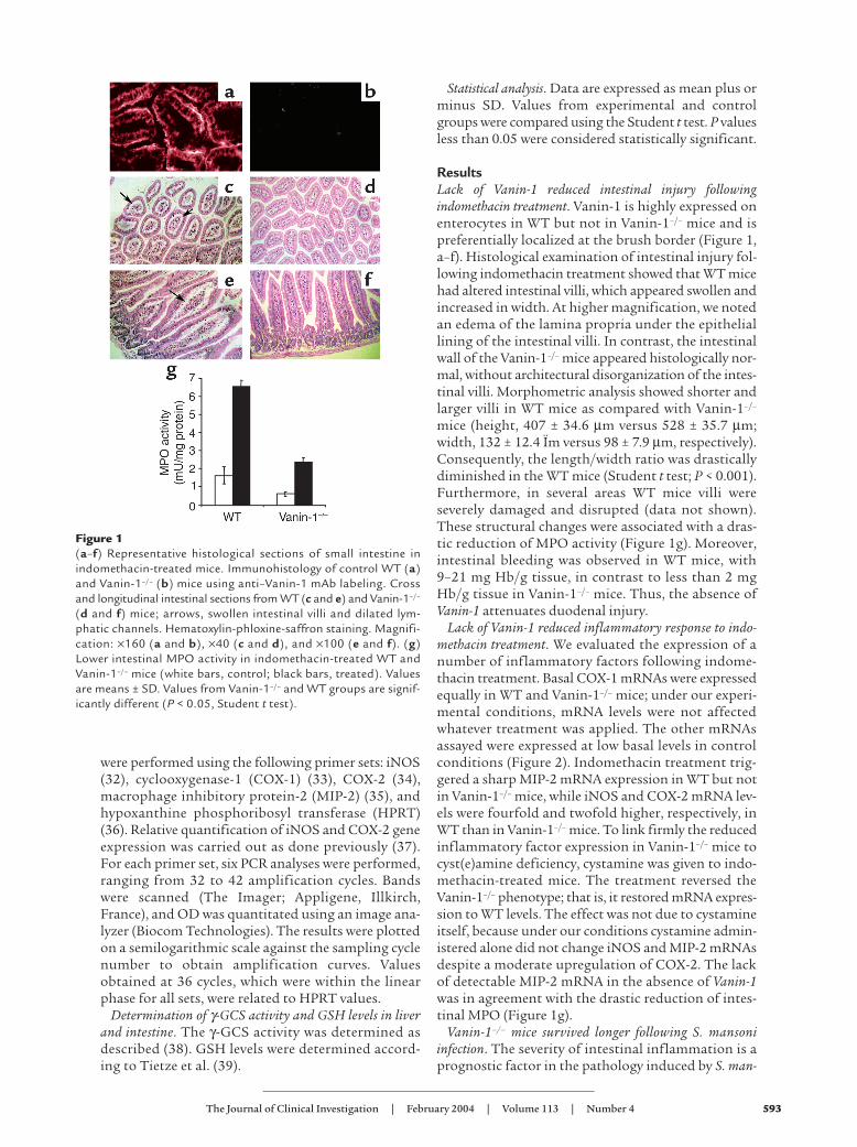
ResultsLack of Vanin-1 reduced intestinal injury followingindomethacin treatment. Vanin-1 is highly expressed onenterocytes in WT but not in Vanin-1–/– mice and ispreferentially localized at the brush border (Figure 1,a–f). Histological examination of intestinal injury fol-lowing indomethacin treatment showed that WT micehad altered intestinal villi, which appeared swollen andincreased in width. At higher magnification, we notedan edema of the lamina propria under the epitheliallining of the intestinal villi. In contrast, the intestinalwall of the Vanin-1–/– mice appeared histologically nor-mal, without architectural disorganization of the intes-tinal villi. Morphometric analysis showed shorter andlarger villi in WT mice as compared with Vanin-1–/–
mice (height, 407 ± 34.6 µm versus 528 ± 35.7 µm;width, 132 ± 12.4 Ïm versus 98 ± 7.9 µm, respectively).Consequently, the length/width ratio was drasticallydiminished in the WT mice (Student t test; P < 0.001).Furthermore, in several areas WT mice villi wereseverely damaged and disrupted (data not shown).These structural changes were associated with a dras-tic reduction of MPO activity (Figure 1g). Moreover,intestinal bleeding was observed in WT mice, with9–21 mg Hb/g tissue, in contrast to less than 2 mgHb/g tissue in Vanin-1–/– mice. Thus, the absence ofVanin-1 attenuates duodenal injury.
Lack of Vanin-1 reduced inflammatory response to indo-methacin treatment. We evaluated the expression of anumber of inflammatory factors following indome-thacin treatment. Basal COX-1 mRNAs were expressedequally in WT and Vanin-1–/– mice; under our experi-mental conditions, mRNA levels were not affectedwhatever treatment was applied. The other mRNAsassayed were expressed at low basal levels in controlconditions (Figure 2). Indomethacin treatment trig-gered a sharp MIP-2 mRNA expression in WT but notin Vanin-1–/– mice, while iNOS and COX-2 mRNA lev-els were fourfold and twofold higher, respectively, inWT than in Vanin-1–/– mice. To link firmly the reducedinflammatory factor expression in Vanin-1–/– mice tocyst(e)amine deficiency, cystamine was given to indo-methacin-treated mice. The treatment reversed theVanin-1–/– phenotype; that is, it restored mRNA expres-sion to WT levels. The effect was not due to cystamineitself, because under our conditions cystamine admin-istered alone did not change iNOS and MIP-2 mRNAsdespite a moderate upregulation of COX-2. The lackof detectable MIP-2 mRNA in the absence of Vanin-1was in agreement with the drastic reduction of intes-tinal MPO (Figure 1g).
Vanin-1–/– mice survived longer following S. mansoniinfection. The severity of intestinal inflammation is aprognostic factor in the pathology induced by S. man-
Figure 1(a–f) Representative histological sections of small intestine inindomethacin-treated mice. Immunohistology of control WT (a)and Vanin-1–/– (b) mice using anti–Vanin-1 mAb labeling. Crossand longitudinal intestinal sections from WT (c and e) and Vanin-1–/–
(d and f) mice; arrows, swollen intestinal villi and dilated lym-phatic channels. Hematoxylin-phloxine-saffron staining. Magnifi-cation: ×160 (a and b), ×40 (c and d), and ×100 (e and f). (g)Lower intestinal MPO activity in indomethacin-treated WT andVanin-1–/– mice (white bars, control; black bars, treated). Valuesare means ± SD. Values from Vanin-1–/– and WT groups are signif-icantly different (P < 0.05, Student t test).

594 The Journal of Clinical Investigation | February 2004 | Volume 113 | Number 4
soni infection (40). Indeed, the 8- to 12-week postin-fection period is the acute phase of S. mansoni–induced inflammation. This period was critical forsurvival of WT mice, with eight out of ten mice dyingat this time. Mortality was delayed in the Vanin-1–/–
group, and only three out of ten mice died duringthis acute-phase period. Survival in the mutantgroup was then stable for 4 weeks, until deathsresumed at 16 weeks after infection (Figure 3a). After20 weeks after infection, only two Vanin-1–/– micewere still alive. Among the eight S. mansoni-infectedWT mice that died during the 8- to 12-week postin-fection period, five showed severe intestinal bleeding,
with a very high Hb level in the feces (Figure 3b). Atautopsy, their cecum and colon were swollen andinfiltrated with blood. Such an appearance was neverseen for Vanin-1–/– mice, which had less abundantand more transient intestinal bleeding than WTmice. Typical intestinal samples from the two groupsare displayed in Figure 3c. Histological and immuno-histological analysis of colon cryosections performed8 weeks after infection showed a severe disorganiza-tion of the mucous membrane of the colon in WTbut not Vanin-1–/– mice. In WT mice, crypts ofLieberkühn were reduced in depth, with increaseddiameter and more mucus production (Figure 4),compared with the Vanin-1–/– mice that presentedhistologically normal crypts of Lieberkühn. In WTmice, peroxidase-positive cells were detected both inthe submucosa and in the lamina propria of mucousmembrane between the crypts of Lieberkühn, where-as these cells remained confined to the submucosa inVanin-1–/– mice. Confocal analysis shows that perox-idase-positive cells were in close contact with dis-rupted Ep-CAM–positive epithelial lining of thecrypts of Lieberkühn in WT mice. In contrast, inVanin-1–/– mice myeloid cells remained in the sub-mucosa. This submucosal localization was noticeableeven in granulomatous areas around parasite eggs inVanin-1–/– mice. Taken as a whole, myeloid cell infil-tration was quantified by measuring the percentageof the total area covered by peroxidase-positive cellson tissue sections of colons (WT = 3.2% versus Vanin-1–/–
= 1.3%; P < 0.01) and by the global MPO activityextracted from whole tissues (WT = 8.5 ± 1.2 versusVanin-1–/– = 6.4 ± 0.5 mU/mg protein; P < 0.01). Allthese findings show that myeloid cell infiltration andtissue damage were more pronounced in WT than inVanin-1–/– infected mice.
Lack of Vanin-1 led to increased GSH levels in liver and intes-tine. In the liver, GSH stores were significantly higher inVanin-1–/– than in WT mice in either healthy or experi-mental animals (Table 1a). Changes in GSH levelsreflected changes in γ-GCS activity, which was, in allcases, significantly higher in Vanin-1–/– than in WT mice,whereas GSH reductase (GR) activity was comparable(56.7 ± 3.1 and 48.7 ± 2.6 mU/mg protein in WT andVanin-1–/– mice, respectively). S. mansoni infection trig-gered a major oxidative stress in WT mice; consequent-ly, liver GSH levels dropped dramatically despite anincrease in γ-GCS enzyme activity. GR activity was alsoreduced to 29.5 ± 3.8 mU/mg protein. Strikingly, inVanin-1–/– mice infection induced a drastic increase inγ-GCS activity, which was significantly higher than inWT mice, whereas GR activity remained unchanged, sug-gesting a better preservation of GSH homeostasis in thisgroup. As a result, GSH levels did not show the decreaseobserved in WT animals. Upon indomethacin treatment,similar variations in γ-GCS activity were observed. In thiscase, GSH stores were maintained at normal levels in WTmice whereas they were enhanced in Vanin-1–/– mice. Theoxidized glutathione/GSH ratio was less than 1% in
Figure 2Reduced expression of inflammatory factors in Vanin-1–/– mice. (a)Representative experiment of semiquantitative RT-PCR analysis ofMIP-2 and COX-1 mRNAs in the small intestine of untreated orindomethacin-treated Vanin-1–/– and WT mice. Cystamine wasadministered when indicated. Amplification of HPRT mRNA tran-script was used as internal control. (b) Relative iNOS and COX-2mRNA levels after PCR amplification.

The Journal of Clinical Investigation | February 2004 | Volume 113 | Number 4 595
untreated mice and less than 2% in inflamed mice (datanot shown). Importantly, cystamine administration dra-matically reduced both γ-GCS activity and GSH levels incontrol mice as well as in indomethacin-treated animals.As a result, cystamine suppressed the difference betweenVanin-1–/– and WT mice.
In the intestine, GSH concentrations paralleled thoseobserved in the liver, however, at a lower level: GSHstores were significantly higher in the absence of Vanin-1in untreated as well as in indomethacin-treated mice.Basal γ-GCS activity needed for constant renewal ofGSH intestinal pools from liver-derived GSH was iden-tical in both types of mice. In contrast, indomethacintreatment induced a significant reduction in γ-GCSactivity in WT mice whereas activity remainedunchanged in Vanin-1–/– mice. As observed in the liver,cystamine administration annihilated the effects ofVanin-1 inactivation; therefore, it reduced intestinalGSH levels in both groups of mice.
Under our experimental conditions, changes in γ-GCSactivity were not associated with changes in enzymeexpression: in liver, as in intestine, Vanin-1–/– mice dis-
played normal levels of γ-GCS catalytic and modifiersubunit mRNAs. The latter remained unchanged afterindomethacin treatment (data not shown).
DiscussionWe showed that Vanin-1–/– mice are less susceptible tointestinal inflammation, either acute (indomethacintreatment) or chronic (S. mansoni infection). In the S.mansoni infection model, a main pathological featureis the presence of diffuse intestinal hemorrhages (40).Such injuries were dramatically reduced in theabsence of Vanin-1, with consecutive prolonged sur-vival after lethal infection. WT and Vanin-1–/– micehad comparable parasite burdens, since equivalentnumbers of eggs were found in both groups (data notshown). Vanin-1–deficient mice that survived intestin-al injury later died from liver disease, since diseaseprogression in the liver proceeded similarly in Vanin-1–/– and WT mice (data not shown). This discrepancybetween liver and intestinal effects of the lack ofVanin-1 could be related to the distinctive form ofchronic liver inflammation induced by S. mansoni,since the inflammatory reaction protects liver againstthe cytotoxic compounds released by the eggs (41).The positive effects of attenuated inflammatoryresponse are counterbalanced by a prolonged survivalof parasite eggs in the organ (our unpublished data).
In the indomethacin inflammatory model, several tran-scripts associated with inflammatory cell activationknown to be induced by this drug (42, 43) were bare-ly expressed or even undetectable in the intestine ofVanin-1–/– mice. Among them is MIP-2, a local chemoat-tractant for neutrophils (44), which plays an importantrole in the progression of indomethacin-induced intes-
Figure 3Prolonged survival of S. mansoni–infected Vanin-1–/– mice. (a) Survivalcurves of Vanin-1–/– (square) and WT (circle) mice infected with alethal dose of S. mansoni cercariae. From 8 to 12 weeks after infection,hemoglobin amount in feces was assayed daily. (b) Reduced intes-tinal hemorrhages in Vanin-1–/– mice following S. mansoni infection.Daily quantification of the Hb amount in infected Vanin-1–/– and WTmice feces. Color scale indicates Hb amount, namely less than 35,35–120, 120–300, and more than 300 ng Hb/mg excrement. +, deadmice. (c) Typical aspect of cecum and colon of WT mice (top) andVanin-1–/– mice (bottom). Scale bars, 1 cm.
Figure 4Cryosections of colon (×100) from three, independent, infectedWT (a–c) or Vanin-1–/– (d–f) mice at 8 weeks after infection. Sec-tions were stained with H&E (a and d) or stained with tyramide-Cy3 (b–e), a peroxidase substrate (yellow/red fluorescence)combined with anti-Ep-CAM (green fluorescence) mAb. Noteincreased mucus production in the crypts of Lieberkühn (*) inWT mice. These pictures are representative of images observedin independent WT (n = 6) or Vanin-1–/– (n = 8) mice, and quan-tification is performed using the Metamorph program asdescribed in the text. Arrowheads indicate disrupted intestinalmucosa; arrows indicate Schistosoma eggs.

596 The Journal of Clinical Investigation | February 2004 | Volume 113 | Number 4
tinal inflammation (24). Absence of MIP-2 expression inVanin-1–/– mice was, as expected, associated with a reducedMPO activity. In agreement, in humans the migratoryfunction of neutrophils involves the Vanin cluster (45).
Protection of Vanin-1–/– mice following indometh-acin treatment was associated with higher GSH lev-els, as compared with WT animals. GSH is wellknown to play crucial regulatory functions in thecontext of inflammation because it is required tomaintain the cellular redox status and to scavengefree radicals. Furthermore, GSH modulates immunefunctions by the regulation of several pathways,including lipid mediator synthesis as a cofactor ofglutathione peroxidases (46, 47). Increase in GSH lev-els in Vanin-1–/– mice likely results from enhancedliver synthesis as indicated by an increase in γ-GCSactivity. Indeed, this organ serves as a central GSH-generating organ that supplies kidney and intestinewith other constituents of GSH resynthesis (48)through the catabolism of GSH by γ-glutamyl-transpeptidase (49). As a consequence, GSH storesare lower in the intestine than in the liver (49–51). Inagreement, we found that GSH levels in the intestinereflected GSH status in the liver, with values signifi-cantly higher in treated Vanin-1–/– than in WT mice.Basal γ-GCS activity, which is required for the main-tenance of a normal GSH pool in the intestine, wascomparable in Vanin-1–/– and WT mice. This suggeststhat under homeostatic conditions the intestinalGSH pool might be dependent mainly upon liverproduction through GSH metabolites. Understressed conditions that enhance Vanin-1 expression(data not shown), however, γ-GCS activity drops inWT and not Vanin-1–/– tissues, reflecting the localinhibitory effect of cysteamine production as occursin the liver. Finally, despite the presence of Vanin-3transcripts in the liver of Vanin-1–/– mice (data notshown), our results seem to suggest that compensa-
tion is not absolute between the twopantetheinase isoforms. This resultis in agreement with the fact thatcysteamine production is unde-tectable in the kidney and liver ofVanin-1–/– mice (6). Vanin-3 is prob-ably secreted and might exert itseffects at distant sites (4).
In vitro studies show that cysta-mine downregulates γ-GCS activity,acting as a glutamine analogue(17–19). Our findings show thatsimilar effects are observed in vivo.Undetectable pantetheinase activityand cysteamine levels in Vanin-1–/–
mice were associated with higher γ-GCS activity in the liver, as com-pared with WT mice, either in con-trol or experimental animals. Inagreement, cystamine administra-tion dramatically reduced the
enzyme activity and GSH levels in such a way that thedifference between the two groups was cancelled.Cystamine action seems to mimic the negative feed-back regulation of γ-GCS by GSH through the GSHbinding site while interacting with a sulfhydryl groupat a second site (52). Other disulfides are unable tofulfill these rather stringent requirements and there-fore are unable to inhibit this enzyme (19). This pro-vides a likely explanation for the gastrointestinalulcerogenic effect of cysteamine (21).
Because of its pleiotropic effects, however, cystaminemight exert a protective effect in some particular cases.Administration of low doses of cystamine is assumed toinhibit transglutaminase activity in the brain and de-creases Huntington disease symptoms in the mouse (53).
Our results show that pantetheinase activity ofVanin-1 molecule is a major regulator of intestinalinflammation, acting through cysteamine release. Thiseffect might be due to the combined effect of down-regulation of GSH levels and enhanced recruitment ofmyeloid cells at inflamed sites; therefore, pantetheinaseinhibitors would have protective effects against intes-tinal inflammatory disorders. These findings provide arationale for a new anti-inflammatory strategy.
AcknowledgmentsWe thank S. Dupré, G. Pitari, A. Greenfield, L. Leser-man, and J. Ewbank for reading the manuscript. Thisstudy was supported by institutional grants from theInstitut National de la Santé et de la Recherche Médi-cale and the Centre National de la Recherche Scien-tifique, as well as charitable trusts from the Associationpour la Recherche contre le Cancer (ARC 5945), theImagerie pour le Petit Animal, and the Association F.Aupetit. F. Malergue, F. Martin, and M.F. Penet arerecipients of a grant from the Ministère de l’EducationNationale, de la Recherche et de la Technologie, and theLigue Nationale contre le Cancer.
Table 1Enhanced γ-GCS activity and GSH levels in the liver and intestine of indomethacin-treat-ed and S. mansoni–infected WT and Vanin-1–/– mice
WT Vanin-1–/–
GSH γ-GCS GSH γ-GCSLiverControls 8.56 ± 1.8 16.5 ± 2.2 12.7 ± 2.4A 23.9 ± 3.4A
Cystamine 5.36 ± 1.1 10.8 ± 0.6 5.55 ± 1.2 11.2 ± 0.5Indomethacin 9.50 ± 1.4 23.4 ± 0.7 16.6 ± 1.2A 37.9 ± 3.3A
Indomethacin + cystamine 7.60 ± 0.6 19.0 ± 4.1 8.30 ± 0.9 21.6 ± 1.7Infection with S. mansoni 3.96 ± 1.8 20.6 ± 2.8 11.09 ± 1.8A 37.2 ± 2.3A
IntestineControls 1.03 ± 0.1 26.5 ± 4.2 1.84 ± 0.2A 23.6 ± 4.5Cystamine 0.90 ± 0.1 20.2 ± 1.2 1.0 ± 0.1 21.5 ± 2.4Indomethacin 0.56 ± 0.1 18.4 ± 2.2 1.37 ± 0.2A 23.7 ± 2.6A
Indomethacin + cystamine 0.65 ± 0.1 17.7 ± 1.4 0.75 ± 0.1 20.6 ± 1.0
Cystamine was administered to compensate for Vanin-1 inactivation. GSH levels and γ-GCS activityare expressed in micromoles per gram of tissue and milliunits per milligram of protein, respectively.AValues significantly different from the WT group (P < 0.05, Student t test).
The Journal of Clinical Investigation | February 2004 | Volume 113 | Number 4 597
1. Aurrand-Lions, M., et al. 1996. Vanin-1, a novel GPI-linked perivascularmolecule involved in thymus homing. Immunity. 5:391–405.
2. Granjeaud, S., Naquet, P., and Galland, F. 1999. An ESTs description ofthe new Vanin gene family conserved from fly to human. Immunogenetics.49:964–972.
3. Galland, F., et al. 1998. Two human genes related to murine vanin-1are located on the long arm of human chromosome 6. Genomics.53:203–213.
4. Martin, F., et al. 2001. Vanin genes are clustered (human 6q22-24 andmouse 10A2B1) and encode isoforms of pantetheinase ectoenzymes.Immunogenetics. 53:296–306.
5. Maras, B., Barra, D., Dupre, S., and Pitari, G. 1999. Is pantetheinase theactual identity of mouse and human vanin-1 proteins? FEBS Lett.461:149–152.
6. Pitari, G., et al. 2000. Pantetheinase activity of membrane-bound Vanin-1:lack of free cysteamine in tissues of Vanin-1 deficient mice. FEBS Lett.483:149–154.
7. Dupre, S., Graziani, M.T., Rosei, M.A., Fabi, A., and Del Grosso, E. 1970.The enzymatic breakdown of pantethine to pantothenic acid and cysta-mine. Eur. J. Biochem. 16:571–578.
8. Miller, S.L., and Schlesinger, G. 1993. Prebiotic syntheses of vitamincoenzymes. I. Cysteamine and 2-mercaptoethanesulfonic acid (coenzymeM). J. Mol. Evol. 36:302–307.
9. Garcia, R.A., Hirschberger, L.L., and Stipanuk, M.H. 1988. Measurementof cyst(e)amine in physiological samples by high performance liquidchromatography. Anal. Biochem. 170:432–440.
10. Ida, S., Tanaka, Y., Ohkuma, S., and Kuriyama, K. 1984. Determinationof cystamine by high-performance liquid chromatography. Anal. Biochem.136:352–356.
11. Kataoka, H., Imamura, Y., Tanaka, H., and Makita, M. 1993. Determi-nation of cysteamine and cystamine by gas chromatography with flamephotometric detection. J. Pharm. Biomed. Anal. 11:963–969.
12. Ricci, G., Nardini, M., Chiaraluce, R., Dupre, S., and Cavallini, D. 1983.Detection and determination of cysteamine at the nanomole level. J. Appl. Biochem. 5:320–329.
13. Siefring, G.E., Jr., Apostol, A.B., Velasco, P.T., and Lorand, L. 1978. Enzy-matic basis for the Ca2+-induced cross-linking of membrane proteins inintact human erythrocytes. Biochemistry. 17:2598–2604.
14. Pontremoli, S., Traniello, S., Enser, M., Shapiro, S., and Horecker, B.L.1967. Regulation of fructose diphosphatase activity by disulfideexchange. Proc. Natl. Acad. Sci. U. S. A. 58:286–293.
15. Namboodiri, M.A., Weller, J.L., and Klein, D.C. 1980. Rapid andreversible activation of acetyl CoA hydrolase in intact pineal cells bydisulfide exchange. Biochem. Biophys. Res. Commun. 96:188–195.
16. Ernest, M.J., and Kim, K.H. 1973. Regulation of rat liver glycogen syn-thetase. Reversible inactivation of glycogen synthetase D by sulfhydryl-disulfide exchange. J. Biol. Chem. 248:1550–1555.
17. Beamer, R.L., Griffith, O.W., Gass, J.D., Anderson, M.E., and Meister, A.1980. Interaction of L- and D-3-amino-1-chloro-2-pentanone withgamma-glutamylcysteine synthetase. J. Biol. Chem. 255:11732–11736.
18. Lebo, R.V., and Kredich, N.M. 1978. Inactivation of human gamma-glu-tamylcysteine synthetase by cystamine. Demonstration and quantifica-tion of enzyme-ligand complexes. J. Biol. Chem. 253:2615–2623.
19. Griffith, O.W., Larsson, A., and Meister, A. 1977. Inhibition of gamma-glutamylcysteine synthetase by cystamine: an approach to a therapy of5-oxoprolinuria (pyroglutamic aciduria). Biochem. Biophys. Res. Commun.79:919–925.
20. Griffith, O.W., and Mulcahy, R.T. 1999. The enzymes of glutathione syn-thesis: gamma-glutamylcysteine synthetase. Adv. Enzymol. Relat. AreasMol. Biol. 73:209–267.
21. Troyer, K.L., et al. 2001. Growth retardation, duodenal lesions, and aber-rant ileum architecture in triple null mice lacking EGF, amphiregulin,and TGF-alpha. Gastroenterology. 121:68–78.
22. Dessein, A.J., et al. 1999. Severe hepatic fibrosis in Schistosoma mansoniinfection is controlled by a major locus that is closely linked to the inter-feron-gamma receptor gene. Am. J. Hum. Genet. 65:709–721.
23. Lambertucci, J.R., et al. 2000. Schistosoma mansoni: assessment of mor-bidity before and after control. Acta Trop. 77:101–109.
24. Beck, P.L., et al. 2000. Mechanisms of NSAID-induced gastrointestinalinjury defined using mutant mice. Gastroenterology. 119:699–705.
25. Abdallahi, O.M., Hanna, S., De Reggi, M., and Gharib, B. 1999. Visual-ization of oxygen radical production in mouse liver in response to infec-tion with Schistosoma mansoni. Liver. 19:495–500.
26. Gharib, B., Abdallahi, O.M., Dessein, H., and De Reggi, M. 1999. Devel-opment of eosinophil peroxidase activity and concomitant alteration ofthe antioxidant defenses in the liver of mice infected with Schistosomamansoni. J. Hepatol. 30:594–602.
27. Pascal, M., et al. 2000. Hyaluronate levels and markers of oxidative stressin the serum of Sudanese subjects at risk of infection with Schistosomamansoni. Trans. R. Soc. Trop. Med. Hyg. 94:66–70.
28. La Flamme, A.C., Patton, E.A., Bauman, B., and Pearce, E.J. 2001. IL-4plays a crucial role in regulating oxidative damage in the liver duringschistosomiasis. J. Immunol. 166:1903–1911.
29. Wynn, T.A., et al. 1998. IL-10 regulates liver pathology in acute murineSchistosomiasis mansoni but is not required for immune down-modu-lation of chronic disease. J. Immunol. 160:4473–4480.
30. Naspetti, M., et al. 2000. A novel anti-Ep-CAM antibody to analyze theorganization of thymic medulla in autoimmunity. Curr. Top. Microbiol.Immunol. 251:109–117.
31. Liaudet, L., et al. 2000. Protection against hemorrhagic shock in micegenetically deficient in poly(ADP-ribose)polymerase. Proc. Natl. Acad. Sci.U. S. A. 97:10203–10208.
32. Lyons, C.R., Orloff, G.J., and Cunningham, J.M. 1992. Molecular cloningand functional expression of an inducible nitric oxide synthase from amurine macrophage cell line. J. Biol. Chem. 267:6370–6374.
33. Ballou, L.R., Botting, R.M., Goorha, S., Zhang, J., and Vane, J.R. 2000.Nociception in cyclooxygenase isozyme-deficient mice. Proc. Natl. Acad.Sci. U. S. A. 97:10272–10276.
34. Scott, D.J., et al. 2001. Lack of inducible nitric oxide synthase promotesintestinal tumorigenesis in the Apc(Min/+) mouse. Gastroenterology.121:889–899.
35. Neumann, B., et al. 1999. Mechanisms of acute inflammatory lunginjury induced by abdominal sepsis. Int. Immunol. 11:217–227.
36. Konecki, D.S., Brennand, J., Fuscoe, J.C., Caskey, C.T., and Chinault, A.C.1982. Hypoxanthine-guanine phosphoribosyltransferase genes of mouseand Chinese hamster: construction and sequence analysis of cDNArecombinants. Nucleic Acids Res. 10:6763–6775.
37. Kinoshita, T., Imamura, J., Nagai, H., and Shimotohno, K. 1992. Quan-tification of gene expression over a wide range by the polymerase chainreaction. Anal. Biochem. 206:231–235.
38. Seelig, G.F., and Meister, A. 1984. Gamma-glutamylcysteine synthetasefrom erythrocytes. Anal. Biochem. 141:510–514.
39. Tietze, F. 1969. Enzymic method for quantitative determination ofnanogram amounts of total and oxidized glutathione: applications tomammalian blood and other tissues. Anal. Biochem. 27:502–522.
40. Cheever, A.W. 1985. Schistosoma japonicum: the pathology of experimen-tal infection. Exp. Parasitol. 59:1–11.
41. Amiri, P., et al. 1992. Tumour necrosis factor alpha restores granulomasand induces parasite egg-laying in schistosome-infected SCID mice.Nature. 356:604–607.
42. Wallace, J.L., et al. 1998. Cyclooxygenase 1 contributes to inflammato-ry responses in rats and mice: implications for gastrointestinal toxicity.Gastroenterology. 115:101–109.
43. Tanaka, A., Araki, H., Hase, S., Komoike, Y., and Takeuchi, K. 2002. Up-regulation of COX-2 by inhibition of COX-1 in the rat: a key to NSAID-induced gastric injury. Aliment. Pharmacol. Ther. 16:90–101.
44. Wolpe, S.D., and Cerami, A. 1989. Macrophage inflammatory proteins1 and 2: members of a novel superfamily of cytokines. FASEB J.3:2565–2573.
45. Suzuki, K., et al. 1999. A novel glycosylphosphatidyl inositol-anchoredprotein on human leukocytes: a possible role for regulation of neu-trophil adherence and migration. J. Immunol. 162:4277–4284.
46. Lu, S.C. 1999. Regulation of hepatic glutathione synthesis: current con-cepts and controversies. FASEB J. 13:1169–1183.
47. Will, Y., et al. 2000. Gamma-glutamyltranspeptidase-deficient knockoutmice as a model to study the relationship between glutathione status,mitochondrial function, and cellular function. Hepatology. 32:740–749.
48. Rana, S.V., Allen, T., and Singh, R. 2002. Inevitable glutathione, then andnow. Indian J. Exp. Biol. 40:706–716.
49. Lieberman, M.W., et al. 1996. Growth retardation and cysteine deficien-cy in gamma-glutamyl transpeptidase-deficient mice. Proc. Natl. Acad. Sci.U. S. A. 93:7923–7926.
50. LeGrand, T.S., and Aw, T.Y. 1996. Chronic hypoxia and glutathione-dependent detoxication in rat small intestine. Am. J. Physiol.270:G725–G729.
51. Griffith, O.W., and Meister, A. 1979. Glutathione: interorgan transloca-tion, turnover, and metabolism. Proc. Natl. Acad. Sci. U. S. A. 76:5606–5610.
52. Huang, C.S., Moore, W.R., and Meister, A. 1988. On the active site thiolof gamma-glutamylcysteine synthetase: relationships to catalysis, inhi-bition, and regulation. Proc. Natl. Acad. Sci. U. S. A. 85:2464–2468.
53. Karpuj, M.V., et al. 2002. Prolonged survival and decreased abnormalmovements in transgenic model of Huntington disease, with admin-istration of the transglutaminase inhibitor cystamine. Nat. Med.8:143–149.