vascular access site bruising - mcmaster university · phd thesis – t. l. cosman; mcmaster...
TRANSCRIPT

VASCULAR ACCESS SITE BRUISING

RELIABILITY OF VASCULAR ACCESS SITE BRUISE MEASUREMENT
AND PATIENT PERCEPTIONS: A MIXED METHODS STUDY
By
TAMMY L. COSMAN RN (EC), BScN, MN
A Thesis
Submitted to the School of Graduate Studies
in Partial Fulfillment of the Requirements
for the Degree of
Doctor of Philosophy
McMaster University
© Copyright by Tammy Cosman, September 2011
PhD Thesis – T. L. Cosman; McMaster University - Nursing
ii
Doctor of Philosophy (2011) McMaster University (Nursing) Hamilton, Ontario TITLE: Reliability of Vascular Access Site Bruise Measurement and Patient Perceptions: A Mixed Methods Study AUTHOR: Tammy L. Cosman RN (EC), B.Sc.N. (University of Western Ontario),
M.N. (University of Toronto) SUPERVISOR: Professor H. M. Arthur NUMBER OF PAGES: xi, 150

PhD Thesis – T. L. Cosman; McMaster University - Nursing
iii
ABSTRACT
Introduction
The most common complication following invasive cardiac procedures is the
development of vascular access site (VAS) bruising. The extent and impact of VAS
bruising is poorly understood and minimally reported in the literature. Research into this
common post-procedure complication is hindered by the lack of a reliable bruise
measurement tool, and the concept that VAS bruising is a minor complication. This
mixed methods study examined the inter-rater reliability of two methods to measure VAS
bruise size. The embedded qualitative descriptive study explored patient perceptions of
VAS bruising.
Methods
Participants having femoral or radial artery puncture for invasive cardiac
procedures were included in this study. Participants reporting VAS bruising completed
self measurement of bruise size using two methods, linear measurement and planimetry.
The principal investigator and research assistant completed bruise measurements at the
same time, and were blinded to participant and each others’ measurements. Following
bruise measurement, the principal investigator conducted semi-structured interviews on a
convenience sample of participants; including both sexes, a range of ages, and bruise
sizes.
Results
Measurements were completed on 40 participants with VAS bruises. Analysis of
inter-rater reliability was done using the intraclass correlation coefficient (ICC), two-way
PhD Thesis – T. L. Cosman; McMaster University - Nursing
iv
random effects model. The inter-rater reliability for both linear measurement and
planimetry between all three measurers was high (.929; .914 respectively). Analysis of
participant narratives uncovered three major themes concerns, impact and mediating
factors, with several sub-themes.
The findings of this study support the reliability of patient VAS bruise
measurement using linear measurement and planimetry. The goals and available
resources for VAS research may determine the choice of measurement approach.
Qualitative descriptive results indicate that patients have concerns related to VAS
bruising and that this bruising may impact activities of daily living. Future research
examining VAS complications should include evaluation of VAS bruising as significant
patient outcome.

PhD Thesis – T. L. Cosman; McMaster University - Nursing
v
ACKNOWLEDGEMENTS
This doctoral thesis is a testament to the consistent support that I have received from numerous individuals in my educational journey. I have many people to thank for their unfailing encouragement. First and foremost I would like express my gratitude to my supervisor, Dr. Heather Arthur. She has been an enduring supporter and advocate from the moment she agreed to be my supervisor to the culmination of this work. She has never failed to provide me with the mentoring and support I required during the years that I have known her.
I would also like to thank my committee members, Dr. Denise Bryant-Lukosius and Dr. Patricia Strachan for their ongoing guidance and expertise. Their probing questions have definitely resulted in a higher quality study.
Krista Craig and Elizabeth Gunn were constant supporters of this research study. Elizabeth freely offered her knowledge and skill with SPSS, and Krista was invaluable in the recruitment and follow up of participants. I could not have completed this study without her.
My sincere appreciation to the nurses working in the Heart Investigation Unit and the post PCI clinic, they were consistently enthusiastic towards this research study and aided in the recruitment of participants. I would also like to acknowledge the participants of this study who were willing to give their time and share their stories to improve our understanding of vascular access site bruising. I would like to thank friends and extended family too numerous to list, who never failed to ask asked “how is school going” and have been understanding as I juggled school, work, family and friends. Finally, I owe my deepest gratitude to my family. My children, Christopher, Bradley and Jessica have provided me with hugs, kisses and encouraging little notes that inspired me to carry on when I needed a little boost. My husband, John, has been unfaltering in his encouragement and motivation from the first discussion of “should I do my PhD”; to entertaining the children out of the house providing me with quiet time to write.

PhD Thesis – T. L. Cosman; McMaster University - Nursing
vi
TABLE OF CONTENTS
ABSTRACT....................................................................................................................... iii ACKNOWLEDGEMENTS................................................................................................ v TABLE OF CONTENTS................................................................................................... vi LIST OF FIGURES ........................................................................................................... ix LIST OF TABLES.............................................................................................................. x LIST OF APPENDICES.................................................................................................... xi Chapter One – Background and Rationale ....................................................................... 1
Femoral Access Site........................................................................................................ 2 Radial Access Site........................................................................................................... 3
Rationale for Measurement Study ...................................................................................... 4 Rationale for Qualitative Study .......................................................................................... 5 Rationale for Mixed Methods Design................................................................................. 5
Reliability Study ............................................................................................................. 6 Qualitative Description ................................................................................................... 6
Research Questions............................................................................................................. 8 Quantitative Questions.................................................................................................... 8 Qualitative Questions...................................................................................................... 8 Mixed Methods Question................................................................................................ 9
Organization of Thesis........................................................................................................ 9 Chapter Two - Literature Review ................................................................................... 11 Importance of Vascular Access Site Bruising .................................................................. 11
Lack of Consistent Definition....................................................................................... 13 Timing and Incidence of Vascular Access Site Bruising.............................................. 14 Impact of VAS Bruising – Resources and Patients ...................................................... 16
VAS Bruise Measurement Tool........................................................................................ 18 Wound Care Literature Review ........................................................................................ 20
Wound Measurement Tools.......................................................................................... 21 Linear measurement.................................................................................................. 22 Planimetry. ................................................................................................................ 23
Reliability of Measurement Tools ................................................................................ 25 Reliability of linear measurement. ............................................................................ 26 Reliability - linear measurement and planimetry...................................................... 27 Limitations. ............................................................................................................... 28
VAS Bruise Measurement Tool Development ................................................................. 29 Requirements for VAS Bruise Measurement Tool....................................................... 30 Bruise Measurement Tools – Two Alternatives ........................................................... 30
Chapter Three - Methods................................................................................................ 33 Study Design..................................................................................................................... 33 Setting ............................................................................................................................... 33

PhD Thesis – T. L. Cosman; McMaster University - Nursing
vii
Population ......................................................................................................................... 34 Inclusion/Exclusion Criteria ......................................................................................... 34 Ethics Approval ............................................................................................................ 34 Participant Recruitment ................................................................................................ 35
Heart investigation unit............................................................................................. 35 Percutaneous coronary intervention follow-up clinic. .............................................. 36
Remuneration................................................................................................................ 37 Management of Adverse Clinical Events ..................................................................... 37
Data Collection ................................................................................................................. 37 Reliability Study ........................................................................................................... 37
Sample size. .............................................................................................................. 37 Demographics and Procedural Details.......................................................................... 39 Instruments.................................................................................................................... 40
Linear measurement.................................................................................................. 40 Planimetry. ................................................................................................................ 41
Bruise Measurement ..................................................................................................... 42 Bruise Measurement Questionnaire.............................................................................. 43 Qualitative Description ................................................................................................. 44
Sample size. .............................................................................................................. 44 Data collection - Interview guide.............................................................................. 45
Data Analysis .................................................................................................................... 46 Measurement Study .......................................................................................................... 46
Demographics and Procedural Details.......................................................................... 46 Reliability Statistics ...................................................................................................... 47
Bruise Measurement Questionnaire.................................................................................. 48 Qualitative Description ..................................................................................................... 48 Chapter Four – Results ................................................................................................... 51 Reliability Study ............................................................................................................... 51
Study Enrollment .......................................................................................................... 51 Participant Demographics and Procedural Details ....................................................... 53 Demographic and Procedural Comparison Between Groups ....................................... 56 Reliability Statistics ...................................................................................................... 59
Linear Measurement. ................................................................................................ 59 Planimetry. ................................................................................................................ 61
Adverse Clinical Events………………………………………………………………….65 Radial Artery Site Bruising............................................................................................... 66 Bruise Measurement Questionnaire.................................................................................. 66
Linear Measurement ..................................................................................................... 67 Planimetry..................................................................................................................... 68
Qualitative Description ..................................................................................................... 69 Study Enrollment .......................................................................................................... 69 Participant Characteristics and Bruise Size .................................................................. 70
Major Themes ................................................................................................................... 70

PhD Thesis – T. L. Cosman; McMaster University - Nursing
viii
Concerns ....................................................................................................................... 70 Body image. .............................................................................................................. 71 Pain. .......................................................................................................................... 71 Surprise or shock....................................................................................................... 71 Questions related to VAS bruising. .......................................................................... 72 No concerns. ............................................................................................................. 72
Impact ........................................................................................................................... 72 Alterations in activities of daily living...................................................................... 73 Self management activities. ...................................................................................... 73 Health seeking activities. .......................................................................................... 73 Minimal impact......................................................................................................... 73
Mediating Factors ......................................................................................................... 73 Participant expectations. ........................................................................................... 74 Information sources. ................................................................................................. 75 Participant misinformation........................................................................................ 75 Participant self explanation....................................................................................... 75 Prior experience. ....................................................................................................... 75 Competing factors. .................................................................................................... 75 Temporal relationship of bruise development. ......................................................... 75 Unplanned procedure. ............................................................................................... 76
Mixed Methods Results .................................................................................................... 76 Conclusions....................................................................................................................... 78 Chapter Five – Discussion .............................................................................................. 80 Reliability of Vascular Access Site Bruise Measurement ................................................ 80
Linear Measurement ..................................................................................................... 81 Planimetry..................................................................................................................... 82 Participant Preferences Between Two Methods ........................................................... 83
Participant Perspective of Vascular Access Site Bruising................................................ 85 Interviewer Bias ............................................................................................................ 86
Mixed Method Question ................................................................................................... 87 Relationship to Prior Research ......................................................................................... 88
Incidence and Size of Vascular Access Site Bruising .................................................. 89 Impact of Vascular Access Site Bruising...................................................................... 92
Implications for Research and Clinical Practice............................................................... 93 Conclusions....................................................................................................................... 96 REFERENCES ................................................................................................................. 98

PhD Thesis – T. L. Cosman; McMaster University - Nursing
ix
LIST OF FIGURES
Figure 1. Participant Flow ……………………………………………………………53

PhD Thesis – T. L. Cosman; McMaster University - Nursing
x
LIST OF TABLES
Table 1. Participant Demographics and Procedural Details............................................ 55 Table 2. Demographic and Procedural Variables Related to Presence of VAS Bruising 58 Table 3. Calculated Bruise Size in Square Centimeters – Linear Measurement.............. 60 Table 4. Intraclass Correlation Coefficients - Linear Measurement ............................... 60 Table 5. Intraclass Correlation Coefficients - Linear Measurement (imputed data)....... 61 Table 6. Intraclass Correlation Coefficient – Comparison of Principle Investigator and Research Assistant Planimetry Measures .......................................................... 62 Table 7. Calculated Bruise Size in Square Centimeters - Principal Investigator Planimetry Measures.......................................................................................... 63 Table 8. Intraclass Correlation Coefficients - Principal Investigator Planimetry Measures …………………………………………………...……..64 Table 9. Intraclass Correlation Coefficients - Planimetry (imputed data) ...................... 64 Table 10. Descriptive Results of Bruise Measurement Questionnaire…………………..67 Table 11. Participant Reported Concerns in Relation to Bruise Size ……………...……77 Table 12. Participant Reported Impact of Vascular Access Site Bruising - Related to Bruise Size...................................................................................... 78

PhD Thesis – T. L. Cosman; McMaster University - Nursing
xi
LIST OF APPENDICES
Appendix A Measurement Techniques …………… .....……………………….….….. 114 Appendix B Research Ethics Board Approval Letters …………………………..….…116 Appendix C Consent Form ………………………………………………….……….. 120 Appendix D Study Information Sheet ………………………………………….......… 126 Appendix E Case Record Form ……………………………………………..……...... 128 Appendix F Participant Instruction Sheet – Femoral ……………………....……...…. 131 Appendix G Participant Instruction Sheet – Radial ………………………………..….135 Appendix H Bruise Measurement Questionnaire ………………………………......…138 Appendix I Interview Participants Characteristics and Bruise Size ………………..…141
Appendix J Interview Guide ……………………………………………….………… 143 Appendix K Contact Summary Sheet …………………………………………….….. 146 Appendix L Planimetry Measurement Data from Both Raters of Tracings ………..…148

PhD Thesis – T. L. Cosman; McMaster University - Nursing
1
Chapter One – Background and Rationale
The primary purpose of this measurement-focused study was to investigate the
reliability of two methods to determine vascular access site (VAS) bruise size seven to
ten days following either femoral or radial arterial puncture for the purpose of cardiac
catheterization (CATH) or percutaneous coronary intervention (PCI). The secondary
purpose was to explore patients’ concerns and their perceptions of the impact of post
procedure bruising following CATH and/or PCI.
This chapter will provide the background and rationale for the study, including
the limitations of current research and the need for a reliable bruise measurement tool for
patients undergoing invasive cardiac procedures of CATH and/or PCI. The choice of
study design will be described. The specific research questions will be presented and
organization of the remainder of the thesis will be outlined.
Cardiovascular disease is the leading cause of death worldwide, with a
predominance of coronary artery disease (CAD) (Gaziano, 2005; World Health
Organization, 2004). Cardiac catheterization is the gold standard invasive diagnostic test
for identification of luminal narrowing within the coronary arteries, also known as CAD.
Identification of CAD can lead to further disease management options including coronary
artery bypass surgery or PCI.
The use of invasive cardiac diagnostics and procedures has risen steadily in
Canada over the past decade (Alter, Stukel, & Newman, 2006). In Canada, rates of
CATH have increased from 359.9 to 471.5 per 100,000 between 1997/1998 and
2001/2002 (Farris, Grant, Galbraith, Gong, & Ghali, 2006). This represents a more than

PhD Thesis – T. L. Cosman; McMaster University - Nursing
2
30% increase in CATH over a period of four years. Percutaneous coronary intervention,
a management option for patients with CAD, is also on the rise throughout Canada. In
2008 over 6,500 of these procedures (CATH or PCI) were performed every 3 months in
Ontario (Cardiac Care network [CCN], 2008). Both CATH and PCI have a variety of
complications associated with them; however, the most common complications are
related to the arterial puncture required to perform the procedure (Applegate et al., 2008;
Ohlow et al., 2009).
Femoral Access Site
Access to the coronary arteries is required to perform CATH and/or PCI. This is
typically performed by accessing the arterial vasculature through either the femoral or
radial artery. The femoral artery has been the dominant access site used for these
procedures for the past 20 years (Jolly, Amlani, Hamon, Yusuf, & Mehta, 2008).
Complications at the femoral VAS range from bruising and hematoma development to
complications requiring external compression. These complications may require
injection of thrombin (to control or stop bleeding), and possible surgical repair of the
femoral artery (Samal & White, 2002). It is now widely understood that major bleeding
following CATH and/or PCI is an independent predictor of morbidity and mortality
(Hamon & Coutance, 2009; Jolly et al., 2008; Manoukian, 2009).
Many studies have examined various methods to reduce the risk of femoral VAS
bleeding complications (Benson et al., 2005; Botti, Williamson, Steen, McTaggart, &
Reid, 1998; Juran et al., 1998; Koreny, Riedmuller, Nikfardjam, Siostrzonek, & Mullner,
2004; Smith & Labriola, 2001). Research studies have also attempted to identify those

PhD Thesis – T. L. Cosman; McMaster University - Nursing
3
patients who are at increased risk of developing femoral VAS complications (Ammann et
al., 2003; Andersen, Bregendahl, Kaestel, Skriver, & Ravkilde, 2005; Cox, et al., 2004;
Dumont, Keeling, Bourguignon, Sarembock, & Turner, 2006; Sulzbach-Hoke, Ratcliffe,
Kimmel, Kolansky, & Polomano, 2010; Yatskar et al., 2007). The majority of research
on VAS complications to date has focused primarily on uncommon, albeit major, femoral
VAS complications. The most commonly occurring VAS complication, significant
bruising, has for the most part been unaddressed in the literature.
Previously completed studies examining femoral VAS complications as an
outcome measure have examined the femoral site prior to patient discharge (Benson et
al., 2005; Jones & McCutcheon, 2003; Sabo, Chlan, & Savik, 2008; Steffenino et al.,
2006). Current practice patterns have resulted in the majority of patients being
discharged home 6 to 24 hours following CATH and/or PCI. There is some evidence in
the literature to suggest that this is too early to adequately assess for bruising at the
femoral VAS (Botti et al., 1998; Cosman, Arthur, & Natarajan, 2011).
Radial Access Site
The use of the radial artery as a VAS for cardiac procedures was introduced in the
late 1980’s and has steadily gained popularity due to the low incidence of deleterious
bleeding complications (e.g., avoidance of retroperitoneal bleeding). Two recent meta-
analyses have demonstrated a reduction in major bleeding complications with radial
access as compared to femoral access for CATH and PCI (Jolly et al., 2008; Vorobcsuk
et al., 2009). Despite an increase in use of the radial artery as a site for CATH and PCI in
some centers, internationally, the majority of these procedures continue to be done using

PhD Thesis – T. L. Cosman; McMaster University - Nursing
4
the femoral artery (Jolly et al., 2008; Nickolaus, Gilchris, & Ettinger, 2001; Ohlow et al.,
2009). The decision about whether to use the radial or the femoral approach is made on
an individual basis by the interventional cardiologist. Research examining complications
related to the radial artery access site is sparse, as this is a relatively new clinical practice.
Some case reports have described the rare development of compartment syndrome, as
well as arteriovenous fistula, pseudoaneurysm and hematoma (Korn et al., 2008;
Nickolaus et al., 2001; Tizon-Marcos & Barbeau, 2008). A longer term issue with radial
artery access cited in the literature is the development of radial artery occlusion, resulting
in an unsuitable conduit for future coronary bypass surgery (Burstein et al., 2007;
Pancholy, Coppola, Patel, & Roke-Thomas, 2008). The development and extent of
bruising following radial artery access has not been reported in the literature.
Rationale for Measurement Study
Studies examining VAS bruise size, at either the femoral or radial sites, are
hampered by inconsistent definitions of hematoma and bruising and, more importantly,
the lack of a standard, reliable method for determining bruise size (Havey & Quinlan,
1998). Bruise size has been reported in the following ways a) a continuous measure
using square centimeters (Behan, Large, Patel, Lloyd, & Sulke, 2007; Botti et al., 1998;
Robb & McLean, 2000), b) by ordered categories identified by familiar objects (quarter,
golf ball etc.) (Cosman et al., 2011), and c) by having patients report bruising as local,
moderate or extensive using line drawings as a guide (Botti et al., 1998). Only three
studies had patients self report their bruise size following discharge (Botti et al., 1998;
Cosman et al., 2011; Robb & McLean, 2000). In general, inconsistent descriptions,

PhD Thesis – T. L. Cosman; McMaster University - Nursing
5
definitions, and reporting of bruise size can limit the interpretation of study findings
(Streiner & Norman, 2003). It is of critical importance that a common definition of
bruising, and a reliable method of measuring VAS bruise size, is utilized in future
research regarding VAS complications. The use of a reliable and cost effective method
of VAS bruise measurement is essential if research into this common complication
following CATH or PCI is to progress. As this complication often develops when
patients are at home, a self administered method of measuring bruise size would be ideal.
Rationale for Qualitative Study
To date there have been no identified studies which examined patients’
perspectives of VAS complications following invasive cardiac procedures.
Understanding the concerns of patients, and the impact of VAS bruising on their lives,
may promote insight and empathy among health care providers who care for these
individuals (Kearney, 2001). Health care providers frequently make comments such as
“it’s only a bruise”, which tends to minimize the importance of this complication without
the necessary information to support or refute such a statement. Creswell and Plano
Clark (2007) also suggest that qualitative methods can “enrich and explain the
quantitative results in the words of participants” (p. 34). The use of qualitative research
to gain understanding of the patient perspective regarding VAS bruising will enhance the
overall understanding of this complication following invasive cardiac procedures.
Rationale for Mixed Methods Design
This study employed an embedded, mixed-method design as described by
Creswell and Plano Clark (2007). Using taxonomy introduced by Morse (2003), the

PhD Thesis – T. L. Cosman; McMaster University - Nursing
6
design is written QUAN + qual. The capital letters indicate the priority of the
quantitative portion of the study and the plus sign (+) indicates the concurrent nature of
two methods. The embedded, mixed-methods design acknowledges the primary goal of
the quantitative component as well as the complementary value of the qualitative
component (Creswell & Plano Clark, 2007).
Reliability Study
The quantitative component of this study evaluated the inter-rater reliability of
two different methods of bruise measurement. Raters were a) the principal investigator
(PI), an advanced practice nurse, b) a research assistant (RA), and c) study participants
who developed VAS bruising following CATH or PCI. This portion of the study
endeavored to meet the key criteria for reliability studies as described by Karanicolas et
al. (2009). Specifically, the raters and participants were representative of those who
would be likely to use measurement tools in either clinical or research settings.
Qualitative Description
The qualitative component of this study explored the impact of VAS bruising as
well as the concerns of patients who developed VAS bruising following CATH and/or
PCI. Qualitative description, as described by Sandelowski (2000; 2010), was the study
method used. The aim of qualitative description is exploratory, and the goal of this study
was to enhance our understanding and awareness of VAS bruising through identification
of themes in patients’ narratives (Dobbins, Jack, Thomas, & Kothari, 2007; Sandelowski,
2000). As little is known about patients’ perceptions related to VAS bruising, qualitative
description is the appropriate qualitative method for this study.

PhD Thesis – T. L. Cosman; McMaster University - Nursing
7
The quantitative and qualitative components of this mixed methods study address
different questions about the phenomenon of VAS bruising. Performing either study
alone would lack the richness of the mixed methods design. A stand alone study
assessing the reliability of VAS bruise measurement fails to acknowledge the individual’s
perspective; a qualitative study alone would fail to identify the optimal method for
measuring VAS bruising. The rationale for mixed methods in this study was to “initiate
new modes of thinking by attending to paradoxes that emerge from two data sources”
(Johnson, Onwuegbuzie & Turner, 2007, p. 115). The results of this mixed methods
study provide a deeper understanding of VAS bruising and will guide future research
examining this phenomenon.
In summary, the most commonly occurring complications following invasive
cardiac procedures are related to the VAS. Bruising at the VAS is the most common
complication following CATH or PCI. The number of patients having these procedures
is increasing, with the majority of patients discharged home within 24 hours of their
procedure. Evidence suggests that VAS bruising becomes apparent several days after the
CATH and/or PCI and will therefore not normally be observed prior to hospital
discharge. Previous studies of VAS bruising are limited by the absence of a reliable
method of bruise measurement and a lack of understanding about the concerns and
impact of post-procedure bruising on patients. A reliable, patient-friendly method of
measuring VAS bruise size is required. Quantitative methods are inadequate to describe
the concerns of patients who develop VAS bruising. The combination of quantitative and
qualitative research methods will provide a better understanding of the phenomenon of

PhD Thesis – T. L. Cosman; McMaster University - Nursing
8
VAS bruising. Using a mixed methods approach to examine these questions will enhance
our understanding of this phenomenon (Creswell & Plano Clark, 2007).
The primary purpose of this study was to determine the reliability of two different
methods of VAS bruise measurement. A further objective was to determine which
method would be easiest for patients to use. The overall goal was to develop a
measurement tool that is both reliable and easy for patient and clinician use in order to
measure and report the extent of VAS bruising that occurs following CATH and/or PCI.
The secondary purpose was to explore the concerns of patients related to VAS
bruising and the impact of VAS bruising on patients following CATH and/or PCI.
Research Questions
Quantitative Questions
1. What is the inter-rater reliability of two different methods for measuring VAS bruise
size? Inter-rater reliability involved comparison of measurements taken by; a) the PI, an
experienced advanced practice nurse; b) the research assistant (RA); and c) the patient
participant.
2. What factors do participants identify as affecting their ability to measure VAS bruise
size (with each measurement method)?
Qualitative Questions
3. What are the concerns of individuals who develop VAS bruising following CATH or
PCI at either radial or femoral sites?
4. What are the patients’ perceptions regarding the impact of VAS bruising following
CATH or PCI, at either the radial or femoral sites, on patients’ activities of daily living?

PhD Thesis – T. L. Cosman; McMaster University - Nursing
9
Mixed Methods Question
5. Does the impact of VAS bruising and related concerns vary according to the size of
VAS bruises?
Organization of Thesis
The thesis is organized into five chapters. This chapter has provided background
information regarding VAS bruising, rationale for the mixed methods study, and briefly
described why a reliable tool is needed for research into VAS bruising.
Chapter two reviews, in detail, the literature regarding VAS bruising and the need
for a reliable tool for VAS bruise measurement. The incidence and impact of VAS
bruising, in terms of utilization of health care resources and effect on patients, will be
addressed. The potential for using currently available measurement tools from the wound
care literature will be examined, including an evaluation of the reliability of these tools.
The requirements for a suitable VAS bruise measurement tool will be addressed and two
potential methods for measurement of VAS bruise size will be put forward.
Chapter three describes the research methods including study design, setting,
population, data collection, and plan for data analysis. The methods for both the
measurement study and the embedded qualitative study are addressed throughout the
chapter.
Chapter four presents the results of the study and is divided into four sections.
The first two sections address the quantitative study results including; description of
study participants, results of the reliability study, and participant questionnaire results.
The third section reviews the results of the embedded qualitative descriptive study. The

PhD Thesis – T. L. Cosman; McMaster University - Nursing
10
major themes identified from the participants’ narrative data are described and exemplars
are used to illustrate the major themes. The fourth section addresses the mixed methods
research question.
The study results, both quantitative and qualitative, are discussed in Chapter five.
How the current study results relate to prior studies and how the current study findings
might be used in future research are examined. Strengths and limitations of the current
study and implications for future research and nursing practice are also considered.

PhD Thesis – T. L. Cosman; McMaster University - Nursing
11
Chapter Two - Literature Review
This chapter reviews the literature related to VAS bruising, as well as literature
related to wound size measurement, which was used to support the development of the
VAS bruise measurement tools. The first section of this chapter provides an overview of
key literature on VAS bruising related to its importance to nursing practice, definitions,
timing, incidence and patient impact of VAS bruising on patients.
The second section of this chapter focuses on the wound care literature;
specifically, a review of wound measurement tools and a critique of reliability studies
conducted on wound measurement tools that are potentially applicable to VAS bruise
measurement.
The chapter concludes by reviewing the requirements for VAS bruise
measurement and proposing two alternatives for VAS bruise measurement.
Importance of Vascular Access Site Bruising
As stated previously, VAS complications are the most common complication
following CATH or PCI and continue to contribute to patient mortality and morbidity.
There has been a large amount of research conducted by both nurses and physicians with
the aim to identify patients at risk for developing major VAS complications, and
strategies to decrease the incidence of such events. Major vascular complications have
traditionally been defined as a significant drop in hemoglobin requiring transfusion (e.g.,
retroperitoneal bleeding), pseudoaneurysm, arteriovenous fistula, peripheral
embolization, and arterial thrombosis (Konstance et al., 2004; Ohlow et al., 2009).
Technological and clinical advances have resulted in a decrease in the incidence of these
PhD Thesis – T. L. Cosman; McMaster University - Nursing
12
major vascular complications (Applegate et al., 2008; Doyle et al., 2008; Ohlow et al.,
2009). At their institution, Applegate et al., (2008) reported a major vascular
complication rate including retroperitoneal bleeding, major bleeding, vascular occlusion,
loss of distal pulse, vascular surgery, and vascular death, of < 1% from 2000 onward.
Development of pseudoaneurysm, arteriovenous fistula and hematoma > 10 cm were
reported as minor complications, occurring < 1.1% of the time over the past ten years.
Ohlow et al., (2009) reported the incidence of pseudoaneurysm and arteriovenous fistula
of 1.2% and 0.6% respectively. While the incidence of these complications is relatively
low, there are limitations to the current body of research regarding VAS complications.
The development of bruising is by far the most common VAS complication
following CATH or PCI, yet has been neglected in the research literature. There are
many potential explanations for this including a) VAS bruising is considered by some to
be a minor complication and an acceptable outcome of CATH or PCI, b) VAS bruising is
not a life threatening complication, c) VAS bruising may not prolong hospital stay and
therefore may not have an obvious impact on human and financial resources of the
hospital, and d) there is currently no reliable method for measuring the extent or impact
of VAS bruising. The lack of a reliable method for measuring VAS bruising can be
traced to two significant issues; a) absence of a clear definition of bruising, as distinct
from hematoma; and b) identification of the optimal timing for assessment of VAS
bruising.

PhD Thesis – T. L. Cosman; McMaster University - Nursing
13
Lack of Consistent Definition
Most published research reporting VAS bruising has neglected to provide clear
definitions of bruising and hematoma; when definitions have been offered, there is
inconsistency among studies (Havey & Quinlan, 1998). A recent randomized controlled
trial (RCT) with a focus on the development and size of bruising following CATH did
not define the difference between bruising and hematoma (Behan et al., 2007). Two
studies examining nursing care and predictors of VAS complications included hematoma
as a minor complication, but used inconsistent definitions of hematoma, and did not
include bruising as an outcome measure (Anderson et al., 2005; Steffenino et al., 2006).
The study by Steffenino et al., (2006) reported hematoma size as < 4 cm, 4 to 8 cm, and >
8 cm, but gives no indication of how the size was measured. Anderson et al., (2005)
reported hematoma size as > 5 to 10 cm, and > 10 cm and describes hematoma
assessment completed by either two experienced nurses or a nurse and a doctor, however
bruising was not included as an outcome measure. In a more recent study of same day
discharge following PCI, 60% of patients self-reported a femoral hematoma; however
there was no definition or description of how patients determined the difference between
hematoma and bruising (Lauck, Johnson, & Ratner, 2009). Bruising at the sheath site
was well defined by Jones and McCutcheon (2003); however there was no definition of
hematoma. There was also no measurement of bruise size, the authors simply reported
the development of bruising during the sheath removal process as a ‘yes’ or ‘no’
outcome. When patients in this study were telephoned five days following CATH, 50%
reported bruising and 14% reported swelling (Jones & McCutcheon, 2003). The study by

PhD Thesis – T. L. Cosman; McMaster University - Nursing
14
Botti et al., (1998) focused on the development of bleeding and bruising with the use of
pressure dressings following CATH. The paper describes how nurses measured local
complications, defined as “bruising or swelling at the groin site” (Botti et al., 1998, p.
362). The inclusion of swelling is not consistent with accepted definitions of bruising. In
contrast, a study by Sabo, Chlan and Savik (2008) clearly defined the VAS complications
of:
oozing (presence of any leakage of blood from the puncture site), ecchymosis (presence of any skin discoloration without a mass), hematoma (presence of a nonpulsatile mass > 4 cm, or pulsatile mass (presence of a palpable mass with movement corresponding to systole and diastole) (p. 192). Robb and McLean (2000) also defined hematoma as “a swelling containing
clotted blood” and bruising as “abnormal discoloration of skin around the puncture site”
(p. 371). It is clear that a clinically accurate and universal definition of VAS bruising is a
priority if it is to be considered an important outcome measure in research designed to
minimize this complication.
Timing and Incidence of Vascular Access Site Bruising
A second limitation of studies examining VAS bruising has been the timing of
VAS assessment. Decreased length of hospital stay has resulted in the majority of
patients being discharged home within 6 to 24 hours of their CATH and/or PCI.
Previously completed studies using VAS complications as an outcome measure have
examined the femoral VAS site prior to patient discharge (Benson et al., 2005; Jones &
McCutcheon, 2003; Steffenino et al., 2006; Sulzbach-Hoke et al., 2010; Walker, Jen,
McCosker, & Cleary, 2008). There is some evidence to suggest that this is too early to
adequately assess for bruising at the VAS.

PhD Thesis – T. L. Cosman; McMaster University - Nursing
15
Botti et al. (1998) found the overall incidence of VAS bruising prior to discharge
was 14.6%. In contrast, 41.9% of patients in this study reported persistent or new
bruising when contacted 6 to 28 days following CATH (Botti et al., 1998). In a RCT
comparing manual compression to vascular closure devices Behan et al., (2007) reported
sequential mean bruise size following CATH. They found an increase in the mean bruise
size from the two hours post-CATH assessment to the one week follow up visit. Mean
bruise size increased from 3.2 cm2 to 82.5 cm2 in patients who had manual compression
and from 0.7 cm2 to 28.5 cm2 in patients who had closure devices. A recent study
specifically examining VAS complications reported an incidence of 6.3% of hematoma
(smaller than 5 cm2) and no bruising at the time of discharge, based on nursing VAS
assessment. However, at the time of telephone follow up, five to seven days following
CATH or PCI, 68.6% of participants reported bruising, with 47% of patients reporting
bruising of 7.5 cm or greater (roughly equivalent to the size of a tennis ball) (Cosman et
al., 2011). In summary, it is reasonable to conclude that VAS bruising is a frequent and
emerging process that occurs over several days to weeks following CATH or PCI.
Bruising at the VAS will not be captured as an outcome of interest if studies continue to
report VAS bruising at the time of hospital discharge, 6 to 24 hours following CATH
and/or PCI.
Three studies in the literature specifically examining the incidence of VAS
bruising found between 41% and 68% of patients reported bruising 3 to 28 days
following CATH or PCI (Cosman et al., 2011; Botti et al., 1998; Robb & Mclean, 2000).

PhD Thesis – T. L. Cosman; McMaster University - Nursing
16
Impact of VAS Bruising – Resources and Patients
It has been suggested in the literature that complications that “do not require
medical attention or affect length of stay” (Sulzbach-Hoke et al., 2010. p. E6), such as
bruising, are not clinically significant. This comment raises questions regarding the
definition of clinical significance in the hospital setting, and from whose perspective
clinical significance is being considered. It would appear that Sulzbach-Hoke et al.
(2010), define a clinically significant event as one that increases length of hospital stay or
requires increased medical or nursing care; the significance of VAS bruising to the
patient has been disregarded. In contrast Amoroso (2005) notes that cardiology nurses
should worry about arterial access complications as nurses are taking an active role in
arterial sheath removal and arterial compression. Amoroso (2005) acknowledges that
“vascular complications, whenever they occur, still have significant impact on morbidity,
mortality and hospital costs” (p. 3). Two studies have recently demonstrated that
approximately 4% of patients with concerns related to VAS, following CATH or PCI,
return to either the emergency department or their family physician for medical follow up
(Cosman et al., 2011; Lauck et al., 2009). In Ontario, this translates to 2,800 health care
visits/year related to patient concerns regarding their VAS following CATH or PCI. The
majority of CATH and PCI procedures are performed at quaternary health care hospitals
and patients may not return to the hospital where the procedure was completed for follow
up. Consequently, staff at the quaternary hospitals may not have a true sense of the
volume of patients presenting for follow up regarding VAS concerns. When patients
present to the family physician or emergency department over VAS concerns, regardless

PhD Thesis – T. L. Cosman; McMaster University - Nursing
17
of the perceived clinical significance of those concerns, there is an increased cost to the
overall health care system. Given the volume of these procedures completed nationally
and internationally, this cost is not insignificant. Bruising at the VAS following invasive
procedures may indeed be a hidden cost of health care utilization following CATH or
PCI.
There is growing acknowledgement that bruising, although not life threatening,
can have an impact upon long term patient outcomes. In the study by Cosman et al.
(2011), it was noted that 41.5% of patients reported pain or discomfort at their VAS but
only 13.4% of patients reported taking any medication for their discomfort. Roy et al.
(2008), recently found that 11% of patients who developed “nuisance bleeding”
following PCI, with a drug eluting stent, discontinued dual antiplatelet therapy without
medical consultation. Nuisance bleeding was defined by the authors as “easy bruising,
bleeding from small cuts, petechia, and ecchymosis” (Roy et al., 2008, p. 1616). The
decision by patients to discontinue antiplatelet medications may have clinical
consequences as short-term studies have shown that early withdrawal of antiplatelet
therapy has been linked to stent thrombosis (Iakovou et al., 2005).
A literature search regarding the impact of VAS complications, specifically
bruising, did not uncover any findings with this patient population. Although not
specifically examined in studies to date, it has been proposed that VAS bruising may be
painful, decrease patient mobility, create increased patient distress, and increase nursing
care time (Behan et al., 2007; Jones & McCutcheon, 2003; Sabo et al., 2008). The results
of a study exploring the experience of day surgery patients’ discharge and recovery found

PhD Thesis – T. L. Cosman; McMaster University - Nursing
18
that some patients were not prepared for the shock of skin discoloration with post
operative bruising. It was suggested that this may have been stressful and perceived as a
threat to body image and appearance for these patients (Gilmartin, 2007).
VAS Bruise Measurement Tool
The use of a reliable measurement tool is of paramount importance when
conducting clinical research (Karanicolas et al., 2009; Streiner & Norman, 2003).
Karanicolas et al. (2009) list seven questions to consider when assessing a reliability
study:
1. Was the research question appropriate? 2. Were the raters representative of the individuals who will apply the instrument in practice? 3. Were the patients or subjects representative of the population that will be rated in practice? 4. Did raters assign the ratings in a clinically relevant manner? 5. Were the data analyzed with use of appropriate reliability statistics? 6. How was the sample size (of raters and subjects) determined? 7. How can the results be interpreted? (p. 100). A reliable measurement tool strengthens the results of research findings, and
provides support for clinicians as they translate study results to the practice setting
(Gethin & Cowman, 2006; Streiner & Norman, 2003). There is no current standard for
measurement of VAS bruising following invasive cardiac procedures in either the clinical
or research setting. A reliable tool for the measurement of VAS bruise size is required
for research into this common complication following CATH or PCI. Prior to beginning

PhD Thesis – T. L. Cosman; McMaster University - Nursing
19
this study a literature search was conducted to determine if there was a reliable tool used
in clinical research to measure VAS bruising.
The following definition for bruise was used when searching for an appropriate
tool to measure bruise size; a bruise is an accumulation of blood in the subcutaneous
tissues as a result of tissue injury. This is evident by the typical discoloration of the skin
surrounding the injured tissue in the absence of a palpable mass. This definition is based
on prior research of VAS complications (Akpinar & Celebioglu, 2008; Robb & McLean,
2000; Sabo et al., 2008). The literature search confirmed the lack of a consistently used
or reliable tool for the purpose of measuring VAS bruising. Two studies were found
which did examine bruise size related to subcutaneous needle puncture. These studies
used wound surface area (WSA) measurement techniques to quantify bruise size (Chan,
2001; Zaybak & Khorshid, 2007). The lack of existing literature related to the
measurement of VAS bruise size demonstrates the perceived lack of significance of VAS
bruising in the health care community.
Following the literature review, it became clear there was no standard reliable
method for measuring VAS bruise size. Examination of the wound care literature lead to
the idea that perhaps modification of existing wound measurement tools would meet the
specific requirement for VAS bruise measurement. Based on clinical experience, the
following attributes of a measurement tool were considered essential to measure VAS
bruising a) ability to measure the surface area of tissue discoloration, b) ability to
measure irregular shapes, and c) ability to measure around body contours.
Considerations for the utility of a bruise measurement tool included the financial costs of

PhD Thesis – T. L. Cosman; McMaster University - Nursing
20
implementing the tool in research. Given the large number of patients undergoing CATH
or PCI, and the clinical practice of discharge within 6 to 24 hours of these procedures, it
would pose a large financial and human resource burden to have patients return to the
hospital for bruise measurement following CATH and/or PCI. It would be ideal if a
reliable VAS bruise measurement tool could be developed for use by patients at home
following discharge. With the above requirements in mind, techniques of WSA
measurement from the wound care literature were considered.
Wound Care Literature Review
To determine the reliability of wound measurement tools, a literature search from
January 1990 to January 2009 was conducted. The Cumulative Index to Nursing and
Allied Health Literature (CINAHL) and Medline databases were searched using the
search terms wound measurement, validity, and reliability, with limits to the English
language. Hand searching the reference lists of all retrieved articles was also done.
Research papers examining the reliability and validity of various wound measurement
tools were reviewed.
Within the wound care literature there exist an abundance of research papers
examining the reliability of various measurement tools. Accurate wound measurement is
essential for researchers in this field because change in wound size is an indicator of
treatment success (Bolton, 2008; Flanagan, 2003; Krouskop, Baker & Wilson, 2002).
Tools exist which measure the two dimensional size of the wound, also known as the
surface area. Three dimensional measures also exist, which incorporate the depth of the
wound. Bruises, as defined in this study, are visualized as two dimensional skin

PhD Thesis – T. L. Cosman; McMaster University - Nursing
21
discolorations apparent at the VAS. A hematoma results when there is a collection of
blood under the skin with the development of a palpable mass. A hematoma is by
definition three dimensional. For the purpose of measuring VAS bruising, as defined in
this study, measurement tool which examined the two dimensional wound size, or surface
area were reviewed. Wound measurement tools which measured the depth of a wound
(three dimensions), or the rate of wound healing were not reviewed.
Wound Measurement Tools
There are a large number of techniques and tools used to assess WSA. Each
technique involves either; a) linear wound measurement, using a ruler; or b) surface area
measurement, based on wound perimeter, referred to as planimetry. Identification of the
wound border can range from the straightforward use of transparent film and a fine tipped
marker to trace the wound perimeter, to sophisticated techniques involving photography,
image processing technology and computer software programs (Ahn, & Salicido, 2008;
Li, 2006). Keeping in mind that one of the attributes of a VAS bruise measurement tool
is ease of use for the patient in the home environment, measurement methods employing
photography and/or computer scanning were not considered viable options. The two
methods of WSA measurement considered reasonable options for VAS bruise
measurement were linear measurement and planimetry. A pictorial representation that
summarizes these two techniques of WSA measurement is provided in Appendix A. The
following is a detailed description of these measurement techniques, including a review
of the reliability of these methods.

PhD Thesis – T. L. Cosman; McMaster University - Nursing
22
Linear measurement.
Linear measurement, also referred to as diameter product, is reported as the most
commonly used WSA measurement in clinical practice (Bryant, Brooks, Schmidt &
Mostow, 2001; Langemo, Anderson, Hanson, Hunter & Thompson, 2008). Using a paper
or plastic ruler, the length and width of the wound is directly measured and the wound
size is reported in either square millimeters (mm2) or centimeters (cm2). With irregularly
shaped wounds the appropriate line of measurement may not be clear. Determining the
axis for measuring length and width has been examined by several authors (Bryant et al.,
2001; Langemo et al., 2008). Four options for length and width measurement of the
wound are outlined:
• perpendicular method – irrespective of axis, measure the maximum wound
length, measure width as the widest, perpendicular to length;
• clockwise method – along the head-to-toe axis, measure the maximum wound
length, measure width at its widest, perpendicular to the head-to-toe axis;
• along the head-to-toe axis, measure maximum wound length, measure width
at its widest point, regardless of angle to each other; and
• irrespective of axis, measure maximum wound length and maximum wound
width regardless of angle to each other (Bryant et al., 2001; Langemo et al.,
2008).
Once the length and width of the wound are determined, the area can be
calculated using one of two formulas. Using the formula, area = length x width, assumes
a rectangular or square shape to the wound and will result in overestimation of wound

PhD Thesis – T. L. Cosman; McMaster University - Nursing
23
size (Ahn & Salcido, 2008; Keast et al., 2004; Majeski, 1992). In many wounds the
surface area has an elliptical shape, and a formula exists for calculating the area of an
ellipse (Langemo, et al., 2008). This method of calculating surface area was rejected for
VAS bruise measurement as clinical experience did not support the notion that the
majority of VAS bruises are elliptical in shape.
Planimetry.
Planimetry is described as the precise measurement of the plane, or flat surface
area, contained within a wound tracing or outline of a wound image (Ahn & Salcido,
2008; Flanagan, 2003). This broad definition has resulted in a number of diverse wound
measurement methods which fall under the umbrella term planimetry. Measurement of
the WSA using planimetry involves two phases.
Phase one involves the delineation of the wound perimeter; this can be done either
directly or indirectly. Direct measurement is often described as the tracing method. This
involves placing an acetate sheet or flexible transparent film directly on the wound. A
fine-tipped indelible marker is then used to outline the wound edge. This fairly
straightforward approach has been used in practice for many years. Wunderlich, Peters,
Armstrong and Lavery (2000) state that this technique was first identified in 1916.
Disadvantages of placing a film over the wound bed include; potential for contamination
of the wound, disruption of granulation tissue and potential distortion of the wound
perimeter with transparency contact and pressure exerted during tracing (Keast et al.,
2004; Krouskop et al., 2002). While measurement of a bruise, where the skin is intact,
will not present the same concerns of contamination and wound disruption, there may be
PhD Thesis – T. L. Cosman; McMaster University - Nursing
24
potential for distortion of bruise perimeter as the bruises often cover curved body
surfaces.
Indirect measurement involves the use of cameras or video equipment (Langemo
et al., 1998; Thawer, Houghton, Woodbury, Keast, & Campbell, 2002). Due to the
complexity and cost involved in these techniques this method was not considered a viable
alternative to direct measurement of the bruise perimeter.
Phase two of planimetry involves calculation of the WSA. Manual calculation
involves placing the wound tracing over graph paper and counting the number of squares
contained within the wound perimeter. Using smaller grid squares, millimeters versus
centimeters, will produce a more precise measure of WSA and would be useful in small
wounds (Langemo et al., 1998). Discrepancies in WSA calculations were found when
observers differed in their decisions about whether to include or exclude squares that
were only partially within the tracing field (Bryant et al., 2001; Richard, Daures, Parer-
Richard, Vannereau, & Boulot, 2000). Richard et al., (2000) suggest including all partial
squares at a rate of 45% (0.45) of the full square area. The manual counting technique
has been described by some as tedious and time consuming (Majeski, 1992; Richard et
al., 2000).
Handheld devices, sometimes referred to as digitizers, or digital planimetry,
automatically calculates surface area of a traced area and has partially eliminated the
need for manual calculation of surface area. Unfortunately the use of digital planimetry,
with the automatic calculation of surface area, is limited by the size of the digital pad (14
cm x 14 cm) (Flanagan, 2003; Goldman & Salcido, 2002; Sugama et al., 2007). Clinical

PhD Thesis – T. L. Cosman; McMaster University - Nursing
25
experience and pilot study data by the author indicated that the size of some VAS bruises
would not be accommodated by the upper size limit of the digital planimetry device.
Reliability of Measurement Tools
Streiner and Norman (2003) refer to reliability as a “fundamental way to reflect
the amount of error, random and systematic, inherent in any measurement” (p. 126).
Random error is a reflection of ‘noise’ within the measurement, whereas systematic error
refers to the accuracy or precision of the measurement tool (DeVon et al., 2007;
McDowell, 2006). One component of reliability is how consistently the tool performs
across patients, observers and time (McDowell, 2006). Measures of reliability reported
in the wound measurement literature include; a) inter-rater reliability, the level of
agreement between different observers using the same measurement tool; b) intra-rater
reliability, the level of agreement within the same observer when the tool is used at least
twice; and c) test-retest reliability, the level of agreement when the tool is used at two
different times (McDowell, 2006; Streiner & Norman, 2003). The inter-rater reliability
of the WSA measurement tools was the focus for consideration when searching for a
VAS bruise measurement tool. The intraclass correlation coefficient (ICC) and Pearson’s
correlation are the two most commonly reported statistics in reliability studies of wound
measurement tools.
The Pearson’s correlation coefficient represents the level of association between
two measurements; it does not reflect absolute agreement between two measures. For
example, with inter-rater reliability one rater may consistently over estimate WSA. If the
raters are consistent in their measures, the Pearson’s correlation between raters (inter-

PhD Thesis – T. L. Cosman; McMaster University - Nursing
26
rater reliability) will be high; however, this does not necessarily reflect agreement
between the raters. The statistical calculation of the ICC accounts for differences
between raters and is therefore the preferred statistic to measure reliability of this type of
tool (Karanicolas et al., 2009; McDowell, 2006; Streiner & Norman, 2008). Thawer et
al., (2002) are very clear in stating that the ICC “reflects both the degree of
correspondence and agreement among two measurement techniques, assessors, or
ratings” (p. 49). The inter-rater reliability of a measurement tool for wound surface area
needs to reflect both consistency and agreement between raters; therefore, the preferred
measure of reliability is the ICC. There is no standard regarding the required reliability
level of a measurement tool, although some authors suggest that an ICC of .75 – 1.00 be
considered excellent, .40 – .74 modest, and .00 – .39 poor (Sugama et al., 2007; Thawer
et al., 2002). Given that the WSA measurement is an objective measure, one would
expect the reliability measures to be excellent.
Reliability of linear measurement.
One study was found which exclusively examined inter-rater reliability of WSA
using the linear measurement technique. Bryant et al., (2001) examined the inter-rater
reliability of 16 raters by having them measure 6 different imitation wounds on a
prosthetic leg, using three different linear methods 1) clockwise, 2) perpendicular, and 3)
their usual method. The largest wound size was 12cm2 (3 cm x 4 cm). Each
measurement was taken two weeks apart by raters who were considered experts in wound
care. The ICC for the clockwise method (.682) and the raters’ usual method (.699) were
both considered acceptable. However the ICC for the perpendicular method was superior

PhD Thesis – T. L. Cosman; McMaster University - Nursing
27
with an ICC of .962. This indicates that the perpendicular method of linear measurement
is more reliable, with 4% of the variance between raters explained by error. The authors
concluded the preferred method of linear measurement was the perpendicular method.
Reliability - linear measurement and planimetry.
Some reliability studies of wound measurement examined multiple measurement
techniques simultaneously. Langemo et al. (1998) had 66 non-expert raters measure
three uniquely shaped plaster of Paris wound models using four different methods
including linear measurement (perpendicular method), and three types of planimetry.
Planimetry included direct measurement with tracing and manual area calculation (full
squares only) and two forms of digital planimetry. Each model was measured twice and
the average measurement was used for reliability calculations. Langemo et al. (1998)
examined inter-rater reliability and reported high inter-rater reliability (ICC .98 – .99) for
all methods when the mean rating was used. However when a single rating was used the
inter-rater reliability decreased for both linear planimetry and planimetry (ICC .30), as
compared to the inter-rater reliability for the two forms of digital planimetry (ICC .53 -
.87). Multiple measures using the same tool should result in improved reliability as the
degree of error is minimized when a tool is administered multiple times (Streiner &
Norman, 2003). The authors concluded that computerized digital planimetry is the most
reliable method when a single measurement is used. While these results do not support
the reliability of linear measurement or basic planimetry, Langemo et al., (1998)
comment that some raters did not follow specific instructions regarding either planimetry
or linear measurement. Raters did not consistently follow written instruction regarding

PhD Thesis – T. L. Cosman; McMaster University - Nursing
28
determining length and width measures specific to the perpendicular method of linear
measurement. As well, some raters counted partial as well as full squares and either
deliberately or randomly placed the transparency grid over the wound which would
impact the calculated surface area. Langemo et al., (1998) comment that “grid placement
can lead to about a 5 square difference in area” (p. 342). The authors also examined
intra-rater reliability using Pearson’s correlations and found that intra-rater correlation
with linear measurement also varied widely (.48 – .68) based on wound shape, the lowest
correlations occurring in the wound that was L shaped. The authors concluded that
wound shape could significantly affect the reliability of measurement tools (Langemo et
al., 1998).
A similar study testing the reliability of both linear measurement and planimetry
had three raters create two tracings of venous stasis ulcers on the lower leg that were no
larger than 4 x 5 cm2 (Majeske, 1992). One rater traced 34 wounds and the remaining
two raters each traced 18 wounds. All tracings occurred on the same day and each rater
calculated wound area using a) linear measurement (method not defined), b) planimetry
with manual calculation (full squares), and c) two different types of digital planimetry.
The authors reported high inter-rater and intra-rater reliabilities with ICC of .97 – .99
across all methods. These findings suggest that each of the four techniques is equally
reliable in wound area calculation.
Limitations.
Overall, the reliability studies of wound measurement tools describe excellent
intra and inter-rater reliabilities for various methods of WSA calculation (Bryant et al.,

PhD Thesis – T. L. Cosman; McMaster University - Nursing
29
2001; Sugama et al., 2007; Majeski, 1992). There are however, limitations to these
reliability studies. As pointed out by Wunderlich et al. (2000), the overall reliability of
tracing and digital planimetry has been confirmed, however “the methods by which these
techniques have been studied are quite inconsistent” (p. 92). Inconsistent definitions and
descriptions of the measurement techniques result in limitations in the interpretation of
reliability as well as reproducibility of these results in research and clinical practice.
There exist a variety of approaches to statistical measurement and reporting of
reliability. Although ICC is the preferred method of analysis and reporting, some authors
report Pearson’s correlation coefficient; others give inadequate description of the
statistical analysis (Haghpanah et al., 2006; Majeski, 1992; Oien et al., 2002; Streiner &
Norman, 2003; Sugama et al., 2007; Thomas & Wysocki, 1990). This creates difficulty
when comparing results across studies.
Lastly, none of the reliability studies examined included a sample size calculation
or considerations when determining the number of raters or the number of wounds to be
measured. Finally, a clear limitation is the scarcity of reliability studies which examine
linear measurement as a technique. Although there are deficiencies in the psychometric
properties of WSA measurement tools, the research that has been done can be
informative when choosing a measurement technique for VAS bruise size.
VAS Bruise Measurement Tool Development
There is no standard method for measuring bruise size as a VAS complication. In
order to perform research related to VAS bruising a reliable measurement tool is
required. A review of the wound care literature has identified two general methods of

PhD Thesis – T. L. Cosman; McMaster University - Nursing
30
surface area measurement which may be applicable to VAS bruise measurement; linear
measurement and planimetry. Streiner and Norman (2008) emphasize that a
measurement tool needs to be assessed for reliability when it is used on a population
other than those for whom it was originally developed.
Requirements for VAS Bruise Measurement Tool
As stated previously, a tool for measuring VAS bruise size would need to be
practical for use, given the size and location of bruising (femoral or radial artery site).
The tool would need to measure an irregularly shaped surface area of tissue discoloration
around body contours. Acknowledging the number of patients undergoing these
procedures and the timing of VAS bruise development, a tool that is reliable for patient
use without excessive training would be ideal. Two methods of measuring surface area
from the wound care literature were considered viable for patient self measurement of
VAS bruise size.
Bruise Measurement Tools – Two Alternatives
Linear measurement and planimetry, with tracing and manual counting of squares,
are both feasible VAS bruise measurement options, each method having unique strengths
and limitations.
Linear measurement calculates surface area based on maximal length and width
measurements. The resulting surface area assumes the shape of a rectangle. This
estimation has been shown to overestimate wound surface area in comparison to
planimetry (Keast et al., 2004; Oien et al., 2002). Specific instructions regarding the
preferred method of linear measurement, the perpendicular method, are essential if linear

PhD Thesis – T. L. Cosman; McMaster University - Nursing
31
measurement is used. Linear measurement however has clear advantages in terms of
time and technology requirements to complete the assessment. Linear measurement
should be considered a potential technique for bruise measurement in clinical research.
Planimetry can range from simple and cost efficient with manual counting, to
complex and expensive, with digital calculations and computer requirements. Digital
planimetry with direct measurement (tracing) and the use of a hand-held digital
planimetry device would be ideal as it performs automatic calculation of area, decreasing
the time and subjectivity associated with manual counting of squares (Shaw, Hughes,
Lagan, Bell, & Stevenson, 2007; Majeski, 1992). However, the use of hand-held devices
for calculating surface area is hindered by the size of the tracing pad. Specific
instructions regarding the placement of the tracing over the grid paper, and the inclusion
or exclusion of partial squares, are essential if manual counting of squares is used.
Planimetry with manual calculation, or counting of squares, is a viable option for the
calculation of VAS bruise surface area. Regardless which of these two methods of
measurement is utilized, the measurement tool will need to be assessed for reliability for
use in VAS bruise size measurement (Streiner & Norman, 2008).
In conclusion, VAS bruising is a common occurrence that generally appears
several days following CATH or PCI. Minimal research has been done examining VAS
bruising and the impact and related concerns of patients related to VAS bruising is
unknown. There is some evidence to suggest that VAS bruising may lead to increased
use of health care resources and possible early discontinuation of proven medical therapy,
and therefore may not be as innocuous as thought by health care professionals. Lack of a
PhD Thesis – T. L. Cosman; McMaster University - Nursing
32
reliable VAS bruise measurement tool and standardized definitions of bruising and
hematoma in the literature has hindered clinical research into VAS bruising. Linear
measurement and planimetry, two reliable methods of WSA measurement may be useful
in the measurement of VAS bruise size. Chapter three will provide the details of both the
reliability study and the qualitative descriptive study examining VAS bruising.

PhD Thesis – T. L. Cosman; McMaster University - Nursing
33
Chapter Three - Methods
Study Design
As previously described, this study used an embedded mixed methods design in
order to address the proposed research questions (Creswell & Plano Clark, 2007). The
primary study was a measurement study, examining the inter-rater reliability to two
different VAS bruise measurement tools. Embedded within the measurement study was a
qualitative descriptive study which examined the impact and concerns of participants
who developed VAS bruising. Participant selection and data collection for both
components of this study occurred simultaneously. Data analysis for the measurement
study occurred following participant recruitment and VAS bruise measurement was
completed. Analysis of the qualitative data commenced following the first patient
interview and progressed in an iterative fashion, in keeping with conventional content
analysis (Hsieh & Shannon, 2005). These methods will be described in detail in this
chapter.
Setting
Hamilton Health Sciences Corporation (HHSC) is a major tertiary care center
which provides invasive cardiovascular testing (CATH and PCI) for persons living within
the Hamilton Niagara Haldimand Brant Local Health Integration Network (LHIN 4).
The Heart Investigation Unit (HIU) provides CATH and PCI services to patients from 16
referring hospitals within LHIN 4. Over 7,000 procedures were completed in the HIU in
2008/2009.

PhD Thesis – T. L. Cosman; McMaster University - Nursing
34
Participants were recruited either prior to or following their procedure. All bruise
measurement appointments were conducted following CATH or PCI in either a private
outpatient examination room, or in one case, an inpatient hospital room. Participant
interviews were conducted following completion of bruise measurements.
Population
Inclusion/Exclusion Criteria
All patients undergoing CATH or PCI using either the femoral or radial artery
were potential participants for this study. However, due to the study requirement of a
return hospital visit, only patients living within Hamilton and the surrounding
communities were approached for participation in the study.
Participants were excluded if they a) were not clinically stable following the
procedure (e.g., requiring inotropic support), b) were booked for urgent coronary artery
bypass surgery, c) were expected to remain in hospital for an extended period, d) did not
speak and read English, e) had language or mental impairments (e.g., aphasia), f) did not
have access to a telephone, or g) were unable to return to the hospital for VAS bruise
measurement.
Ethics Approval
Study protocols were submitted and approved by the Joint Research Ethics Board
of McMaster University and Hamilton Health Sciences. The study approval letters are
included (Appendix B) as is the participant consent form for this study (Appendix C).

PhD Thesis – T. L. Cosman; McMaster University - Nursing
35
Participant Recruitment
In a previous pilot study at HHS, 68% of patients developed some degree of
bruising at their VAS five to seven days following CATH or PCI (Cosman et al., 2011).
With over 500 CATH or PCI procedures carried out per month in the HIU, it was
estimated that recruitment could be completed within 15 to 20 weeks. However a large
number of patients were either not eligible, or were unwilling to participate in the study
due to the required return visit for bruise measurement. The initial strategy for
participant recruitment involved recruiting patients either prior to or following their
procedure in the HIU. Following REB amendment and approval participants were
recruited to take part in this study through three different avenues, described as follows.
Heart investigation unit.
i) Participants living within the Hamilton region were recruited in the HIU either
prior to or following their cardiac procedure. Nurses within the patients’ circle of care,
but at arms length from the study, obtained permission from the patient to have either the
RA or PI speak to the patient to a) determine study eligibility and b) fully explain the
study. If acceptable to the patient, the RA or PI obtained consent for study participation
(Appendix C). Consenting participants were contacted by telephone two to four days
following their procedure. Participants who reported a bruise at their VAS returned to
the hospital 3 to 11 days after their procedure for bruise measurement. Participant
interviews, if conducted, were completed following bruise measurement.
ii) It was postulated that potential participants who initially declined participation
in the study may later choose to participate if they developed a VAS bruise following
PhD Thesis – T. L. Cosman; McMaster University - Nursing
36
discharge from the HIU. A flyer describing the study and providing the name of the PI
and telephone contact number was given to eligible patients who declined study
enrollment while in the HIU (Appendix D). If an individual was interested in
participating in the study after he/she returned home they could contact the PI to arrange
outpatient bruise measurement.
Percutaneous coronary intervention follow-up clinic.
iii) A post-PCI clinic was created at HHSC following the initiation of this
research study. Conversations with the Nurse Clinicians working in the clinic revealed
that the majority of this population presented with VAS bruising when seen in clinic
seven to ten days following PCI. These potential participants were not being captured in
the HIU as they may have been clinically unstable pre-procedure, and post procedure
would have been taken directly to the Coronary Care Unit. Standard practice was for the
Nurse Clinicians to see these patients in hospital and book a post-PCI clinic appointment
prior to discharge from the hospital. The Nurse Clinicians provided participants with a
study information sheet (Appendix D) prior to discharge and obtained permission from
the patient to have either the RA or PI speak to the patient to a) determine study
eligibility and b) explain the study. If acceptable to the patient, the RA or PI fully
explained the study and obtained consent for study participation following post-PCI
clinic visit. Bruise measurement and participant interview, as appropriate, were
completed following post-PCI clinic visit.
All participants consenting to this study were made aware that they would be
required to return to the hospital for follow up VAS bruise measurement. All participants

PhD Thesis – T. L. Cosman; McMaster University - Nursing
37
enrolled were also aware that they may be asked to take part in a patient interview
following bruise measurement. Participants had the right to decline to be interviewed,
even if they took part in the bruise measurement component of the study.
Remuneration
From the time of initial recruitment into this study all participants received a
parking voucher to cover the cost of parking for the return visit to the hospital. Due to
slower than expected recruitment, and subsequent to REB amendment and approval,
participants also received remuneration in the sum of $50 to cover the approximate
expense of driving up to 50 kilometers for participation in this study.
Management of Adverse Clinical Events
In the previously completed pilot study of 172 patients following CATH or PCI
no participants reported complications requiring medical attention (Cosman et al., 2011).
In the event that a participant arrived for bruise measurement and had a) clinical signs
indicating loss of femoral/pedal pulses, b) numbness or pain in the affected extremity (leg
or hand), or c) worrisome signs and symptoms at the VAS, the PI would escort the
participant to the Emergency Department for further assessment.
Data Collection
Reliability Study
Sample size.
This reliability study assessed the inter-rater reliability of two VAS bruise
measurement tools. The sample size is reflective of the number of bruises measured, not
the number of participants enrolled in the study. Some participants had more than one

PhD Thesis – T. L. Cosman; McMaster University - Nursing
38
VAS (e.g., femoral and radial) and therefore more than one bruise measurement. When
assessing the reliability of a measurement tool it is important that there is variability in
the measured outcome (Streiner & Norman, 2008). In this study the cardiac procedure
and location of the bruise were less important for the sample size calculation than
variability in bruise size. Pilot work completed in 2005 demonstrated a range of bruise
sizes as reported by the participants of the study; from < 2.5 cm2 to >7.5 cm2 (Cosman et
al., 2011).
The calculation of sample size for reliability studies is based on an estimate of the
reliability coefficient of the measurement tool (Streiner & Norman, 2008). The planned
statistical analysis, and sample size calculation, was based on using the ICC to determine
the reliability of each measurement tool. To compute the required sample size an
estimate of the likely ICC was hypothesized as well as the likely standard error (or
confidence interval). Sample size calculation was dependent upon three factors the
hypothesized reliability, the standard error, and the number of raters (Streiner & Norman,
2008). The number of raters for this study was based on generalizability and feasibility
as outlined by Karanicolas et al. (2009). Having raters that were representative of those
who would use the instrument in research and practice required at least one professional
rater along with the participant rater. The feasibility of performing multiple measures on
human subjects also limits the number of raters and negates the option of performing test-
retest reliability in this study. For these reasons three raters were used in this study.
The primary objective of this study was to determine the most reliable
measurement tool for nurses or other health professionals to use in measuring VAS

PhD Thesis – T. L. Cosman; McMaster University - Nursing
39
bruises. A secondary objective was to determine which measurement tool was most
reliable when patients themselves, or family members, measure VAS bruise size. In
regard to the primary objective, there were two professional raters, the PI and RA. It was
hypothesized that there would be high agreement between these two raters. An estimated
reliability of .90 with a standard error of 0.05 was used in calculating sample size. Using
these data, the required sample size was approximately 30 bruise measurements to assess
reliability between the professional raters (Streiner & Norman, 2008). In regard to the
secondary objective, there were three raters; two professionals (PI and RA) and one
participant. It was hypothesized that the reliability between participants and professional
raters would be lower than between professional raters alone. Therefore a reliability
coefficient of .80 and a standard error of 0.05 were hypothesized when measurement
involved the participant. Using these data, the required sample size was approximately
42 bruise measurements. In order to have sufficient numbers for determination of
reliability related to both study objectives, a target of 42 bruise measurements was set for
the reliability study. Based on literature reporting post procedure bruising rates between
41% and 68%, it was estimated that approximately 100 participants would need to be
enrolled in the study (Cosman et al., 2011; Botti et al., 1998; Robb & Mclean, 2000).
Demographics and Procedural Details
Participant demographics and procedural details were obtained either directly
from the participants at the time of consent or from the participant’s chart following the
procedure. Demographic and procedural details were collected on a case record form
created for each participant (Appendix E).

PhD Thesis – T. L. Cosman; McMaster University - Nursing
40
Instruments
Linear measurement and planimetry were the two methods used to measure VAS
bruise size in this study. Each of these methods was modified to accommodate the
unique characteristics of VAS bruise measurement. Written instructions, specific for
femoral and radial VAS, provided to the participants instructed them to a) remove any
bandages at the VAS, b) measure purple/blue discoloration only, and c) place a towel or
sheet in line with their inner thigh to provide privacy during femoral VAS bruise
measurement (Appendix F, G). Bruising that was concealed by the towel with femoral
VAS, was not to be measured. The decision to discount bruising occurring in the pubic
region may hamper the accuracy of bruise measurement; however it was deemed
necessary in order to maintain participant privacy, minimize potential embarrassment for
participants and enhance ease of bruise measurement for both participant and
professional raters.
Linear measurement.
Linear measurement involved using a measuring tape and directly measuring the
length and width of the bruise. Research has demonstrated the perpendicular method of
linear measurement as the most reliable (Bryant, Brooks, Schmidt, & Mostow, 2001).
The perpendicular method involves measuring the maximum bruise length, irrespective
of axis, and measuring the bruise width at its widest point, perpendicular to length
(Langemo et al., 2008). Therefore the instructions provided to the participants were
written such that the participant would measure bruise length and width using the

PhD Thesis – T. L. Cosman; McMaster University - Nursing
41
perpendicular method. The perpendicular method of bruise measurement was used by
both APN and RA when completing linear measurement.
Planimetry.
The second bruise measurement method involved placing a sheet of acetate
directly over the VAS bruise. A permanent black marker was used to trace the outline of
the bruised area. As with linear measurement, bruising concealed by towel placement
was not included in the tracing and therefore was not part of the bruise measurement. If
participants were unable to trace the full extent of their VAS bruise on a single
transparency sheet, two transparency sheets were taped together. Participants were not
asked to calculate bruise surface area using either measurement method.
In order to calculate the surface area of the tracings generated by the three raters it
was necessary to manually count the number of square centimeters contained within the
perimeter of each tracing (Richard et al., 2000). Manual counting was completed
independently by both the PI and RA for all tracings. The graph paper used for manual
counting of squares was the same size as the acetate (8.5 inches x 11inches) with a grid in
square centimeters on the paper. In order to minimize subjectivity with placement of the
graph paper, it was consistently placed in line with the corners of the acetate tracing prior
to counting. The number of complete square centimeters within the tracing perimeter
was recorded as FULL. In addition, any squares that were more than 50% within the
tracing perimeter were counted as a partial square. The number of FULL and PARTIAL
squares counted for each tracing was entered into the database and used in the final
calculation of the bruise surface area.

PhD Thesis – T. L. Cosman; McMaster University - Nursing
42
Bruise Measurement
Participants who returned to hospital for bruise measurement were met by the PI
and RA and taken to a private examination room for bruise measurement. If the
participant arrived with a family member or individual to assist with bruise measurement
they were encouraged to assist as needed. Assistance was recorded on the case record
form (Appendix E). Written instructions with diagrams, specific for femoral or radial
VAS, were given to the participant (Appendix F, G). Participants were supplied with all
necessary equipment to complete both methods of bruise measurement including;
instruction sheet, pen, one to two sheets of 8.5 inches x 11 inches acetate for tracing,
permanent black marker, flexible plastic measuring tape (centimeters and inches on
opposite sides), towel or sheet to ensure modesty with bruise measurement, bruise
measurement questionnaire and opaque envelope. The instruction sheet, tracing acetate,
bruise measurement questionnaire and envelope were all labeled with the participant
identification number (ID) and the letters PT. Participants were asked to write the linear
measurement results on the instruction sheet and insert the tracing and instruction sheet
into the opaque letter size envelope once completed. Participants were asked to complete
the bruise measurement questionnaire following bruise measurement (Appendix H).
Bruise measurements were completed without the assistance of the PI or RA. The PI and
RA left the examination room to allow the participant, with or without assistance from
family member, to complete the bruise measurement in privacy.
Once the participant completed the measurements the PI and RA returned to the
examination room. The RA then completed both methods of bruise measurement.

PhD Thesis – T. L. Cosman; McMaster University - Nursing
43
Linear measurement results and tracings were recorded independent of participant results
and were placed in an opaque envelope identified by the participant ID number and the
letters RA (research assistant).
Following RA measurements the PI then completed VAS bruise measurements
using both methods. Linear measurement results and tracings were recorded independent
of participant and RA results and were placed in an opaque envelope identified by the
participant ID number and the letters PI (principal investigator). The PI and RA
remained blinded to both the participant and each other’s measurements.
Following all bruise measurements the PI examined the VAS for any indication of
vascular compromise. Peripheral arterial circulation was assessed and femoral VAS was
assessed for the presence of a bruit.
All demographic and procedural data entry were completed by the RA. A third,
independent research coordinator, not directly involved with this study, entered the
measurement data for both linear measurement and planimetry. A data audit was
performed by the RA following all demographic, procedural and measurement data entry.
All measurement data (100%) were checked for accuracy of data entry. Random
selections of 20% of the participants’ demographic and procedural data were checked for
accuracy of data entry.
Bruise Measurement Questionnaire
In order to determine the ease of use of each of these methods of bruise
measurement by the participants, a short questionnaire was developed (Appendix H).
This short two page questionnaire used a five point Likert scale to examine a) participant
PhD Thesis – T. L. Cosman; McMaster University - Nursing
44
understanding of the measurement instructions and b) ease of each method of bruise
measurement. Participants were also asked their preference for either linear measurement
or planimetry as a method of measuring VAS bruise size.
Qualitative Description
Sample size.
The participants in the qualitative study were purposively selected from those
who took part in the measurement study. Sandelowski (2000) recommends maximum
variation sampling to gather narrative data from a range of participants. In order to attain
maximum variation in the sample of participants interviewed, the PI kept a log which
identified interview participants in terms of their demographics, VAS and extent of
bruising (Appendix I). Participants in the measurement study were asked to take part in
the qualitative study following bruise measurement if the PI felt they met the following
criteria a) would be able to provide a rich description of their experience; b) provided
some variability in terms of the range of participants related to age, gender, VAS and
bruise size; and c) there was adequate time to properly engage the participant in a
meaningful dialogue regarding their VAS bruise.
Sample size for the qualitative descriptive study was not specified a priori,
however it was estimated that between 10 and 15 interviews would be conducted.
Participant interviews continued until it was determined that data saturation had been
reached (Guest, Bunce & Johnson, 2006).

PhD Thesis – T. L. Cosman; McMaster University - Nursing
45
Data collection - Interview guide.
All interviews were conducted by the PI in a private examination room following
bruise measurement. Interviews were audio-taped and were conducted using a semi-
structured interview guide (Appendix J). The focus of the interview questions was
related to participant concerns related to VAS bruising and impact of VAS bruising on
functional abilities or physical activities (e.g., return to work, driving). The data
collection and analysis process was iterative; that is data analysis began following the
first participant interview and that analysis was used to inform subsequent interview
questions and probes. The interview questions evolved following analysis and
discussions with one of the PI’s supervisory committee members, an experienced
qualitative researcher (PS) (Appendix J).
Prior to transcription, the PI reviewed all audio-tapes and erased any names or
comments considered to be direct identifiers of the participants. The audio-files of
participant interviews were identified only by the study ID number assigned to the
participant. These files were sent to the transcriptionist via email. No one except the
transcriptionist and the PI listened to the audio-taped interviews. The transcribed
interview files were returned to the PI and the transcriptionist was instructed to delete any
audio-tape files of the interviews. Before beginning data analysis all transcriptions were
checked against the original audio-tape for accuracy by the PI. Any required corrections
to the transcriptions were made by the PI.
Following each interview the PI listened thoughtfully to each interview and
completed a contact summary sheet (Appendix K). The aim of this exercise was

PhD Thesis – T. L. Cosman; McMaster University - Nursing
46
threefold, to assist the investigator to identify particularly meaningful comments or
interactions within the interview, identifying new or persistent areas of investigation, and
to enrich the interview skills of the PI as a novice qualitative researcher.
An audit trail was kept by the PI. Thoughts and reflections regarding the data
collection process, discussions with supervisory committee members, and decisions
regarding interview questions, data analysis and coding were recorded in the audit trail.
Support and guidance with the participant interviews, data analysis, and coding was
provided by an experienced qualitative researcher (PS). Data collection and analysis did
not bring to light any new themes following the eighth interview. Discussion with the
qualitative methods expert resulted in the suggestion that concentration be placed on
completing interviews that increased the variation of the sample, specifically to interview
male participants and those having radial access sites. Data collection was concluded
following the tenth participant interview.
Data Analysis
This section will provide a brief overview of the data analysis procedures utilized
in this study. Detailed description of the analysis procedures and their rationale will be
provided along with the study results in chapter four.
Measurement Study
Demographics and Procedural Details
All data analyses were conducted using SPSS statistical software (17.0).
Descriptive analysis, including means, standard deviations and frequency distributions, as
appropriate, were conducted on demographic and procedural data (e.g., age, sex,

PhD Thesis – T. L. Cosman; McMaster University - Nursing
47
procedure completed, access site, sheath size), of the entire sample. In addition, a
comparison between participants who developed bruising versus those who did not report
bruising was undertaken. Demographic and procedural details were compared between
groups using the t-test and Chi square statistic as appropriate. Details specific to the
bruise measurement group were also assessed.
Reliability Statistics
Calculation of bruise surface area for both linear measurement and planimetry
was completed using SPSS software. Bruise surface area of linear measurement was
calculated by creating a new variable in SPSS and using the formula length multiplied by
width (L x W) (Appendix A). Bruise surface area of planimetry was calculated by
creating a new variable in SPSS, and using the formula FULL squares plus PARTIAL
squares multiplied by 0.5 (FULL + (PARTIAL x 0.5)) (Appendix A).
Analysis of inter-rater reliability for linear measurement and planimetry was done
using the ICC, two way random effects model looking for absolute agreement
(Karanicolas et al., 2009; McGraw & Wong, 1996; Streiner & Norman, 2008). This
statistical analysis of inter-rater reliability looks not only at correlation between measures
but also at agreement between raters (Karanicolas et al., 2009; McGraw & Wong, 1996;
Streiner & Norman, 2008). Calculation of the ICC for planimetry involved a two step
process. Step one involved calculating the ICC between PI and RA for planimetry
measures for each individual tracing. It was hypothesized that there would be a high
correlation (ICC > .90) between PI and RA planimetry measures. In step two, given a

PhD Thesis – T. L. Cosman; McMaster University - Nursing
48
high correlation between professional raters, the calculation of ICC between participant,
PI and RA planimetry measures was undertaken using the PI planimetry measures.
Two participants were unable to complete linear measurement and three
participants were unable to complete the bruise tracings (planimetry), these data were
excluded from the original analysis. Secondary analysis of the ICC was undertaken
following imputation for the missing data. Missing data were imputed with the mean
bruise size for participants matched for sex, vascular access site (femoral or radial),
procedure completed, and age (mean ± SD).
Bruise Measurement Questionnaire
Descriptive statistics including; mean, standard deviation and range for each item
on the questionnaire was conducted. Narrative comments by participants relating to
difficulties with each particular method, and preference for measurement method, were
categorized by similar related themes for reporting. Direct quotes of participant
comments were used to highlight the strengths and limitations of each method of
measuring VAS bruising.
Qualitative Description
Conventional content analysis, as described by Hsieh and Shannon (2005) was the
specific method of content analysis used to examine the interview data in this study.
Content analysis takes a large volume of narrative data and condenses it to a manageable
number of themes and exemplars from the actual data. This is the least interpretive of the
many techniques available for qualitative analysis and is grounded in the data. Content
analysis is the method of choice for analysis when little known about the phenomenon of

PhD Thesis – T. L. Cosman; McMaster University - Nursing
49
interest; there are no predefined codes and the codes and themes are developed from, and
grounded in, the data (Hsieh & Shannon, 2005; Sandelowski, 2000). There were no
predetermined codes identified prior to the first interview in this study.
Following transcription of the interviews and checking for accuracy of
transcription, the coding of the narrative data was begun by the PI. Coding of interviews
was completed as described by Hsieh & Shannon (2005) and Miles and Huberman
(1994). Initial codes were developed following the first participant interview. The
transcripts were read a second, third and occasionally a fourth time to identify general
topics within the narrative data. Narrative segments of the transcripts that identified a
specific idea or concept were highlighted, and a code (word or phrase) was developed to
identify the idea or concept. Initial codes were reviewed and discussed with the
experienced qualitative researcher (PS). Consensus was obtained regarding the initial
codes before proceeding with further data analysis. Subsequent to the first interview and
discussion with PS, contact summaries (Appendix K), as described by Miles and
Huberman (1994) were completed following each interview. Word files were created for
each code developed in the process of qualitative data analysis. Coded segments from
within the narrative data were copied and pasted into word files specific for that code. It
was possible for narrative data elements to be given more than one code.
Subsequent interviews and ongoing data analysis utilized the constant comparison
method of data analysis (Hsieh & Shannon, 2005; Thorne, 2000). Interviews were coded
and compared to the initial codes for new ideas/thoughts within the transcripts. New
codes were developed as new themes emerged from the data. Four further meetings were

PhD Thesis – T. L. Cosman; McMaster University - Nursing
50
held between the PI and PS to discuss and come to consensus on a) the interview guide
questions and prompts, b) code development and definitions of the coded segments, c)
combining, splitting and deleting codes, d) data saturation, and e) themes emerging from
the data.

PhD Thesis – T. L. Cosman; McMaster University - Nursing
51
Chapter Four – Results
This chapter presents the results of the study in four distinct sections. The first
section focuses on the results of the reliability study. Descriptive statistics regarding the
participants and procedural details, including details related to timing and assistance with
bruise measurement, are described. Comparison of the demographic and procedural
details between participants who developed VAS bruising, versus those who did not, is
also reported. The results of the reliability analysis, including all secondary analyses, are
reviewed.
The second section of this chapter summarizes the results of the participant
questionnaire that was administered following bruise measurement. Descriptive statistics
were used to illustrate participant ease of use of both methods of bruise measurement.
Participant comments are discussed further in chapter five.
The third section of this chapter provides the results of the embedded qualitative,
descriptive study. Major themes and sub-themes emerging from the data are outlined,
along with participant narratives as exemplars.
The final section of this chapter will addresses the mixed methods question which
focused on the relationship between VAS bruise size, its impact, and the concerns of
participants related to bruising.
Reliability Study
Study Enrollment
A convenience sample of participants was recruited and followed up between
November 2009 and June 2010. Participants were recruited in the HIU, prior to and

PhD Thesis – T. L. Cosman; McMaster University - Nursing
52
following their procedure, as well as in the post PCI clinic. Ninety five participants were
enrolled in the HIU and four participants were enrolled through the post PCI clinic. In
total 99 participants were enrolled in this study. Two participants each had two sites for
vascular access, resulting in a total of 101 potential vascular access bruise sites. One
participant was lost to follow-up and 54 participants reported no VAS bruising at the time
of telephone contact. Six participants either declined to return for follow up bruise
measurement or did not present for bruise measurement at their booked follow up
appointment. Forty VAS bruise measurements were completed (Figure 1).
Study recruitment ceased prior to completing the target of 42 bruise
measurements. This study required the availability of both the PI and RA to complete
bruise measurements. Due to scheduling challenges, coordinating availability of both
professional raters, combined with slow study recruitment, recruitment for the study
closed prior to reaching the study target of 42 bruise measurements.
When the data were audited by the PI it was noted that some of the participant’s
linear measurement numbers were much smaller than the linear measures of both the
research assistant and the principal investigator. A detailed review of each case record
form and tracing outlines revealed that six participants recorded linear measurements
using inches, not centimeters as instructed. This data collection error was rectified prior
to data analysis.

PhD Thesis – T. L. Cosman; McMaster University - Nursing
53
Figure 1. Participant Flow
Participant Demographics and Procedural Details
Prior to analysis all demographic and procedural data were examined for
normality of the distributions. The original source data were checked for accuracy and to
identify any results that appeared to be outliers (e.g., age > or < mean ± 2 SD).
Participant characteristics and procedural details are summarized in Table 1. The
sample included 70 males and 31 females with a mean age of 62.5 years. The mean body
mass index of the sample was 29.4, with more than half of the participants having cardiac
catheterization (53.5%). Most of the procedures were done using the right femoral
46 reported bruising
40 Bruise measurements completed
10 Participant Interviews
6 no bruise measurement
101 potential bruise sites
1 Lost to Follow up
54 reported no bruising

PhD Thesis – T. L. Cosman; McMaster University - Nursing
54
approach (61.4%), with the majority completed using a size 6 French sheath (98%). A
small proportion of participants received a femoral closure device (5.9%). At the time of
telephone contact 46 (45.5%) participants reported a VAS bruise.

PhD Thesis – T. L. Cosman; McMaster University - Nursing
55
Table 1. Participant Demographics and Procedural Details Variable N %
101
Age (mean ± SD) 62.5 ± 10.3 Body Mass Index (mean ± SD) 29.4 ± 5.2 Sex Male 70 69.3 Female 31 30.7 Level of Education < grade 8 9 8.9 Completed grade 8 27 26.7 Completed high school 28 27.7 Completed College/University 36 35.6 Completed Graduate Education 1 1.0 Procedure Completed Cardiac Catheterization 54 53.5 Percutaneous Coronary Intervention 45 44.6 Left and Right Heart Catheterization 2 2.0 Vascular Access Location Right Femoral 62 61.4 Left Femoral 6 5.9 Right Radial 32 31.7 Left Radial 1 1.0 Sheath Size 6 French 99 98.0 5 French 2 2.0 Closure Device used No 95 94.1 Yes 6 5.9 Bruise Present at telephone contact Yes 46 45.6 No 54 53.5 Unknown 1 1.0

PhD Thesis – T. L. Cosman; McMaster University - Nursing
56
Bruise measurements were completed on all participants between 3 to 11 days
post-procedure. The mean number of days to bruise measurement was 6.2 days, with a
standard deviation of 1.5 days. The majority of bruise measurements were completed on
day six post procedure (n = 17; 42.5%). The original intent was to complete bruise
measurements between three and ten days. One participant, having agreed to participate
in the study, arrived in the post PCI clinic 11 days post procedure with a radial VAS
bruise and was included in the study. Self measurement was completed by 29
participants (72.5%); the remainder received assistance with bruise measurement from a
family member or individual who accompanied the participant to the follow up
appointment.
Demographic and Procedural Comparison Between Groups
This study was not a RCT; however it was felt that a comparison of demographic
and procedural details between those participants who developed VAS bruising and those
who did not would be useful in order to assess the generalizability of the sample and to
identify possible factors associated with the development of a bruise versus no bruise.
The Chi Square statistic was used to compare the bruise and no bruise groups on ordinal
and nominal level variables including gender, procedure type, sheath size, vascular access
location, level of education, and the use of closure devices. The independent T test was
used to compare the bruise and no bruise groups for the continuous data variables of age
and body mass index. Table 2 provides the details of the comparison between groups.
There was a statistically significant difference between the bruise and no bruise
groups in relation to the procedure completed (p = .000), and age (p = .05). These

PhD Thesis – T. L. Cosman; McMaster University - Nursing
57
findings indicate that a bruise is more likely to develop following PCI as compared to
CATH. Younger patients were also more likely to develop a bruise compared to older
patients.

PhD Thesis – T. L. Cosman; McMaster University - Nursing
58
Table 2. Demographic and Procedural Variables Related to Presence of VAS Bruising
Variables Bruise n = 46
No Bruise n = 54
Unknown n = 1
p value
Sex .600
Male 30 39 1 Female 16 15 Level of Education .560
< grade 8 2 7 Completed grade 8 14 12 1 Completed high school 12 16 Completed College/University 17 19 Completed Graduate Education 1
Procedure completed .000*
Cardiac Catheterization 22 32 Percutaneous Coronary Intervention 24 21 Left & Right Heart Catheterization 1 1 Vascular Access Location .948
Right Femoral 29 32 1 Left Femoral 3 3 Right Radial 14 18 Left Radial 0 1 Sheath Size .295
6 French 44 54 1 5 French 2 0 Closure Device used .949
Yes 3 3 No 43 51 1 Age .050*
Mean ± SD 60.1 ± 8.8 64.2 ±11.1 Minimum – Maximum 34.2 - 76.3 35.7 - 86.7 Body Mass Index .260
Mean ± SD 28.9 ± 4.7 30.0 ± 5.6 Minimum – Maximum 21.0 - 41.5 19.5 - 42.5
* Statistically significant

PhD Thesis – T. L. Cosman; McMaster University - Nursing
59
Reliability Statistics
The primary objective of this study was to determine the inter-rater reliability of
two different tools for measuring VAS bruise size. Analysis of inter-rater reliability was
done using the ICC, two-way random effects model, looking for absolute agreement
(Karanicolas et al., 2009; McGraw & Wong, 1996; Streiner & Norman, 2008). The
hypothesis of the study was that the ICC between professional raters would be high ( >
.90, standard error 0.05), and the ICC between professional raters and the participants
would be lower ( > .80, standard error 0.05).
Linear Measurement.
The final calculation of bruise surface area, based on linear measurement, was
calculated in SPSS using the formula, bruise surface area = length x width (Appendix A).
Descriptive analysis of the linear measurement results is presented in Table 3. The mean
bruise size ranged from 155 cm2 to 188 cm2 with standard deviations of 253 cm2 to 331
cm2. The calculated ICC for linear measurements are presented in Table 4. The ICC
between the PI and RA for linear measurement was .955 (95% CI = 0.92, 0.96). The ICC
between PI and participant, as well as the RA and participant, was > .80. The ICC
between all three raters was also high at .929 (CI = .88, .86).

PhD Thesis – T. L. Cosman; McMaster University - Nursing
60
Table 3. Calculated Bruise Size in Square Centimeters – Linear Measurement
Raters Mean SD Minimum Maximum
Participant bruise measurement n = 38
155 253 1 1025
PI bruise measurement n = 40
169 261 12 1152
RA bruise measurement n = 40
188 331 18 1400
Note: PI Principle Investigator; RA Research Assistant
Table 4. Intraclass Correlation Coefficients - Linear Measurement
Raters ICC 95% Confidence Interval
PI - RA
.955 .917 - .976
PI - participant
.946 .899 - .947
RA - participant
.889 .795 - .941
PI - RA - participant
.929 .882 - .957
Note: PI Principle Investigator; RA Research Assistant
Two participants were unable to perform linear measurement and were excluded
from the original analysis. Secondary analysis was performed using imputed numbers for
the two participants who were unable to complete linear measurement. The mean bruise
size for participants, matched for sex, vascular access site (femoral or radial), procedure
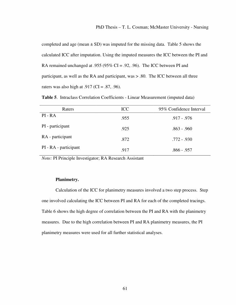
PhD Thesis – T. L. Cosman; McMaster University - Nursing
61
completed and age (mean ± SD) was imputed for the missing data. Table 5 shows the
calculated ICC after imputation. Using the imputed measures the ICC between the PI and
RA remained unchanged at .955 (95% CI = .92, .96). The ICC between PI and
participant, as well as the RA and participant, was > .80. The ICC between all three
raters was also high at .917 (CI = .87, .96).
Table 5. Intraclass Correlation Coefficients - Linear Measurement (imputed data)
Raters ICC 95% Confidence Interval
PI - RA
.955 .917 - .976
PI - participant
.925 .863 - .960
RA - participant
.872 .772 - .930
PI - RA - participant
.917 .866 - .957
Note: PI Principle Investigator; RA Research Assistant
Planimetry.
Calculation of the ICC for planimetry measures involved a two step process. Step
one involved calculating the ICC between PI and RA for each of the completed tracings.
Table 6 shows the high degree of correlation between the PI and RA with the planimetry
measures. Due to the high correlation between PI and RA planimetry measures, the PI
planimetry measures were used for all further statistical analyses.

PhD Thesis – T. L. Cosman; McMaster University - Nursing
62
Table 6. Intraclass Correlation Coefficient – Comparison of Principle Investigator and Research Assistant Planimetry Measures
Raters ICC 95% Confidence Interval
Participant Tracings
.913 .839 - .954
PI Tracings
.970 .945 - .989
RA Tracing
.959 .924 - .978
Note: PI Principle Investigator; RA Research Assistant
The final calculation of bruise surface area, based on planimetry, was calculated
in SPSS using the formula bruise surface area = FULL squares + (PARTIAL squares x
0.5) (Appendix A). Planimetry measures were done for each tracing by both the PI and
the RA. Descriptive analysis of the planimetry results, based on PI planimetry measures,
is presented in Table 7. The mean bruise size ranged from 80 cm2 to 95 cm2 across
raters, with standard deviations from 115 to 151 cm2.

PhD Thesis – T. L. Cosman; McMaster University - Nursing
63
Table 7. Calculated Bruise Size in Square Centimeters - Principal Investigator Planimetry Measures
Raters Mean SD Minimum Maximum
Participant bruise measurement n = 37
80 116 0.5 433
PI bruise measurement n = 40
95 151 5 616
RA bruise measurement n = 40
80 115 6.5 477
Note: PI Principle Investigator; RA Research Assistant
Given the very high correlation between PI and RA; step two, the calculation of
ICC between measurers was done using the PI planimetry measures. The calculated ICC
for planimetry is presented in Table 8. The ICC between the PI and RA for planimetry
was .942 (95% CI = .89, .97). The ICC between both the PI and participant, and the RA
and participant pair, was > .80. The ICC between all three raters was also high at .914
(CI = .86, .95).

PhD Thesis – T. L. Cosman; McMaster University - Nursing
64
Table 8. Intraclass Correlation Coefficients - Principal Investigator Planimetry Measures
Raters ICC 95% Confidence Interval
PI - RA
.942 .888 - .970
PI - participant
.877 .771 - .935
RA - participant
.925 .860 - .961
PI - RA- participant
.914 .856 - .951
Note: PI Principle Investigator; RA Research Assistant
Three participants were unable to perform planimetry and were excluded from the
original analysis. Secondary analysis was performed using imputed numbers for the
three participants who were unable to complete the tracing for planimetry. The mean
bruise size for participants who were matched based on sex, vascular access site (femoral
or radial), procedure completed and age (mean ± SD) was imputed for the missing data.
Table 9 shows the calculated ICC with the imputation measures.
Table 9. Intraclass Correlation Coefficients - Planimetry (imputed data)
Raters ICC 95% Confidence Interval
PI - RA
.942 .888 - .970
PI - participant
.848 .732 - .917
RA - participant
.887 .797 - .939
PI - RA - participant
.893 .829 - .938
Note: PI Principle Investigator, RA Research Assistant

PhD Thesis – T. L. Cosman; McMaster University - Nursing
65
Careful examination of the final planimetry data revealed that some bruise
measurements between PI and RA had larger than expected discrepancies. These data are
presented in Appendix L. Irrespective of the high inter-rater reliability between PI and
RA planimetry measures, examining the raw data it is apparent that the largest
measurement discrepancies occur with larger bruises.
Adverse Clinical Events
During the course of the study two participants presented with atypical symptoms
related to their VAS. The first participant presented for bruise measurement following
CATH via right radial approach. At the time of assessment, six days following the
procedure, there were no signs of vascular compromise in the affected extremity. The
participant contacted the PI by telephone 5 days later to inform me that although the
bruising was receding she was experiencing increasing pain in her right arm which was
causing her to wake at night. I spoke directly with the cardiologist who performed the
procedure. The patient was assessed by the cardiologist who performed the procedure,
including ultrasound of the radial artery, the following day.
The second participant arrived for bruise measurement following CATH via the
right femoral approach with a vascular closure device. In the examination room the
participant stated that she was not sure if she actually had a bruise. Prior to the
participant beginning bruise measurement I examined the VAS. The patient did not have
a VAS bruise; however the area was reddened, tender to touch, with a small amount of
exudate. The participant also reported having warm spells in the evening. Due to the

PhD Thesis – T. L. Cosman; McMaster University - Nursing
66
potential of VAS infection I escorted the participant to the Emergency room for further
assessment and spoke with the cardiologist who performed the procedure the following
day. The participant was seen in follow up in the HIU by the cardiologist.
Radial Artery Site Bruising
While it was not the intention of this study to specifically examine radial site
bruising, as a newer technique with minimal understanding of the consequences of radial
artery puncture for CATH or PCI, it is worthy to comment on the findings from this study
related to the radial access site and bruising. Thirty three participants (32.7%) had a
radial artery access for either CATH or PCI. Fourteen of the participants having radial
artery puncture reported bruising at the VAS, representing 42% of those who had a radial
artery puncture. Two participants in the qualitative study had radial VAS, and reported
very different experiences, from no discomfort at all, to pain in the wrist which limited
the participant’s ability to drive and keyboard at work. These are new insights into the
concerns of patients and potential impact of radial access site bruising.
Bruise Measurement Questionnaire
This section will review the results of the participant survey conducted following
bruise measurement (Appendix H). The goal of the survey was to discern a) if the
instructions for each measurement method were clear, b) the ease of completing each
measurement method, and c) participant preference for either linear or planimetry
measurement of bruise size. Each participant completed the bruise questionnaire,
resulting in 39 questionnaires completed. One participant had two VAS however only
completed one questionnaire. The questionnaire used a five point Likert scale to
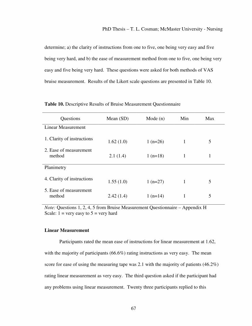
PhD Thesis – T. L. Cosman; McMaster University - Nursing
67
determine; a) the clarity of instructions from one to five, one being very easy and five
being very hard, and b) the ease of measurement method from one to five, one being very
easy and five being very hard. These questions were asked for both methods of VAS
bruise measurement. Results of the Likert scale questions are presented in Table 10.
Table 10. Descriptive Results of Bruise Measurement Questionnaire
Questions Mean (SD) Mode (n) Min Max
Linear Measurement
1. Clarity of instructions 1.62 (1.0) 1 (n=26) 1 5
2. Ease of measurement method
2.1 (1.4) 1 (n=18) 1 1
Planimetry
4. Clarity of instructions 1.55 (1.0) 1 (n=27) 1 5
5. Ease of measurement method
2.42 (1.4) 1 (n=14) 1 5
Note: Questions 1, 2, 4, 5 from Bruise Measurement Questionnaire – Appendix H Scale: 1 = very easy to 5 = very hard
Linear Measurement
Participants rated the mean ease of instructions for linear measurement at 1.62,
with the majority of participants (66.6%) rating instructions as very easy. The mean
score for ease of using the measuring tape was 2.1 with the majority of patients (46.2%)
rating linear measurement as very easy. The third question asked if the participant had
any problems using linear measurement. Twenty three participants replied to this

PhD Thesis – T. L. Cosman; McMaster University - Nursing
68
question as follows a) five participants replied ‘No’, b) two participants replied ‘Yes’, c)
three participants commented that it would be easier with a mirror, d) four participants
commented that it was easier, or would be easier, with the assistance of a second person.
The remaining comments summarize the specific reasons participants gave regarding
difficult bruise measurement:
ID 155 “because of the massive size of the bruise, trying to carry out a complete measurement was not quite easy”;
ID 140 “large stomach hard to see”; ID 105 “unable to do because of bruise location”; and ID 174 “bruise went around leg and I couldn’t see it”.
Planimetry
Participants rated the mean ease of instructions for planimetry at 1.55, with the
majority of participants (71.1%) rating instructions as very easy. The mean score for ease
of using the tracing method (planimetry) was 2.42 with the majority of patients (36.8%)
rating planimetry as very easy. The third question asked if the participant had any
problems using planimetry. Twenty-nine participants replied to this question with
comments. Ten participants replied ‘No’, one participant commented that it would be
easier with a mirror, four participants commented that it was easier, or would be easier,
with the assistance of a second person, and three participants commented that it was
difficult to handle the tracing sheet due to size and/or location of the bruise. The
remaining comments summarize the specific reasons participants gave regarding difficult
bruise measurement:
PhD Thesis – T. L. Cosman; McMaster University - Nursing
69
ID 174 “can’t see all bruise to measure”; ID 135 “awkward as I had bruising on both sides of the arm”; and ID 150 “it wasn’t clear how to position the acetate – picture confused things”. The final question on the bruise measurement questionnaire was a check box for
participants to indicate preference for either linear measurement or planimetry.
Seventeen participants completed the final question. Eight participants preferred using
the measuring tape, seven participants preferred doing the tracing, one participant
indicated both methods, and one participant did not indicate a preference.
Qualitative Description
Study Enrollment
Following REB amendment and approval, participants recruited for the
measurement portion of this study were also considered potential participants for the
embedded qualitative, descriptive study. Participants were asked to take part in the
interview following bruise measurement if; a) the PI felt the individual would provide a
rich source of information; b) there was variability in participants in terms of age, sex,
VAS, procedure completed and bruise size; and c) there was sufficient time to engage the
participant in a meaningful conversation. Ten semi-structured interviews were completed
between February 3rd, 2010 and June 23rd, 2010 (Figure 1). No new themes or sub-
themes emerged following the eighth interview. A further two interviews were
completed in an effort to include more male participants and those with radial access
sites. The interviews were concluded following the tenth interview.

PhD Thesis – T. L. Cosman; McMaster University - Nursing
70
Participant Characteristics and Bruise Size
The interview participant characteristics are depicted in Appendix I. There were
an equal number of men and women interviewed with ages ranging from 34 to 74 years.
Two participants had radial access sites with the remaining participants having femoral
access sites. The majority of patients underwent CATH, with three participants having
PCI, one having primary PCI. Four participants had previous experience with either
CATH or PCI. The bruise size identified in Appendix I was based on the participant
linear measurement, and indicates a range of bruise sizes among the interviewed
participants.
Major Themes
The major themes and sub-themes will be presented in this section. Each of the
major themes will be described, with discussion of the component sub-themes, in turn.
Analysis of the participant narrative data uncovered three major themes with several sub-
themes. The three major themes identified from the participant interviews included a)
concerns, b) impact, and c) mediating factors. Within each major theme several sub-
themes were also identified. The major themes and sub-themes are described below with
exemplars from participant interviews.
Concerns
The major theme of concerns encompasses participants’ comments ranging from
no concerns to comments indicating worries, anxieties, apprehension, alarm, distress and
unease related to VAS bruising. My inherent biases lead to an initial notion that
participant ‘concerns’ would be such that they might be inclined to seek and use

PhD Thesis – T. L. Cosman; McMaster University - Nursing
71
additional health care resources. However with further interviews, reflection, and
discussions with co-supervisor (PS) I came to understand that participant concern was a
larger theme that included several sub-themes. The sub-themes were evident in the
participant narrative data as they commented not on their ‘concerns’ but general
discussion regarding the development of VAS bruising. Narrative data analysis
uncovered participant comments regarding body image, pain and discomfort at the VAS,
surprise and shock regarding the size of the bruising and questions related to the VAS
bruising. Concern as a major theme incorporates the sub-themes of a) body image, b)
pain or discomfort at the VAS, c) surprise or shock, d) questions related to VAS bruising,
and e) no concerns. Participant comments related to each of the sub-themes are provided
below. Each participant is identified by a code indicating their study ID number, sex,
access site (femoral or radial), and bruise size in square centimeters. For example 174 FF
912 indicates a female participant with a femoral access with a bruise size of 912 square
centimeters.
Body image.
174 FF 912 I don’t want people staring at me or looking at it, it’s pretty ugly and it’s so big you know it’s a public pool. I guess I have some feelings about that Pain.
138 MF 49.5 there was no pain, absolutely no pain whatsoever 157 FR 12 it was shooting up my arm at one point. I’d say, 10 being the worst I would say about 8 or 9 …it went on till about Tuesday night (5 days) Surprise or shock.
170 FF 165 This is not just a little bruise you know … you get all … concerned that the bruising is as extensive

PhD Thesis – T. L. Cosman; McMaster University - Nursing
72
146 FF 49.5 Well it’s ugly, it’s um I was shocked. I am bruising all over the place Questions related to VAS bruising.
138 MF 49.5 It’s three separate bruises, why were the other two? What causes a bruise? They just putting a needle in, why is it so big? No concerns.
131 MF 39 She asks me if I was worried and I said no cause there is no real great amount pain it’s there and it’s noticeable but it’s nothing that would send me running These narratives indicate that although many of the participants stated they had no
concerns, there may have been some degree of distress related to VAS bruising that did
not reach the level of ‘concern’ for that individual.
Impact
As the interviews progressed the analysis of the data revealed how VAS bruising
was affecting the participants in terms of impacting their daily activities. Data analysis
resulted in many coded files related to how participants managed their VAS bruising.
Each participant, in their unique and individual way was impacted by the VAS bruising.
The impact of VAS bruising was described by many of the participants as minor,
however they also described modifying their normal activities, most commonly
decreasing physical activity, and using self management methods, such as icing the VAS
or using pain medication. One participant went to urgent care and then sought out the
family physician due to concerns about the VAS bruising. These sub-themes coalesced
to form the major theme of impact. In this study ‘impact’ was defined as any participant
description of how VAS bruising did or did not influence each individual as they carried

PhD Thesis – T. L. Cosman; McMaster University - Nursing
73
out their activities of daily living following CATH or PCI. The major theme of impact
included four sub-themes of a) alterations in activities of daily living, b) self-management
activities, c) health seeking activities, and d) minimal impact.
Alterations in activities of daily living.
157 FR 12 Going back to work Monday at the keyboard typing. I had to take rests cause it was starting to throb … [on] the weekend, I didn’t drive because the pain was so, it was painful and I was too scared to aggravate it even more so I kinda waited til Monday 174 FF 912 I was going to go back swimming next week but I don’t think I will Self management activities.
170 FF 165 I did use ice on a couple of occasions and then … on the Tuesday took a Bufferin cause I was out for the evening Health seeking activities.
174 FF 912 I went to a walk in clinic at that point in time and the doctor there said that you know this is normal (accentuated) Minimal impact.
188 MR 108.75 No, I wasn’t shocked by the bruising, I was you know, I saw it and I thought holy moly look at the bruise but I had bruises, I’ve had lots of bruises … so bruising and cuts and bangs don’t really bother me as much as they might bother someone else but this is really nothing to me
Mediating Factors
Data analysis revealed a multitude of participant comments relating to
factors which appeared to mediate how participants and family members perceived and
interpreted VAS bruising. Participants described prior experience of having CATH or
PCI, their expectations, both formal and informal education regarding the procedure, as
well as the development and healing of the VAS bruise as influencing their concerns.

PhD Thesis – T. L. Cosman; McMaster University - Nursing
74
Some participants had an inaccurate understanding about the procedure and subsequent
VAS bruising, and others spoke of their personal interpretations of why the bruising
occurred and how it should be managed. Participants also described competing factors;
such as sciatica; knee pain; or their pleasure with the outcome of CATH or PCI; the latter
overshadowed the impact of VAS bruising. One participant interviewed in this study had
a large radial bruise following successful PCI. This patient (ID 188 MR 108.75),
identified his bruising as “the red badge of courage”. With one participant the influence
of an unplanned emergency procedure was found to influence concerns regarding VAS
bruising.
Collapsing codes into meaningful sub-themes produced several sub-themes which
described individual concerns, response to these concerns, and ultimately the impact of
VAS bruising on the individual. These sub-themes were combined in the larger theme of
mediating factors. Sub-themes within mediating factors included a) participant
expectations, b) information sources (formal and informal), c) participant misinformation,
d) participant self explanation e) prior experience, f) competing factors, g) temporal
relationship of bruise development, and h) unplanned procedure.
Participant expectations.
138MF 49.5 I was expecting it to be much more painful but the whole procedure was not painful at all 161 FF 108.5 They (nursing staff) told me that I could bruise or could have a bruise. Didn’t tell me how big or how much

PhD Thesis – T. L. Cosman; McMaster University - Nursing
75
Information sources.
170FF 165 I went on the internet and punch up hematoma and you know you see somebody’s leg with this huge bruise but of course you can’t always depend on the internet for maybe a 100% correct information Participant misinformation.
169 MF 957 I mean it’s like blood clots right? … so I was thinking, can that get to my heart? Cause we were away and a friend we were down in Florida … and a friend of ours, a guy that my son played hockey with at school, he was playing hockey and he got a bruise in his shin … and that caused it to go to the heart … and that’s all it was a blood clot cause he got a bruise and I’m thinking so that this definitely had to be a lot bigger than what he had on his shin so … so I’m thinking is this going to happen to me or could it happen? Participant self explanation.
146 FF 49.5 I think it’s Placel?? …plavix and ah ah aspirin … I think that’s the culprits that’s doing it
Prior experience.
131 MF 39 This time around with the groin it’s been more sensitive you know there’s swelling there wasn’t before. There is bruising more bruising more sensitivity and its just taken longer … its lasting longer, the first time around it was maybe two days until I was able to get rid of the bandaids and stuff like that Competing factors.
186 MF 63.25 I keep walking cause I have a sore knee and I gotta make sure that that gets better 188 MR 108.75 I mean when you think of the whole procedure that I had, I couldn’t care if my whole arm was black and blue … I feel so good about you know … so this doesn’t bother me at all …I had a stint put in the main artery feeding my heart
Temporal relationship of bruise development.
186 MF 63.25 Initially it wasn’t as big but it got bigger you know the first day it wasn’t there, but the day after when I think the Friday it was there … I could see, and I didn’t have any bruise and the first day after I didn’t have anything it was on the second day home … that I went to change ah ah the plaster, and I saw the

PhD Thesis – T. L. Cosman; McMaster University - Nursing
76
bruise and only part of it was showing above the tape and when I took it off I saw it was bigger and by the time the third day came it was quite, well it was almost as big as where it is now Unplanned procedure.
169 MF 957 (spouse) things were moving along pretty fast for him on the Sunday and he was sedated, um morphine he was having so I’m not sure how much he remembered it and we were kind of just in shock The results of the qualitative study clearly indicate that VAS bruising has the
potential to cause a range of concerns and responses from patients. While some patients
may carry on with life, only modestly altering their activity level, others may be
concerned enough to seek out health care assistance from urgent care or their family
physician. Several individualized mediating factors were identified which may serve to
temper the patients concerns and impact of VAS bruising.
Mixed Methods Results
The mixed methods query of this study addressed the question of a relationship
between VAS bruise size and consequent concerns and impact of VAS bruising. Ten
participants with a variety of bruise sizes were interviewed and each individual had
specific concerns and noted impact of VAS bruising. Tables 11 and 12 identify the range
of bruise sizes and associated qualitative data findings related to the various themes and
sub-themes of concern and impact. While some participants clearly stated they were
concerned about their VAS bruise size many indicated they were not concerned. Despite
stating they had “no concerns” these same individuals described shock, discomfort, had
questions related to VAS bruise size, or concerns related to body image. The impact of
VAS bruising ranged from slight alterations in activities of daily living (e.g. decreased

PhD Thesis – T. L. Cosman; McMaster University - Nursing
77
walking distance, more cautious getting into and out of a chair) to taking medications for
pain, or seeking out health care professional consultation.
Table 11. Participant Reported Concerns in Relation to Bruise Size
Bruise Size
(cm2) Concerns
No Concerns
Surprise/ Shock
Pain/ Discomfort
Patient Questions
Body Image
12 √ √
39 √ √ √
49.5 √ √ √ √
49.5 √ √ √ √
63.25 √ √
108.5 √ √ √ √
108.75 √ √
165 √ √ √ √
912 √ √ √
957 √ √ √

PhD Thesis – T. L. Cosman; McMaster University - Nursing
78
Table 12. Participant Reported Impact of Vascular Access Site Bruising - Related to Bruise Size
Bruise Size
(cm2)
minimal impact
alterations in activities of daily living
self-management
activities
health seeking activities
12 √ √
39 √ √
49.5 √
49.5 √ √
63.25 √
108.5 √ √
108.75 √
165 √ √ √ √
912 √ √
957 √ √
Conclusions
As hypothesized, the ICC for both linear measurement and manual planimetry
was > .90 between professional raters, and > .80 between professional raters and
participants. Results of the bruise measurement questionnaires did not clearly indicate a
participant preference for linear measurement or manual planimetry. Qualitative data
analysis uncovered three major themes regarding VAS bruising; participant concerns,
impact of VAS bruising and mediating factors. Each major theme is composed of several

PhD Thesis – T. L. Cosman; McMaster University - Nursing
79
sub-themes which represent the diversity of participant concerns, impact of VAS bruising
and mediating factors.

PhD Thesis – T. L. Cosman; McMaster University - Nursing
80
Chapter Five – Discussion
This chapter discusses the results of the reliability study as well as the qualitative
descriptive study. Strengths and weaknesses of this study are addressed throughout the
discussion. A comparison of how the results of the current study relate to previous
studies is also undertaken. Finally, implications for future research and nursing practice
will be identified.
Reliability of Vascular Access Site Bruise Measurement
This study has enhanced our knowledge regarding VAS bruising in two important
and distinct ways. Primarily it has demonstrated high inter-rater reliability of both health
care professional and participant VAS bruise measurement. The use of a standard,
reliable method for VAS bruise measurement can increase consistency in assessment of
VAS bruise size as an outcome measure and aid in the comparison of this outcome across
studies. With a clear definition of bruising and a reliable measurement method, VAS
bruise size can be used in both research and clinical practice as a quantifiable outcome
measure. One of the overall strengths of this reliability study was the variation in bruise
size, an important criterion in the establishment of reliability. A further strength of this
study is how it meets most of the seven key criteria put forth by Karanicolas et al. (2009)
in the design and completion of a reliability study. Specifically, this study utilized raters
(patients) who would use these measurement tools in practice, the patients were typical of
those who develop VAS bruising, the data was analyzed using ICC, the most suitable
statistical analysis of reliability with continuous data, and the sample size was determined
based on hypothesized reliability, standard error and number of raters (Streiner &

PhD Thesis – T. L. Cosman; McMaster University - Nursing
81
Norman, 2008). While both linear measurement and planimetry had high inter-rater
reliability scores, differences in each method warrant further discussion.
Linear Measurement
As hypothesized, this study demonstrated high inter-rater reliability between
professional raters (ICC > .90) as well as high inter-rater reliability between professional
raters and participants (ICC > .80) with linear measurement. Secondary analysis, with
imputation for missing data, did not alter the findings.
The calculation of bruise size with linear measurement assumes the bruise is
rectangular in shape. Using the perpendicular method, maximum bruise length by
maximum bruise width at a perpendicular angle, will inevitably result in a calculated
bruise size that overestimates the size of the actual bruise. This is apparent when
examining the reported mean bruise size using linear measurement (155 cm2 to 188 cm2)
compared to planimetry (80 cm2 to 95 cm2) in this study. Such discrepancies are likely to
be more pronounced with significantly large bruises that are irregular in shape.
A data collection error occurred with linear measurement in that participants
measured bruise size in inches, not centimeters as instructed. This error may have
occurred because the measuring tape provided to participants had inches on one side of
the tape, and centimeters on the other side. Had this error not been identified it may have
resulted in underestimation of bruise size. The opportunity for this error could be
avoided in future studies, especially if measurement is conducted by participants in their
home environment, by providing all participants with a measuring tape with only the unit
of measure required for the study, in this case centimeters.

PhD Thesis – T. L. Cosman; McMaster University - Nursing
82
Planimetry
Due to the method of calculating bruise surface area with planimetry the
calculation of ICC involved a two step process. The inter-rater reliability of planimetry
measures between the PI and RA of all tracings was high with ICC > .90. Consequently
the calculation of inter-rater reliability between tracings was completed using the PI
planimetry measurements. As hypothesized, this study demonstrated high inter-rater
reliability between professional raters (ICC > .90) as well as high inter-rater reliability
between professional raters and participants (ICC > .80) with planimetry. Secondary
analysis, with imputation for missing data, did not alter the findings.
Despite the fact that there was high inter-rater reliability between the PI and RA
in planimetry measures, examination of PI and RA planimetry measures, based on
counting of squares, revealed larger than expected discrepancies with large bruise sizes
(Appendix L). The method of counting FULL and PARTIAL square centimeters, using a
grid, is time and labor intensive, significantly so with large bruises, which may lead to
fatigue during with manual counting. This can potentially lead to systematic error in the
final calculation of bruise surface area. This issue became apparent in the current study
when comparing planimetry measures between PI and RA of the same tracing
The calculation of bruise size using planimetry will result in a bruise surface area
that is closer to the exact size of the VAS bruise; however the potential for error inherent
in manually counting squares needs to be acknowledged. Alternative methods to
calculate bruise surface area from tracings could be explored in future research as options
to minimize this potential for error. The potential for a larger size digitizer for the

PhD Thesis – T. L. Cosman; McMaster University - Nursing
83
automatic calculation of bruise surface area could be explored with industry. Given the
exponential growth in digital technology, it may be possible in the future to utilize this
technology for a patient friendly, cost effective and reliable method of VAS bruise
measurement.
Participant Preferences Between Two Methods
In terms of participant preference for linear measurement or planimetry, neither
method was identified as superior by study participants. Participants found the
instructions for measurement clear, with the ease of completing planimetry only slightly
more difficult than linear measurement. Some participants indicated they found linear
measurement easier to accomplish than tracing due to manual dexterity issues with
holding a tracing film and drawing at the same time. Participant comments reflected that
bruise size and location were issues which hampered bruise measurement with both
methods. Several participants commented that bruise measurement would be easier if
given a mirror, or with assistance from a second person. These comments support the
notion that VAS bruise measurement could be carried out at home by patients and family
members following CATH or PCI. Potential challenges with either method include
ensuring participants understand the instructions and providing participants with
appropriate measurement tools.
In summary, potential challenges related to linear measurement of VAS bruise
size include, overestimation of bruise size and potential data collection error if
appropriate equipment is not provided to participants. Challenges with manual
planimetry measurement of VAS bruise size include increased personnel resources for

PhD Thesis – T. L. Cosman; McMaster University - Nursing
84
manual calculation of bruise size, as well as fatigue and potential systematic errors
related to manual calculation of bruise size. With both linear measurement and
planimetry some participants found it difficult to complete VAS bruise measurement due
to the location of the VAS bruising or challenges related to manual dexterity. Although
not apparent in this study, a potential challenge for both linear measurement and
planimetry might be related to participant difficulty in visualizing the VAS bruising
related to impaired vision.
Choosing a Bruise Measurement Method
Both linear measurement and planimetry offer inexpensive and reliable methods
for collection of data related to VAS bruise size, and both are viable options for VAS
bruise measurement. Strengths and weaknesses are inherent in each particular method.
Practical advantages of linear measurement over planimetry include relatively quick
calculation of bruise surface area once linear measurement completed. Planimetry may
yield bruise size measurements closer to the actual bruise size, but at the cost of potential
systematic errors and increased time and resources in the calculation of bruise surface
area. It is essential to consider the purpose of VAS bruise measurement and the available
resources prior to deciding to use either linear measurement or planimetry. For example,
if conducting a RCT comparing compression techniques with sheath removal, a more
exact determination of bruise size might be preferred. In contrast, if the goal was to
determine the incidence and extent of VAS bruising of the current state of practice in a
given clinical setting, a large scale preliminary audit using linear measurement may be
preferred.

PhD Thesis – T. L. Cosman; McMaster University - Nursing
85
Participant Perspective of Vascular Access Site Bruising
My clinical background and experience with patients who developed VAS
bruising following CATH or PCI was one component which drove the initial stimulus for
this embedded qualitative study. The goal of the embedded qualitative study was to
enhance our understanding and awareness of VAS bruising through exploration of
participant narratives and identification of themes within patients’ narratives. This is the
first study of this kind to specifically explore the patient perspective of VAS bruising.
Participant narratives demonstrate that many patients did have concerns related to
VAS bruising. These data, revealing the breadth of participant concerns were somewhat
unexpected. I found the commonly reported surprise and shock at the extent of bruising,
even from participants who had prior experience with CATH or PCI, surprising.
Previous studies have indicated that participants continued to have pain or discomfort at
the femoral VAS; however comments from participants with radial VAS regarding pain,
which impacted their ability to work, were unanticipated.
The qualitative data identified how VAS bruising has the potential to affect
patients’ activities of daily living. While some participants stated VAS bruising had no
impact, many went on to describe their how they had altered their normal activities. I
found it intriguing that participants indicated they had no concerns, but went on to
indicate they had limited their daily activity level. Although previously conducted
research found that 4% of patients seek out health care professional advice regarding
VAS concerns, I was surprised to find that one of the participants went to urgent care,
called the family physicians office, had VAS bruise measurement with myself, and had

PhD Thesis – T. L. Cosman; McMaster University - Nursing
86
plans to see the family physician two days later. This participant personified how
individual patients use health care resources to manage concerns related to VAS bruising.
Novel and unforeseen findings were the numerous and individualized mediating factors
identified by participants of this study. These factors appeared to influence the
participants’ concern and impact of VAS bruising. While participant expectations, prior
experience, and information or knowledge of participants were expected mediating
factors, there were some unpredicted findings. Of particular note was participant
misinformation and self explanations regarding not only VAS bruising but their
understanding of the procedure they had undergone less than a week before the interview.
The results of this qualitative study have enhanced awareness and understanding
of the range of concerns, impact, and numerous mediating factors which individual
patients may experience with VAS bruising.
Interviewer Bias
As the interviews progressed I made a conscious effort to remain open to the
participants and explore their comments related to their VAS bruising. As a clinician
involved with the overall care of this patient population I was surprised by the lack of
understanding patients demonstrated regarding the procedure and the possibility of
subsequent bruising. I felt the pull of clarifying misinformation during the interviews,
which may have distracted me from fully probing the meaning of VAS bruising with
some of the participants. A potential sub-theme that I could have explored further was
the participants’ perception of the success or failure of the procedure as a mediating
factor influencing participant concerns and impact of VAS bruising.

PhD Thesis – T. L. Cosman; McMaster University - Nursing
87
This qualitative descriptive study reflects the narrative data from ten men and
women who developed VAS bruising following CATH or PCI. The participants were a
diverse group in terms of sex, age, type of procedure and VAS utilized. While this study
and the findings are unique, they represent the data from this specific group of
participants. As an exploratory study, where little is known about the phenomenon, the
aim of this qualitative study was to provide a deeper understanding of patient perceptions
of VAS bruising (Sandelowski, 2010). The goal in most qualitative research is not to
generalize the findings, but to create a deeper understanding of a phenomenon (Polit &
Beck, 2010). The findings of the qualitative descriptive study provides an opening for
future dialogue between health care professionals and patients regarding the concerns of
patients and the impact of bruising that may occur following CATH or PCI.
Mixed Method Question
One of the goals of this study was to see if there was a relationship between VAS
bruise size and the concerns and impact of VAS bruising on the participant. The results
of this study do not support the notion of a relationship between bruise size and
participant concerns, or bruise size and impact on participant. Some participants with
quite large bruises were very nonchalant about the bruise size and even touted the bruise
as their badge of honor, whereas other participants with smaller bruises were disturbed by
the VAS bruising to the point of seeking out professional health care advice.
This finding highlights the importance of thorough nursing assessment and
education of patients and family regarding their expectations related to VAS bruising
following CATH or PCI. While advances in techniques and changes to nursing practice

PhD Thesis – T. L. Cosman; McMaster University - Nursing
88
may eventually minimize the incidence and extent of VAS bruising, individual patients
may continue to have concerns related to VAS bruising that will impact their activities of
daily living.
Relationship to Prior Research
Prior to the initiation of this study an extensive literature search was conducted
looking for methods of measuring VAS bruise size following CATH or PCI. The vast
majority of the research has focused on major VAS complications examining; a) factors
which may lead to increased risk of major complications, and therefore the ability to
predict who will develop significant complications; and b) clinical practice of sheath
removal, with the goal of minimization of VAS complications, and defining optimal
nursing practice related to sheath removal. No research literature was identified which
focused specifically on VAS bruising, and very few studies acknowledged VAS bruising
as a complication occurring after CATH or PCI. There has been very little high quality
research on VAS bruising, the most commonly occurring complication following CATH
or PCI.
As this is the first known study to specifically examine the reliability of VAS
bruise measurement techniques there are limited studies for comparison. The study by
Botti et al., (1998) briefly addressed inter-rater reliability of VAS bruise measurement.
In their study examining the impact of pressure dressings following CATH, Botti et al.,
(1998) reported completing a pilot study to examine the inter-rater reliability of VAS
bruise measurement. Using linear measurement on bruise tracings they reported a high
correlation coefficient (.92) with bruises < 50 cm2. However the information in the paper

PhD Thesis – T. L. Cosman; McMaster University - Nursing
89
did not describe the pilot study with enough detail to evaluate the method of bruise
measurement, the statistical analysis of reliability, the participants completing the pilot
study or the number of participants. This study did not clearly define the terms bruising
or hematoma and used them interchangeably throughout the paper. Changes in clinical
practice related to sheath removal limit the applicability of these findings in the current
era. Technology related to CATH and PCI is an evolving field and, as such, knowledge
related to the development of VAS complications, including bruising, needs to
continually advance. Although no other studies were found that specifically examined
reliability of VAS bruise measurement, there have been some studies that have examined
the incidence and size of VAS bruising.
Incidence and Size of Vascular Access Site Bruising
The reported incidence of VAS bruising in the current study, based on participant
self report, was 45.6% (n = 46). In a recent study of same day discharge following PCI,
60% of the participants reported VAS hematoma (Lauck et al., 2009) at the time of
telephone contact, two to five days after discharge. Unfortunately the authors did not
define how the patients identified VAS hematoma versus VAS bruising and the
hematoma was not objectively assessed, as bruising was in this study.
A RCT of 1,075 patients examined the effect of pressure dressings following
CATH and found at 41.9% of participants reported new or persistent VAS bruising at the
time of telephone follow up (Botti et al., 1998). In this study Botti et al., (1998) had
participants identify the extent of VAS bruising as local, moderate or extensive.
Participants indicated the size of bruising based on 3 line drawings with increasing

PhD Thesis – T. L. Cosman; McMaster University - Nursing
90
degrees of bruising. This study found that 33.2% of the participants who had no evidence
of VAS bruising 12 hours after CATH reported VAS bruising at the time of telephone
follow-up, and that 21% of those patients reported moderate to extensive bruising. The
findings regarding the timing of bruise development are similar to the findings reported
by Cosman et al. (2011).
An observational study of VAS bruising found that 68.6% of participants reported
VAS bruising five to seven days following CATH or PCI (Cosman et al., 2011). Similar
to the study by Botti et al., (1998) only 6.3% of participants had any indication of VAS
complication at the time of discharge.
In a study examining the use of pressure dressings Robb and McLean (2000) had
patients trace both their VAS bruise and hematoma 72 hours after CATH. The incidence
of bruising in this study was 61% with pressure dressing and 59% without pressure
dressing. The median bruise size was reported as 19 cm2 in the group receiving a
pressure dressing and 30 cm2 in the group with no pressure dressing. This is an older
study and there have been significant clinical practice changes in CATH since this study
was reported.
A study comparing manual compression with a closure device reported VAS
bruise sizes from 28.5 cm2 to 82.5 cm2 one week following CATH (Behan et al., 2007).
The method of bruise measurement was poorly described in this study and the statistical
analysis and results were unclear (Behan et al., 2007).
These studies support the notion that VAS bruising occurs in the range of 41.9%
to 68.6% of patients following either CATH or PCI (Botti et al., 1998, Cosman et al.,

PhD Thesis – T. L. Cosman; McMaster University - Nursing
91
2011, Lauck et al., 2009). Studies also indicate that significant VAS bruise formation
most frequently occur following hospital discharge, even in those patients who have no
evidence of VAS bruising at the time of discharge (Behan, et al., 2007; Botti et al., 1998;
Cosman et al., 2011; Robb & McLean, 2000).
There are several potential explanations for the wide range in the incidence of
VAS bruising reported in the literature. One potential explanation is the patient
population being examined for VAS bruising. The study by Lauck et al. (2009) included
participants having PCI, the study by Botti et al. (1998) included participants undergoing
CATH and the study by Cosman et al. (2011) included both procedures, as well as radial
access sites. It is well documented in the literature that the incidence of VAS
complications is higher with higher doses of anticoagulation and extended indwelling
sheath times associated with PCI (Konstance et al., 2004).
The procedures of CATH and PCI have undergone technological advances which
have reduced major vascular complications. Smaller sheath size and changes in
anticoagulation practices, as well as the use of the radial artery for vascular access may
impact the incidence and size of VAS bruising.
Because there is no globally accepted best practice for sheath removal, institutions
have developed site-specific practices related to sheath removal which vary widely across
institutions both locally and internationally. An Australian study demonstrated a range of
practice patterns related to method of sheath removal, time to ambulation, patient
positioning and pain relief (Rolley, Salamonson, Dennison, & Davidson, 2010). In the
multi-site study by Botti et al., (1998) patients were on bed rest for either 6 or 12 hours
PhD Thesis – T. L. Cosman; McMaster University - Nursing
92
following CATH, varying with the clinical practice of the specific site. In the study by
Lauck et al. (2009), the mean length of stay following PCI was 9 hours and 50 minutes,
whereas during the completion of this study practice at Hamilton Health Sciences was to
have post PCI patients observed in the hospital overnight.
While the incidence of bruising in this study is somewhat lower than other studies
it should be acknowledged that the goal of the current study was not to ascertain the
incidence of VAS bruising following CATH or PCI. Participants in this study had either
CATH or PCI through both the femoral approach and the newer radial approach. In this
study six participants reported bruising but did not complete the bruise measurement.
Two of the participants declined return visit to the hospital for bruise measurement, and
four participants failed to appear at their scheduled time for bruise measurement. As all
participants did not return to visually verify the presence or absence of VAS bruising, it is
possible that some participants stated they did not have bruising in order to avoid a return
visit to the hospital.
Impact of Vascular Access Site Bruising
A search of the literature did not find any qualitative studies which focused
specifically on the patient experience of VAS bruising. Some quantitative studies were
found that did report findings related to patient concerns about VAS bruising. Lauck et
al. (2009) found that 37.6% of participants reported groin pain with a mean pain score of
2.5 (SD = 1.7), where 0 represented no pain and 10 represented the worst pain. At the
time of telephone contact 52% of the participants reported limitations in their physical
activity at home and 4% had been to the emergency department for management of VAS

PhD Thesis – T. L. Cosman; McMaster University - Nursing
93
concerns within 24 hours of discharge. Cosman et al. (2011) also found 25.7% of
participants reported ongoing VAS pain or discomfort at the time of telephone contact
five to seven days post procedure. However, only 13.3% of participants reported taking
any medication for pain.
In a recent phenomenological study examining patients’ experience following day
surgery, the authors reported that some participants were not prepared for the skin
discoloration associated with bruising (Gilmartin, 2007). The bruising was described as
shocking and the authors suggest that this might be perceived as a “threat to body image
and appearance” (Gilmartin, 2007, p. 117). Participants in this study also described the
VAS bruising as shocking and stated that the bruising was extensive enough to prohibit
them from taking part in particular activities, like swimming, due to concerns about the
appearance of the bruising.
Implications for Research and Clinical Practice
The results of this study have several implications for both research and nursing
practice. The development of a reliable measurement tool to assess VAS bruising is a
step forward for research examining methods to minimize VAS bruising. A tool that can
be used by patients to measure VAS bruise size opens the door to collecting data on an
outcome that occurs in a large number of patients outside of the traditional ‘time of
discharge’ VAS assessment. The qualitative study findings acknowledge VAS bruising
as an important outcome for patients, and therefore as an essential outcome measure in
future research related to VAS bruising. The qualitative descriptive study was a first
exploratory step towards a greater understanding of the concerns of patients related to

PhD Thesis – T. L. Cosman; McMaster University - Nursing
94
their VAS. Future qualitative research might focus on the significance of pain, or the
impact of education related to the procedure and potential for bruising post procedure in
relation to patient concerns. An example of how VAS bruise size could be used in future
research follows.
Historically research comparing two different compression techniques with sheath
removal would have examined the VAS at the time of discharge for major vascular
complications, such as retroperitoneal bleeding, bleeding requiring transfusion or
development of a pseudoaneurysm. If this study found no difference between techniques
in terms of major vascular complications the recommendation of the study might be to
use the technique which was less time consuming or less expensive. If the study included
VAS bruise size and use of health care resources to manage patient concerns related to
VAS bruising as outcome measures, the recommendations of the study might be altered.
For example, in the study by Botti et al., (1998) although participants without pressure
dressings had a statistically higher incidence of bleeding, the authors also examined other
less common complications associated with femoral sheath removal. Participants who
had pressure dressings following CATH were found to have a higher incidence of back,
groin and leg pain, nausea and urinary difficulties. The final recommendation of the
authors was not to use pressure dressings after sheath removal following CATH.
The findings of this study draw attention to the role of the nurse caring for
patients undergoing CATH or PCI. In practice, VAS bruising may be an outcome that is
directly impacted by clinical practice changes. As such nurses involved in the care of
these patients need to be aware that VAS bruising is common following CATH and PCI

PhD Thesis – T. L. Cosman; McMaster University - Nursing
95
despite the fact that the patients may have no indication of VAS bleeding or bruising at
the time of discharge. Nurses also need to be cognizant of the fact that some patients do
have concerns related to VAS bruising, and that this may impact their daily lives. The
importance of educating both the patients and the families cannot be overlooked when
explaining not only the procedure but also what the patient can expect after discharge.
Rolley et al., (2010) found that psychosocial care following CATH was rated as a lower
priority than other tasks by cardiovascular nurses in Australia and New Zealand.
Many of the mediating factors identified in the qualitative study point to the
importance of ensuring patients are educated regarding the procedure, as well as what to
expect following the procedure. The importance of informing both the patient and family
members regarding the potential development of VAS bruising should not be
underestimated. Patients should be made aware of the potential size of the bruising to
expect, not just “you might get a bruise”. Specific information regarding symptoms,
other than bruising, that warrant early follow-up should be discussed. The nurse should
ensure that the patient and family members have an understanding of what may happen at
the VAS following discharge. For example, explaining to patients that as they stand up
and walk around at home the bruise may be displaced and migrate down their leg may
alleviate some of the concern related to ‘growing’ bruise size. Lastly, these patients may
be given medications for pain or anxiety and are often under considerable stress as they
undergo these procedures. Any teaching provided to the patient and family should be
supplemented with written materials which provide specific information regarding the

PhD Thesis – T. L. Cosman; McMaster University - Nursing
96
potential for VAS bruising, and details regarding symptoms that warrant health care
personnel follow up.
Conclusions
This reliability study demonstrated high inter-rater reliability of VAS bruise
measurement, using either linear measurement or planimetry. These findings support
patient self measurement of VAS bruise size, the most common complication following
CATH or PCI. Research examining nursing actions to minimize VAS complications,
specifically bruising, can utilize patient VAS bruise size as an outcome measure. This
study confirmed two important aspects related to VAS bruising a) it typically occurs
beyond the conventional timeframe used in most studies, and b) though this complication
is often considered minor by most health care professionals, it is of concern to the
patients experiencing VAS bruising. Participants’ responses did not indicate a preference
for either linear measurement or planimetry as the method of choice for VAS bruise
measurement. The researcher or practitioner should consider the goal of VAS bruise
measurement and the resources available when choosing either linear measurement or
planimetry as an outcome measure in research or clinical practice.
Participants with a range of bruise sizes and concerns were interviewed in the
embedded qualitative descriptive study. Participant interviews revealed that many
patients do have concerns related to VAS bruising, and that VAS bruising impacts their
activities of daily living. Some participants sought out health care consultation related to
VAS bruising. Acknowledging the significance of VAS bruising from the patient
perspective is a significant advancement in thinking from the idea that VAS bruising is

PhD Thesis – T. L. Cosman; McMaster University - Nursing
97
not a clinically significant issue. This study did not identify a relationship between the
size of the bruise and the concerns of impact of VAS bruising. Mediating factors, unique
to each individual, were identified as influencing the concerns and impact of VAS
bruising on the participants.
This study has demonstrated the reliability of new and innovative methods for
measuring a common, often overlooked, complication following CATH or PCI. It has
highlighted the significance of VAS bruising from the perspective of the patient, and it
has reminded us that the unique characteristics of each individual will influence how a
person will respond to a surprising consequence of CATH or PCI, such as VAS bruising.
There is no doubt that patient VAS bruise measurement can be a reliable and useful
method for measuring VAS bruise size in both clinical practice and research.

PhD Thesis – T. L. Cosman; McMaster University - Nursing
98
REFERENCES
Ahn, C., & Salcido, R. (2008). Advances in wound photography and assessment
methods. Advances in Skin & Wound Care, 21(2), 85-95.
Akpinar, R. B., & Celebioglu, A. (2008). Effect of injection duration on bruising
associated with subcutaneous heparin: A quasi-experimental within-subject design.
International Journal of Nursing Studies, 45(6), 812-817.
Alter, D., Stukel, T. A., & Newman, A. (2006). Proliferation of cardiac technology in
Canada: A challenge to the sustainability of medicare. Circulation, 113(3), 380-387.
Ammann, P., Brunner-La Rocca, H. P., Angehrn, W., Roelli, H., Sagmeister, M., &
Rickli MD, H. (2003). Procedural complications following diagnostic coronary
angiography are related to the operator’s experience and the catheter size.
Catheterization and Cardiovascular Interventions, 59(1), 13-18.
doi:10.1002/ccd.10489
Amoroso, G. (2005) Why cardiology nurses should worry about vascular complications
and arterial access. European Journal of Cardiovascular Nursing, 5, 3-4.
Andersen, K., Bregendahl, M., Kaestel, H., Skriver, M., & Ravkilde, J. (2005).
Haematoma after coronary angiography and percutaneous coronary intervention via
the femoral artery frequency and risk factors. European Journal of Cardiovascular
Nursing, 4(2), 123-127. doi:DOI: 10.1016/j.ejcnurse.2005.02.003

PhD Thesis – T. L. Cosman; McMaster University - Nursing
99
Applegate, R. J., Sacrinty, M. T., Kutcher, M. A., Kahl, F. R., Gandhi, S. K., Santos, R.
M., & Little, W. C. (2008). Trends in vascular complications after diagnostic cardiac
catheterization and percutaneous coronary intervention via the femoral artery, 1998
to 2007. JACC: Cardiovascular Interventions, 1(3), 317-326.
Behan, M. W., Large, J. K., Patel, N. R., Lloyd, G. W., & Sulke, A. N. (2007). A
randomised controlled trial comparing the routine use of an angio-seal STS device
strategy with conventional femoral haemostasis methods in a district general
hospital. International Journal of Clinical Practice, 61(3), 367-372.
Benson, L. M., Wunderly, D., Perry, B., Kabboord, J., Wenk, T., Birdsall, B., . . .
Manguba, G. (2005). Determining best practice: Comparison of three methods of
femoral sheath removal after cardiac interventional procedures. Heart & Lung: The
Journal of Acute and Critical Care, 34(2), 115-121. doi:DOI:
10.1016/j.hrtlng.2004.06.011
Bolton, L. (2008). LETTERS. Advances in Skin & Wound Care, 21(10), 450.
Botti, M., Williamson, B., Steen, K., McTaggart, J., & Reid, E. (1998). The effect of
pressure bandaging on complications and comfort in patients undergoing coronary
angiography: A multicenter randomized trial. Heart & Lung: The Journal of Acute
and Critical Care, 27(6), 360-373.

PhD Thesis – T. L. Cosman; McMaster University - Nursing
100
Bryant, J. L., Brooks, T. L., Schmitd, B., & Mostow, E. N. (2001). Reliability of wound
measuring techniques in an outpatient wound center. Ostomy Wound Management,
47(4), 44-51.
Burstein, J. M., Gidrewciz, D. Hutchinson, S. J., Holmes, K., Jolly, S., & Cantor, W. J.
(2007). Impact of radial artery cannulation for cornonary artery angiography and
angioplasty on radial artery function. American Journal of Cardiology, 99(4), 457-
459.
Cardiac Care Network (CCN). (2008). Angioplasty Statistics 2008-01-03 & Cardiac Cath
Statistics 2008-01-03, Retrieved March 10th, 2009, from
http://www.ccn.on.ca/pdfs/st-pci-2008-01-03.pdf, http://www.ccn.on.ca/pdfs/st-cath-
2008-01-03.pdf
Chan, H. (2001). Effects of injection duration on site-pain intensity and bruising
associated with subcutaneous heparin. Journal of Advanced Nursing, 35(6), 882-892.
Cosman, T. L., Arthur, H. M., & Natarajan, M. K. (2011). Prevalence of bruising at the
vascular access site one week after elective cardiac catheterisation or percutaneous
coronary intervention. Journal of Clinical Nursing, 20(9-10), 1349-1356.
doi:10.1111/j.1365-2702.2010.03595.x

PhD Thesis – T. L. Cosman; McMaster University - Nursing
101
Cox, N., Resnic, F., Popma, J., Simon, D., Eisenhauer, A., & Rogers, C. (2004).
Comparison of the risk of vascular complications associated with femoral and radial
access coronary catheterization procedures in obese versus nonobese patients.
American Journal of Cardiology, 94(9), 1174-1177.
doi:10.1016/j.amjcard.2004.07.088
Creswell, J.W. & Plano Clark, V.L. (2007). Designing and Conducting mixed methods
research. Thousand Oaks, CA: Sage.
DeVon, H. A., Block, M. E., Moyle-Wright, P., Ernst, D. M., Hayden, S. J., Lazzara, D.
J., . . . Kostas-Polston, E. (2007). A psychometric toolbox for testing validity and
reliability. Journal of Nursing Scholarship, 39(2), 155-164.
Dobbins, M., Jack, S., Thomas, H., & Kothari, A. (2007). Public health decision-makers'
informational needs and preferences for receiving research evidence. Worldviews on
Evidence-Based Nursing, 4(3), 156-163. doi:10.1111/j.1741-6787.2007.00089.x
Doyle, B. J., Ting, H. H., Bell, M. R., Lennon, R. J., Mathew, V., Singh, M., . . . Rihal, C.
S. (2008). Major femoral bleeding complications after percutaneous coronary
intervention. JACC: Cardiovascular Interventions, 1(2), 202-209. Retrieved from
http://resolver.scholarsportal.info/resolve/19368798/v01i0002/202_mfbcapci

PhD Thesis – T. L. Cosman; McMaster University - Nursing
102
Dumont, C., Keeling, A. W., Bourguignon, C., Sarembock, I. J., & Turner, M. (2006).
Predictors of vascular complications post diagnostic cardiac catheterization and
percutaneous coronary interventions. Dimensions of Critical Care Nursing, 25(3),
137-142.
Farris, P. D., Grant, F. C., Galbraith, P. D., Gong, Y., & Ghali, W. A. (2006). Diagnostic
cardiac catheterization and revascularization rates for coronary heart disease. In J. V.
Tu, W. A. Ghali, L. Pilote & S. Brien (Eds.), CCORT Canadian cardiovascular atlas
(pp. 135-141) Pulsus Group and the Institute for Clinical Evaluative Sciences.
Flanagan, M. (2003). Improving accuracy of wound measurement in clinical practice.
Ostomy/Wound Management, (49). Retrieved from
http://www.o-wm.com.libaccess.lib.mcmaster.ca/article/2121
Gaziano T. A., (2005). Cardiovascular disease in the developing world and its cost-
effective management. Circulation 112, 3547-3553.
doi: 10.1161/CIRCULATIONAHA.105.591792
Gethin, G., & Cowman, S. (2006). Wound measurement comparing the use of acetate
tracings and visitrak digital planimetry. Journal of Clinical Nursing, 15(4), 422-427.
Gilmartin, J. (2007). Contemporary day surgery: Patients’ experience of discharge and
recovery. Journal of Clinical Nursing, 16(6), 1109-1117. doi:10.1111/j.1365-
2702.2007.01548.x

PhD Thesis – T. L. Cosman; McMaster University - Nursing
103
Goldman, R. J., & Salcido, R. (2002). More than one way to measure a wound: An
overview of tools and techniques. Advances in Skin & Wound Care, 15(5), 236-243.
Guest, G., Bunce, A., & Johnson, L. (2006). How many interviews are enough? An
experiment with data saturation and variability. Field Methods, 18(1), 59-82.
Haghpanah, S., Bogie, K., Wang, X., Banks, P. G., & Ho, C. H. (2006). Reliability of
electronic versus manual wound measurement techniques. Archives of Physical
Medicine & Rehabilitation, 87(10), 1396-1402.
Hamon, M., & Coutance, G. (2009). Transradial intervention for minimizing bleeding
complications in percutaneous coronary intervention. The American Journal of
Cardiology, 104(5), 55C-59C. Retrieved from
http://resolver.scholarsportal.info/resolve/00029149/v104i0005_s/55c_tifmbcipci
Havey, S., & Quinlan, B. (1998). Development of a hematoma classification tool for
assessment and documentation post femoral artery puncture. Canadian Journal of
Cardiovascular Nursing, 9(1), 17-21.
Hsieh, H., & Shannon, S. (2005). Three approaches to qualitative content analysis.
Qualitative Health Research, 15(9), 1277-1288. doi:10.1177/1049732305276687
Iakovou, I., Schmidt, T., Bonnizzoni, E., Ge, L., Sangiorgi, G. M., Stankovic, G., …
Colombo, A. (2005). Incidence, predictors and outcome of thrombosis after
successful implantation of drug-eluting stents. JAMA, 293(17), 2126-2130.

PhD Thesis – T. L. Cosman; McMaster University - Nursing
104
Jolly, S. S., Amlani, S., Hamon, M., Yusuf, S., & Mehta, S. R. (2008). Radial versus
femoral access for coronary angiography or intervention and the impact on major
bleeding and ischemic events: A systematic review and meta-analysis of randomized
trials. American Heart Journal, 157(1), 132-140. doi:DOI:
10.1016/j.ahj.2008.08.023
Johnson, R. B., Onwuegbuzie, A. J., & Turner, L. A. (2007). Toward a definition of mixed methods research. Journal of Mixed Methods Research, 1(2), 112-133.
Jones, T., & McCutcheon, H. (2003). A randomised controlled trial comparing the use of
manual versus mechanical compression to obtain haemostasis following coronary
angiography. Intensive and Critical Care Nursing, 19(1), 11-20. doi:DOI:
10.1016/S0964-3397(03)00005-3
Juran, N. B., Rouse, C.L., Smith, D.D., O’Brien, M.A., DeLuca, S.A., & Sigmon, K.
(1998). Nursing interventions to decrease bleeding at the femoral access site after percutaneous coronary intervention. American Journal of Critical Care, 8,303-313.
Karanicolas, P. J., Bhandari, M., Kreder, H., Moroni, A., Richardson, M., Walter, S. D., .
. . Guyatt, G. H. (2009). Evaluating agreement: Conducting a reliability study.
Journal of Bone & Joint Surgery, American Volume, 91, 99-106.
doi:10.2106/JBJS.H.01624

PhD Thesis – T. L. Cosman; McMaster University - Nursing
105
Kearney, M. H. (2001). Levels and applications of qualitative research evidence.
Research in Nurisng & Health, 24(2), 145-153.
Keast, D. H., Bowering, C. K., Evans, A. W., Mackean, G. L., Burrows, C., & D'Souza,
L. (2004). MEASURE: A proposed assessment framework for developing best
practice recommendations for wound assessment. Wound Repair and Regeneration,
12(Supplement), S1-S17.
Konstance, R., Tcheng, J. E., Wightman, M. B., Kelly, L. P., Moore, A., Harrison, J. K.,
& Sketch, M., J. (2004). Incidence and predictors of major vascular complications
after percutaneous coronary intervention in the glycoprotein IIb/IIIa platelet inhibitor
era. Journal of Interventional Cardiology, 17(2), 65-70. Retrieved from
http://search.ebscohost.com/login.aspx?direct=true&db=cin20&AN=2005042629&s
ite=ehost-live
Koreny, M., Riedmuller, E., Nikfardjam, M., Siostrzonek, P., & Mullner, M. (2004).
Arterial puncture closing devices compared with standard manual compression after
cardiac catheterization - systematic review and meta-analysis. Jama-Journal of the
American Medical Association, 291(3), 350-357. doi:10.1001/jama.291.3.350
Korn, H. V., Ohlow, M. A., Yu, J., Huegl, B., Schulte, W., Wagner, A., . . . Lauer, B.
(2008). A new vascular closure device for the transradial approach: The D stat radial
system. Journal of Interventional Cardiology, 21(4), 337-341. doi:10.1111/j.1540-
8183.2008.00366.x

PhD Thesis – T. L. Cosman; McMaster University - Nursing
106
Krouskop, T. A., Baker, R., & Wilson, M. S. (2002). A noncontact wound measurement
system. Journal of Rehabilitation Research & Development, 39(3), 337-345.
Langemo, D., Anderson, J., Hanson, D., Hunter, S., & Thompson, P. (2008). Measuring
wound length, width, and area: Which technique? Advances in Skin and Wound
Care, 21(1), 42-45.
Langemo, D., Melland, H., Hanson, D., Olson, B., Hunter, S., & Henly, S. (1998). Two-
dimensional wound measurement: Comparison of 4 techniques. Advances in Wound
Care, 11(7), 337-343.
Lauck, S., Johnson, J. L., & Ratner, P. A. (2009). Self-care behaviour and factors
associated with patient outcomes following same-day discharge percutaneous
coronary intervention. European Journal of Cardiovascular Nursing, 8(3), 190-199.
Retrieved from
http://search.ebscohost.com/login.aspx?direct=true&db=cin20&AN=2010417762&s
ite=ehost-live
Li, D. (2006). Design and implementation for wound measurement application. Primary
Intention, 14(3), 56-58, 60-63, 66.
Majeske, C. (1992). Reliability of wound surface area measurements. Physical Therapy,
72(2), 138-141.

PhD Thesis – T. L. Cosman; McMaster University - Nursing
107
Manoukian, S. V. (2009). Predictors and impact of bleeding complications in
percutaneous coronary intervention, acute coronary syndromes, and ST-segment
elevation myocardial infarction. The American Journal of Cardiology, 104(5,
Supplement 1), 9C-15C. doi:DOI: 10.1016/j.amjcard.2009.06.020
McDowell, I. (2006). The theoretical and technical foundations of health measurement.
In Measuring health A guide to rating scales and questionnaires (third ed., pp. 10 -
54). New York: Oxford University Press.
McGraw, K. O., & Wong, S. P. (1996). Forming inferences about some intraclass
correlation coefficients. Psychological Methods, 1(1), 30-46. doi:10.1037//1082-
989X.1.1.30
Miles, M. B., & Huberman, A. M. (1994). Early steps in analysis. In, Qualitative Data
Analysis (pp. 50 – 62). Thousand Oaks, CS: Sage. Morse, J. M. (2003). Principles of mixed methods and multimethod research design. In A. Tashakkori & C. Teddlie (Eds) (pp. 189-208). Handbook of Mixed Methods in
Social & Behavioral Research. Thousand Oaks, CA: Sage. Nickolaus, M., Gilchris, I., & Eittinger, S. (2001). The way to the heart is all in the wrist:
Transradial catheterization and interventions. AACN: Clinical issues: Advanced
practice in Acute & Critical Care, 12(1), 62-71.

PhD Thesis – T. L. Cosman; McMaster University - Nursing
108
Ohlow, M., Secknus, M., von Korn, H., Neumeister, A., Wagner, A., Yu, J., & Lauer, B.
(2009). Incidence and outcome of femoral vascular complications among 18,165
patients undergoing cardiac catheterisation. International Journal of Cardiology,
135(1), 66-71. doi:10.1016/j.ijcard.2008.03.035
Oien, R. F., Hakansson, A., Hansen, B. U., & Bjellerup, M. (2002). Measuring the size of
ulcers by planimetry: A useful method in the clinical setting. Journal of Wound
Care, 11(5), 165-168.
Pancholy, S., Coppola, J., Patel, T., & Roke-Thomas, M. (2008). Prevention of radial
artery occlusion-patent hemostasis evaluation trial (PROPHET study): A randomized
comparison of traditional versus patency documented hemostasis after transradial
catheterization. Catheterization and Cardiovascular Interventions, 72(3), 335-340.
doi:10.1002/ccd.21639
Polit, D. F., & Beck, C. T. (2010). Generalization in quantitative and qualitative research:
Myths and strategies. International Journal of Nursing Studies, 47(11), 1451-1458.
doi:10.1016/j.ijnurstu.2010.06.004
Richard, J., Daures, J., Parer-Richard, C., Vannereau, D., & Boulot, I. (2000). Of mice
and wounds: Reproducibility and accuracy of a novel planimetry program for
measuring wound area. Wounds, 12(6), 148-154.

PhD Thesis – T. L. Cosman; McMaster University - Nursing
109
Robb, C., & McLean, S. (2000). Using pressure dressings after femoral artery sheath
removal. Professional Nurse, 15(6), 371-374.
Rolley, J. X., Salamonson, Y., Dennison, C. R., & Davidson, P. M. (2010). Nursing care
practices following a percutaneous coronary intervention: Results of a survey of
australian and new zealand cardiovascular nurses. Journal of Cardiovascular
Nursing, 25(1), 75-84. Retrieved from
http://search.ebscohost.com/login.aspx?direct=true&db=cin20&AN=2010545076&s
ite=ehost-live
Roy, P., Bonello, L., Torguson, R., de Labriolle, A., Lemesle, G., Slottow, T. L., . . .
Waksman, R. (2008). Impact of "nuisance" bleeding on clopidogrel compliance in
patients undergoing intracoronary drug-eluting stent implantation. American Journal
of Cardiology, 102(12), 1614-1617.
Sabo, J., Chlan, L. L., & Savik, K. (2008). Relationships among patient characteristics,
comorbidities, and vascular complications post-percutaneous coronary intervention.
Heart & Lung, 37(3), 190-195. Retrieved from
http://search.ebscohost.com/login.aspx?direct=true&db=cin20&AN=2009932865&s
ite=ehost-live
Samal, A. K., & White, C. J. (2002). Percutaneous management of access site
complications. Catheterization and Cardiovascular Interventions, 57(1), 12-23.
doi:10.1002/ccd.10179

PhD Thesis – T. L. Cosman; McMaster University - Nursing
110
Sandelowski, M. (2000). Whatever happened to qualitative description? Research in
Nursing & Health, 23(4), 334-340. doi:10.1002/1098-240X(200008)23:4<334::AID-
NUR9>3.0.CO;2-G
Sandelowski, M. (2010). What's in a name? Qualitative description revisited. Research in
Nursing & Health, 33(1), 77-84. doi:10.1002/nur.20362
Shaw, J., Hughes, C.M., Lagan, K. M. Bell, P. M., & Stevenson, M. R. (2007). An
evaluation of three wound measurement technique in diabetic foot wounds. Diabetes
Care, 30(10), 264-2.
Smith, T., & Labriola, R. (2001). Developing best practice in arterial sheath removal for
registered nurses. Journal of Nursing Care Quality, 16(1), 61-67.
Steffenino, G., Dutto, S., Conte, L., Dutto, M., Lice, G., Tomatis, M., . . . LaScala, E.
(2006). Vascular access complications after cardiac catheterisation: A nurse-led
quality assurance program. European Journal of Cardiovascular Nursing, 5(1), 31-
36. doi:DOI: 10.1016/j.ejcnurse.2005.06.001
Streiner, D. L., & Norman, G. R. (2003). Health measurement scales A practical guide to
their development and use (3rd ed.). New York: Oxford University Press.
Streiner, D. L., & Norman, G. R. (2008). Health measurement scales A practical guide to
their development and use (4th ed.). Oxford: Oxford University Press.
PhD Thesis – T. L. Cosman; McMaster University - Nursing
111
Sugama, J., Matsui, Y., Sanada, H., Konya, C., Okuwa, M., & Kitagawa, A. (2007). A
study of the efficiency and convenience of an advanced portable wound
measurement system (VISITRAKTM). Journal of Clinical Nursing, 16, 1267-1269.
doi: 10.1111/j.1365-2702.2006.01790.x
Sulzbach-Hoke, L. Ratcliffe, S. J., Kimmel, Kolansky, D. M., & Polomano, R. (2010).
Predictors of complications following sheath removal with percutaneous coronary
intervention. Journal of Cardiovascular Nursing, 25(3), E1-E8. Retrieved from
http://ovidsp.ovid.com/ovidweb.cgi?T=JS&CSC=Y&NEWS=N&PAGE=fulltext&D
=yrovftk&AN=00005082-201005000-00016
Thawer, H. A., Houghton, P. E., Woodbury, G., Keast, D., & Campbell, K. A. (2002). A
comparison of computer-assisted and manual wound size measurement.
Ostomy/Wound Management, 48(10), January 27, 2009.
Thomas, A. C., & Wysocki, A. B. (1990). The healing wound: A comparison of three
clinically useful methods of measurement. Decubitus, 3(1), 18-25.
Thorne, S. (2000). Data anlaysis in qualitative research. Evidence Based Nursing, 3, 68-
70. doi: 10.1136/ebn.3.3.68
Tizon-Marcos, H., & Barbeau, G. R. (2008). Incidence of compartment syndrome of the
arm in a large series of transradial approach for coronary procedures. Journal of
Interventional Cardiology, 21(5), 380-384. doi:10.1111/j.1540-8183.2008.00361.x

PhD Thesis – T. L. Cosman; McMaster University - Nursing
112
Vorobcsuk, A., Kónyi, A., Aradi, D., Horváth, I. G., Ungi, I., Louvard, Y., & Komócsi,
A. (2009). Transradial versus transfemoral percutaneous coronary intervention in
acute myocardial infarction: Systematic overview and meta-analysis. American
Heart Journal, 158(5), 814-821. doi:DOI: 10.1016/j.ahj.2009.08.022
Walker, S., Jen, C., McCosker, F., & Cleary, S. (2008). Comparison of complications in
percutaneous coronary intervention patients mobilized at 3, 4, and 6 hours after
femoral arterial sheath removal. Journal of Cardiovascular Nursing, 23(5), 407-413.
World Health Organization. The Atlas of Heart Disease and Stroke. Geneva, Switzerland:
World Health Organization; 2004. Available at: http://www.who.int/cardiovascular_diseases/en/cvd_atlas_01_types.pdf Accessed June February 14, 2011.
Wunderlich, R. P., Peters, E. J. G., Armstrong, D. G., & Lavery, L. A. (2000). Reliability
of digital videometry and acetate tracing in measuring the surface area of cutaneous
wounds. Diabetes Research and Clinical Practice, 49(2-3), 87-92.
Yatskar, L., Selzer, F., Feit, F., Cohen, H. A., Jacobs, A. K., Williams, D. O., & Slater, J.
(2007). Access site hematoma requiring blood transfusion predicts mortality in
patients undergoing percutaneous coronary intervention: Data from the national
heart, lung, and blood institute dynamic registry. Catheterization and
Cardiovascular Interventions, 69(7), 961-966. doi:10.1002/ccd.21087

PhD Thesis – T. L. Cosman; McMaster University - Nursing
113
Zaybak, A., & Khorshid, L. (2007). A study on the effect of the duration of subcutaneous
heparin injection on bruising and pain. Journal of Clinical Nursing, 17, 378-385.

PhD Thesis – T. L. Cosman; McMaster University - Nursing
114
Appendix A
Measurement Techniques

PhD Thesis – T. L. Cosman; McMaster University - Nursing
115
Manual counting
FULL squares or FULL and PARTIAL
Phase ONE Measuring perimeter
Direct Measurement (Manual)
Acetate or Transparent film
Phase TWO Calculation of Area
Linear Measurement
Length x Width, head-to-toe axis, regardless of angle
Clockwise method
Perpendicular method
Length x Width, regardless of axis, regardless of angle
Formulas for area calculations: Rectangle/square Area = Length x Width
Formula for area calculation: Area = FULL squares + (PARTIAL squares x 0.5)
Planimetry

PhD Thesis – T. L. Cosman; McMaster University - Nursing
116
Appendix B
Research Ethics Board Approval Letters
September 17th, 2009 December 3rd, 2009
April 13th, 2010

PhD Thesis – T. L. Cosman; McMaster University - Nursing
117
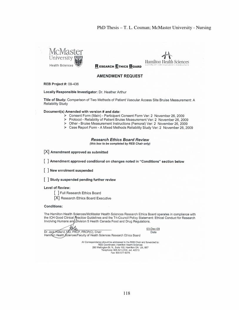
PhD Thesis – T. L. Cosman; McMaster University - Nursing
118

PhD Thesis – T. L. Cosman; McMaster University - Nursing
119

PhD Thesis – T. L. Cosman; McMaster University - Nursing
120
Appendix C
Consent Form

PhD Thesis – T. L. Cosman; McMaster University - Nursing
121
PARTICIPANT INFORMATION SHEET
Title of Study: Comparison of Two Methods of Patient Vascular Access Site Bruise
Measurement: A Mixed Methods Reliability Study
Study Sponsor:
HSFO/Michael G. DeGroote Endowed Chair in Cardiovascular Nursing Principal Investigator:
Tammy Cosman RN(EC), MN, Advanced Practice Nurse Surgical Oncology & Orthopedics Program, Hamilton Health Sciences, Henderson Site
You are being invited to participate in a research study conducted by Tammy Cosman RN(EC), MN, PhD student. This is a student research project conducted under the supervision of Dr. H. Arthur RN, PhD. The study will help the student learn more about the topic area and develop a measurement tool which may be used in future studies requiring bruise measurement. In order to decide whether or not you want to be a part of this research study, you should understand what is involved and the potential risks and benefits. This form gives detailed information about the research study, which will be discussed with you. Once you understand the study, you will be asked to sign this form if you wish to participate. Please take your time to make your decision. Feel free to discuss it with your friends and family. WHY IS THIS RESEARCH BEING DONE?
Having a cardiac catheterization (CATH) or percutaneous coronary intervention (PCI) requires the doctor to place a small tube in an artery that leads to the heart. Occasionally patients develop complications where the tube was placed. The most common problem seen by patients is a bruise in that area. Many patients get bruising after they are sent home from the hospital. There is no standard tool used for measuring bruises that happen when the patient gets home. Research to decrease bruising is difficult without a standard way to measure bruises. This study will compare how patients and nurses measure bruises in two different ways.

PhD Thesis – T. L. Cosman; McMaster University - Nursing
122
Patients will tell us how easy it is to measure bruises with each method. The impact of bruising following CATH or PCI on patients is not clear. This study will explore how bruising impacts your ability to perform daily activities and how you perceive the bruise.
WHAT IS THE PURPOSE OF THIS STUDY? This study will give us information about two different ways to measure bruises at the wrist or groin site. This study will also help us determine if patients have the ability to measure and report bruise size. This research may support the use of a standard tool for patient bruise measurement. Understanding the impact on patients of bruising that develops after hospital discharge will expand our understanding of how patients cope with bruising once at home.
WHAT WILL MY RESPONSIBILITIES BE IF I TAKE PART IN THE STUDY? If you volunteer to participate in this study, you will be asked to do the following
things: The study investigator/research assistant will approach you to talk about your participation in this study. The study will be explained to you in detail, and you will have an opportunity to ask any questions. If you are eligible for the study and you wish to participate, you will be asked to sign a consent form. If you chose not to participate in the study you may be provided with a study information/contact sheet should you decide to participate at a later time. Your care will not change in any way if you chose to participate in this study. You will be contacted by telephone after your procedure. If you have no bruising, nothing further will be requested of you. If you have any bruising at all you will be asked to return to the hospital for a follow up visit. If you are returning to the hospital for bruise measurement you will be given directions to an outpatient area where you will be met by the study investigator or research assistant. If are returning to the hospital for a pre-booked follow up appointment by staff in the Heart Investigation Unit you may be given a study information sheet by a nurse. If you agree, you will be contacted by the study investigator/research assistant regarding your participation in this study. If you choose to participate, the investigator/research assistant will explain the study in detail and have you sign the consent form at the time of your scheduled appointment. The study will be completed at the end of your pre-booked clinic visit and will take no longer than 30 minutes. You will be given written instructions (with diagrams) showing you how to measure any bruising at your access site (either wrist or groin). You will be asked to measure the bruise using two different methods. One method will use a measuring tape; the second method involves tracing the outline of the bruise onto a clear plastic film. This will occur in a private exam room. Following your measurement both a research assistant and the study investigator, an Advanced Practice Nurse, will repeat the bruise measurements using both methods. Before you leave you will asked to complete a 5-minute

PhD Thesis – T. L. Cosman; McMaster University - Nursing
123
questionnaire regarding the two measurement tools. This entire visit should take no longer than 30 minutes. If you experience bruising you may be asked to provide some further information about how this bruising has, or has not, affected you and the quality of your home and work life. An interview with the study investigator will be conducted following the bruise measurement. The interview will be tape recorded for research purposes. The tape recordings will only be heard by the study investigator, research assistant and transcriptionist. The interview should take no longer than 30 minutes. You may choose to not answer any of the questions and remain in the study. Your name will not be used in any documents related to this study. You will have an assigned number so that all the information you give us remains anonymous and confidential.
WHAT ARE THE POSSIBLE RISKS AND DISCOMFORTS? There are no known risks associated with this research. All other aspects of your care remain unchanged. If you choose to take part in this study, you will be told about any new information which might affect your willingness to continue to participate in this research.
HOW MANY PEOPLE WILL BE IN THIS STUDY?
There will be approximately 50 people enrolled in this study.
WHAT ARE THE POSSIBLE BENEFITS FOR ME AND/OR FOR SOCIETY? We cannot promise any personal benefits to you from your participation in this study. However, your participation may help other people in the future who are having cardiac procedures. Having a standard method for bruise measurement, done by patients, will help with future research to decrease the amount of bruising after cardiac procedures.
IF I DO NOT WANT TO TAKE PART IN THE STUDY, ARE THERE OTHER
CHOICES?
It is important for you to know that you can choose not to take part in the study. Choosing not to participate in this study will in no way affect your care or
treatment.
WHAT INFORMATION WILL BE KEPT PRIVATE? Your data will not be shared with anyone except with your consent or as required by law. All personal information such as your name, address, phone number will be removed from the data and will be replaced with a study number. A list linking the number with your name will be kept in a secure place, separate from your file. The data, with identifying information removed, will be securely stored in a locked office. The data for this research study will be retained for a maximum of 10 years.

PhD Thesis – T. L. Cosman; McMaster University - Nursing
124
For the purposes of ensuring the proper monitoring of the research study, it is possible that a member of the Hamilton Health Sciences/FHS McMaster University Research Ethics Board may consult your research data and medical records. However, no records, which identify you by name or initials, will be allowed to leave the hospital. By signing this consent form you authorize such access.
If the results of the study are published, your name will not be used and no information that discloses your identity will be released or published without your specific consent to the disclosure. However, it is important to note that this original signed consent form and the data, which follows, may be included in your health record. CAN PARTICIPATION IN THE STUDY END EARLY?
If you volunteer to be in this study, you may withdraw at any time and this will in no way affect the quality of care you receive at this institution. You may also refuse to answer any questions you don’t want to answer and still remain in the study. The investigator may withdraw you from this research if circumstances arise which warrant doing so. WILL I BE PAID TO PARTICIPATE IN THIS STUDY? If you agree to take part in this study you will be provided with a parking voucher for the hospital parking ramp to cover the parking costs associated with your return visit to the hospital. You will also receive $50 to cover the transportation costs associated with your return to the hospital. WHAT HAPPENS IF I HAVE A RESEARCH-RELATED INJURY?
It is highly doubtful that you will experience a research-related injury in this particular study. However, if you are injured as a direct result of taking part in this study, all necessary medical treatment will be made available to you at no cost. Financial compensation for such things as lost wages, disability or discomfort due to this type of injury is not routinely available. However, if you sign this consent form it does not mean that you waive any legal rights you may have under the law, nor does it mean that you are releasing the investigator(s), institution(s) and/or sponsor(s) from their legal and professional responsibilities. IF I HAVE ANY QUESTIONS OR PROBLEMS, WHOM CAN I CALL?
If you have any questions about the research now or later, or if you think you have a research-related injury, please contact Dr. Heather M. Arthur, McMaster University, 905-525-9140, Ext. 22270. If you have any questions regarding your rights as a research participant, you may contact the Office of the Chair of the Hamilton Health Sciences / Faculty of Health Sciences Research Ethics Board at 905-521-2100, ext. 42013.

PhD Thesis – T. L. Cosman; McMaster University - Nursing
125
CONSENT STATEMENT
SIGNATURE OF RESEARCH PARTICIPANT/LEGALLY-AUTHORIZED REPRESENTATIVE
I have read the preceding information thoroughly. I have had the opportunity to
ask questions, and all of my questions have been answered to my satisfaction. I
agree to participate in this study. I understand that I will receive a signed copy of
this form.
______________________________________ Name of Participant ________________________________________ ______________ Signature of Participant or Legally Authorized Representative Date
Consent form administered and explained in person by:
_____________________________________ Name and title _____________________________________ ______________ Signature Date

PhD Thesis – T. L. Cosman; McMaster University - Nursing
126
Appendix D
Study Information Sheet

PhD Thesis – T. L. Cosman; McMaster University - Nursing
127
BRUISE MEASUREMENT STUDY
Patients having an angiogram or angioplasty may develop bruising in their wrist or groin area after they are discharged from the hospital. There is no standard way for patients to report the size of the bruising once they are at home. This study will test two bruise measurement tools for patients to use at home.
• You will be asked to return to the hospital one time for bruise measurement 3-10 days after your procedure
• You will be given instructions on how to measure your bruise in a private exam room
• An Advanced Practice Nurse and research assistant will also measure your bruise
• You may be asked to participate in a interview after you measure your bruise
• You will receive a parking pass and $50 for your participation in this study
If you experience a bruise following your angiogram or angioplasty, and are able to return to the hospital for bruise measurement on: _______________________________________________________ Please contact:
Tammy Cosman RN (EC), MN Principal Investigator Phone: 905 527-4322 extension - 43894 You will receive a call from the principal investigator or research assistant to explain the study in more detail.
What is the Bruise Measurement Study?
What is involved?
How can I help?

PhD Thesis – T. L. Cosman; McMaster University - Nursing
128
Appendix E
Case Record Form

PhD Thesis – T. L. Cosman; McMaster University - Nursing
129
Comparison of Two Methods of Patient Vascular Access Site Bruise Measurement:
A Mixed Methods Reliability Study
CASE RECORD FORM Study ID: _____
Procedure Date (dd/mm/yyyy): _____________________
Telephone Follow up Date (dd/mm/yyyy): ____________________
DEMOGRAPHICS: DOB (dd/mm/yyyy): __________________
Sex: 01 Male 02 Female
Highest Level of Education Achieved: 01 less than grade 8 02 completed grade 8 03 completed high school 04 completed College/University Undergraduate 05 completed Graduate education
Anthropometrics: Height (cm): _____ Weight (kg): _____ PROCEDURE INFORMATION:
Procedure Completed 01 Cardiac Catheterization 02 Percutaneous Coronary Intervention
Vascular Access Location (Arterial) 01 Right Femoral Artery 02 Left Femoral Artery 03 Right Radial Artery 04 Left Radial Artery
Sheath Size: 01 6 French 02 7 French 04 Other
Vascular Closure Device used (Femoral only) 01 Yes 02 No

PhD Thesis – T. L. Cosman; McMaster University - Nursing
130
Study ID: ____
Follow up Visit – VAS Bruise Measurement: Follow up Visit Date (dd/mm/yyyy): ________________
Patient Measurements: 01 Self Measurement 02 Assisted Measurement
Linear Method: Length (cm) ___________ Width (cm) ____________ Tracing Method: Number of FULL squares in tracing perimeter: ____________________ Number of PARTIAL squares in tracing perimeter: _________________

PhD Thesis – T. L. Cosman; McMaster University - Nursing
131
Appendix F
Participant Instruction sheet – Femoral

PhD Thesis – T. L. Cosman; McMaster University - Nursing
132

PhD Thesis – T. L. Cosman; McMaster University - Nursing
133

PhD Thesis – T. L. Cosman; McMaster University - Nursing
134

PhD Thesis – T. L. Cosman; McMaster University - Nursing
135
Appendix G
Participant Instruction Sheet – Radial
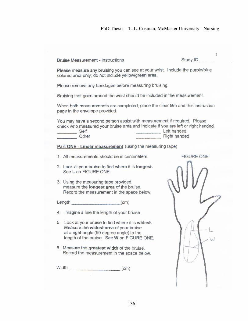
PhD Thesis – T. L. Cosman; McMaster University - Nursing
136

PhD Thesis – T. L. Cosman; McMaster University - Nursing
137

PhD Thesis – T. L. Cosman; McMaster University - Nursing
138
Appendix H
Bruise Measurement Questionnaire

PhD Thesis – T. L. Cosman; McMaster University - Nursing
139
Thank you for your involvement in this study. Your help is appreciated with this important research. Please take the time to complete both pages of this questionnaire about the two ways to measure bruising. Linear measurement (using a measuring tape) On a scale of 1-5 please rate each question. 1 = very easy 5 = very hard
1. How easy was it to figure out the instructions about how to use the tape measure?
1 = very easy 5 = very hard 1 2 3 4 5
2. How easy was it to measure the bruise using the tape measure?
1 = very easy 5 = very hard 1 2 3 4 5
3. Did you have any problems measuring your bruise using the tape measure? Please describe below.
_____________________________________________________________
_____________________________________________________________
_____________________________________________________________
_____________________________________________________________
_____________________________________________________________

PhD Thesis – T. L. Cosman; McMaster University - Nursing
140
-2-
Bruise outline measurement (tracing method) On a scale of 1-5 please rate each question. 1 = very easy 5 = very hard
4. How easy was it to figure out the instructions about how to trace the bruise?
1 = very easy 5 = very hard 1 2 3 4 5
5. How easy was it to measure the bruise using the tracing method?
1 = very easy 5 = very hard 1 2 3 4 5
6. Did you have any problems measuring your bruise by tracing the outline? Please describe below.
______________________________________________________________________________________________________________________________________________________________________________________________________________________________________________________________________________________________________________________________________________________________________________
Did you prefer to use: □ the measuring tape or
□ tracing the outline of the bruise?
_____________________________________________________________
_____________________________________________________________
_____________________________________________________________
Thank you for your participation in this study!

PhD Thesis – T. L. Cosman; McMaster University - Nursing
141
Appendix I
Interview Participant Characteristics and Bruise Size

PhD Thesis – T. L. Cosman; McMaster University - Nursing
142
Interview Participant Characteristics and Bruise Size
Study
ID Sex Site
Age
Bruise
Size
(cm2)
Procedure
Completed
Prior
Procedure
131 M F 56 39 Cath Yes
138 M F 74 49.5 Cath Yes
146 F F 72 49.5 PCI Yes
157 F R 34 12 Cath No
161 F F 65 108.5 Cath Yes
169 M F 55 957 Primary
PCI No
170 F F 69 165 Cath No
174 F F 59 912 Cath No
186 M F 62 63.25 Cath No
188 M R 69 108.75 PCI No
Note: Sex M = male, F = female; Site F = femoral, R = radial, Procedure Completed Cath = cardiac catheterization, PCI = percutaneous coronary intervention, Primary PCI = primary percutaneous coronary intervention

PhD Thesis – T. L. Cosman; McMaster University - Nursing
143
Appendix J
Interview Guide

PhD Thesis – T. L. Cosman; McMaster University - Nursing
144
1. Can you tell me about the bruise at your (name specific) site?
Probe: How did you notice that you had a bruise? When did you notice and what made you notice the bruise?
Does the bruise bother you in any way? What are your thoughts about the bruising? Have you ever had this type of procedure and developed a bruise in the past? How has this influenced what you think about this bruise?
Added after seventh interview:
What does a bruise mean to you – what do you think causes it?
2. What action(s) did you take, if any, once you noticed the bruise forming?
Probe: At what point would the bruise have been bad enough to do something? How bad would the bruise have to be before you would seek advice from a health care professional (GP, Emergency dept, other)?
3. Please describe any symptoms or concerns you have related to your bruise.
Probe: Have you had any pain or discomfort at your bruise site? Have you noticed any other symptoms?
4. Can you describe what you have done/will do to deal with this (name symptom)?
Probe: Did this work? What other things did you try? What are you thinking of trying?
5. How has this bruise affected your ability to perform your normal activities? Probe: How has the bruise limited any of your work, home or recreational activities?
6. What did your family members or friends think about the bruise? Probe: Did you ask any family or friends what they thought you should do?
Did any of your family or friends suggest things you should do? Did any family or friends encourage you to seek medical attention?

PhD Thesis – T. L. Cosman; McMaster University - Nursing
145
Added after first interview following discussion with committee member:
Do you remember being told what to expect at your (wrist/groin) site after
discharge?
What were you told to expect regarding bruising? Anything else
regarding the access site?
7. If you were helping someone prepare for this procedure, what would you tell them
about bruising? 8. Do you have anything to add?

PhD Thesis – T. L. Cosman; McMaster University - Nursing
146
Appendix K
Contact Summary Sheet
PhD Thesis – T. L. Cosman; McMaster University - Nursing
147
Questions to pose to self following interview:
Main issues or themes that struck you? Summarize information obtained for each question New or remaining questions Anything that struck you as important Am I asking the right questions to get the information I want?

PhD Thesis – T. L. Cosman; McMaster University - Nursing
148
Appendix L
Planimetry Measurement Results - PI and RA Measures

PhD Thesis – T. L. Cosman; McMaster University - Nursing
149
Planimetry Measurement Results – PI and RA Measures
Study ID number
PI measures patient Planimetry
RA measures patient Planimetry
PI measures RA Planimetry
RA measures RA Planimetry
PI measures PI Planimetry
RA measures PI Planimetry
174 433 462 469.5 696 616.5 747
165 419.5 472.5 215 213.5 294 387
168 385 380.5 477 457.5 502.5 383
155 293.5 612 355 397 532.5 540
140 185 183 240.5 241 375 248.5
170 156.5 77.5 85 81 88.5 88
169 133.5 143 134.5 130.5 171 171
134 86.5 74.5 85.5 96 91 85.5
150 79.5 72 68 72.5 73.5 70
188 73 68 40 35 78 77.5
135 67.5 61.5 93 87.5 74 68
161 63 59 56 56.5 56 60.5
116 55 45 142 137.5 137.5 129.5
207 50 46.5 61.5 56.5 54 48
107 48 47 51 45.5 52 54
128 39 36.5 54 50 41.5 37
138 37.5 36 23 24 29.5 25.5
186 34.5 32 32.5 31 37 36
146 34 34 18 17 28.5 26
159 28.5 27.5 23.5 22.5 34 31.5
Note: PI Principle Investigator, RA Research Assistant

PhD Thesis – T. L. Cosman; McMaster University - Nursing
150
Planimetry Measurement Results – PI and RA Measures (continued)
Study ID number
PI measures patient Planimetry
RA measures patient Planimetry
PI measures RA Planimetry
RA measures RA Planimetry
PI measures PI Planimetry
RA measures PI Planimetry
148 28 25.5 19 18.5 31.5 28.5
108 27 26 27 25 7.5 6
190 26.5 26.5 38 35 30 29.5
126 22 19 26 23 16.5 15.5
177 21 20.5 35.5 36.5 28 29
185 19 19 22 23 24 21
127 18.5 17 41.5 37 23 17
162 18.5 18.5 15 15.5 11.5 11.5
100 18 18 20 20 13 13
121 16 13.5 21 20.5 12 13
119 12.5 6 26.5 26 29.5 28
145 11.5 10 12 10 7 4.5
178 10.5 8 40 40 47.5 41
144 10 10.5 8.5 7.5 40.5 37
131 8.5 6.5 14.5 13.5 12 10.5
106 7.5 7.5 13.5 11 10.5 11
157 0.5 0.5 6.5 5.5 5 3.5
105 30 29.5 31 27
182 31 30.5 31 30.5
1211 22.5 23.5 17.5 16.5
Note: PI Principle Investigator, RA Research Assistant