vascular tumors of the head and neck - welcome to utmb ... · pdf filevascular tumors of the...
TRANSCRIPT

Vascular Tumors of the Head and Neck
Russell D. Briggs, M.D.
Faculty Advisor: Anna M. Pou, M.D.
The University of Texas Medical Branch
Department of Otolaryngology
Grand Rounds Presentation
April 2001

Introduction
Many different types of neoplasms
Share common etiology with vascular system
Benign, malignant, others

Benign Tumors
Vascular Birthmarks
Hemangiomas
Vascular Malformations
Nasopharyngeal angiofibromas

Vascular Birthmarks
History

Vascular Birthmarks
Classification system
Hemangioma vs. malformation
Based on clinical, cellular, biologic factors
Older terms – “capillary”, “juvenile”, “strawberry”, “cavernous”
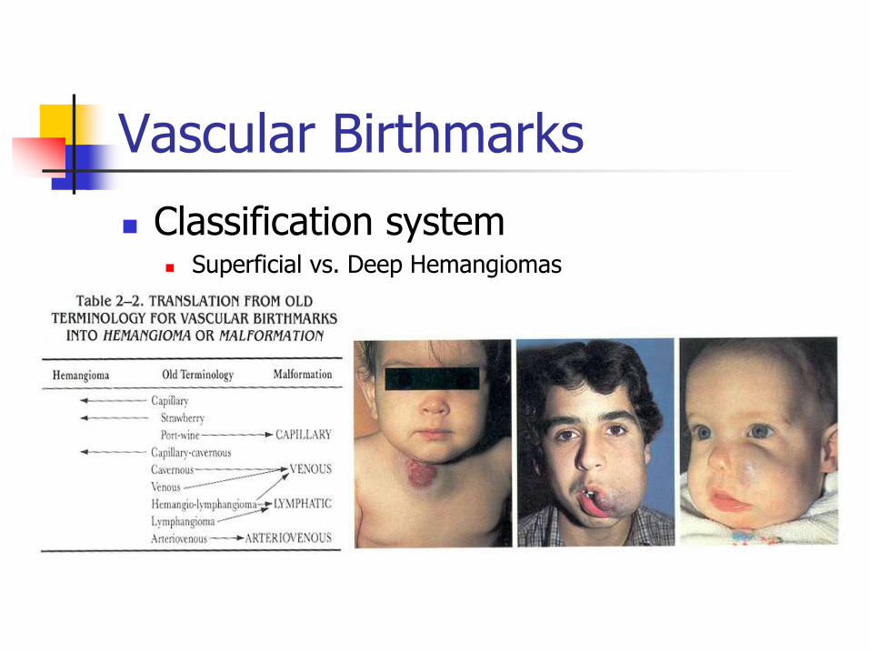
Vascular Birthmarks
Classification system Superficial vs. Deep Hemangiomas
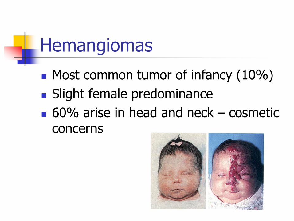
Hemangiomas
Most common tumor of infancy (10%)
Slight female predominance
60% arise in head and neck – cosmetic concerns

Hemangiomas
Clinical presentation for diagnosis
Not seen at birth
Precursor lesion
Proliferative phase
Involution phase
Superficial vs. deep

Complications from Hemangiomas
Occur in 20%
Ulceration
Compression of vital structures
High-output cardiac failure
Bleeding
Kasabach-Merritt syndrome

Laryngeal Hemangiomas
Usually in the subglottis
Healthy infant with biphasic stridor (croup)
Behave similarly
50% with cutaneous counterpart

Diagnosis of Hemangiomas
History and physical examination
Certain cases – radiology
Ultrasound, MRI
Large facial hemangiomas– Dandy-Walker

Treatment of Hemangiomas
Why and when to treat?
Normal skin in 50% that involute within 5 years
Other 50%-- 80% substantial deformity
Pro’s and Con’s

Treatment of Hemangiomas
Observation Serial photography important to document
involution
Regular visits with reassurance
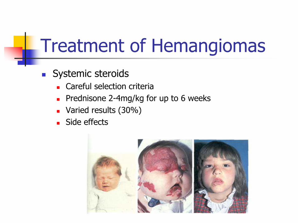
Treatment of Hemangiomas
Systemic steroids
Careful selection criteria
Prednisone 2-4mg/kg for up to 6 weeks
Varied results (30%)
Side effects

Treatment of Hemangiomas
Intralesional steroids
Usually for vision threatening lesions
Combination of beta-methasone and triamcinolone

Treatment of Hemangiomas
Pulse-dye lasers
Useful for superficial variety
Good for ulcerations/residual cosmesis

Treatment for Hemangiomas
Surgery
Eyelid lesions, bulky lesions, vermillion border, nasal tip, eyebrow
CO2 laser for subglottis

Treatment of Hemangiomas
Arterial embolization
Radiation therapy
Alpha-2b interferon

Vascular malformations
Capillary, venous, arterial, lymphatic, mixed
By definition– present at birth
No proliferative or involution phase
Commensurate growth
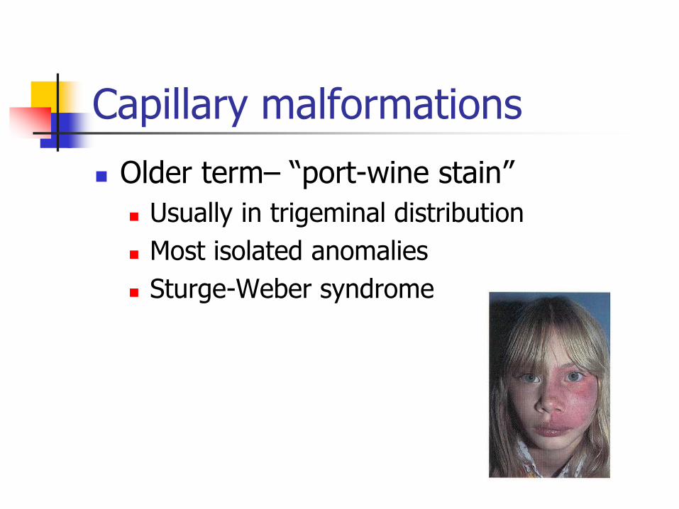
Capillary malformations
Older term– “port-wine stain”
Usually in trigeminal distribution
Most isolated anomalies
Sturge-Weber syndrome
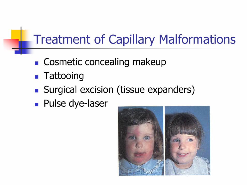
Treatment of Capillary Malformations
Cosmetic concealing makeup
Tattooing
Surgical excision (tissue expanders)
Pulse dye-laser

Venous Malformations
Diagnosis is clinical – palpation
Treatment dependent on location (surgery and sclerotherapy)
Lymphatic malformations
Older terms– “cystic hygroma”, “lymphangioma”
Can expand with URI
Surgical treatment is mainstay
Picibanil (OK-432)

Arteriovenous malformations
Usually clinically apparent
Embolization and surgical resection
Nasopharyngeal Angiofibroma
Most common benign tumor of nasopharynx
Older term– “juvenile nasopharyngeal angiofibroma”, “JNA”
Presentation: recurrent epistaxis/nasal congestion, hearing loss, orbital, CN
Arise where sphenoidal process of palatine bone meets horizontal ala of vomer

Nasopharyngeal Angiofibroma
Diagnosis is made by clinical and radiographic findings
CT/MRI
Biopsy- rarely indicated
Angiography

Nasopharyngeal Angiofibroma
Angiography Histology
Nasopharyngeal Angiofibroma
Treatment Embolization and surgery
Autologous blood/Cell Saver
Approaches Transnasal endoscopic, lateral rhinotomy/MFD with
medial maxillectomy or LeFort I, transpalatal, facial translocation/maxillary swing, infratemporal approaches, craniotomy
Radiation therapy
Chemotherapy
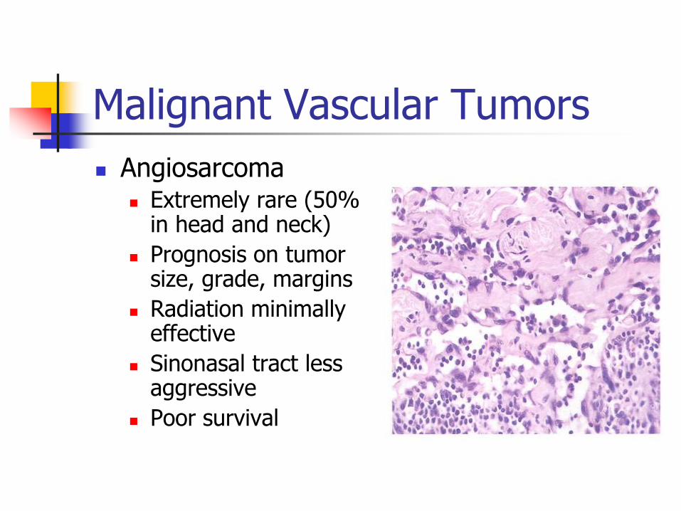
Malignant Vascular Tumors
Angiosarcoma Extremely rare (50%
in head and neck)
Prognosis on tumor size, grade, margins
Radiation minimally effective
Sinonasal tract less aggressive
Poor survival

Malignant Vascular Tumors
Hemangiopericytoma
Pericytes of Zimmerman
25% in head and neck
Surgical treatment
Grade important on prognosis
Radiation/chemotherapy for selected cases

Malignant Vascular Tumors
Kaposi’s Sarcoma
Viral-induced
Four entities
Classic
Endemic
Immunosuppressed
AIDS-related
Surgery, chemo, radiation, sclerotherapy

Paragangliomas
Named for anatomic location
Arise in paraganglionic tissue (neural crest)
Type I cells (chief) – APUD cells – catecholamines
Type II cells (sustentacular)
Clusters together– “Zellballen”
Malignancy is clinical
Carotid paragangliomas
Most common of head and neck (60%)
Multicentric 10%, malignant 10%
Familial (AD) 20% -- more multicentric
Painless mass at SCM, immobile superior-inferior direction
May produce catecholamines

Carotid paragangliomas

Carotid paragangliomas
Treatment
Surgery
Mortality 8%, >5cm tumors had more complications
Preop workup key – vascular surgeon, anesthesia
Embolization – controversial
Radiation

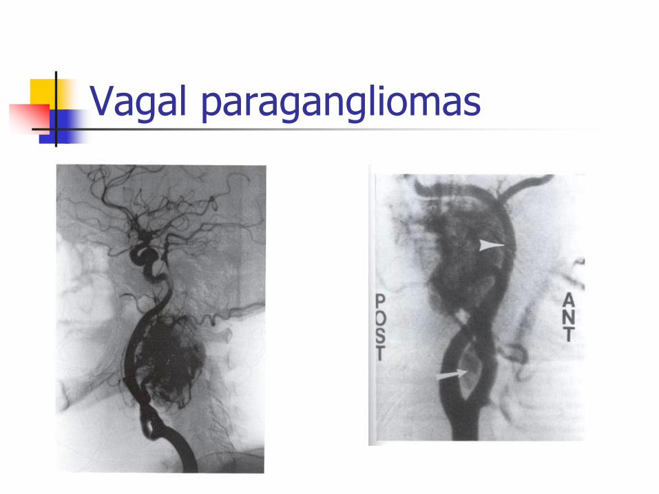
Vagal paragangliomas
Most commonly at nodose ganglion
Painless mass at angle of mandible present for many years – enlarging may get Horner’s, CN XII, hoarseness
More multicentric (25%)
Malignancy (18%)
None produce catecholamines
Vagal paragangliomas

Laryngeal paragangliomas
Usually from superior laryngeal paraganglia from aryepiglottic fold
Hoarseness and dysphagia common
High rates of malignancy
Wide local excision or partial laryngectomy