embryology of the neck & neck masses steven t. wright, m.d. shawn newlands, m.d., ph.d, m.b.a...
TRANSCRIPT

Embryology of the Neck & Neck Masses
Steven T. Wright, M.D.Shawn Newlands, M.D., Ph.D,
M.B.AUTMB Dept of Otolaryngology
Grand Rounds June 8, 2005
Neck Masses
A mass in the neck is a common clinical finding.
Benign NeoplasmMalignant NeoplasmInfectiousCongenital

Neck Masses
An appreciation for the embryological development of the cervical structures must be made to competently understand and treat the disorders of the neck.
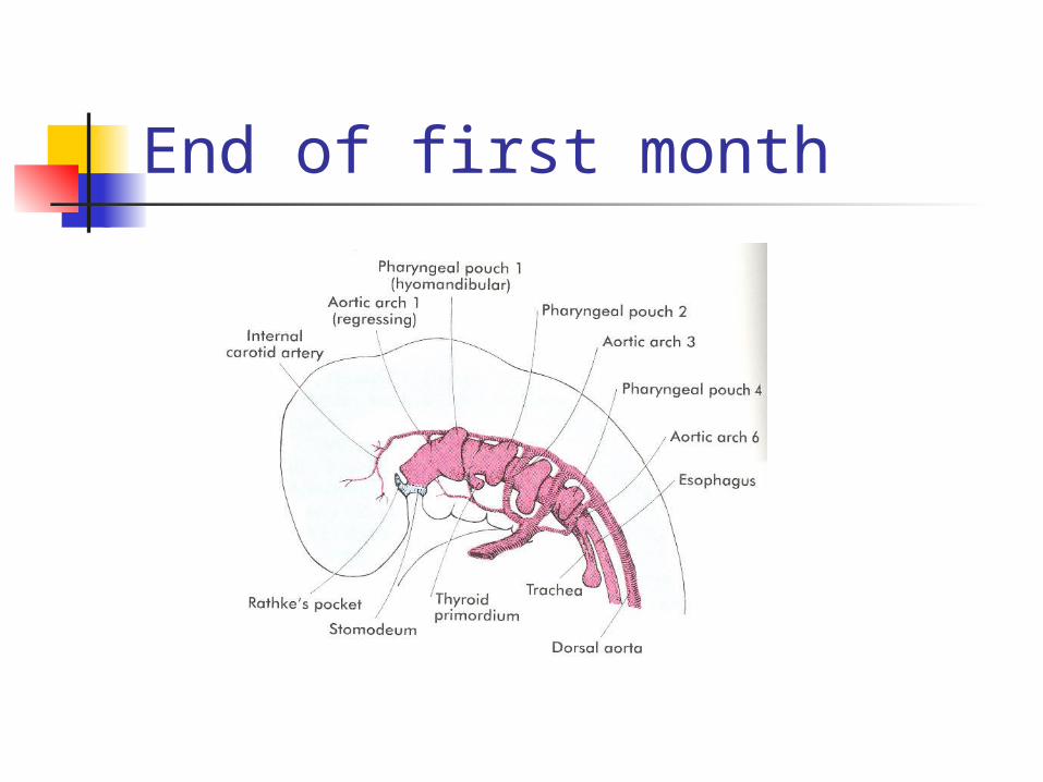
End of first month
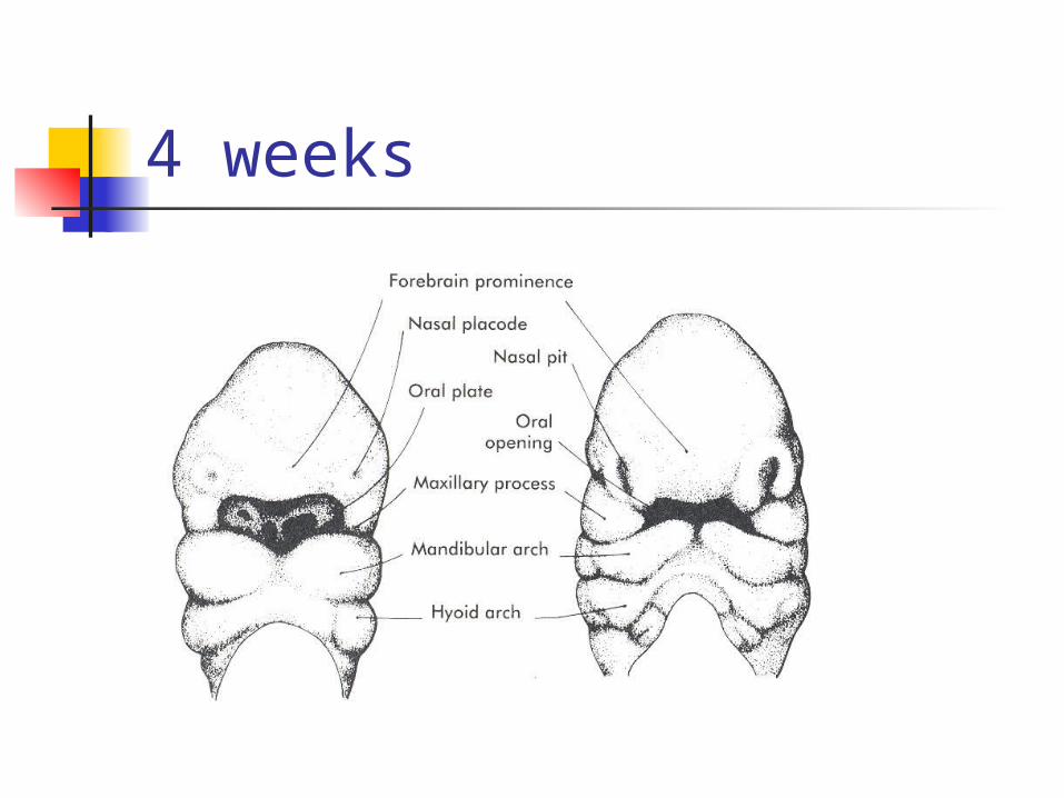
4 weeks
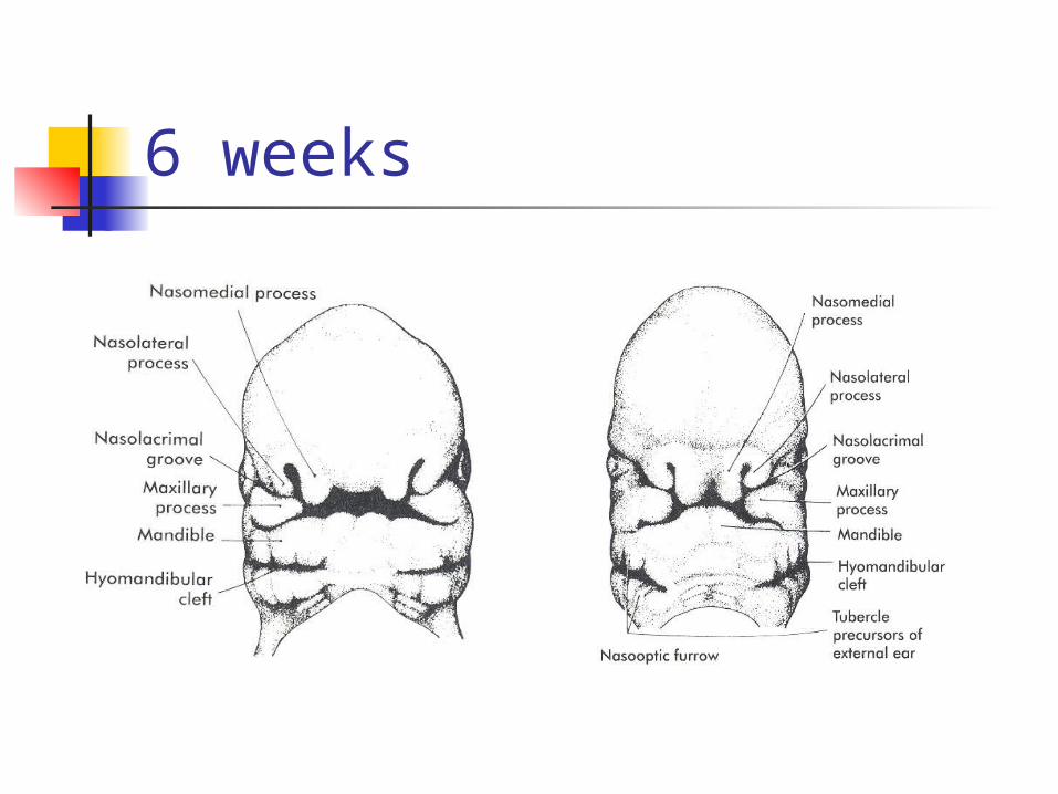
6 weeks
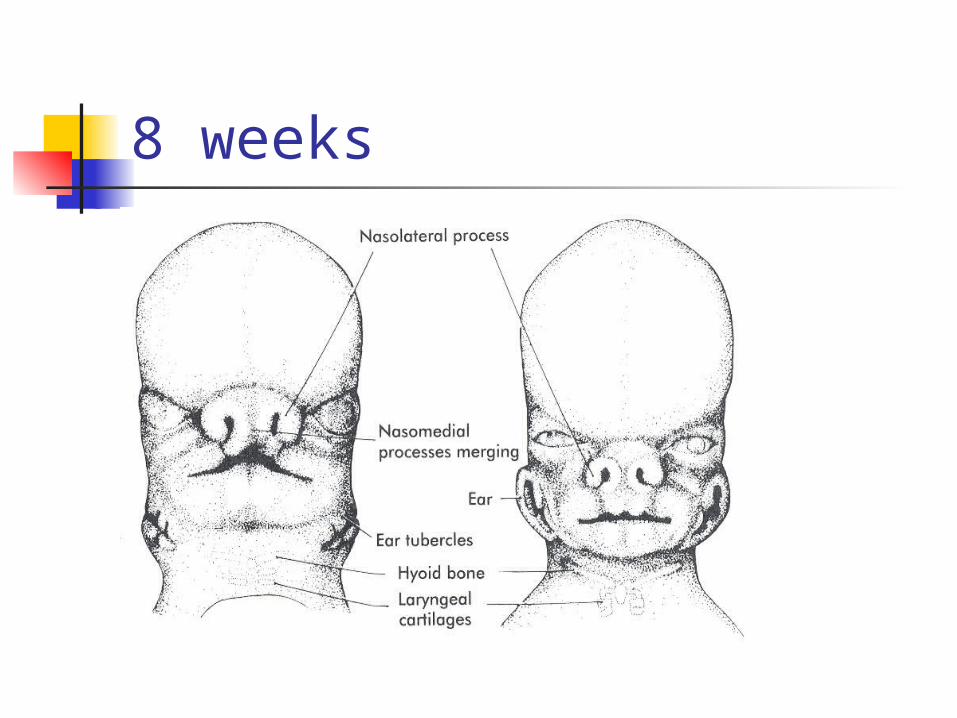
8 weeks
Embryology and Anatomy
Branchial System- 6 pairs of pharyngeal arches separated by endodermally lined pouches and ectodermally lined clefts.
Each arch consists of a nerve, artery, and cartilaginous structures.
The remaining neck musculature gains contributions from cervical somites.

Branchial system First Branchial arch
Maxillary and mandibular (Meckel’s) process regress to leave the malleus and incus.
Ossification around Meckel’s cartilage gives rise to the mandible, sphenomandibular ligament, and anterior malleolar ligaments.
Muscles- temporalis, masseter, pterygoids, mylohyoid, ant belly of digastric, tensor tympani, tensor veli palatini
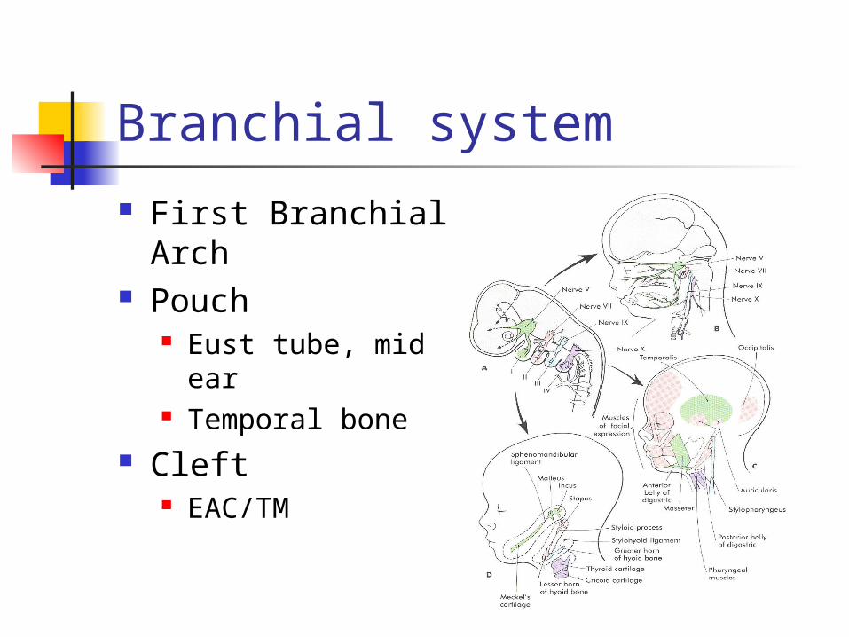
Branchial system First Branchial
Arch Pouch
Eust tube, mid ear Temporal bone
Cleft EAC/TM

Branchial system
Second Branchial Arch Reichert’s cartilage contributes to the
superstructure of the stapes, the upper body and lesser cornu of the hyoid, the styloid process and stylohyoid ligament.
Muscles- platysma, muscles of facial expression, posterior belly of digastric, stylohyoid, and stapedius
Nerve- 7th cranial nerve Artery- stapedial artery
Branchial system
Third Branchial Arch Lower body of the hyoid and greater
cornu. Muscles- stylopharyngeus, superior
and middle pharyngeal constrictors. Nerve- 9th cranial nerve Artery- common carotid and proximal
portions of the internal and external carotid.
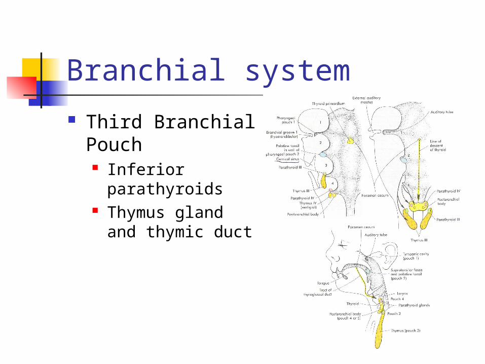
Branchial system Third Branchial
Pouch Inferior
parathyroids Thymus gland and
thymic duct
Branchial system Fourth and Sixth Branchial arches fuse
to form the laryngeal cartilages. Fourth Arch
Muscles- cricothyroid, inferior pharyngeal constrictors
Nerve- Superior Laryngeal Nerve Artery- Right Subclavian, Aortic arch
Fourth Pouch- superior parathyoid glands and parafollicular thyroid cells
Branchial system
Sixth Branchial Arch Muscles- remaining/intrinsic laryngeal
musculature Nerve- Recurrent Laryngeal Nerve Artery- Pulmonary Artery and ductus
arteriosus
Branchial system
Epipericardial ridge- mesodermal elements of the sternocleidomastoid, trapezius, and lingual and infrahyoid musculature. Nerve- hypoglossal and spinal
accessory nerve Cervical Sinus of His
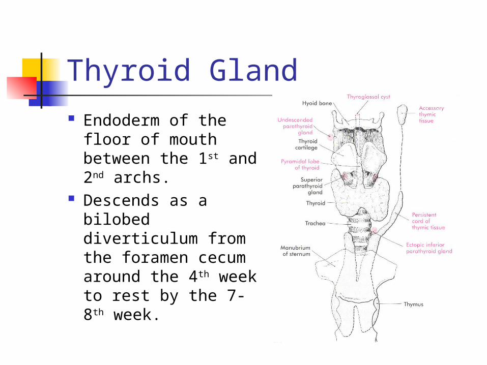
Thyroid Gland Endoderm of the
floor of mouth between the 1st and 2nd archs.
Descends as a bilobed diverticulum from the foramen cecum around the 4th week to rest by the 7-8th week.

Oral Cavity
Neck Masses Midline Neck Masses
Thyroid nodules Cervical Lymphadenopathy Thyroglossal Duct cyst Thymus gland anomalies Plunging ranula
Lateral Neck Masses Branchial cleft anomalies Laryngoceles Dermoid and Teratoid Cysts
Midline Neck Masses
Thyroid nodules Thyroglossal duct cyst Cervical Thymic Cyst Plunging ranula
Thyroid Nodules 4% of population 1/20 will harbor Cancer H&P combined with FNA is crucial for
diagnosis FNA
Malignant Suspicious Benign Indeterminate
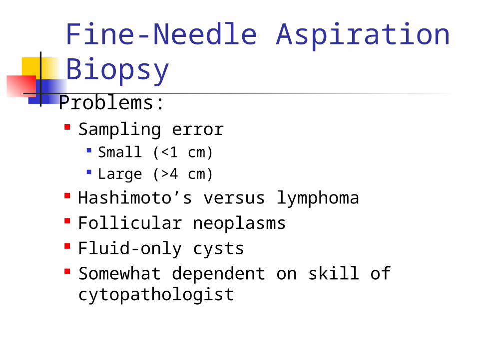
Fine-Needle Aspiration Biopsy
Problems: Sampling error
Small (<1 cm) Large (>4 cm)
Hashimoto’s versus lymphoma Follicular neoplasms Fluid-only cysts Somewhat dependent on skill of
cytopathologist
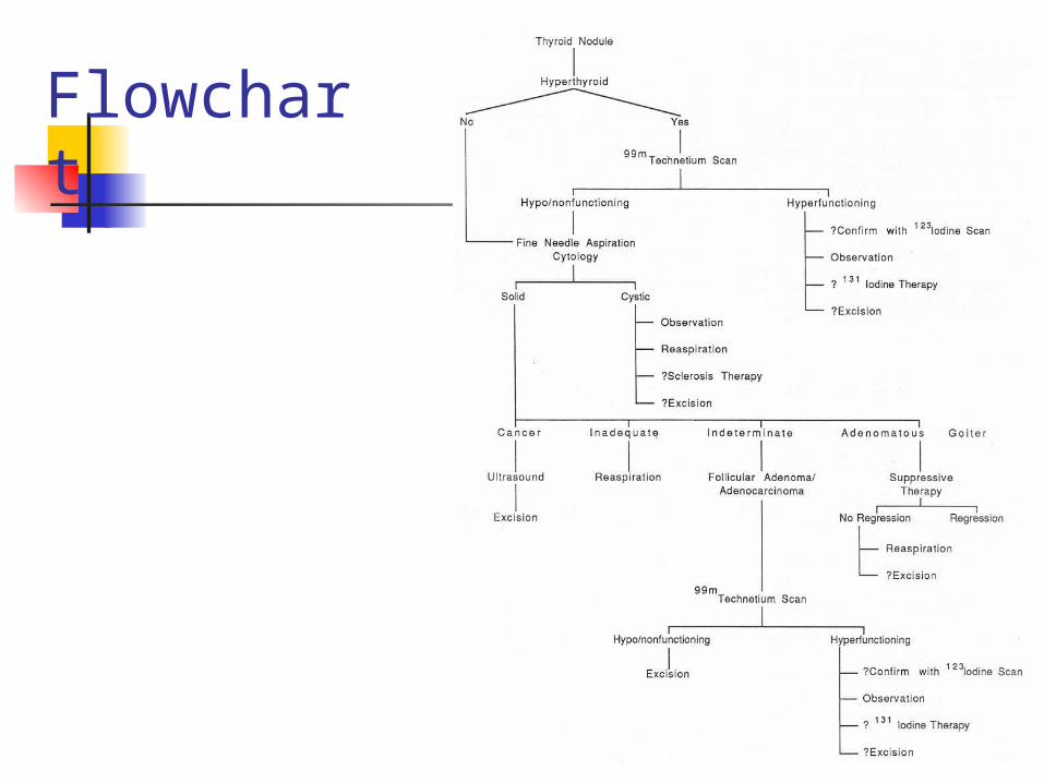
Flowchart
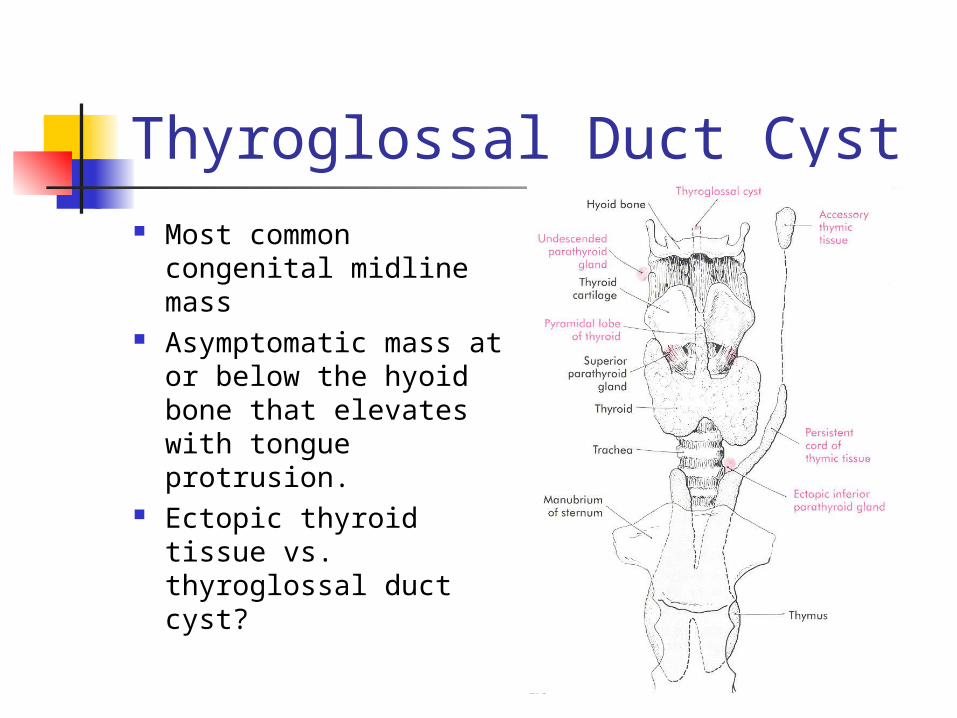
Thyroglossal Duct Cyst Most common
congenital midline mass
Asymptomatic mass at or below the hyoid bone that elevates with tongue protrusion.
Ectopic thyroid tissue vs. thyroglossal duct cyst?

Thyroglossal Duct Cyst
Thyroglossal Duct Cyst 1-2% have Ectopic Thyroid glands so
imaging is indicated to document presence of a normal or ectopic thyroid gland
Simple Excision leads to high recurrence rate
Sistrunk Procedure Patients at high risk for recurrence-
Modified Sistrunk Procedure
TGDC Carcinoma
Uncommon, 1% 94% Thyroid- Papillary 6% Squamous Cell
TGDC Carcinoma or a Metastatic Cystic Thyroid Carcinoma in a Midline Lymph node?
TGDC Carcinoma Patel et al.
“incidentally discovered, well-differentiated thyroid CA in a low risk patient (<45yrs, <4cm, no local/regional invasion) can be adequately managed by Sistrunk.
In presence of a clinically/radiographically normal thyroid.
Other Convincing evidence: Lack of Lymph tissue Presence of Columnar or Squamous epithelium
Total thyroidectomy with or without neck dissection.
Ectopic Thyroid
90% are lingual 1/3rd are hypothyroid- elevated
TSH- goiter Symptoms are of base of tongue
obstruction, dysphagia Surgical Excision

Lateral Nonmalignant Thyroid Tissue True Embyrologic rest of normal thyroid
tissue as a result a migration error or is it a metastatic well differentiated thyroid carcinoma?
ANY suspicious findings should favor a metastatic deposit rather than LNTT.
Strict criteria must be followed for LNTT: must be small, with only a few thyroid follicles no atypical nuclear features of papillary
carcinoma should be present only in the capsular region
of the node

Cervical Thymic Cysts
Failure of involution of the cervical thymopharyngeal ducts.
Firm, mobile masses found in the lower aspects of the neck.
CXR, CT scan Surgical Excision- Inferior limit of
dissection is the brachiocephalic v.
Plunging Ranula Simple ranula- unilateral oral cavity cystic
lesion. Plunging ranula- pierce the mylohyoid to
present as a paramedian or lateral neck mass.
Cyst aspirate- high protein, amylase levels CT scan/MRI Treatment is intraoral excision to include
the sublingual gland of origin.
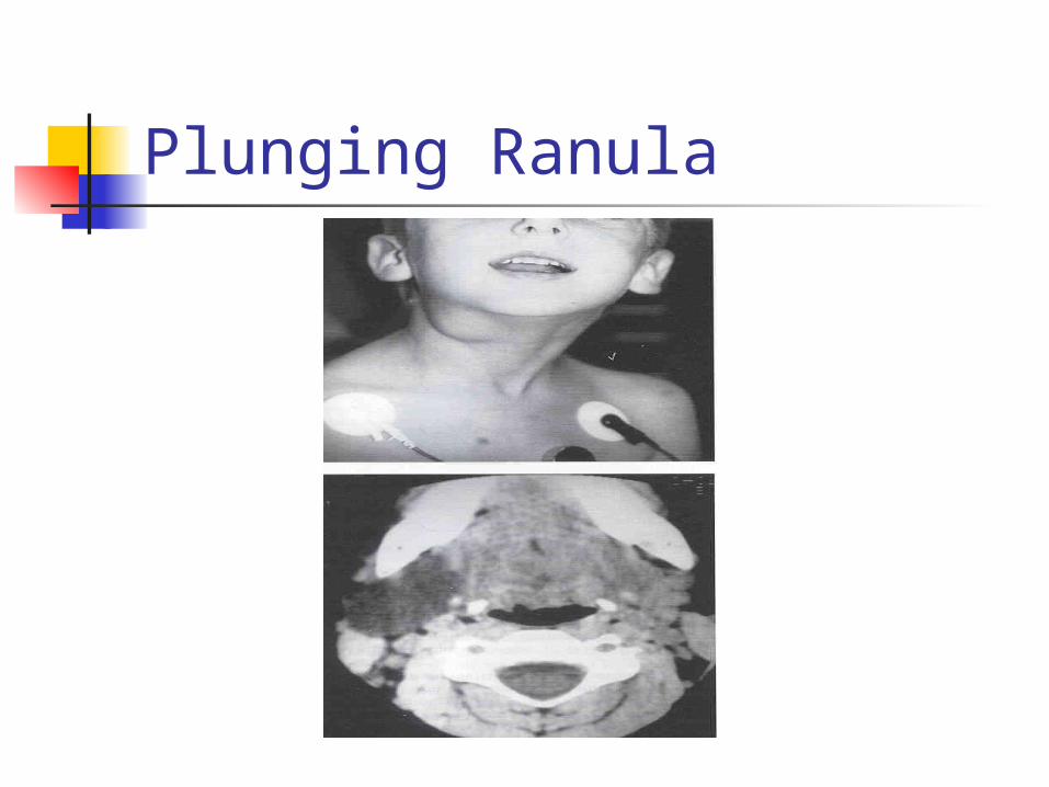
Plunging Ranula
Lateral Neck Masses
Branchial cleft anomalies Laryngoceles Dermoid and Teratoid Cysts Sternocleidomastoid Pseudotumor
of Infancy
First Branchial Cleft Cysts
Type I Ectodermal Duplication anomaly of
the EAC with squamous epithelium only.
Parallel to the EAC Pretragal, post auricular Connection with TM or Malleus>Incus Surgical Excision
First Branchial Cleft Cysts Type II
Squamous epithelium and other ectodermal components
Anterior neck, superior to hyoid bone. Courses over the mandible and through the
parotid in variable position to the Facial Nerve.
Terminates near the EAC bony-cartilaginous junction.
Surgical excision- superficial parotidectomy

First Branchial Cleft Cysts

Second Branchial Cleft Cysts Most Common (90%) branchial anomaly Painless, fluctuant mass in anterior
triangle Inferior-middle 2/3 junction of SCM, deep
to platysma, lateral to IX, X, XII, between the internal and external carotid and terminate in the tonsillar fossa
Surgical treatment may include tonsillectomy
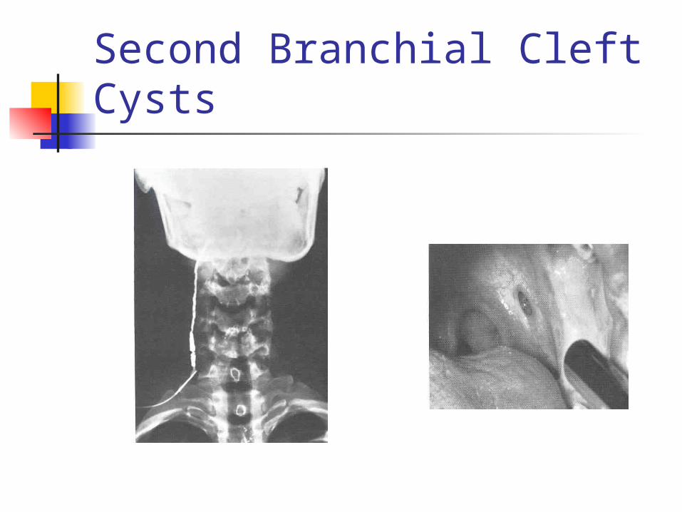
Second Branchial Cleft Cysts
Third Branchial Cleft Cysts Rare (<2%) Similar external presentation to 2nd BCC Internal opening is at the pyriform sinus,
then courses cephalad to the superior laryngeal nerve through the thyrohyoid membrane, medial to IX, lateral to X, XII, posterior to internal carotid
Surgical approach must visualize recurrent layngeal nerves- Thyoidectomy incision
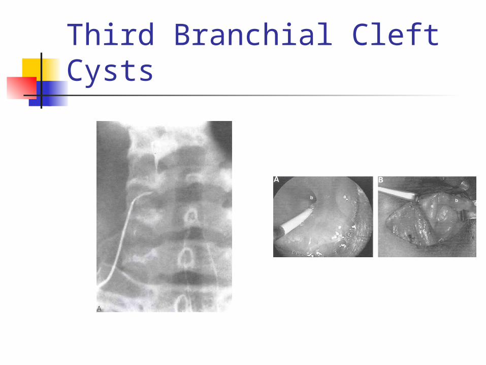
Third Branchial Cleft Cysts
Fourth Branchial Cleft Cysts
Courses from pyriform sinus apex caudal to superior laryngeal nerve, to emerge near the cricothryoid joint, and descend superficial to the recurrent laryngeal nerve.
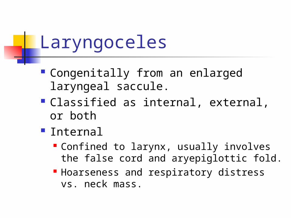
Laryngoceles
Congenitally from an enlarged laryngeal saccule.
Classified as internal, external, or both
Internal Confined to larynx, usually involves the
false cord and aryepiglottic fold. Hoarseness and respiratory distress vs.
neck mass.
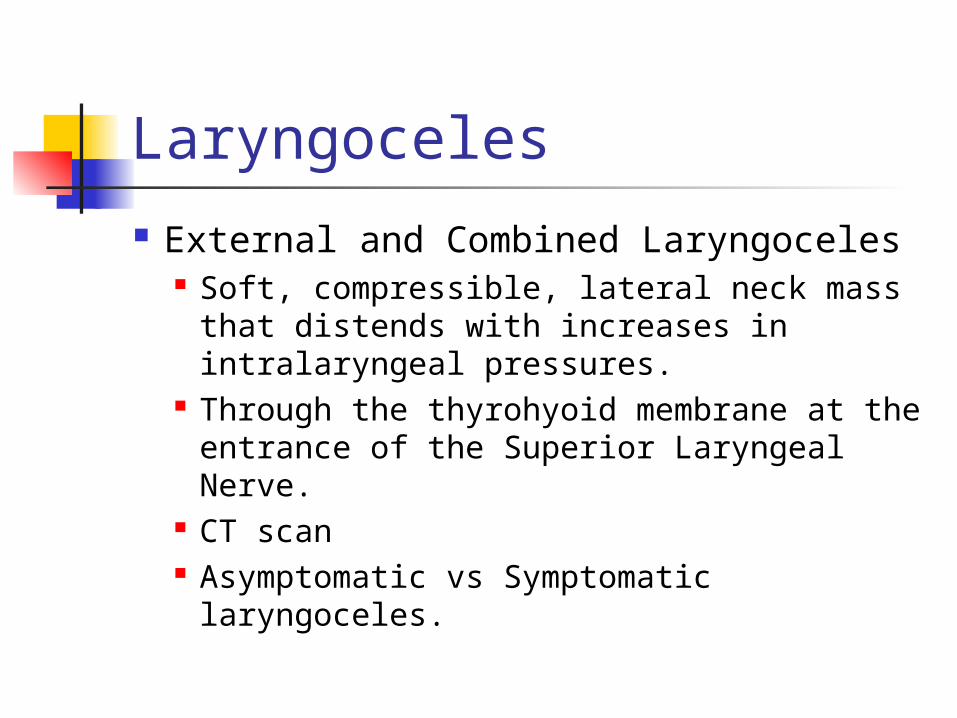
Laryngoceles
External and Combined Laryngoceles Soft, compressible, lateral neck mass that
distends with increases in intralaryngeal pressures.
Through the thyrohyoid membrane at the entrance of the Superior Laryngeal Nerve.
CT scan Asymptomatic vs Symptomatic
laryngoceles.
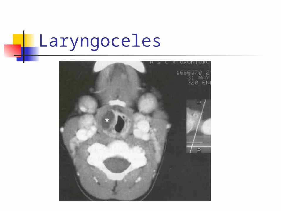
Laryngoceles
Laryngoceles
1-3% of Laryngoceles will harbor an underlying laryngeal carcinoma
ALL adult patients should undergo direct laryngoscopy at the time of surgical intervention.
Dermoid and Teratoid Cysts
Developmental anomalies composed of different germ cell layers.
Isolation of pluripotent stem cells or closure of germ cell layers within points of failed embryonic fusion lines.
Classified according to composition.
Dermoid Cysts
Mesoderm and Ectoderm Midline, paramedian, painless
masses that usually do not elevate with tongue protrusion.
Commonly misdiagnosed as Thyroglossal Duct Cysts.
Treatment is simple surgical excision
Teratoid Cysts and Teratomas All three germ cell layers- Endoderm,
mesoderm and ectoderm. Larger midline masses, present earlier in
life. 20% associated maternal
polyhydramnios Unlike adult teratomas, they rarely
demonstrate malignant degeneration. Surgical excision.

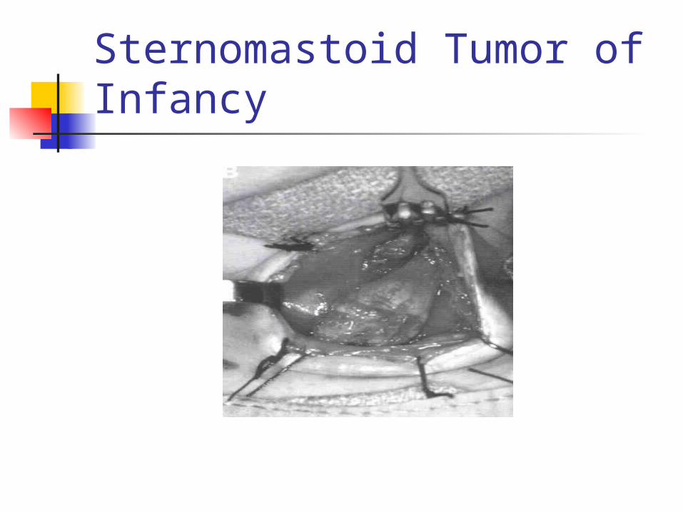
Sternomastoid Tumor of Infancy(Psuedotumor)
Firm mass of the SCM, chin turned away and head tilted toward the mass.
Hematoma with subsequent fibrotic replacement.
Ultrasound Physical therapy is very successful. Myoplasty of the SCM only if refractory
to PT.
Sternomastoid Tumor of Infancy
Conclusions
Neck masses are very common Approach with History and Physical
exam will commonly lead to the correct diagnosis
An understanding of cervical embryology is crucial in treatment of these masses