©verlag ferdinand berger & söhne ges.m.b.h., horn ... plants of selaginella martensii spring,...
TRANSCRIPT

PHYTONANNALES REI BOTANICAE
VOL. 45, FASC. 1
Phyton (Horn, Austria) Vol. 45
PAG. 1-
Fasc. 1
-144
1-8
30.
30.
6
6.
. 2005
2005
The Gametophyte-Sporophyte Junction in Selaginellamartensii SPRING (Selaginellales, Lycopodiophyta)
By
Hartmut H. HILGER*), Nancy KAPUSKAR**) and Wolfgang FREY*)
With 6 Figures
Received August 20, 2004
Key words : Lycopodiophyta, Pteridophyta, Selaginella, Selaginellales. -;omy, gametophyte-sporophyte junction, placental space. - Electron microscopy.Anatomy, game
Summary
HILGER H. H., KAPUSKAR N. & FREY W. 2005. The gametophyte-sporophyte
junction in Selaginella martensii SPRING (Selaginellales, Lycopodiophyta). - Phyton(Horn, Austria) 45 (1): 1-8, with 6 figures. - English with German summary.
The gametophyte-sporophyte junction in Selaginella martensii (Selaginellales,Lycopodiophyta) consists of a sporophytic conical foot embedded in the maternalgametophytic tissue. Both generations are separated by a narrow placental space
*) Prof. Dr. H. H. HILGER, Prof. Dr. W. FREY, Institut für Biologie - SystematischeBotanik und Pflanzengeographie - der Freien Universität Berlin, Altensteinstrae 6,D-14195 Berlin, Germany; e-mail: [email protected], [email protected]
**) Dr. N. KAPUSKAR, Institut für Spezielle Botanik und Botanischer Garten derJohannes Gutenberg-Universität, Bentzelweg 2, D-55099 Mainz;e-mail: [email protected]
©Verlag Ferdinand Berger & Söhne Ges.m.b.H., Horn, Austria, download unter www.biologiezentrum.at

filled with thin-walled collapsed cells of gametophytic origin. Gametophytic andsporophytic placental cells lack wall ingrowths, respectively transfer cells. Neitherinterdigitation nor intermingling of placental cells nor nacreous thickenings are de-veloped. The structure of the gametophyte-sporophyte junction in Selaginella mar-
tensii resembles that of Isoetes boliviensis, the second lycopodiopside investigated,and thus differs from the junction described for pteridopsides and lycopods. Incontrast to Isoetes the sporophytic epidermis of the placental region shows enlargedcells, apparently adapted for enhanced uptake of nutrients from the gametophyte.The finding corroborates the view of Lycopodiophyta as having evolved as an in-dependent microphyllous lineage in land plants.
Zusammenfassung
HILGER H. H., KAPUSKAR N. & FREY W. 2005. Die Gametophyt-Sporophyt-
Brücke von Selaginella martensii SPRING (Selaginellales, Lycopodiophyta). - Phyton(Horn, Austria) 45 (1): 1-8, mit 6 Abbildungen. - Englisch mit deutscher Zusam-menfassung.
Die Sporophyt-Gametophyt-Brücke von Selaginella martensii {Selaginellales,
Lycopodiophyta) besteht aus einem konischen Sporophytenfuß, der in das mütter-liche Gametophytengewebe eingebettet ist. Beide Generationen sind durch einenengen placentalen Spalt getrennt, der sich mit kollabierten, dünnwandigen, gameto-phytischen Zellen anfüllt. Die gametophytischen und sporophytischen Placenta-Zellen verzahnen sich weder, noch bilden sie an ihren Grenzflächen Transferzellenaus. Der Aufbau der Sporophyt-Gametophyt-Brücke in Selaginella martensii ähneltdem von Isoetes boliviensis, der zweiten bisher untersuchten Lycopodiopside, und istsomit ebenfalls anders, als von den Pteridopsiden und Lycopodien beschrieben. ImGegensatz zu Isoetes sind die epidermalen Zellen der sporophytischen, placentalenZellschicht bedeutend großlumiger, was wohl der verbesserten Aufnahme von Nähr-stoffen aus dem Gametophyten dient. Mit den hier präsentierten Daten wird einweiteres Argument dafür geliefert, dass die mikrophyllen Lycopodiophyta und diemegaphyllen Pteridophyta (s. str.) zwei getrennte Entwicklungslinien darstellen.
Introduct ion
The contact zone between the nursing gametophyte and the young
sporophyte in archegoniate plants - the gametophyte-sporophyte junction
- is one of the main characteristics indicating the relationships between
the major land plant groups. Detailed work within the last three decades
has revealed a good knowledge and an evolutionary interpretation espe-
cially in the bryophytes [Bryophytina with Hepaticae (liverworts) (March-
antiopsida and Jungermanniopsida), Bryopsida (mosses), and Anthocer-
otopsida (hornworts)]; for reviews see LIGRONE & al. 1993, FREY & al. 2001].
Recently (DUCKETT & LIGRONE 2003), the gametophyte-sporophyte
junction in a number of leptosporangiate ferns was investigated, revealing
a distinct junction type for this plant group and indicating a clear cut be-
tween the Euphyllophyta and the Lycopodiophyta. For the latter group, up
to now the ultrastructural features of only three species are fragmentarily
©Verlag Ferdinand Berger & Söhne Ges.m.b.H., Horn, Austria, download unter www.biologiezentrum.at

known. In the two species of Lycopodiales investigated [L. appressum(PETERSON & WHITTIER 1991), L. cernuum (DUCKETT & LIGRONE 1992)] the
generations are separated by a thin layer of electron-dense material, lackintermingling of gametophytic and sporophytic placental cells, and bothgenerations develop one layer of placental cells, each with extensive la-byrinth wall ingrowths. In contrast, the third species investigated, Isoetesboliviensis (HILGER & al. 2002), representing the order Isoetales, standsalone amongst the pteridophytes and lycopodiopsides investigated to date:both generations are separated by a placental space filled with thin-walledcollapsed cells of gametophytic origin. Gametophytic and sporophyticplacental cells lack wall ingrowths.
Among the pteridophytes s.l. the Selaginellales - together with eu-sporangiate ferns - is the last taxon for which ultrastructural features ofthe gametophyte-sporophyte junction are not known in detail. With theresults here presented for Selaginella (Selaginellales), one of the last majorgaps in knowledge of the ultrastructure of the gametophyte-sporophytejunction in archegoniate land plants is closed and a more substantial basisfor the discussion of an evolutionary line of its own, the microphyllouslycopodiopsides, is given.
Mater ia l and Methods
Adult plants of Selaginella martensii SPRING, S. pilifei'a A. BRAUN, andS. kraussiana (KUNZE) A. BRAUN were grown in the greenhouse of the BotanicalGarden of the Johannes-Gutenberg Universität Mainz. Herbarium specimens aredeposited at the Herbarium of the Botanical Garden Mainz. Mature micro-andmegaspores were collected, sown on wet filter paper in Petri dishes and kept attemperatures between 23 °C and 30 °C. Megaspores for further treatment werecollected on a daily basis starting with megaspore germination until emergingsporophytes could be observed. The megaspores were either processed for lightmicroscopy (LM: 7 jam Leica Historesin microtome sections stained with toluidineblue), scanning electron microscopy (SEM: razor-sectioned, CO2 dehydrated, criti-cally-point-dried, sputtered with gold, and analyzed with a LEO 430 SEM) andtransmission electron microscopy (TEM: OsO4 fixed material embedded in Araldite,dissected with a Reichert Ultracut, contrasted with lead citrate, and investigatedwith a Zeiss EM 109) following the standard procedure.
Results
Mature spherical megaspores of Selaginella pilifera (Fig. 2) are about0.3-0.4 mm, and of S. martensii 0.2-0.35 mm in diameter. S. kraussiana hasslightly egg shaped megaspores ranging from 0.4 to 1.1 mm in length. Inculture, the microspores adhere to the megaspores (Fig. 1). The megasporesusually germinate over a period of several weeks. Germination (rupture ofspore wall) begins earliest in S. pilifera (after 3 to 5 days), and later in£. martensii and S. kraussiana (after 5-8 days). S. pilifera shows the most
©Verlag Ferdinand Berger & Söhne Ges.m.b.H., Horn, Austria, download unter www.biologiezentrum.at
rapid embryo development and the first embryos emerge from the mega-gametophyte (megaprothallium) three weeks after megaspore germination.In S. martensii, it takes four weeks for the first embryos to break throughthe megagametophyte, and six weeks in S. kraussiana.
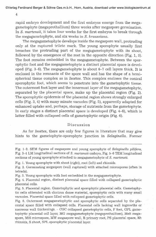
The megagametophyte develops inside the megaspore wall, protrudingonly at the ruptured trilete mark. The young sporophyte usually firstbreaches the protruding part of the megagametophyte with its shoot,followed by the emergence of the root in the opposite direction (Fig. 1,3).The foot remains embedded in the megagametophyte. Between the spor-ophytic foot and the megagametophyte a distinct placental space is devel-oped (Fig. 3-6). The megagametophyte is about 6-7 cell layers thick. It isenclosed in the remnants of the spore wall and has the shape of a hemi-spherical tissue complex as in Isoetes. This complex encloses the conicalsporophytic foot, which seems to penetrate into the gametophytic tissue.The outermost foot layer and the innermost layer of the megagametophyte,separated by the placental space, make up the placental region (Fig. 2).The sporophytic epidermis of the placental region shows strongly enlargedcells (Fig. 3, 4) with many minute vacuoles (Fig. 5), apparently adapted forenhanced uptake and, perhaps, storage of nutrients from the gametophyte.In early stages a distinct placental space is developed (Fig. 4-6), which islatter filled with collapsed cells of gametophytic origin (Fig. 6).
Discussion
As for Isoetes, there are only few figures in literature that may givehints to the gametophyte-sporophyte junction in Selaginella. Former
Fig. 1-2. SEM figures of megaspore and young sporophyte of Selaginella pilifera,Fig. 3-4 LM longitudinal sections of S. martensii embryo, Fig. 5-6 TEM longitudinalsections of young sporophyte attached to megagametophyte of S. martensii.
Fig. 1. Young sporophyte with shoot (right), root (left) and rhizoids.Fig. 2. Germinating megaspore (wall ruptured) with attached microspores (often intetrads).Fig. 3. Young sporophyte with foot embedded in the megagametophyte.Fig. 4. Placental region, distinct placental space filled with collapsed gametophyticplacental cells.Fig. 5. Placental x-egion. Gametophytic and sporophytic placental cells. Gametophy-tic cells oliterated with electron dense material, sporophytic cells with many smallvacuoles. Placental space filled with collapsed gametophytic cells.Fig. 6. Outermost megagametophytic and sporophytic cells separated by the pla-cental space filled with collapsed cells. Placental cells lacking wall ingrowths ornacreous wall thickenings. - CGC collapsed gametophytic cells, F foot, GPL game-tophytic placental cell layer, MG megagametophyte (megaprothallium), MeS mega-spore, MiS microspore, MW megaspore wall, R primary root, PS placental space, Rhrhizoids, S shoot, SPL sporophytic placental layer.
©Verlag Ferdinand Berger & Söhne Ges.m.b.H., Horn, Austria, download unter www.biologiezentrum.at

©Verlag Ferdinand Berger & Söhne Ges.m.b.H., Horn, Austria, download unter www.biologiezentrum.at

6
authors such as BRUCHMANN 1909 were apparently only interested in theinvestigation of the female gametophyte and/or the development of theembryo. From a picture in BOLD & al. 1987: Fig. 14-34 we are only able toconclude that there is a distinct cleft between megagametophyte andembryo. Indeed, the placental cleft in combination with collapsed game-tophytic cells and lacking transfer cells, is the characteristic feature of thejunction.
In a recent publication (DUCKETT & LIGRONE 2003) the authors havebroadened our knowledge of the gametophyte-sporophyte junctions ofleptosporangiate ferns by investigating another five taxa. The junctionsare highly distinctive and show single-celled sporophytic haustoria inter-digitating with gametophytic tissue (absent only in Azolla) and an earlyappearance of wall ingrowths in both generations, but lack both intra-placental spaces and degenerating gametophytic cells. Thus, leptospor-angiate fern placentas resemble those of Tmesipteris (Psüotophyta, FREY &al. 1994a, 1994b) and Equisetum.
On the other hand, they are very different from the gametophyte-sporophyte junction in Lycopodiophyta. In Lycopodium [L. appressum(PETERSON & WHITTIER, 1991), L. cernuum (DUCKETT & LIGRONE, 1992)] thetwo generations are separated by smooth surfaces. In Isoetes, anotherrepresentative of Lycopodiophyta, a distinct placental space containingcollapsed gametophytic cells is present (HILGER & al., 2002). There is nointermingling or interdigitating of gametophytic or sporophytic cells. Withour findings of a similar arrangement in Selaginella we can show that bothheterosporous representatives of the Lycopdiopsida are identical in thisfeature, but differ from homosporous Lycopodium. In the placental spacethe cells of both generations are so loosely attached that the sporophyticfoot can easily be separated from the surrounding gametophytic tissue. Theresults of the recent investigations are summarized in table 1.
The findings with the characters mentioned are so different from thosefound in the fern group, that they corroborate the view of Lycopodiophytaas having evolved as an independent microphyllous lineage in land plants,an idea already proposed, e.g. by PRYER & al. 2001. Unfortunately, detailedknowledge (TEM investigations) about the junctions in eusporangiateferns and Equisetum are still wanting.
Acknowledgements
We thank Ms. E. SCHEREE for technical assistance and M. WEIGEND (both Berlin)for discussion.
References
BOLD H. C, ALEXOPOULOS C. J. & DELEVORYAS T. 1987. Morphology of plants and
fungi. - 5th ed. Harper & Row: New York.
©Verlag Ferdinand Berger & Söhne Ges.m.b.H., Horn, Austria, download unter www.biologiezentrum.at
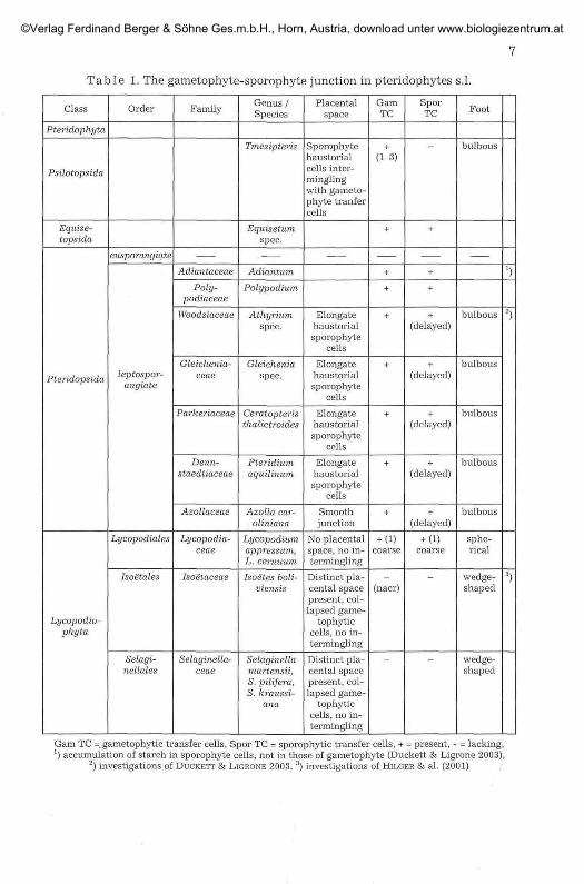
Table 1. The gametophyte-sporophyte junction in pteridophytes s.l.
Class
Pteridophyta
Psilotopsida
Equise-topsida
Pteridopsida
Lycopodio-phyta
Order
eusporangiate
leptospor-angiate
Lycopodiales
Isoetales
Selagi-nellales
Family
Adiantaceae
Poly-podiaceae
Woodsiaceae
Gleichenia-ceae
Parkeriaceae
Denn-staedtiaceae
Azollaceae
Lycopodia-ceae
Isoetaceae
Selaginella-ceae
Genus/Species
Tmesipteris
Equisetumspec.
Adiantum
Polypodium
Athyriumspec.
Gleicheniaspec.
Ceratopteristhalictroides
Pteridiumaquilinum
Azolla car-oliniana
Lycopodiumappressum,L. cernuum
Isoetes boli-viensis
Selaginellamartensii,S. pilifera,S. kraussi-
ana
Placentalspace
Sporophytehaustorialcells inter-minglingwith gameto-phyte tranfercells
Elongatehaustorialsporophyte
cells
Elongatehaustorialsporophyte
cells
Elongatehaustorialsporophyte
cells
Elongatehaustorialsporophyte
cells
Smoothjunction
No placentalspace, no in-termingling
Distinct pla-cental spacepresent, col-lapsed game-
tophyticcells, no in-termingling
Distinct pla-cental spacepresent, col-lapsed game-
tophyticcells, no in-termingling
GamTC
+(1-3)
+
+
+
+
+
+
+
+
+ (Dcoarse
(nacr)
SporTC
+
+
+
+(delayed)
+
(delayed)
+(delayed)
+
(delayed)
+(delayed)
+ (1)coarse
Foot
bulbous
bulbous
bulbous
bulbous
bulbous
bulbous
sphe-rical
wedge-shaped
wedge-shaped
2)
3)
Gam TC = gametophytic transfer cells, Spor TC = sporophytic transfer cells, + = present, - = lacking,:) accumulation of starch in sporophyte cells, not in those of gametophyte (Duckett & Ligrone 2003),
2) investigations of DUCKETT & LIGRONE 2003, 3) investigations of HILGER & al. (2001)
©Verlag Ferdinand Berger & Söhne Ges.m.b.H., Horn, Austria, download unter www.biologiezentrum.at

BRUCHMANN H. 1909. Vom Prothallium der großen Spore und von der Keimesent-wicklung einiger Selaginella-Arten. - Flora 99: 12-51.
— 1912. Zur Embryologie der Selaginellaceen. - Flora NF 4: 180-224.DUCKETT J. G. & LIGRONE R. 1992. A light and electron microscopy study of the fungal
endophytes in the sporophyte and gametophyte of Lycopodium cernuum L.with observations on the gametophyte-sporophyte junction. - Canad. J. Bot.70: 58-72.
— & — 2003. The structure and development of haustorial placentas in lep-tosporangiate ferns provide a clear-cut distinction between euphyllophytesand lycophytes. - Ann. Bot. 92: 513-521.
FREY W., CAMPBELL E. O. & HILGER H. H. 1994a. Structure of the sporophyte-game-tophyte junction in Tmesipteris elongata P. A. DANGEARD (Psilotaceae, Psilo-topsida) and its phylogenetic implications - A SEM analysis. - Nova Hedwigia59: 21-32.
— , — & — 1994b. The sporophyte-gametophyte junction in Tmesipteris{Psilotaceae, Psilotopsida). - Beitr. Biol. Planzen 68: 105-111.
— , HOFMANN M. & HILGER H. H. 2001. The gametophyte-sporophyte junction inApotreubia hortonae {Treubiaceae, Hepaticophytina): Structure and systema-tic implications. - Nova Hedwigia 72: 339-345
HILGER H. H., WEIGEND M. & FREY W. 2002. The gametophyte-sporophyte junction inIsoetes boliviensis WEBER (Isoe'tales, Lycopodiophyta). - Phyton (Horn, Aus-tria) 42:149-157.
LIGRONE R., DUCKETT J. G. & RENZAGLIA K. S. 1993. The gametophyte-sporophyte
junction in land plants. - Adv. bot. Res. 19: 231-317.PETERSON R. L. & WHITTIER D. P. 1991. Transfer cells in the sporophyte-gametophyte
junction of Lycopodium appressum. - Canad. J. Bot. 69: 222-226.PRYER K. M., SCHNEIDER H., SMITH A. R., CRANFILL R., WOLF P. G., HUNT J. S. & SIPES
S. D. 2001. Horsetails and ferns are a monophyletic group and the closestliving relatives to seed plants. - Nature 409: 618-622.
©Verlag Ferdinand Berger & Söhne Ges.m.b.H., Horn, Austria, download unter www.biologiezentrum.at