eprints.whiterose.ac.ukeprints.whiterose.ac.uk/112304/1/de_caso_et_al_brain_cognition.docx · web...
TRANSCRIPT
In press at Brain and Cognition
Full title: Knowing me, knowing you: Resting-state functional connectivity of ventromedial
prefrontal cortex dissociates memory related to self from a familiar other
Running title: vmPFC connectivity and memory for self and other
Irene de Casoa, Theodoros Karapanagiotidisa, Elena Aggius-Vella b, Mahiko Konishia, Daniel S
Marguliesc, Elizabeth Jefferiesa, Jonathan Smallwooda
a Department of Psychology/York Neuroimaging Centre, University of York, Heslington, York, United
Kingdom
b Unit for Visually Impaired People, Istituto Italiano di Tecnologia, Genova, Italy
c Max Planck Research Group: Neuroanatomy & Connectivity, Max Planck Institute for Human
Cognitive and Brain Sciences, Leipzig, Germany
Corresponding Author: Irene de Caso, [email protected], Department of Psychology,
University of York, Heslington, York, YO10 5DD. 07810 060 455
Suggested reviewers:
Andreas Roepstorff, Department of Clinical Medicine - Center of Functionally Integrative
Neuroscience, Aarhus University. [email protected]
Neil Macrae, School of Psychology, University of Aberdeen. [email protected]
Arnaud D'Argembeau, Department of Psychology – Cognition and Behavior, University of
Liège. [email protected]
1
Abstract
Material related to the self, as well as to significant others, often displays mnemonic superiority
through its associations with highly organized and elaborate representations. Neuroimaging studies
suggest this effect is related to activation in regions of medial prefrontal cortex (mPFC). Incidental
memory scores for trait adjectives, processed in relation to the self, a good friend and David
Cameron were collected. Scores for each referent were used as regressors in seed-based analyses of
resting state fMRI data performed in ventral, middle and dorsal mPFC seeds, as well as hippocampal
formation. Stronger memory for self-processed items was predicted by FC between ventral mPFC,
angular gyrus and middle temporal gyri. These regions are within the default mode network, linked
to relatively automatic aspects of memory retrieval. In contrast, memory for items processed in
relation to best friends, was better in individuals whose ventral mPFC showed relatively weak
connectivity with paracingulate gyrus as well as positive connectivity with lateral prefrontal and
parietal regions associated with controlled retrieval. These results suggest that mechanisms
responsible for memory related to ourselves and personally-familiar people are partially dissociable
and reflect connections between ventral mPFC, implicated in schema-based memory, and regions
implicated in more automatic and controlled aspects of retrieval.
Keywords: default mode network, medial prefrontal cortex, self and other
Abbreviations: a: anterior; AG: Angular Gyrus; ATL: Anterior Temporal Pole; d: dorsal; DAN: Dorsal Attention Network; DMN: Default Mode Network; FC: Functional Connectivity; HF+: Hippocampal Formation; Occ: Occipital;Paracing: Paracingulate Gyrus; l:left, mPFC: medial prefrontal cortex; MTG: Medial Temporal Gyrus; r: right; SMG: Supramarginal Gyrus; v:ventral
2

1. Introduction
A fundamental aspect of the brain is its ability to encode, update and retrieve information, processes
that can occur in an automatic manner or through the application of conscious effort. Both encoding
and retrieval are more likely when the information is personally relevant. Strong automatic effects
on memory are illustrated by the self reference effect when incidental memory for material that is
related to the self tends to be higher than for other types of material, such as items related to others
(Kuiper & Rogers, 1979; Kelley et al., 2002) or semantically judged material (Rogers, Kuiper &
Kirker,1977). The strong automatic encoding that occurs during self-reference is thought to reflect
the rich associative structure of knowledge about who we are (Symons & Johnson, 1997). Knowledge
of oneself provides a powerful schema through which information can be organised during encoding
and retrieval. In contrast, memory for information with a less rich associative structure is more
difficult to encode and retrieve.
There is a growing body of evidence that memories with a rich associative structure depend upon
the default mode network (DMN), a large-scale network anchored by medial regions in the medial
prefrontal cortex and the posterior cingulate cortex (Andrews-Hanna, 2012). The DMN, and in
particular the mPFC, show high levels of activation during tasks that require self-reference (Johnson
et al., 2002; Kelley et al., 2002; Macrae et al., 2004; D'Argembeau et al., 2005; Northoff et al., 2006)
as well as for personally familiar referents, such as a close friend (Mitchell, Banaji, & Macrae, 2005),
and when retrieving dominant semantic associations of words that come to mind relatively
automatically (Binder et al., 2009; Davey et al., 2015). In all this cases memory encoding and
retrieval are aided by the presence of previously formed schemas which are thought to be supported
by, at least in part, the vmPFC (van Kesteren et al., 2010a; Ghosh et al., 2014). The notion that the
DMN has an important role in the retrieval of information is also supported by studies that show
strong coupling between the DMN and the hippocampus during successful retrieval (van Kesteren et
al., 2010b; van Kesteren et al., 2012; Huijbers et al., 2011) as well as by studies that show that
3

activity in the mPFC during the encoding phase of a self-reference paradigm predicts subsequent
memory scores for items encoded during self-reference (Macrae et al., 2004). Moreover a related
literature has shown stronger responses within the DMN during spontaneous retrieval states such as
mind-wandering (Mason et al., 2007; Christoff et al., 2009; Starwarzyck et al., 2011) in which
internally generated information is processed. Activity in the DMN often leads to errors during tasks
that depend on a detailed processing of perceptual input (Weissman et al., 2006; Li, Bergguist
&Sinh,2007) and shows patterns of anticorrelation with regions involved in tasks involving controlled
external attention at rest (Fox et al., 2005). These converging literatures are often taken as evidence
that DMN can support spontaneous and undirected retrieval that interferes with ongoing processes
requiring cognitive control (Anticevic et al., 2012). Together these parallel literatures implicate the
DMN in the encoding and retrieval of personally relevant information into and from memory.
However, recent research has also indicated that DMN sites can couple with regions implicated in
executive control in situations that require memory retrieval to be controlled to suit the current
demands (Spreng et al., 2014). These and other findings (e.g. Konishi et al., 2015, Krieger-Redwood,
et al., 2016, Vatansever et al., 2015) suggest the DMN plays a more flexible role in memory
processing than may have be recognised in the past.
To elucidate a more nuanced view of the role of the DMN in memory retrieval the current study
explored whether different patterns of FC could predict incidental memory scores and in particular,
whether these differ for material with different levels of personal relevance. We asked participants
who had already participated in a neuroimaging session in which we recorded resting state activity
to return to the laboratory to perform an incidental memory task. They made decisions about
whether trait adjectives applied to three different referents: themselves, their best friend or David
Cameron (UK Prime Minister). These referents differ on their strength of personal associations which
should result in higher incidental memory scores for items related to the self than their best friend
and the lowest retrieval for David Cameron. In addition, since memory for similar others are known
to elicit similar DMN activation and may be organised using similar or overlapping schema (Mitchell
4
et al., 2006), accurately retrieving information about a best friend may require that competition
from self-processed items may be overcome, which have been encoded in a similar way. In contrast,
items processed in relation to David Cameron will be more distinct and experience less interference.
Individual variations in these scores were used to predict the FC in three sub-regions of the mPFC
(ventral, middle, dorsal) taken from a decomposition of the DMN (Andrews-Hanna et al., 2010).
Given evidence that the hippocampal formation is important in retrieval of information from
memory, and this region is also a member of a subsystem of the DMN (Andrews-Hanna et al., 2010)
this region was also selected as a seed region. In the decomposition of Andrews-Hanna et al. (2010),
the hippocampal formation showed stronger connectivity to ventral mPFC than the other seed
locations, and ventral mPFC has also been implicated in schema-based memory (van Kesteren et
al.,2012; Spalding et al., 2015), giving rise to the prediction that this site may be particularly critical
for self and best friend memory. In addition, we measured executive control via the stop signal
response time task (SSRT, (Logan & Cowan, 1984; Verbruggen & Logan, 2009)) to explore whether
strong automatic retrieval underpinning the self-reference effect was associated with problems in
executive control.
2. Methods
2.1 Participants
Forty healthy right-handed participants were recruited through advert and either received a
monetary reward of £20 or course credits. One participant had to be excluded from all analyses due
to irregularities observed during fMRI scanning. Two further participants were excluded due to poor
task performance, one from each task. Separate FC maps for each task were calculated with a total
of 38 participants (21 males) with an average age of 22.5 (SD = 2.9) years. Approval for this project
was granted by the York Neuroimaging Centre (YNiC) Ethics Committee and was in accordance with
the ethical standards of the responsible committee on human experimentation (institutional and
national) and with the Helsinki Declaration of 1975, as revised in 2008.
5

2.2 Procedure
2.21. Self-reference paradigm.
This laboratory task involved an evaluation and a retrieval phase. During the evaluation phase
(Figure 1A, top) participants were asked to make decisions about the association between adjectives
and one of three referents (‘Self’, ‘Best Friend’ and ‘David Cameron’). Adjectives were presented
sequentially on-screen and participants were required to indicate whether each adjective applied to
a particular referent by pressing ‘Y’ with the index finger of the right hand for ‘yes’ or ‘N’ with the
index finger of the left hand for ‘no’. For each category, participants were presented with a list of 18
unique adjectives presented in separate blocks and the order in which each category was presented
was counterbalanced across participants. Each of the 18-item lists was also rotated across the
different referents and the order of item presentation within each block was randomised. Stimuli
were separated by an inter-stimulus interval of 2500ms during which participants were shown a
blank screen with a fixation cross. Following the evaluation phase, subjects were presented with a
surprise retrieval test in which they were sequentially shown words and asked whether or not that
particular item had been presented in the previous phase. This retrieval phase (Figure 1A, bottom)
contained all the words from the previous stage of the experiment, plus an equal number of new
words. Items were presented in a random order and participants had to either press ‘Y’ if they
thought the word had appeared before or ‘N’ if they thought it was a new word. All words were
selected from a pool of normalized personality trait adjectives with meaningfulness and likeability
ratings (Anderson, 1968). Positive, negative and neutral adjectives with the highest meaningfulness
rating were selected for this experiment. Correct memory for each referent was calculated by
subtracting the relative number of false alarms from the total number of correctly retrieved items.
2.22. Stop signal response time task (SSRT).
6

We developed a version of a stop-signal task (Logan & Cowan, 1984; Verbruggen & Logan, 2009)
using PsychoPy (Peirce, 2007). Figure 1B presents a schematic representation of the trial sequence
for the task. The task featured arrowheads pointing either to the left (<) or to the right (>) staying on
the screen for 1000 msec independently of RT and interleaved by a 500 msec fixation cross.
Participants were instructed to respond as quickly as they could, using the left and right arrow keys
for the left and right arrowheads, respectively. Participants were also instructed to withhold their
respond when they heard a beeping sound (the stop signal) accompanying the arrowhead stimuli,
which occurred in 20% of the trials (stop signal trials); the latency between the beep and the
arrowhead presentation (stop signal delay or SSD) was initially set at 250 msec and was then varied
with a staircase tracking procedure: when inhibition was successful and participants correctly
withheld response in stop signal trials, SSD was increased by 50 msec; when inhibition was
unsuccessful, SSD was decreased by 50 msec. Participants initially received on-screen instructions,
followed by a brief practice session (20 trials) and then moved on to the experimental session, which
was composed of 150 trials divided in two equal blocks, allowing participants a quick break in
between. The whole task lasted approximately 7 minutes.
For each participant, a Stop Signal Response Time (SSRT) score was calculated by subtracting mean
SSD from the untrimmed mean RT (Logan, Schachar, & Tannock, 1997). Given the wide variance of
error percentage in participants a Stop Signal Efficiency score was also calculated by dividing the
SSRT score by the proportion of correct stop-signal trial responses. One participant with a stop-signal
trial error percentage higher than 33% was excluded from the analysis.
2.3. Resting state
2.31. Scan acquisition
Functional MRI data was acquired independent of task stimulus on a 3 Tesla GE scanner. Participants
observed a fixation cross for a scan that lasted 7 minutes. The scan had a repetition time of 2
7
seconds, resulting in 210 volumes. We used interleaved slice-timing and isotropic voxel dimensions
of 3 mm3 (matrix size of 64 X 64, 192mm field of view, and 32 slices) with a 0.5mm gap between
slices.
2.32. Pre-processing
All fMRI preprocessing and analysis was performed using FSL. We extracted the brain from the skull
using the BET toolbox for both the flair and the structural T1 weighted images and these scans were
registered to standard MNI152 (2mm) space using FLIRT (Jenkinson & Smith, 2001). Prior to
conducting the functional connectivity analysis, the following pre-statistics processing was applied to
the resting state data; motion correction using MCFLIRT (Jenkinson et al., 2002); slice-timing
correction using Fourier-space time-series phase-shifting; non-brain removal using BET (Smith 2002);
spatial smoothing using a Gaussian kernel of FWHM 6mm; grand-mean intensity normalisation of
the entire 4D dataset by a single multiplicative factor; highpass temporal filtering (Gaussian-
weighted least-squares straight line fitting, with sigma = 100 s; Gaussian lowpass temporal filtering,
with sigma = 2.8s.)
2.33. First level analysis
Following these steps, the time series of 4 regions of interest (ROI) were extracted. The seed regions
corresponded to 3mm radius spheres centred around the following MNI coordinates: ventromedial
Prefrontal Cortex (vmPFC 0,26,-18), anteriomedial Prefrontal Cortex (amPFC, -6,52,-2), dorsomedial
Prefrontal Cortex (dmPFC 0,52,26) and the hippocampal formation (HF+, -22,-20,-26). These
locations were selected based on previous literature (Andrews-Hanna et al., 2010) that decomposes
the DMN into three subsystems, each mPFC location belonging to a different subsystem. The time
series for each location were averaged and used as an explanatory variable in a subject-level FC
analysis, which also included the following nuisance regressors: the first five principal time-series
components extracted from white matter (WM) and cerebrospinal fluid (CSF) masks in accordance
8

with the CompCor method (Behzadi, Restom et al. 2007) and six motion parameters. WM and CSF
masks were generated by segmenting each individual’s high-resolution structural image (using FAST
in FSL). The default tissue probability maps, referred to as Prior Probability Maps (PPM), were
registered to each individual’s high-resolution structural image (T1 space) and the overlap between
these PPM and the corresponding CSF and WM maps was identified. Finally, these maps were
thresholded (40% for the CSF and 66% for the WM), binarized and combined. The six motion
parameters were calculated in the motion-correction step during pre-processing. Linear
displacements in each of the three Cartesian directions (x, y, z) and rotations around three axes
(pitch, yaw, roll) were included for each individual. No global signal regression was performed
(Murphy, Birn et al. 2009).
2.34. Second-level analysis.
To understand how our psychological measures varied with the connectivity of the DMN seed
regions, we used FSL to conduct a group-level regression of the connectivity matrices of each seed
region. In this analysis we included the mean centred scores for the retrieval of items recalled for
each item type as regressors of interest, and the mean movement during the scanning was included
as a covariate of no interest. This procedure was repeated in an independent analysis using the SSRT
scores instead of the self-other reference task scores. In these analyses the data were processed
using FEAT version 5.98 part of FSL (FMRIB's Software Library, www.fmrib.ox.ac.uk/fsl) and the
analyses were carried out using FMRIB's Local Analysis of Mixed Effects (FLAME). A grey matter
mask with a probability threshold of 40% was used as a pre-thresholding mask and the cluster-
forming threshold was set as z-score of 2.3. For these analyses we controlled for Type I errors by
controlling for the number of voxels in the brain (Worsley 2001), as well as the number of ROIs and
the two tailed nature of our comparisons yielding an alpha value of P<.005 FWE. The unthresholded
maps from the contrasts reported in this paper are available at Neurovault at the following link:
http://neurovault.org/collections/1373/
9

2.4 Neurosynth meta-analyses
In order to study how the patterns of functional connectivity predictive of memory obtained in the
current study were related to previous neuroimaging investigations, we performed a meta-analysis
using the online Neurosynth database (Yarkoni et al., 2011). We performed a meta-analytic
decoding of the unthresholded maps produced in this study by uploading them onto Neurosynth.
This allows the identification of the cognitive terms that are most likely to be associated with the
specific image. We display the results of these terms in the form of word clouds in the relevant
figures. We also performed a specific meta-analysis of the relationship between the maps produced
by our experiment, and the spatial maps that are generated by studies exploring the self. We
performed a meta-analysis (903 studies) of the term “self”
(http://neurosynth.org/analyses/terms/self/) and compared the corresponding map to the
connectivity maps predictive of self memory obtained in the current study.
10
3. Results
3.1 Behavioural results
A one-way analysis of variance (ANOVA) indicated a significant effect of referent on incidental
memory performance (F (2, 76) = 21.58, p < .001, see Figure 1C), as measured during the retrieval
phase of the self-other reference paradigm. Post-hoc Bonferroni corrected comparisons indicated
that words referred to the Self were recalled better than best friend words (p < .01), and these were
better recalled than David Cameron (p = .05) items. In addition, examination of the confidence
intervals for memory for David Cameron suggested it was at chance (95% CI [.46,.54], whereas
memory for best friend (95% CI [.52,.60]) and self (95% CI [.61,.68]) were both above chance . Next
we examined how the process of self-reference was associated with a participant’s tendency for
behavioural inhibition as measured by their efficiency on the SSRT. A linear regression with
incidental memory for items processed in relation to the Self, Best Friend and David Cameron as
independent variables and the SSRT inefficiency as the dependent variable, revealed a model that
accounted for a 22% of the variance in behavioural inhibition scores [F (3, 36) = 3.14, p< .05, r2
= .22]. Higher memory for the Self was associated with less efficiency on the SSRT (standardized beta
= .47, t(33) = 2.7, p < .01) (Figure 1D). Memory scores for best friend and David Cameron items were
not a reliable predictor (standardized beta = .153, t(33) = 0.9, p = .35; standardized beta = -.34, t(33)
=-1.9, p = 0.58, respectively).
Figure 1: Behavioural results. A) Schematic representation of the self-reference task. Top row: Evaluation phase. Bottom row: Retrieval phase. B) Schematic representation of the stop signal response time task. C) Proportion of hits for each referent and error bars. Asterisks represent significant differences in memory performance across referents. D) Scatterplot reflecting the positive
11

correlation between memory for self items and SSRT inefficiency. Acronyms: BF- Best friend, DC – David Cameron.
3.2 RS fMRI analyses
We conducted a series of multiple regressions in which the functional connectivity map of each
region was the dependent variable. For each seed region we entered a measure of retrieval
performance for each referent type (Self, Best Friend, David Cameron) as an explanatory variable.
Independently we performed the same analysis using the SSRT efficiency scores instead of the
memory scores. This SSRT measure did not reveal any patterns of functional connectivity predictive
of inhibitory control for any of the seeded locations.
Figure 2 displays the FC group maps for each seed location and Table 1 summarises the clusters that
were predictive of memory performance and that passed correction for multiple comparisons,
including correction for whole-brain analysis, two-tailed tests and the number of seeded locations.
Clusters that passed the first two corrections but did not pass correction for the number of seeded
locations are still included in the results but are presented separately (in yellow in Figure 3 and
Figure 4).
12
Figure 2: Seed regions (left column) and associated functional connectivity (FC) group maps (right column). A) Dorsal medial Prefrontal Cortex (dmPFC) 0,52,26. B) Anterior medial Prefrontal Cortex (amPFC) -6,-52,-2. C) Ventral medial Prefrontal Cortex (vmPFC) 0,26,-18. D) Hippocampal formation (HF+) -22,-20,-26.
3.21 vmPFC
The FC of the vmPFC seed region predicted memory for self and best friend items. In particular,
memory performance for self items was related to the FC between vmPFC and 3 clusters: right
middle temporal lobe (rMTG) (Figure 3A, first row), left superior angular gyrus (lAG) (Figure 3B, first
row), and left medial and anterior temporal lobe (lMTG cluster) (Figure 3c, first row). In all cases,
stronger FC between the vmPFC and these clusters predicted better memory for self-related items as
seen in the corresponding scatterplots in Figure 3. The rMTG cluster did not pass correction for the
number of seeded locations (and is therefore shown in yellow).
13
Figure 3: Association between ventromedial prefrontal cortex (vmPFC) connectivity and better self memory (Panel A, B & C) and worse Best Friend (BF) memory (Panel D). A) A region in Right Middle Temporal Gyrus (rMTG) was more coupled to vmPFC for individuals with better memory for self-related items. This cluster does not pass correction for the number of seeded locations. B) A Region of Left Angular Gyrus (lAG) showed stronger FC to vmPFC in individuals with stronger memory for self-related items. C) A Region of Left Middle Temporal Gyrus showed stronger FC to vmPFC for individuals with better memory for self-related items. D) A region of Paracingulate gyrus showed stronger FC to the vmPFC for individuals with reduced memory for BF items. Second Row: Scatterplots reflecting relationship between memory performance and FC between ROI and cluster. Third row: Overlap (yellow) between clusters and Default Mode Network (green). Grey panel: Pie charts and legend reflecting the percentage of the cluster that overlaps with each one the Yeo networks.
In order to study the association between the clusters found and resting state networks, these
clusters were overlaid with the Yeo networks (Yeo et al., 2011). The pie charts presented in the grey
panel in Figure 3 illustrate the overlap with the DMN (Network 7), Frontoparietal Control (FPN)
(Network 6) and Limbic Network (Network 5) as defined by Yeo and colleagues (2011). These show
14

the greatest overlap with the DMN (indicated in red) suggesting that functional coupling within the
DMN is associated with increased memory for self-related items. In addition, the region in the rMTG
shows overlaps with the FPN (indicated in orange).
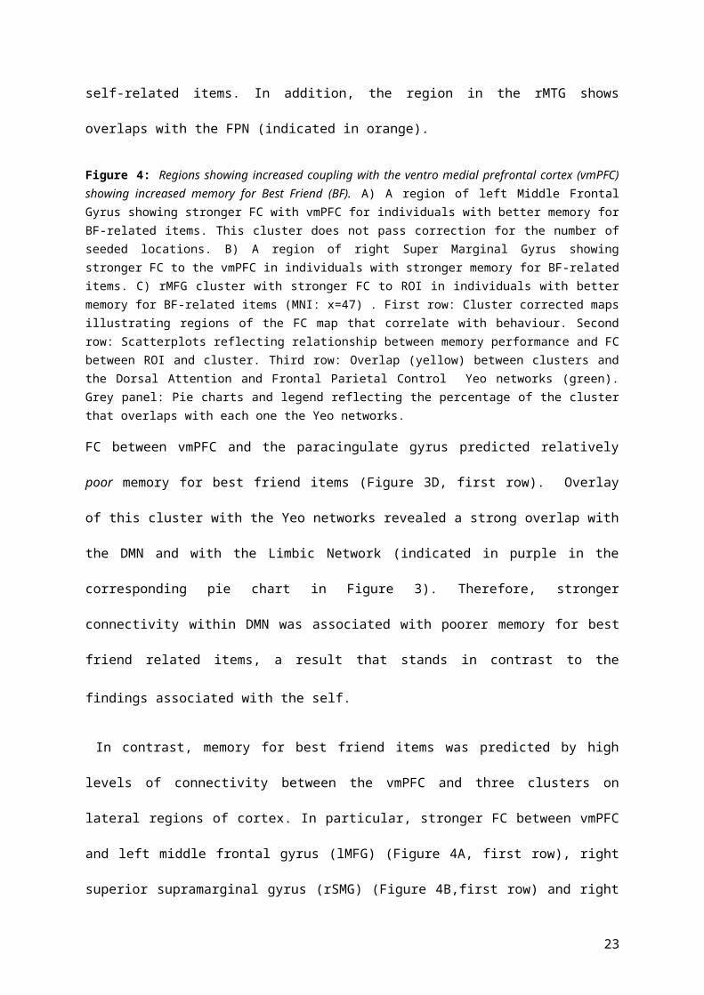
Figure 4: Regions showing increased coupling with the ventro medial prefrontal cortex (vmPFC) showing increased memory for Best Friend (BF). A) A region of left Middle Frontal Gyrus showing stronger FC with vmPFC for individuals with better memory for BF-related items. This cluster does not pass correction for the number of seeded locations. B) A region of right Super Marginal Gyrus showing stronger FC to the vmPFC in individuals with stronger memory for BF-related items. C) rMFG cluster with stronger FC to ROI in individuals with better memory for BF-related items (MNI: x=47) . First row: Cluster corrected maps illustrating regions of the FC map that correlate with behaviour. Second row: Scatterplots reflecting relationship between memory performance and FC between ROI and cluster. Third row: Overlap (yellow) between clusters and the Dorsal
15

Attention and Frontal Parietal Control Yeo networks (green). Grey panel: Pie charts and legend reflecting the percentage of the cluster that overlaps with each one the Yeo networks.
FC between vmPFC and the paracingulate gyrus predicted relatively poor memory for best friend
items (Figure 3D, first row). Overlay of this cluster with the Yeo networks revealed a strong overlap
with the DMN and with the Limbic Network (indicated in purple in the corresponding pie chart in
Figure 3). Therefore, stronger connectivity within DMN was associated with poorer memory for best
friend related items, a result that stands in contrast to the findings associated with the self.
In contrast, memory for best friend items was predicted by high levels of connectivity between the
vmPFC and three clusters on lateral regions of cortex. In particular, stronger FC between vmPFC and
left middle frontal gyrus (lMFG) (Figure 4A, first row), right superior supramarginal gyrus (rSMG)
(Figure 4B,first row) and right middle and inferior frontal gyrus (rMFG) (Figure 4C,first row) predicted
stronger memory for best friend items. The lMFG cluster did not pass correction for the number of
seeded locations. These clusters were again overlaid with the 7 Yeo resting state networks. The
overlapping proportion of each cluster and the Yeo networks is displayed in the pie charts in Figure
4. These clusters generally overlap with regions that are important in tasks that demand externally
oriented attention such as the FPN and the dorsal attention network (DAN). This overlap can be
observed in the third row of Figure 4, in which the DAN and FPN have been displayed with the same
colour (green) for visualization purposes. Unlike a heightened memory for self, better retrieval of
trait adjectives related to a best friend was associated with coupling in regions involved in executive
control that largely fall outside the DMN.
3.22. amPFC
The FC of this brain location did not predict individual differences in memory for any of the three
referents.
3.23. dmPFC
16
The FC of the dmPFC ROI predicted individuals’ memory for self-related items. Stronger FC between
dmPFC and a cluster located in the right occipital lobe was correlated with better memory for self-
referent items (Figure 5A, first row).
This cluster overlapped with the
visual network as defined by Yeo et
al.’s (2011) resting state network
analysis (see Figure 5).
Figure 5: Functional connectivity of dorsal medial prefrontal cortex (dmPFC) and the hippocampal formation (HF+) associated with stronger memory for the self.A) A region of the right occipital (rOcc) cortex that showed stronger FC with the dmPFC for individuals with better memory for self-related items. B) A region of left ventral Anterior Temporal Lobe (lvATL) that showed stronger coupling with the HF+ for participants with a better memory for items related to the self. First row: Cluster corrected maps illustrating regions of the FC map that correlate with behaviour. Second row: Scatterplot reflecting relationship between memory
performance and FC between ROI and cluster. Third row: A) Overlap (yellow) between limbic Yeo network (green) and rOcc cluster (red). B) Overlap (yellow) between Yeo visual network (green) and lvATL cluster. Grey panel: Pie charts and legend reflecting the percentage of the clusters that overlap with each of the Yeo networks.
3.24. Hippocampal formation
The regression analyses performed on the HF+ seed revealed effects for self-related items: in
particular stronger FC between the seed region and a cluster in left ventral anterior temporal lobe
17

(lvATL) (Figure 5B, first row) resulted in a better memory for self-related items. This cluster showed
strong overlap with the Limbic Yeo Network (see right pie chart in Figure 5). Visualization of this
overlap can be observed in the third row of Figure 5B.
3.3.Neurosynth decoding meta-analysis
To provide a quantitative inference of our experimental data, the connectivity maps obtained for
each ROI and for each regressor was decoded using NeuroSynth’s dataset
(http://www.neurosynth.org/decode/). Figure 6 displays all the functional terms from which the
corresponding neuroimaging data from the database had correlation values bigger than 0.1 for each
contrast in our data. From this meta-analytic decoding it can be seen that the functional
connectivity map obtained for stronger memory for self-related items was associated with studies
from the database containing terms such as retrieval, autobiographical, emotion, mentalizing,
semantics and theory of mind. In contrast, connectivity maps obtained for better memory for best
friend were associated with terms such as working memory, working and task. Importantly the term
self-referential was positively associated with the maps obtained for self memory and negatively
associated with those obtained for best friend memory. These patterns of associations are
consistent with the proposal that the map associated with self-related memory is associated with
relatively automatic processes, and ones that are characteristic of the DMN, while the map
associated with memory for a best friend is associated with relatively controlled processes.
Figure 6: Neurosynth meta-analysis of the unthresholded images obtained for each significant contrast. A) Ventral Medial Prefrontal Cortex (vmPFC) map B) Hippocampal formation (HF+) and C) Dorsal Medial Prefrontal Cortex (dmPFC) maps associated with better memory for self-related items. D) vmPFC map
18

associated with better memory for Best Friend (BF) related items. E) vmPFC map associated with worse memory for BF items.
3.4.Neurosynth “self” map activations
Finally, we formally compared the data produced through the individual difference analysis of
resting state functional connectivity approaches with a spatial meta-analysis of peak activations
performed by Neurosynth (search term: “self”; 903 contributing studies;
http://neurosynth.org/analyses/terms/self/). The overlap between the meta-analytical map and the
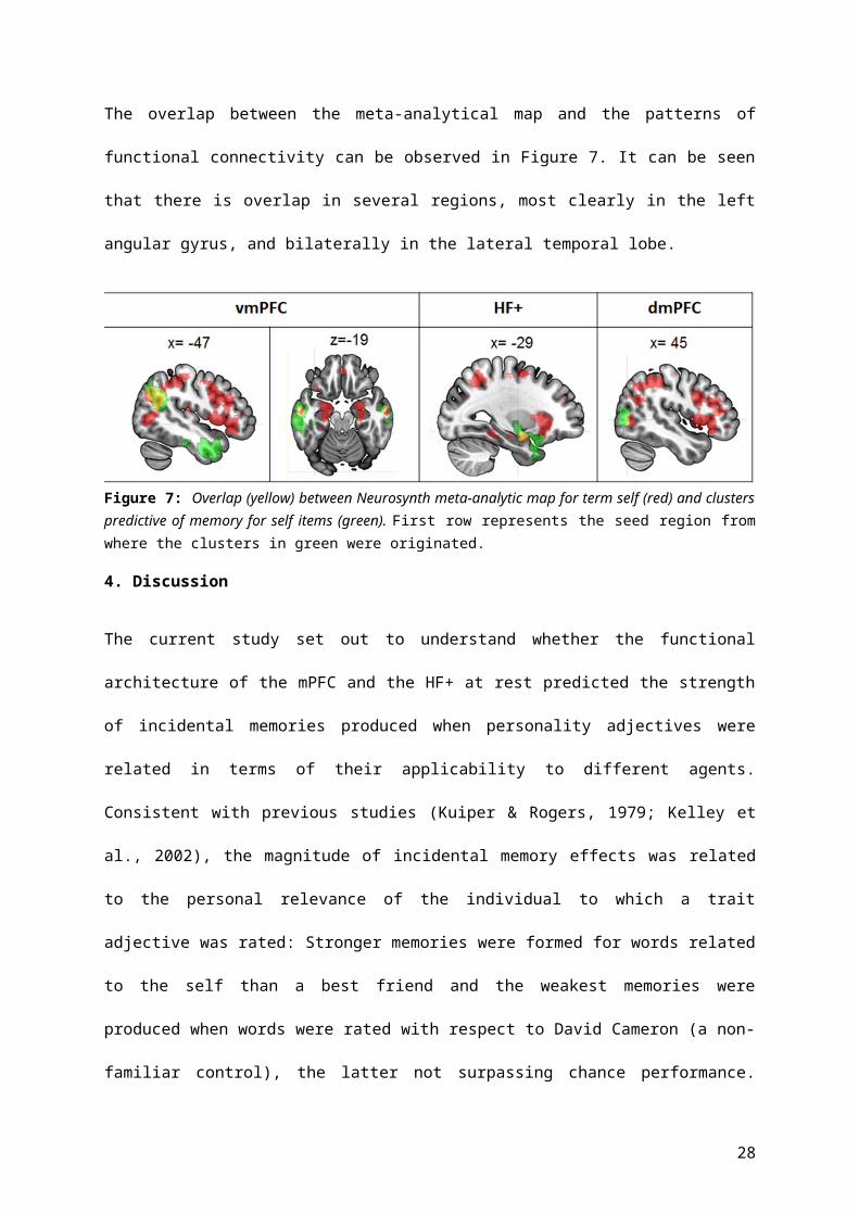
patterns of functional connectivity can be observed in Figure 7. It can be seen that there is overlap in
several regions, most clearly in the left angular gyrus, and bilaterally in the lateral temporal lobe.
Figure 7: Overlap (yellow) between Neurosynth meta-analytic map for term self (red) and clusters predictive of memory for self items (green). First row represents the seed region from where the clusters in green were originated.
4. Discussion
The current study set out to understand whether the functional architecture of the mPFC and the
HF+ at rest predicted the strength of incidental memories produced when personality adjectives
were related in terms of their applicability to different agents. Consistent with previous studies
(Kuiper & Rogers, 1979; Kelley et al., 2002), the magnitude of incidental memory effects was related
to the personal relevance of the individual to which a trait adjective was rated: Stronger memories
were formed for words related to the self than a best friend and the weakest memories were
19

produced when words were rated with respect to David Cameron (a non-familiar control), the latter
not surpassing chance performance. Using the scores for each referent as regressors in an individual
difference analysis of resting state FC, we found that stronger memories following self-related
processing were related to stronger functional coupling between the vmPFC and bilateral mid
temporal lobe, and left angular gyrus, as well as coupling between HF+ and regions of lvATL. We also
found that stronger memory for self related items was linked to coupling between the dmPFC seed
and a region of medial visual cortex, a region that falls at the boundary of the DMN and the visual
cortex. In contrast, successful retrieval of words encoded with respect to the best friend was linked
to decoupling between the ventral prefrontal cortex and the paracingulate gyrus, plus coupling with
lateral parietal and prefrontal regions. No patterns of FC predicted memory scores for items related
to David Cameron possibly due to retrieval for these items being at chance. Finally, a meta-analytic
decoding of the connectivity maps predictive of self and best friend memory supported our
distinction between individuals who excel at memory for themselves, rather than their best friends:
Memory for self was associated with terms such as theory of mind, autobiographical or self-
referential whereas enhanced memory for best friend was associated with terms like working
memory.
It is often assumed that the reason why items that are referred to the self form strong memories is
because of the rich associative structure that is associated with our knowledge of who we are
(Symons & Johnson, 1997): this self-knowledge provides a strong schema to support memory
encoding and retrieval allowing it to be retrieved efficiently and automatically. Prior work has shown
that schema-based memory engages vmPFC (van Kesteren et al., 2010a; van Kesteren et al., 2012;
Ghosh et al., 2014; Mckenzie et al., 2014; Spalding et al., 2015) and our study suggests that a strong
bias to remembering information rated to ourselves depends on forming a network between this
region and lateral and anterior regions of the temporal lobe and the angular gyrus – regions that
together make up the DMN (Raichle et al., 2001; Raichle & Snyder, 2007). Functional studies often
implicate the DMN in situations when information from memory is often retrieved effortlessly, such
20

as making global semantic associations (Bar et al., 2007; Wirth et al., 2011) periods of spontaneous
thought (Mason et al., 2007) and the process of self-reference itself (Gusnard et al., 2001; Macrae et
al., 2004; Northoff et al., 2006). These are all states that can involve the automatic retrieval of
information from memory. Behaviourally we observed that self-memory was correlated with
relatively poor performance on the SSRT. The SSRT is a measure of inhibitory control and previous
studies have shown that errors in response inhibition are linked to a lack of DMN deactivation (Li et
al., 2007). Moreover SSRT can be used to distinguish subjects with ADHD from normal controls
(Sendereka et al., 2012) and previous research on ADHD has revealed reduced DMN deactivations
during complex tasks (Fassbender et al., 2009) supporting the notion that successful executive
control requires DMN deactivation. In addition, high activity in the DMN precedes lapses in
cognitively demanding tasks (Weissman et al., 2006). Altogether our results therefore are consistent
with the idea that self-relevant memories are supported by integrated activity within the DMN, a
state that promotes the automatic and elaborated processing of associative information from
memory that can at times be hard to inhibit.
Information related to best friends was retrieved more effectively than for the David Cameron
control items. However, this type of memory was associated with a different network of regions
than those observed for strong memories of the self. Better memory for a best friend involved a
network that spanned the lateral surface of parietal and frontal cortex, including middle frontal and
inferior frontal gyrus and supramarginal gyrus. Overlap with the Yeo networks, revealed that
although this network was anchored in the vmPFC, these regions are a part of the dorsal attention
and frontoparietal control networks, large-scale systems that are often activated by attention-
demanding tasks (Collette et al., 1999; Corbetta & Shulman, 2002; Duncan, 2010). Thus unlike a
strong memory for the self, a tendency to remember items related to one’s best friend was linked to
a coupling between ventral regions of the mPFC and regions beyond the broader DMN that are
involved in goal-directed attention. Studies have shown that the lateral prefrontal cortex,
particularly the inferior frontal gyrus, often activates when participants make semantic decisions
21

that are more difficult either because the meaning is ambiguous or because participants must make
links between stimuli that are only weakly related together (Noonan et al., 2013). More generally,
co-activation between the DMN and the lateral prefrontal cortex occurs when novel or complicated
decisions have to be made based on memory such as during creativity (Beaty et al., 2014) or when
we plan the future (Spreng et al., 2010). Together the enhanced retrieval for best friend relative to
the David Cameron control, as well as a functional connectivity network anchored in the vmPFC
seed, suggest that memory for the best friend is likely to also benefit from an elaborate schema,
perhaps one that is similar to that of the self (e.g. Mitchell et al., 2005). Importantly, this similarity
with the self may mean that an accurate memory for close personal acquaintances is not only
hampered by the weaker traces formed at encoding but may also depend on overcoming
interference from associations with self memories and requires regions outside of the DMN that
may function to guide retrieval in the face of interference. This possibility is supported by previous
research which has commonly found inferior and dorsolateral prefrontal gyri, regions predictive of
best friend memory in this study, to be involved in working memory processes (Curtis & D’Esposito,
2003). This hypothesis should be examined in future studies.
One general implication of our results is that vmPFC may act as a hub whose FC determines how
schematic information is represented in the cortex. As well as connections to other regions of the
mPFC, it can be seen from Figure 2 that this region of cortex is connected to medial aspects of the
temporal lobe, as well as other limbic regions. In topographical terms this region is therefore well
placed to integrate affective and episodic information into the broader prefrontal cortex. Consistent
with this view, our data shows that, across people, the nature of the patterns of connectivity it
exhibits at rest has implications for aspects of social memory: A strong memory for self-relevant
information was associated with greater integration within the DMN, whereas a stronger memory
for best friend required integration with regions important for executive control. One implication of
this view is that the vmPFC exhibits modes of cortical processing that reflect how different aspects of
mnemonic and affective information dominate cognition. Although our current data are consistent
22
with this hypothesis, it is impossible to infer whether these patterns exert their effect on memory
during encoding or retrieval since the current study explored individual differences in resting state
FC rather than measuring online neural activity. Future studies exploring different patterns of FC
during different types of social and non social memory retrieval will help to address this question.
It is worth considering certain limitations with the current data. Our study shows that better
memory for different referents is associated with distinct patterns of functional connectivity
however, the current study is unable to decipher whether the different patterns of functional
connectivity predictive of memory for self and best friend items are indeed capturing the processing
differences in referents per se, or whether instead they are reflecting differences in general memory
strength. Future studies using a control memory task matched in accuracy to the reference task but
instead employing a different memory manipulation such as elaborative semantic encoding will be
able to address this issue.
Regardless of these issues, our results suggest that information related to the self and to one’s best
friend is supported by different patterns of FC with the vmPFC. Whereas information exclusively
related to the self relies on integration between these region and the DMN, remembering
information about a similar other, benefits from integration between the vmPFC and executive
control regions. We argue that this occurs because there are different strengths of association for
the different types of memory. Memories associated with a best friend have weaker associations
than do self-related. Consequently, remembering information about a personally significant other
will requires additional executive control directed either to retrieve the weaker memory trace, or to
correctly select the appropriate memory despite interference from the stronger, and often
associated, memories about the self.
5. Acknowledgements
Elizabeth Jefferies was supported by grants from BBSRC (BB/J006963/1) and the European Research
Council (SEMBIND – 283530) and Jonathan Smallwood was supported by European Research Council
23

(WANDERINGMINDS – 646927). This publication was also made possible through the support of a
grant from the John Templeton Foundation, “Prospective Psychology Stage 2: A Research
Competition” to Martin Seligman. The opinions expressed in this publication are those of the
author(s) and do not necessarily reflect the views of the John Templeton Foundation.
References
Anderson, N. H. (1968). Likableness ratings of 555 personality-trait words.Journal of personality and
social psychology, 9(3), 272.
Andrews-Hanna, J. R. (2012). The brain’s default network and its adaptive role in internal
mentation. The Neuroscientist, 18(3), 251-270.
Andrews-Hanna, J. R., Smallwood, J., & Spreng, R. N. (2014). The default network and self-generated
thought: Component processes, dynamic control, and clinical relevance. Annals of the New
York Academy of Sciences, 1316(1), 29–52.
Andrews-Hanna, J. R., Reidler, J. S., Sepulcre, J., Poulin, R., & Buckner, R. L. (2010). Functional-
Anatomic Fractionation of the Brain’s Default Network. Neuron, 65(4), 550–562.
Anticevic, A., Cole, M. W., Murray, J. D., Corlett, P. R., Wang, X. J., & Krystal, J. H. (2012). The role of
default network deactivation in cognition and disease. Trends in cognitive sciences, 16(12), 584-
592.
Bar, M., Aminoff, E., Mason, M., & Fenske, M. (2007). The units of thought.Hippocampus, 17(6), 420-
428.
Beaty, R. E., Benedek, M., Wilkins, R. W., Jauk, E., Fink, A., Silvia, P. J., … Neubauer, A. C. (2014).
Creativity and the default network: A functional connectivity analysis of the creative brain at
rest. Neuropsychologia, 64C, 92–98.
24

Behzadi, Y., Restom, K., Liau, J., & Liu, T. T. (2007). A component based noise correction method
(CompCor) for BOLD and perfusion based fMRI.Neuroimage, 37(1), 90-101.
Binder, J. R., Desai, R. H., Graves, W. W., & Conant, L. L. (2009). Where is the semantic system? A
critical review and meta-analysis of 120 functional neuroimaging studies. Cerebral
Cortex, 19(12), 2767-2796.
Collette, F., Salmon, E., Van Der Linden, M., Chicherio, C., Belleville, S., Degueldre, C., … Franck, G.
(1999). Regional brain activity during tasks devoted to the central executive of working
memory. Cognitive Brain Research, 7(3), 411–417.
Corbetta, M., & Shulman, G. L. (2002). Control of Goal-Directed and Stimulus-Driven Attention in the
Brain. Nature Reviews Neuroscience, 3(3), 215–229. http://doi.org/10.1038/nrn755
Curtis, C. E., & D'Esposito, M. (2003). Persistent activity in the prefrontal cortex during working
memory. Trends in cognitive sciences, 7(9), 415-423.
D'Argembeau, A., Collette, F., Van der Linden, M., Laureys, S., Del Fiore, G., Degueldre, C., ... &
Salmon, E. (2005). Self-referential reflective activity and its relationship with rest: a PET
study. Neuroimage, 25(2), 616-624.
Davey, J., Cornelissen, P. L., Thompson, H. E., Sonkusare, S., Hallam, G., Smallwood, J., & Jefferies, E.
(2015). Automatic and Controlled Semantic Retrieval: TMS Reveals Distinct Contributions of
Posterior Middle Temporal Gyrus and Angular Gyrus. The Journal of Neuroscience, 35(46),
15230-15239.
Duncan, J. (2010). The multiple-demand (MD) system of the primate brain: mental programs for
intelligent behaviour. Trends in cognitive sciences,14(4), 172-179.
25

Fassbender, C., Zhang, H., Buzy, W. M., Cortes, C. R., Mizuiri, D., Beckett, L., & Schweitzer, J. B.
(2009). A lack of default network suppression is linked to increased distractibility in
ADHD. Brain research, 1273, 114-128.
Fox, M. D., Snyder, A. Z., Vincent, J. L., Corbetta, M., Van Essen, D. C., & Raichle, M. E. (2005). The
human brain is intrinsically organized into dynamic, anticorrelated functional networks.
Proceedings of the National Academy of Sciences of the United States of America , 102(27),
9673–8
Ghosh, V. E., Moscovitch, M., Colella, B. M., & Gilboa, A. (2014). Schema representation in patients
with ventromedial PFC lesions. The Journal of Neuroscience, 34(36), 12057-12070.
Gusnard, D. A., Akbudak, E., Shulman, G. L., & Raichle, M. E. (2001). Medial prefrontal cortex and
self-referential mental activity: relation to a default mode of brain function. Proceedings of the
National Academy of Sciences of the United States of America, 98(7), 4259–64.
http://doi.org/10.1073/pnas.071043098
Huijbers, W., Pennartz, C. M. A., Cabeza, R., & Daselaar, S. M. (2011). The hippocampus is coupled
with the default network during memory retrieval but not during memory encoding. PLoS ONE,
6(4).
Jenkinson, M., & Smith, S. (2001). A global optimisation method for robust affine registration of brain
images. Medical image analysis, 5(2), 143-156.
Jenkinson, M., Bannister, P., Brady, M., & Smith, S. (2002). Improved optimization for the robust and
accurate linear registration and motion correction of brain images. Neuroimage, 17(2), 825-
841.
26

Johnson, S. C., Baxter, L. C., Wilder, L. S., Pipe, J. G., Heiserman, J. E., & Prigatano, G. P. (2002).
Neural correlates of self‐reflection. Brain, 125(8), 1808-1814.
Kelley, W. M., Macrae, C. N., Wyland, C. L., Caglar, S., Inati, S., & Heatherton, T. F. (2002). Finding the
self? An event-related fMRI study. Journal of Cognitive Neuroscience, 14(5), 785–94.
Konishi, M., McLaren, D. G., Engen, H., & Smallwood, J. (2015). Shaped by the past: the default mode
network supports cognition that is independent of immediate perceptual input. PloS one,
10(6), e0132209.
Krieger-Redwood, K., Jefferies, E., Karapanagiotidis, T., Seymour, R., Nunes, A., Ang, J. W. A., ... &
Smallwood, J. (2016). Down but not out in posterior cingulate cortex: Deactivation yet
functional coupling with prefrontal cortex during demanding semantic cognition. Neuroimage,
141, 366-377.
Kuiper, N. A., & Rogers, T. B. (1979). Encoding of personal information: Self–other
differences. Journal of Personality and Social Psychology, 37(4), 499.
Li, C. S. R., Yan, P., Bergquist, K. L., & Sinha, R. (2007). Greater activation of the “default” brain
regions predicts stop signal errors. Neuroimage, 38(3), 640-648.
Logan, G. D., & Cowan, W. B. (1984). On the ability to inhibit thought and action: A theory of an act
of control. Psychological review, 91(3), 295.
Logan, G. D., Schachar, R. J., & Tannock, R. (1997). Impulsivity and inhibitory control. Psychological
Science, 8(1), 60-64
Macrae, C. N., Moran, J. M., Heatherton, T. F., Banfield, J. F., & Kelley, W. M. (2004). Medial
prefrontal activity predicts memory for self. Cerebral Cortex, 14(6), 647–654.
27
Mason, M. F., Norton, M. I., Horn, J. D. Van, Wegner, D. M., Grafton, S. T., Macrae, C. N., … Macrae,
C. N. (2007). Wandering minds: Stimulus-independent thought. Science, 315(January), 393–
395.
McKenzie, S., Frank, A. J., Kinsky, N. R., Porter, B., Rivière, P. D., & Eichenbaum, H. (2014).
Hippocampal representation of related and opposing memories develop within distinct,
hierarchically organized neural schemas.Neuron, 83(1), 202-215.
Mitchell, J. P., Banaji, M. R., & Macrae, C. N. (2005). The link between social cognition and self-
referential thought in the medial prefrontal cortex. Journal of Cognitive Neuroscience, 17(8),
1306–15.
Mitchell, J. P., Macrae, C. N., & Banaji, M. R. (2006). Dissociable medial prefrontal contributions to
judgments of similar and dissimilar others. Neuron,50(4), 655-663.
Murphy, K., Birn, R. M., Handwerker, D. A., Jones, T. B., & Bandettini, P. A. (2009). The impact of
global signal regression on resting state correlations: are anti-correlated networks
introduced?. Neuroimage, 44(3), 893-905.
Noonan, K. A., Jefferies, E., Garrard, P., Eshan, S., & Lambon Ralph, M. A. (2013). Demonstrating the
qualitative differences between semantic aphasia and semantic dementia: A novel exploration
of nonverbal semantic processing. Behavioural Neurology, 26(1-2), 7–20.
Northoff, G., Heinzel, A., de Greck, M., Bermpohl, F., Dobrowolny, H., & Panksepp, J. (2006). Self-
referential processing in our brain-A meta-analysis of imaging studies on the self. NeuroImage,
31(1), 440–457.
Peirce, J. W. (2007). PsychoPy—psychophysics software in Python. Journal of neuroscience
methods, 162(1), 8-13.
28
Raichle, M. E., MacLeod, A. M., Snyder, A. Z., Powers, W. J., Gusnard, D. A., & Shulman, G. L. (2001).
A default mode of brain function. Proceedings of the National Academy of Sciences, 98(2), 676-
682.
Raichle, M. E., & Snyder, A. Z. (2007). A default mode of brain function: a brief history of an evolving
idea. Neuroimage, 37(4), 1083-1090.
Rogers, T. B., Kuiper, N. A., & Kirker, W. S. (1977). Self-reference and the encoding of personal
information. Journal of personality and social psychology, 35(9), 677.
Senderecka, M., Grabowska, A., Szewczyk, J., Gerc, K., & Chmylak, R. (2012). Response inhibition of
children with ADHD in the stop-signal task: An event-related potential study. International
Journal of Psychophysiology,85(1), 93-105.
Smith, S. M. (2002). Fast robust automated brain extraction. Human brain mapping, 17(3), 143-155.
Spalding, K. N., Jones, S. H., Duff, M. C., Tranel, D., & Warren, D. E. (2015). Investigating the Neural
Correlates of Schemas: Ventromedial Prefrontal Cortex Is Necessary for Normal Schematic
Influence on Memory.The Journal of Neuroscience, 35(47), 15746-15751.
Spreng, R. N., Stevens, W. D., Chamberlain, J. P., Gilmore, A. W., & Schacter, D. L. (2010). Default
network activity, coupled with the frontoparietal control network, supports goal-directed
cognition. NeuroImage, 53(1), 303–317.
Spreng, R. N., DuPre, E., Selarka, D., Garcia, J., Gojkovic, S., Mildner, J., ... & Turner, G. R. (2014).
Goal-congruent default network activity facilitates cognitive control. The Journal of
Neuroscience, 34(42), 14108-14114.
Symons, C., & Johnson, B. (1997). The self-reference effect in memory: a meta-analysis.
Psychological Bulletin, 121(1987), 371–394
29

van Kesteren, M. T., Rijpkema, M., Ruiter, D. J., & Fernández, G. (2010a). Retrieval of associative
information congruent with prior knowledge is related to increased medial prefrontal activity
and connectivity. The Journal of Neuroscience, 30(47), 15888-15894.
van Kesteren, M. T., Fernández, G., Norris, D. G., & Hermans, E. J. (2010b). Persistent schema-
dependent hippocampal-neocortical connectivity during memory encoding and postencoding
rest in humans. Proceedings of the National Academy of Sciences, 107(16), 7550-7555.
van Kesteren, M. T., Ruiter, D. J., Fernández, G., & Henson, R. N. (2012). How schema and novelty
augment memory formation. Trends in neurosciences, 35(4), 211-219.
Vatansever, D., Menon, D. K., Manktelow, A. E., Sahakian, B. J., & Stamatakis, E. A. (2015). Default
mode network connectivity during task execution. Neuroimage, 122, 96-104.
Weissman, D. H., Roberts, K. C., Visscher, K. M., & Woldorff, M. G. (2006). The neural bases of
momentary lapses in attention. Nature neuroscience,9(7), 971-978.
Worsley, K. J. (2001). Statistical analysis of activation images. Functional MRI: An introduction to
methods, 14, 251-270.
Yarkoni, T., Poldrack, R. A., Nichols, T. E., Van Essen, D. C., & Wager, T. D. (2011). Large-scale
automated synthesis of human functional neuroimaging data. Nature methods, 8(8), 665-670.
30