viral exanthems 21 nov 2013 danize buemio mary april chan
TRANSCRIPT

VIRAL EXANTHEMS
21 Nov 2013Danize Buemio
Mary April Chan

MEASLES
Measles
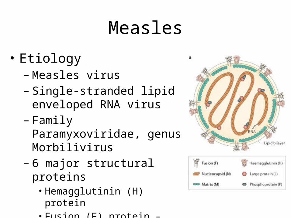
• Etiology– Measles virus– Single-stranded lipid enveloped
RNA virus– Family Paramyxoviridae, genus
Morbilivirus– 6 major structural proteins• Hemagglutinin (H) protein • Fusion (F) protein – antibodies limit
proliferation
Measles
• Transmission– Contact with large droplets or small droplet
aerosols in which virus is suspended– Entry into respiratory tract or conjunctivae– Approx. 90% of exposed individuals develop
measles– Face-fece contact is not necessary– Viable virus may be suspended in the air up to 1
hour
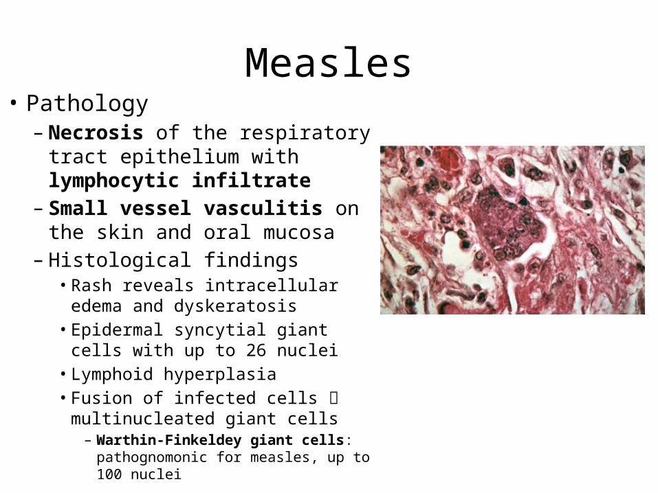
Measles• Pathology– Necrosis of the respiratory tract
epithelium with lymphocytic infiltrate– Small vessel vasculitis on the skin and
oral mucosa– Histological findings
• Rash reveals intracellular edema and dyskeratosis
• Epidermal syncytial giant cells with up to 26 nuclei
• Lymphoid hyperplasia• Fusion of infected cells multinucleated
giant cells– Warthin-Finkeldey giant cells:
pathognomonic for measles, up to 100 nuclei
Measles
Incubation Period
Prodromal Illness
Exanthematous Phase Recovery

Measles
Incubation Period
Prodromal Illness
Exanthematous Phase Recovery
• Incubation period: 8-12 days• Virus migrates to regional lymph nodes• Primary viremia disseminates the virus to the
reticuloendothelial system• Secondary viremia spreads virus to body surfaces
Measles
Incubation Period
Prodromal Illness
Exanthematous Phase Recovery
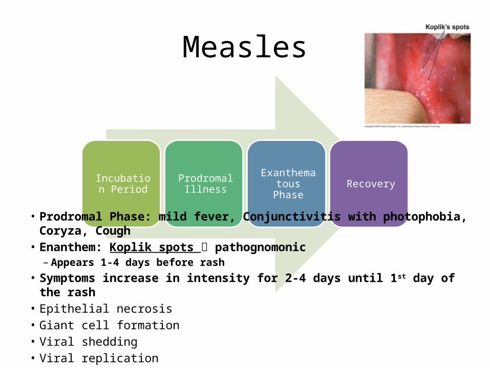
• Prodromal Phase: mild fever, Conjunctivitis with photophobia, Coryza, Cough• Enanthem: Koplik spots pathognomonic
– Appears 1-4 days before rash• Symptoms increase in intensity for 2-4 days until 1st day of the rash• Epithelial necrosis • Giant cell formation• Viral shedding• Viral replication
Measles
Incubation Period
Prodromal Illness
Exanthematous Phase Recovery
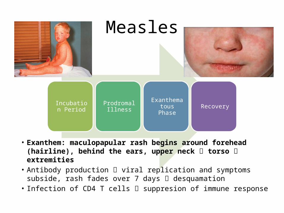
• Exanthem: maculopapular rash begins around forehead (hairline), behind the ears, upper neck torso extremities
• Antibody production viral replication and symptoms subside, rash fades over 7 days desquamation
• Infection of CD4 T cells suppresion of immune response
Inapparent Measles Infection
• Subclinical form of measles• Individuals with passively acquired antibody– Infants, recepients of blood products
• Rash may be indistinct, brief or absent• Do not shed measles virus or transmit
infection
Atypical measles
• Original formalin-inactivated measles vaccine• More severe form• High onset of fever and headache• Maculopapular rash on the extremities petechial
and purpuric– Progress in centripetal direction
• Frequently complicated by pneumonia and pleural effusion
• Circulating immune complexes due to abnormal response to vaccine
Diagnostics
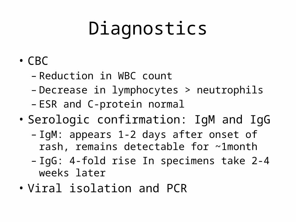
• CBC– Reduction in WBC count– Decrease in lymphocytes > neutrophils– ESR and C-protein normal
• Serologic confirmation: IgM and IgG– IgM: appears 1-2 days after onset of rash, remains
detectable for ~1month– IgG: 4-fold rise In specimens take 2-4 weeks later
• Viral isolation and PCR
Differential Diagnoses
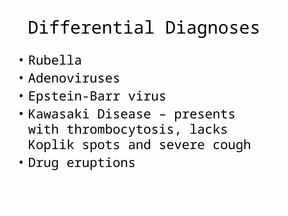
• Rubella• Adenoviruses• Epstein-Barr virus• Kawasaki Disease – presents with
thrombocytosis, lacks Koplik spots and severe cough
• Drug eruptions
Treatment
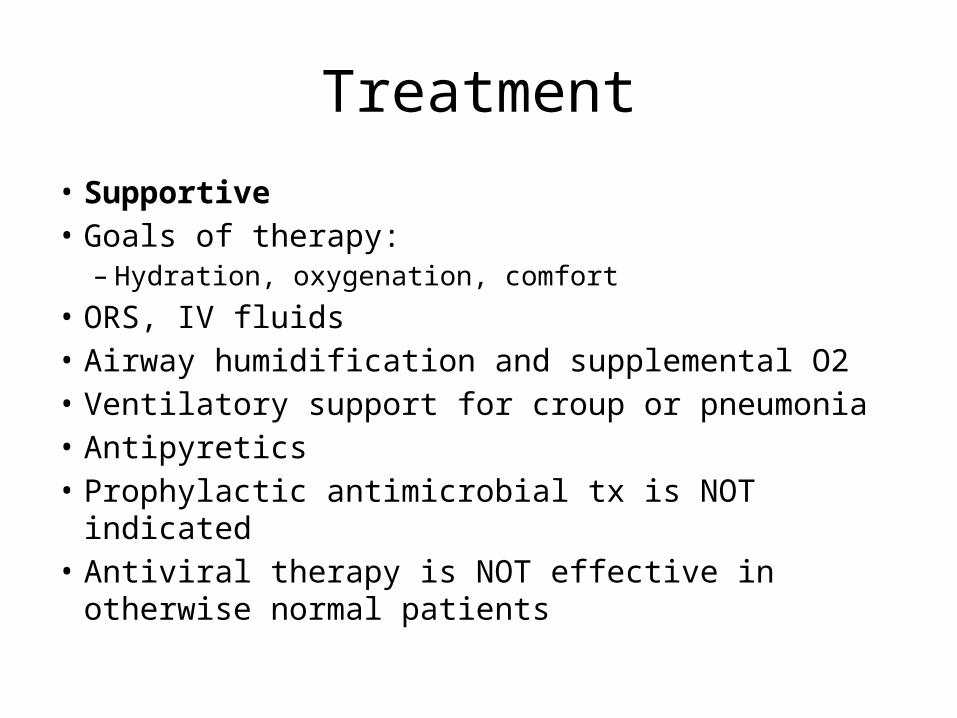
• Supportive• Goals of therapy:– Hydration, oxygenation, comfort
• ORS, IV fluids• Airway humidification and supplemental O2• Ventilatory support for croup or pneumonia• Antipyretics• Prophylactic antimicrobial tx is NOT indicated• Antiviral therapy is NOT effective in otherwise normal
patients
Treatment
• Measles in immunocompromised is highly lethal– Ribavarin with or without intravenous gamma
globulin
Treatment
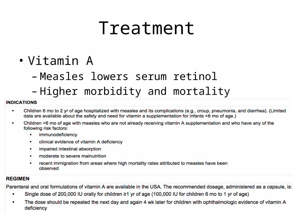
• Vitamin A– Measles lowers serum retinol– Higher morbidity and mortality
Complications
• Morbidity and mortality are greatest in <5 years and >20 years
• Crowding – larger inoculum dose• Severe malnutrition – suboptimal immune
response• Measles in immunocompromised

Complications
• Dehydration– Fever, diarrhea, vomiting
• Known to suppress PPD skin test responsiveness– May have an increased rate of PTB activation
Complications• Pneumonia – most common cause of death
– Giant cell pneumonia caused by virus– Superimposed bacterial infection– Final pathway: bronchiolitis obliterans– Croup, tracheitis
• Otitis Media – most common complication• Sinusitis, mastoiditis, retropharygeal abscess• Encephalitis
– Postinfectious immunologically mediated process– Seizures (56%), lethargy (45%), coma (28%), irritability (26%)– CSF: lymphocytic pleocytosis, elevated protein– Approx 15% death, 20-40% mental retardation, deafness, motor disabilities
• Hemorrhagic or “black measles”: hemorrhagic skin eruption, fatal• Myocarditis: rare• Subacute Sclerosing Paraencephalitis (SSPE)
Subacute Sclerosing Paraencephalitis (SSPE)
• Rare disease• Results from a persistent infection with an
altered measles virus harbored in CNS for years• After 7-10 years, virus regains virulence and
attacks cells in CNS that offered the virus protection
• Inflammation and cell death neurodegenerative process
Subacute Sclerosing Paraencephalitis (SSPE)
• Age of onset: <1 to <30 years• Usually in children in adolescents• Measles at an early age favors SSPE development• Males affected twice as often as females• Defective measles virus• Defective or immature immune system• Immature virus able to reside within neural cells
for long periods
Subacute Sclerosing Paraencephalitis (SSPE)
• Symptoms begin 7-13 years after primary measles infection• 1st stage: Subtle changes in behavior
– Irritability, reduced attention span, temper outbursts– Fever, headache, signs of encephalitis are absent
• 2nd stage: massive myoclonus– Extension of inflammatory process to deeper brain structures
including basal ganglia• 3rd stage: choreoathetosis, immobility, dystonia, lead pipe
rigidity– Sensorium deteriorates: dementia stupor coma– involuntary movements disappear
• 4th stage– Loss of centers supporting breathing, heart rate, BP– Death

MUMPS
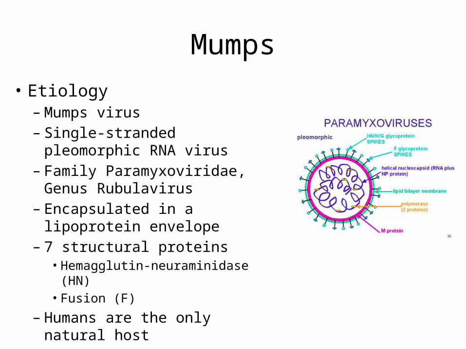
Mumps• Etiology– Mumps virus– Single-stranded pleomorphic
RNA virus– Family Paramyxoviridae, Genus
Rubulavirus– Encapsulated in a lipoprotein
envelope– 7 structural proteins
• Hemagglutin-neuraminidase (HN)• Fusion (F)
– Humans are the only natural host
Mumps
• Transmission– Spread from person-person via respiratory
droplets– Virus appears in saliva 7 days before and 7 days
after the onset of parotid swelling– Period of maximum infectiousness: 1-2 days
before to 5 days after parotid swelling
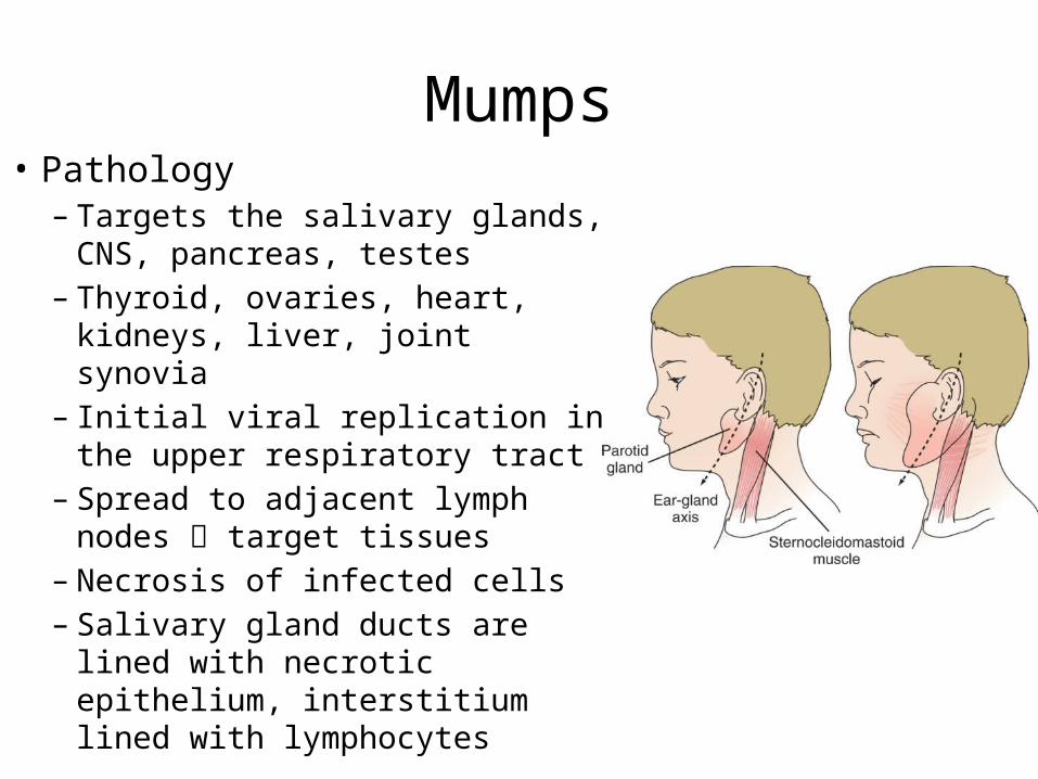
Mumps• Pathology– Targets the salivary glands, CNS,
pancreas, testes– Thyroid, ovaries, heart, kidneys,
liver, joint synovia– Initial viral replication in the
upper respiratory tract– Spread to adjacent lymph nodes target tissues
– Necrosis of infected cells – Salivary gland ducts are lined with
necrotic epithelium, interstitium lined with lymphocytes
Mumps
Incubation Period
Prodromal Illness
Exanthematous Phase Recovery
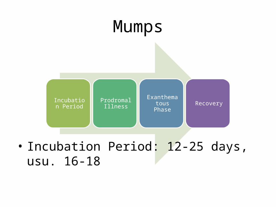
• Incubation Period: 12-25 days, usu. 16-18
Mumps
Incubation Period
Prodromal Illness Parotitis Recovery
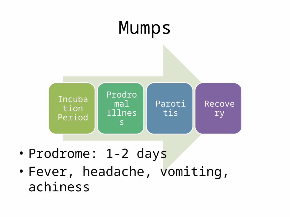
• Prodrome: 1-2 days • Fever, headache, vomiting, achiness
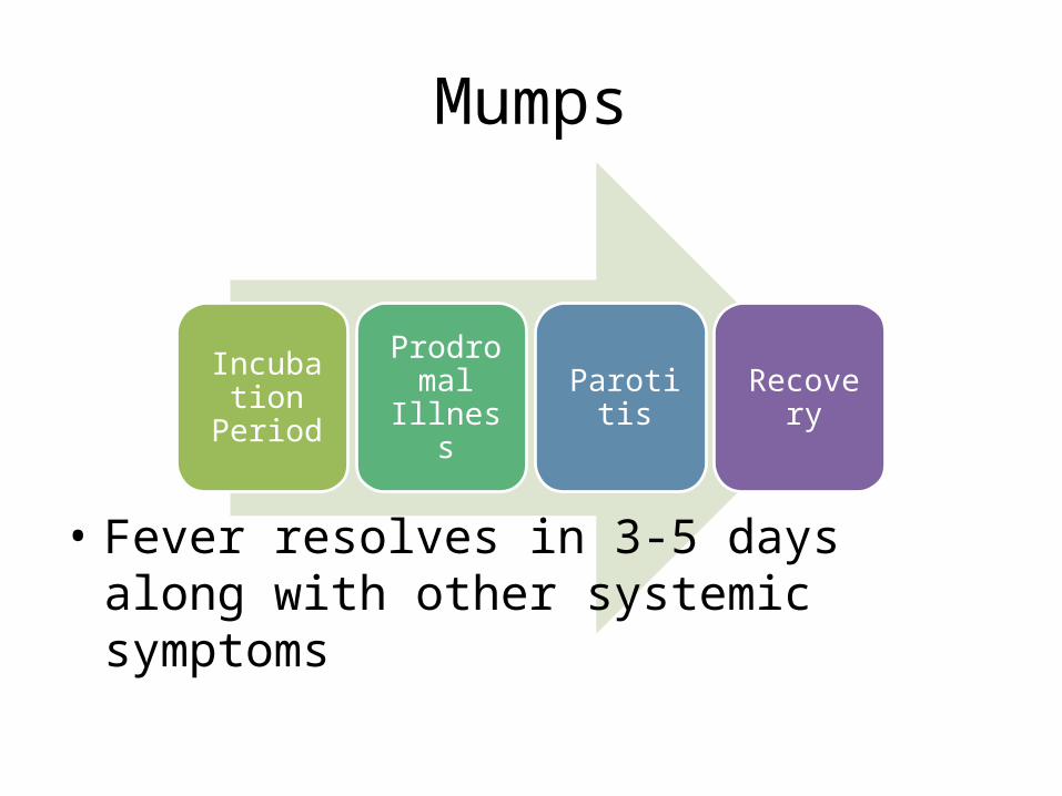
Mumps
Incubation Period
Prodromal Illness Parotitis Recovery
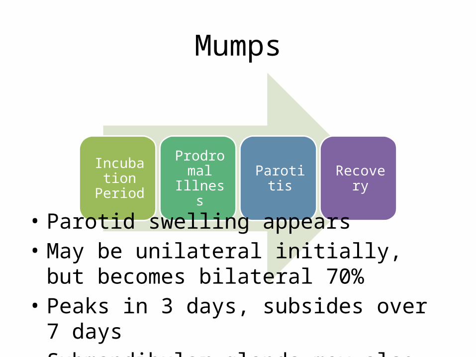
• Parotid swelling appears• May be unilateral initially, but becomes
bilateral 70%• Peaks in 3 days, subsides over 7 days • Submandibular glands may also be involved
Mumps
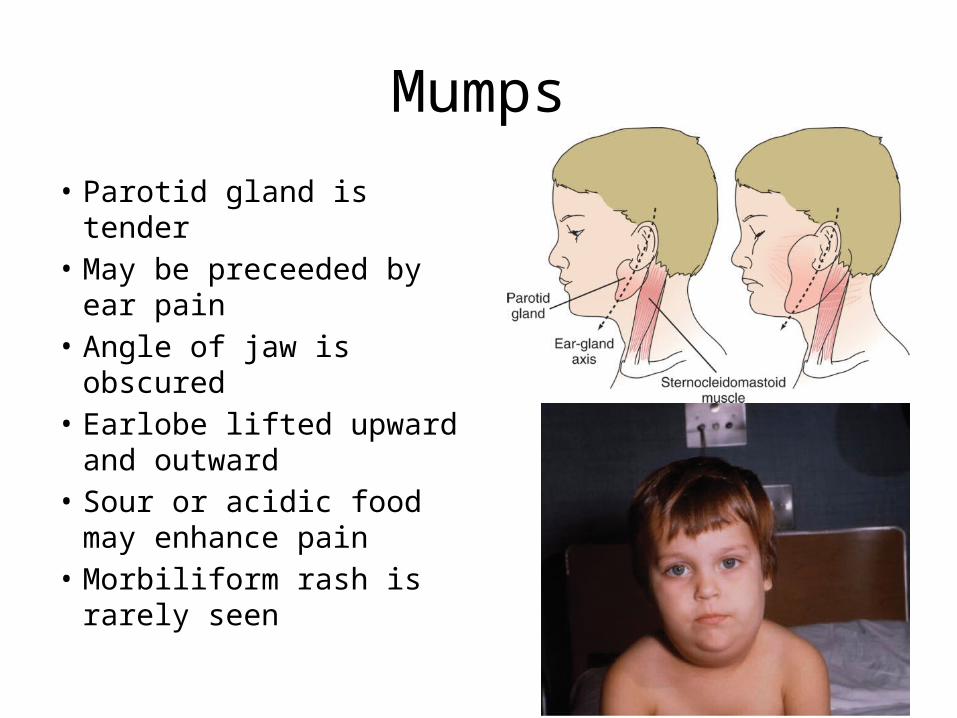
• Parotid gland is tender• May be preceeded by ear
pain• Angle of jaw is obscured• Earlobe lifted upward
and outward• Sour or acidic food may
enhance pain• Morbiliform rash is rarely
seen
Mumps
Incubation Period
Prodromal Illness Parotitis Recovery
• Fever resolves in 3-5 days along with other systemic symptoms
Diagnostics
• History of exposure to mumps infection• Clinical findings• Elevated amylase level• Relative lymphocytosis• Serologic testing– Increase in mumps IgG, IgM– IgG may cross react with antibodies to parainfluenza virus
• Isolation of the virus in cell culture, PCR, immunofluorescence
Treatment
• No specific antiviral therapy is available for mumps
• Pain control• Adequate hydration• Antipyretics for fever
Complications
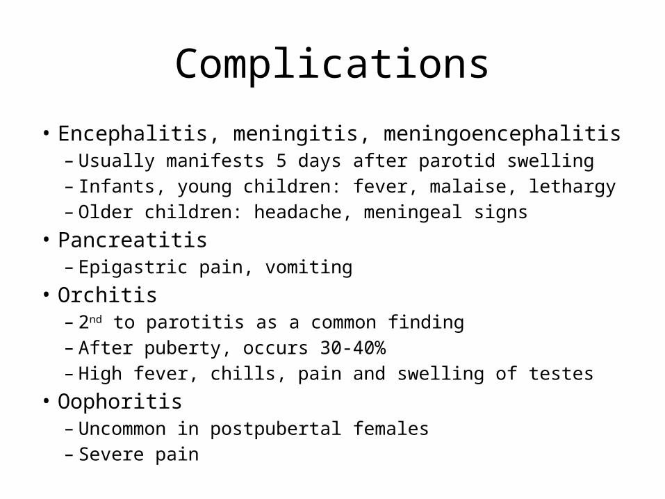
• Encephalitis, meningitis, meningoencephalitis– Usually manifests 5 days after parotid swelling– Infants, young children: fever, malaise, lethargy– Older children: headache, meningeal signs
• Pancreatitis– Epigastric pain, vomiting
• Orchitis– 2nd to parotitis as a common finding– After puberty, occurs 30-40%– High fever, chills, pain and swelling of testes
• Oophoritis– Uncommon in postpubertal females– Severe pain
Prognosis
• Excellent• Some fatal cases of encephalitis were reported
Prevention
• 2 dose MMR vaccine– MMR1: 12-15 mos– MMR2: 4-6 years

ROSEOLA
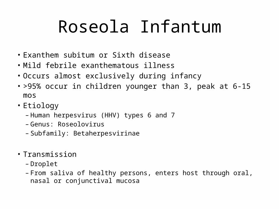
Roseola Infantum• Exanthem subitum or Sixth disease • Mild febrile exanthematous illness• Occurs almost exclusively during infancy• >95% occur in children younger than 3, peak at 6-15 mos• Etiology
– Human herpesvirus (HHV) types 6 and 7– Genus: Roseolovirus– Subfamily: Betaherpesvirinae
• Transmission– Droplet– From saliva of healthy persons, enters host through oral, nasal or
conjunctival mucosa
Roseola Infantum
• Prodrome– Usually asymptomatic– Mild URTI signs: minimal rhinorrhea, slight
pharyngeal inflammation, mild conjunctival redness
– Mild cervical or occipital lymphadenoathy– Mild palpebral edema
Roseola Infantum
• High fever (average: 39C), lasts for 3-5 days• Rhinorrhea, sore throat, abdominal pain, vomiting,
diarrhea• Nagayama spots: ulcers at uvulopalatoglossal
junction• Rash appears within 12-24 hours of fever lysis– Rose-colored, discrete, 2-5mm, slightly raised lesions on
trunk neck face proximal ext– Usu. Non-pruritic, no vesicles or pustules– Fades after 1-3 days

Clinical Manifestation
Abrupt onset of high fever with fussiness
Rash: faint pink or rose-colored, nonpruritic, 2- to 3-mm morbilliform rash
Diagnostics
• Clinical presentation• Serology• Virus culture• PCR
Treatment
• Supportive therapy• HHV-6 inhibit by ganciclovir, cidofovir, foscarnet
(but not acyclovir) • HHV-7 inhibited by cidovir and foscarnet• No approved treatment• Treatment is warranted for
immunocompromised children with severe disease– Gangciclovir, cidofovir for 2-3 weeks

RUBELLA
Rubella
• German measles• Three-day measles
• Rash similar to that of mild rubeola or scarlet fever
• Enlargement and tenderness of the postoccipital, retroauricular, and posterior cervical lymph nodes
• Rubella in early pregnancy may cause the congenital rubella syndrome
Etiology
• RNA virus• Genus Rubivirus• Family Togaviridae
Epidemiology
• Humans are the only natural host of rubella virus
• Distributed worldwide, affects both sexes equally
• Spread by:– Oral droplet– Transplacentally to the fetus
Epidemiology
• Peak incidence: 5-14 years of age• In closed populations, such as institutions and
military barracks, almost 100% of susceptible individuals may become infected.
• In family settings, 50-60% of susceptible family members acquire the disease.
Epidemiology
• Virus is shed in nasopharyngeal secretions, blood, feces, and urine
• Virus is present in the nasopharynx 7 days before exanthem, and 7-8 days after its disappearance
• Subclinical disease is also infectious
Pathogenesis
• Not well understood• Virus can be found from both infected and
uninfected areas of the skin– Immune processes may be important
Pathogenesis
• Primary maternal infection during the 1st trimester: greatest risk of congenital defects
• <11th wk AOG: 90% • End of trimester: 10-20%• >16th wk AOG: low risk for congenital defects• Overall risk for first trimester infection: 70%
Clinical Manifestations
• Incubation period: 14-21 days• Prodromal phase of mild catarrhal symptoms
is shorter than that of measles
Clinical Manifestations
• Retroauricular, posterior cervical, and postoccipital lymphadenopathy– Most characteristic
• Evident at least 24 hours before the onset of rashes
• May last up to 1 week
Clinical Manifestations
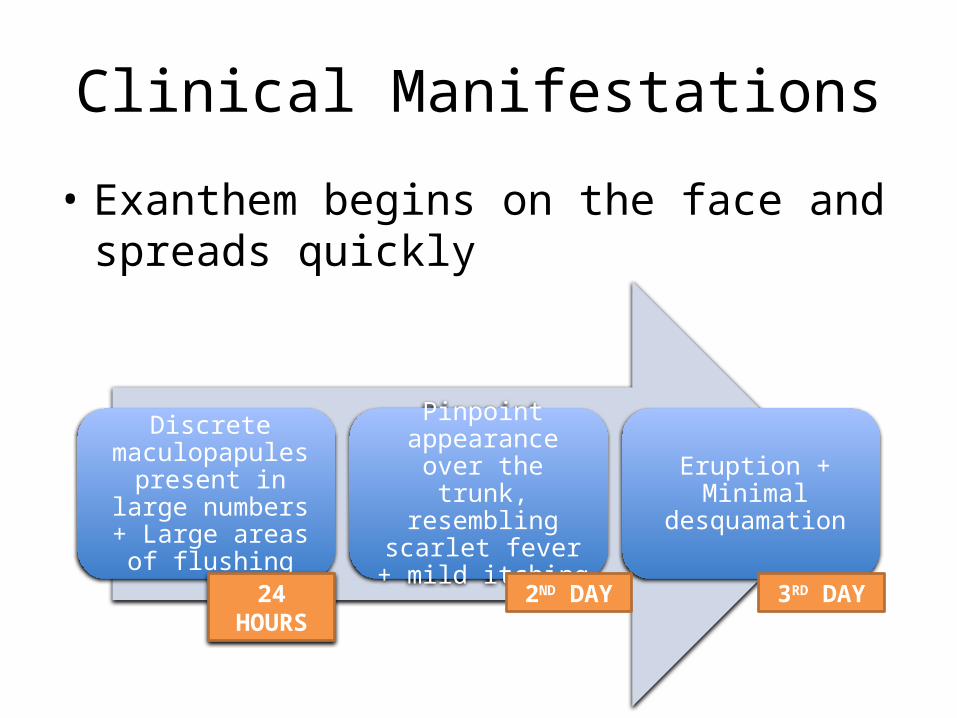
• Exanthem begins on the face and spreads quickly
Discrete maculopapules present in large numbers + Large areas of flushing
Pinpoint appearance over the trunk,
resembling scarlet fever + mild itching
Eruption + Minimal desquamation
24 HOURS 2ND DAY 3RD DAY
Clinical Manifestations
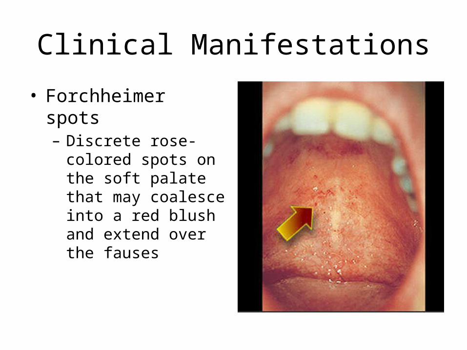
• Forchheimer spots– Discrete rose-colored
spots on the soft palate that may coalesce into a red blush and extend over the fauses
Clinical Manifestations
• Pharyngeal mucosa and conjunctivae are slightly inflamed
• No photophobia (in contrast to rubeola)• Fever is low grade or absent and usually lasts
only for 3 days• Polyarthritis may occur with arthralgia, swelling,
tenderness, and effusion but usually without any residuum
• NOT common: headache, malaise, anorexia
Differential Diagnosis
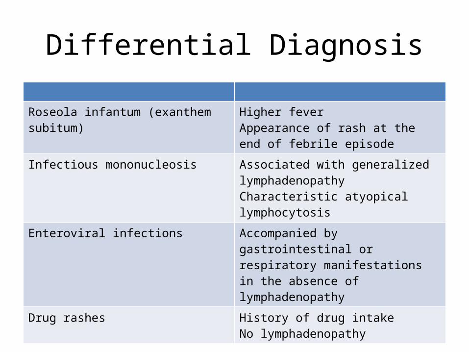
Roseola infantum (exanthem subitum) Higher feverAppearance of rash at the end of febrile episode
Infectious mononucleosis Associated with generalized lymphadenopathyCharacteristic atyopical lymphocytosis
Enteroviral infections Accompanied by gastrointestinal or respiratory manifestations in the absence of lymphadenopathy
Drug rashes History of drug intakeNo lymphadenopathy
Diagnosis
• Clinical• Objective findings:– Enlarged spleen– Normal or slightly reduced WBC count– Thrombocytopenia (rare)
• Confirmed by serology or virus culture
Diagnosis
• Rubella virus can be cultured from the nasopharynx and blood.
• It is detected by the ability of rubella-infected African green monkey kidney (AGMK) cells to resist challenge with enterovirus.
Diagnosis
• Hemagglutination-inhibition (HI) antibody • Latex agglutination • enzyme immunoassay• passive hemagglutination, and • fluorescent immunoassay
Diagnosis
• Immunoglobulin (Ig) M antibodies – detectable in the first few days of illness– Also useful for the diagnosis of congenital rubella
syndrome. • Seroconversion, or a fourfold increase in IgG
titer, is diagnostic.
Treatment
• Supportive treatment• No specific antiviral therapy• Antipyretics for fever
Prognosis
• Excellent.• Infection usually confers permanent immunity,
although reinfection may occur.
Complications
• Encephalitis– Occurs in about 1 in 6,000 cases– Overall mortality rate of 20%– Symptoms usually resolve within 1-3 wk without
neurologic sequelae• Thrombocytopenic purpura occurs at an
overall rate of 1 in 3,000 cases.
Complications
• The most important consequence of rubella in a pregnant woman is congenital rubella syndrome.
• Progressive rubella panencephalitis is a persistent, slowly progressive rubella infection of the central nervous system

Congenital Rubella Syndrome
• Intrauterine growth retardation: most common manifestations
• Common findings:– Cataracts– Myocarditis and structural
cardiac defects– Blueberry muffin skin
lesions– Sensorineural hearing loss– Meningoencephalitis
Congenital Rubella Syndrome
• Diagnosis:– Rubella-specific IgM antibody in the neonatal
serum, or by culturing rubella virus from the infant (nasopharynx, urine, or tissues)
– Isolating the virus from amniotic fluid or by identification of rubella-specific IgM in cord blood.
Congenital Rubella Syndrome
• Prognosis– Better for infants with fewer stigmata of the
syndrome, presumably those who were initially infected later in gestation.
– Only 30% of infants with encephalitis appear to escape residual neuromotor deficitis, including an autistic syndrome
Reinfection
• The incidence of reinfection– 3-10% on exposure to wild virus among those with
a history of previous rubella – 14-18% among those immunized with the RA 27/3
vaccine. • May lead only to an IgG booster response, to
both an IgM and IgG response, or to clinical rubella.
Prevention
• Rubella vaccine is derived from the RA 27/3 strain of rubella virus
• Induces antibody in more than 99% of seronegative recipients and has protective efficacy in more than 90%
• Vaccine virus may be shed from the nasopharynx in low titers for as long as 18-25 days after vaccination
Prevention
• MMR– 1st: recommended at 12-15 mo of age. – 2nd: recommended routinely at 4-6 yr of age but may
be administered at any time during childhood provided at least 4 wk have elapsed since the first dose.
• Pregnant women should not be given live rubella virus vaccine and should avoid becoming pregnant for 3 mo after they have been vaccinated
Prevention
• Symptoms that may follow rubella immunization include – Fever (5-15%), – Rash (5%), – Lymphadenopathy, and– Arthralgias and arthritis
Postexposure prophylaxis
• Nonpregnant susceptible contacts of persons with rubella should be vaccinated– does not prevent infection but ensures protection
for future rubella exposures• All pregnant women, regardless of
immunization history, should make every effort to avoid exposure to rubella.
ERYTHEMA INFECTIOSUM
Parvovirus B19
• Erythema infectiosum• Fifth disease
Etiology
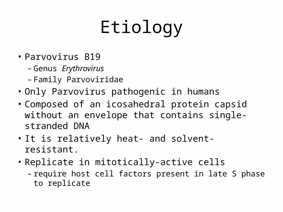
• Parvovirus B19 – Genus Erythrovirus – Family Parvoviridae
• Only Parvovirus pathogenic in humans• Composed of an icosahedral protein capsid without
an envelope that contains single-stranded DNA• It is relatively heat- and solvent-resistant. • Replicate in mitotically-active cells– require host cell factors present in late S phase to
replicate
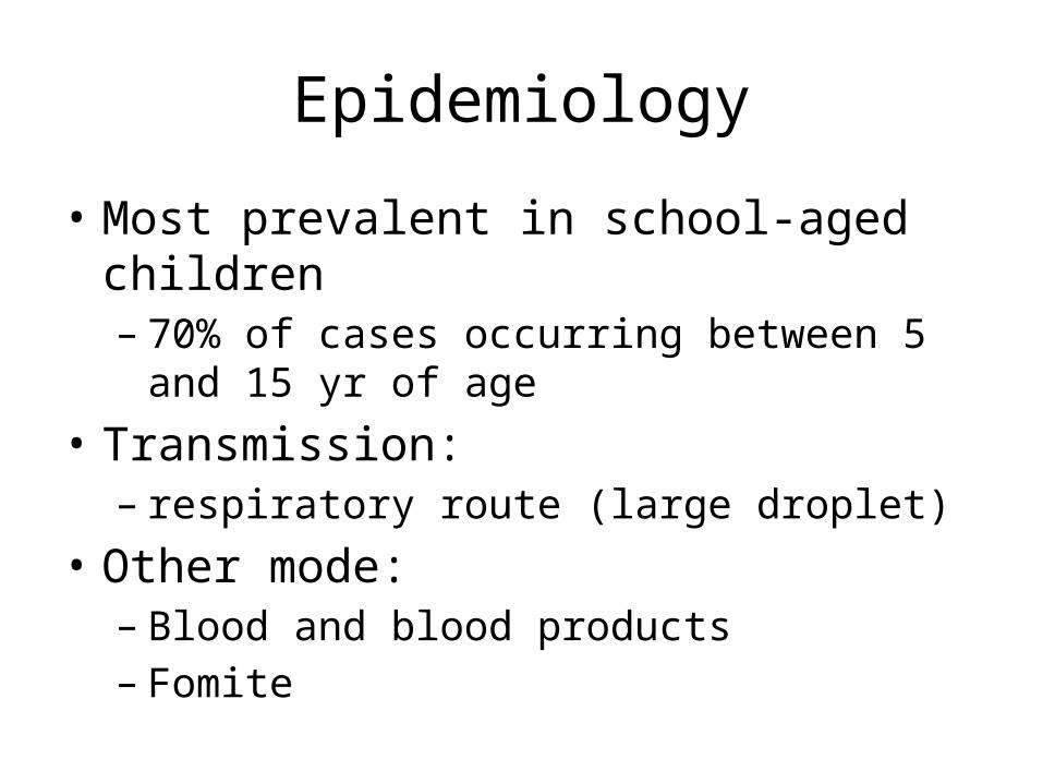
Epidemiology
• Most prevalent in school-aged children– 70% of cases occurring between 5 and 15 yr of age
• Transmission:– respiratory route (large droplet)
• Other mode:– Blood and blood products– Fomite
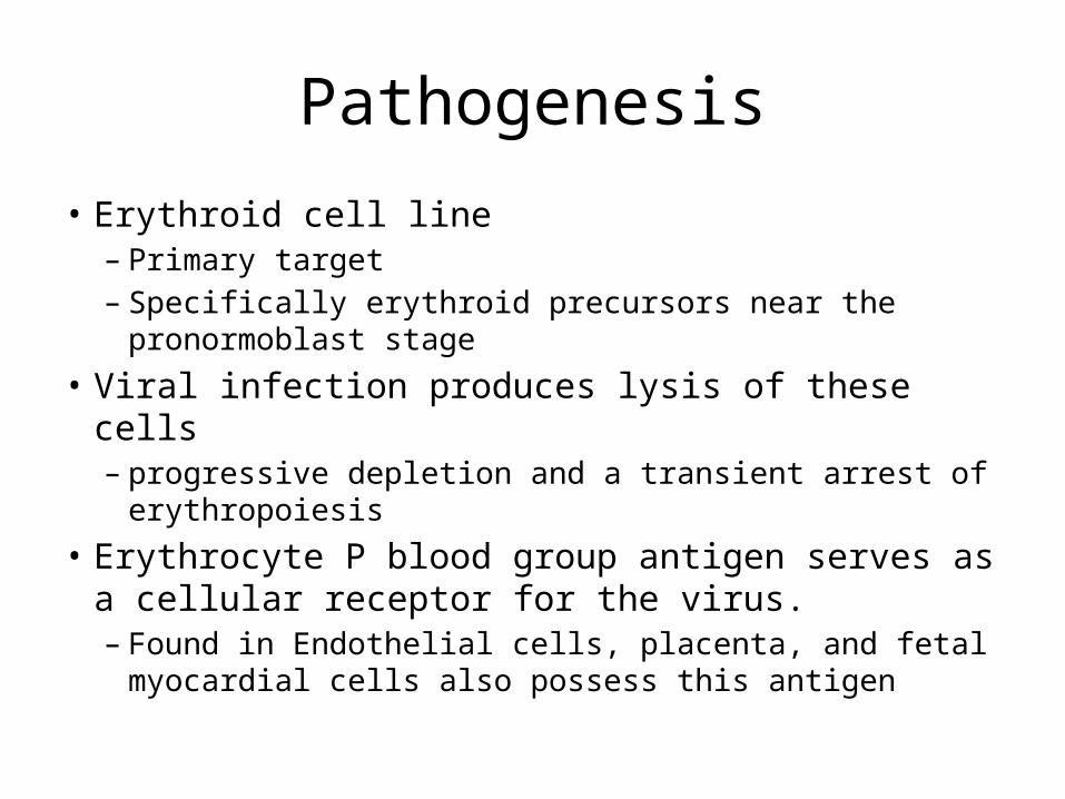
Pathogenesis
• Erythroid cell line– Primary target– Specifically erythroid precursors near the pronormoblast
stage• Viral infection produces lysis of these cells– progressive depletion and a transient arrest of
erythropoiesis• Erythrocyte P blood group antigen serves as a cellular
receptor for the virus. – Found in Endothelial cells, placenta, and fetal myocardial
cells also possess this antigen
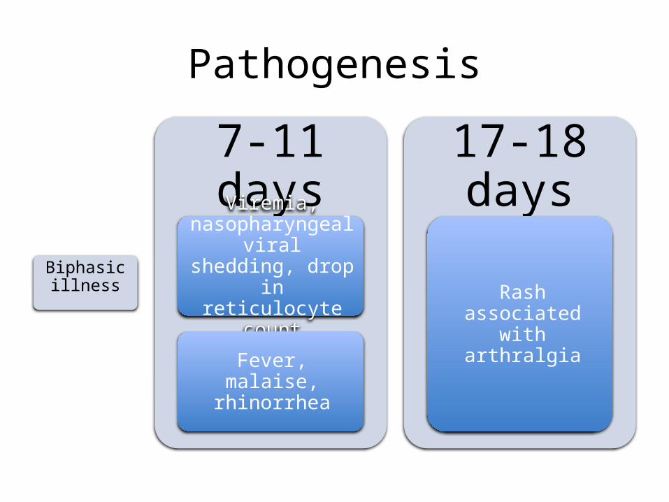
Pathogenesis
Biphasic illness
7-11 daysViremia,
nasopharyngeal viral shedding, drop in reticulocyte count
Fever, malaise, rhinorrhea
17-18 days
Rash associated with arthralgia
Pathogenesis
• B19 is associated with nonimmune fetal hydrops and stillbirth in women experiencing a primary infection
• Not teratogenic
Clinical Manifestation
• Most common manifestation: erythema infectiosum
• Benign self-limited exanthematous• Incubation period: 4-28 days (ave: 16-17 days)
Clinical Manifestation
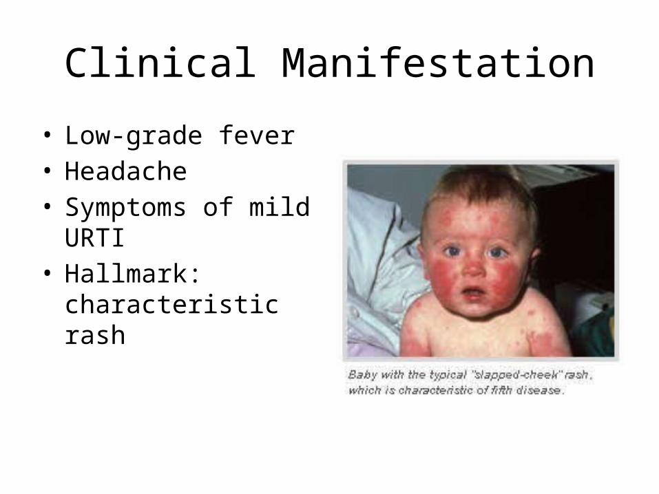
• Low-grade fever• Headache• Symptoms of mild URTI• Hallmark: characteristic
rash
Clinical Manifestation
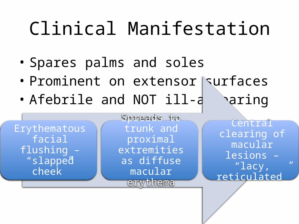
• Spares palms and soles• Prominent on extensor surfaces• Afebrile and NOT ill-appearing
Erythematous facial flushing – “slapped
cheek”
Spreads to trunk and proximal extremities
as diffuse macular erythema
Central clearing of macular lesions – “lacy, reticulated”
Clinical Manifestation
• Papular-purpuric "gloves and socks" syndrome (PPGSS)– Fever– Pruritus– Painful edema and erythema localized to the distal
extremities in a distinct glove and sock distribution– Acral petechiae and oral lesions
Diagnosis
• Clinical• Serologic tests– B19-specific IgM: persists for 6-8 wk– Anti-B19 IgG: marker of past infection/immunity– Anti-B19 IgM: best marker of recent/acute
infection – seroconversion of anti-B19 IgG antibodies can also
be used to confirm recent infection
Diagnosis
• Cannot be isolated by culture– PCR or nucleic acid hybridization are necessary
Differential Diagnosis
• Rubella• Measles• Enteroviral infections• Drug reactions• Juvenile rheumatoid arthritis• SLE
Treatment
• No specific antiviral therapy• Administration of IVIG may give only a
temporary remission, and periodic re-infusions may be required.
Complications
• Arthralgia or arthritis– May persist after the rash
• Thrombocytic purpura• Aseptic meningitis• Virus-associated hemophagocytic syndrome
Prevention
• Children with erythema infectiosum are not likely to be infectious at presentation because the rash and arthropathy represent immune-mediated, postinfectious phenomena.
• No vaccine• No data to support post-exposure prophylaxis

VARICELLA
Varicella-Zoster
• Primary– Varicella (chickenpox)– Establishment of lifelong latent infection of
sensory ganglion neurons• Reactivation – herpes zoster (shingles)
Etiology
• Neurotropic human herpesvirus • Double-stranded DNA genomes
Epidemiology
• Within households, transmission to susceptible individuals occurs at a rate of 65-86%;
• More casual contact is associated with lower attack rates among susceptible children
Epidemiology
• Contagious period: – From 24-48 hr BEFORE the rash appears – Until 3-7 days after onset of rash (crusted vesicles)
• Susceptible children may also acquire varicella after close, direct contact with adults or children who have herpes zoster.
Epidemiology
• Lifetime risk for herpes zoster is 10-15%• 75% of cases occurring after 45 yr of age
Pathogenesis
• Transmission:– Respiratory secretions (airborne)– Fluid of skin lesions (direct contact)
Pathogenesis
• Incubation period: 10-21 days– Virus replicates in the respiratory tract followed by
a brief subclinical viremia– During the late incubation period, virus is
transported back to respiratory mucosal sites facilitating transmission even before appearance of rash
• Second viremic phase: Widespread cutaneous lesions
Pathogenesis
• VZV establishes latent infection in sensory ganglia cells in all individuals who experience primary infection.
• Subsequent reactivation: herpes zoster– a vesicular rash that usually is dermatomal– necrotic changes may be produced in the
associated ganglia
Clinical Manifestations
• Acute febrile rash illness• Variable severity but usually self-limited• Herpes zoster, uncommon in children, causes
localized cutaneous symptoms
Clinical Manifestations
• Illness usually begins 14-16 days after exposure
Clinical Manifestations
• Fever, malaise, anorexia, headache, and occasionally mild abdominal pain may occur 24-48 hr before the rash appears.
• Fever and other systemic symptoms persist during the first 2-4 days after the onset of the rash.
Clinical Manifestations
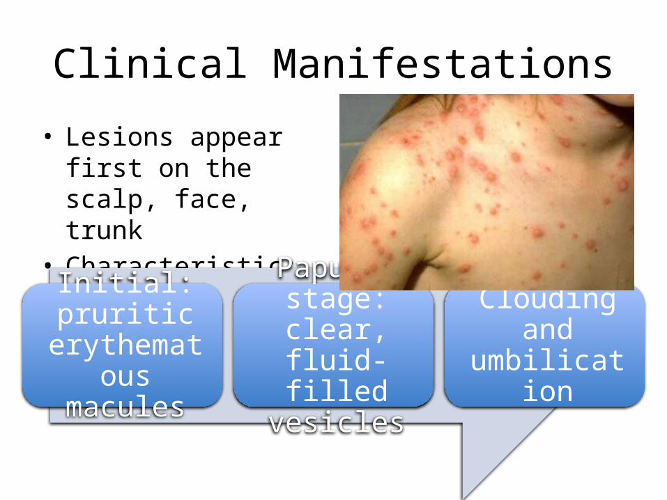
• Lesions appear first on the scalp, face, trunk
• Characteristic: lesions in various stages
Initial: pruritic erythematous
macules
Papular stage: clear, fluid-
filled vesicles
Clouding and umbilication
Clinical Manifestations
• Distribution: central or centripetal– As opposed to smallpox where rashes are
prominent on the face and distal extremities
Clinical Manifestations
• Hypopigmentation or hyperpigmentation of lesion sites persists for days to weeks in some children, but severe scarring is unusual unless the lesions were secondarily infected
Differential Diagnosis
• Includes vesicular rashes caused by other infectious agents– herpes simplex virus– enterovirus, or – Staphylococcus aureus;
• Drug reactions• Contact dermatitis• Insect bites.
Diagnosis
• Leukopenia is typical during the first 72 hours; it is followed by a relative and absolute lymphocytosis. Results of liver function tests are also usually (75%) mildly elevated
Diagnosis
• VZV can be identified quickly by direct fluorescence assay (DFA) of cells from cutaneous lesions, which is widely available, and by polymerase chain reaction (PCR) amplification testing. Although multinucleated giant cells can be detected with nonspecific stains (Tzanck smear), they have poor sensitivity and do not differentiate VZV and herpes simplex virus infections.
Diagnosis
• Infectious virus may be recovered using tissue culture methods; newer methods have decreased time needed for culture from 7-10 days to 3-4 days. VZV immunoglobulin G (IgG) antibodies can be detected by several methods and a 4-fold rise in IgG antibodies is also confirmatory of acute infection. VZV IgG antibody tests can also be valuable to determine the immune status of individuals whose clinical history of varicella is unknown or equivocal.
Treatment
• Antiviral treatment modifies the course of both varicella and herpes zoster.
• Acyclovir• Foscarnet for acyclovir-resistant VZV
Treatment
• Oral therapy with acyclovir (20 mg/kg/dose; maximum: 800 mg/dose) given as 4 doses per day for 5 days should be used to treat uncomplicated varicella
• To be most effective, treatment should be initiated as early as possible, preferably within 24 hr of the onset of the exanthem.
Complications
• Bacterial super-infection• Pneumonia• Encephalitis• Bleeding disorders• Congenital infection• Life-threatening perinatal infection