visible light-driven photophysics and photochemistry of ... · pdf filea department of general...
TRANSCRIPT

1
Accepted manuscript of Coord. Chem. Rev. 325 (2016) 59–66
http://dx.doi.org/10.1016/j.ccr.2015.12.011
Visible light-driven photophysics and photochemistry of water-soluble
metalloporphyrins
Ottó Horváth,a* Zsolt Valicsek,a Melinda A. Fodor,a Máté M. Major,a Muhammad Imran,a
Günter Grampp,b Alexander Wankmüllerb
a Department of General and Inorganic Chemistry, Institute of Chemistry,
University of Pannonia, P.O.Box 158, 8201 Veszprém, Hungary, b Institute of Physical and Theoretical Chemistry, Graz University of Technology, 8010 Graz,
Stremayrgasse 9., Austria
Contents
1. Introduction……………………………………………………………………………......00
2. Photophysics and photoredox chemistry of water-soluble in-plane metalloporphyrins…..00
2.1 Photophysics and photoredox chemistry of cationic manganese(III) and cobalt(III)
porphyrins…………………………………………………………………………...00
2.1.1 Photophysics…………………………………………………………………….00
2.1.2 Photochemistry………………………………………………………………….00
2.2 Photophysics and photoredox chemistry of a cationic nickel(II) porphyrin……...….00
2.2.1 Photophysics………………………………………………………...………..…00
2.2.2 Photochemistry………………………………………………………………….00
3. Photophysics and photochemistry of water-soluble out-of-plane lanthanide(III)
metalloporphyrins.………………………………………………………………………....00
3.1 Photophysics ……………………………..………………………………….………00
3.2 Photochemistry……………………………...……………………………………….00
4. Summary and conclusions…………………………………………………………………00
Acknowledgment………………………………………………...………………………00
References……………………………………………………………………………….00
__________________________________________________________________________
Abstract
Metal ions can form normal (in-plane) metalloporphyrins, fitting into the central hole of
the porphyrin ring, or several of them are located out of the ligand plane, resulting in sitting-
atop (SAT) complexes. Kinetically inert water-soluble complexes of Mn(III), Co(III), and
Ni(II) with 5,10,15,20-tetrakis(1-methylpyridinium-4-yl)porphyrin display a weak, short-lived
fluorescence. This can be affected by elongation of the alkyl substituent and using micellar
environment in the case of Mn(III) porphyrins. In the presence of a suitable electron donor
(triethanolamine, TEOA) and acceptor (methylviologen, MV2+), these metalloporphyrins
proved to be efficient photocatalysts transferring electrons between the ground-state donor
and acceptor via outer-sphere mechanism. In these systems triplet excited-state Mn(III) and
2
Co(III) porphyrins are dynamically quenched with TEOA. The Mn(II) and Co(II) complexes
formed in this way need also photoexcitation for the transfer of electron to the ground-state
acceptor. However, the triplet excited state of Ni(II)TMPyP4+ cannot be dynamically
quenched with TEOA. Instead, this electron donor forms an associate with Ni(II)TMPyP4+ in
a ground state equilibrium. The lifetime of the triplet excited state of this species is much
longer than that of the nickel(II) porphyrin alone, and it can undergo an efficient dynamic
oxidative quenching with MV2+. Thus, a one-step electron transfer can be realized between
the electron donor and acceptor, generating MV+, which can be utilized for hydrogen
generation from water.
Lanthanide(III) porphyrins are of typical SAT complexes, the photophysical and –chemical
features of which can be tuned by the size of the metal center. Anionic, early lanthanide(III)
mono- and bisporphyrin complexes exhibit very similar photoinduced properties as a
consequence of a special type of aggregation, through the peripheral substituents. The rather
slow formation of complexes and transformation between the mono- and bisporphyrins can be
accelerated by the irradiation of the system. These by-processes play considerable roles beside
the photoredox degradation and demetalation. Depending on the wavelength of irradiation,
two types of photoproducts can appear: during the photolysis at the Soret-band, a radical type
intermediate can be observed, which disappears in dark. However, irradiation at the Q-bands,
generates the formation of a new, stable photoproduct.
Keywords: metalloporphyrins; photochemistry; out-of-plane compexes; in-plane complexes;
photocatalysis; lanthanide ions
1. Introduction
Metalloporphyrins display special spectral and coordination features [1, 2]. Their
advantageous photoinduced properties can also be exploited in various photocatalytic
procedures [3, 4]. Water-soluble derivatives can be utilized in environmentally benign
systems not containing organic solvents.
Kinetically inert in-plain metalloporphyrins, in which the metal center is coplanarly located in
the cavity of the ligand, may offer promising possibilities for realization of photocatalytic
systems based on outer-sphere electron transfer [5]. The so-called hyper-porphyrins can be
particularly interesting in this respect, due to their distorted structure, which may increase the
(photo)redox reactivity of these complexes. In these complexes the metal center is smaller
than the ligand cavity, hence it shrinks the porphyrin ring, resulting in a saddle or ruffled
distortion.
Metal ions can be located out of the ligand plane too, resulting in kinetically labile out-of-
plane (OOP or sitting-atop=SAT) complexes with dome-distorted structure, displaying typical
photophysical and photochemical properties [3, 6]. Besides, the OOP position promotes the
formation of bis- or oligoporphyrins [7]. Lanthanide(III) complexes can be typical examples
of metallo-oligoporphyrins, in which the π-π interaction depends on the distance between the
3
macrocycles, strongly influenced by the size of the metal center [8, 9]. Therefore,
lanthanide(III) ions are good candidates for fine tuning of the out-of-plane distances, due to
the lanthanide contraction.
The typical in-plane metalloporphyrins show blueshifts in the Soret-range compared to the
corresponding free-base porphyrins because the atomic orbitals of their metal center
covalently bonded in the plane can overlap more strongly with the occupied molecular
orbitals (the highest in energy is the HOMO) of the ligand, resulting in a stronger reduction in
energy; while the unoccupied MOs (the lowest is the LUMO) do not change. Accordingly, the
energy gaps between the excited and ground states increase.
In the out-of-plane complexes, the atomic orbitals of the more weakly bonded metal ions may
slightly influence the unoccupied MOs and lesser the occupied ones, resulting in the decrease
of the energy gaps, i.e. the increase of the corresponding wavelengths.
The aim of this review is to exemplify the considerable difference between the photoinduced
features of the kinetically inert in-plane and the labile sitting-atop (SAT) or out-of-plane
(OOP) complexes, due to their fundamental constructional deviation. Fig. 1 shows the
structures of the anionic and cationic porphyrins involved in this work. In the case of the
cationic porphyrins, additionally (beside the methyl) hexyl and dodecyl derivatives were also
investigated.
Fig. 1. Structures of anionic and cationic porphyrins: H2TSPP4- = 5,10,15,20-tetrakis(4-
sulfonatophenyl)porphyrin, H2TMPyP4+ = 5,10,15,20-tetrakis(1-methyl-4-
pyridinium)porphyrin.
2. Photophysics and photoredox chemistry of water-soluble in-plane metalloporphyrins
In the case of most of the in-plane metalloporphyrins the size of the metal center corresponds
to that of the ligand cavity, thus it is located in the porphyrin plane and does not distort it.
However, if the ionic radius (ri) of the central metal is significantly smaller than the core of
the porphyrin, i.e. ri < 70 pm, it shrinks the ligand and, hence, causes its distortion. The type
of such a distortion may be saddle, wave or ruffled. This structural change, compared to the
normal in-plane metalloporphyrins, considerably modifies the visible absorption spectrum of
these complexes, especially in the range of the Soret-bands. While the Soret-bands of most of
the in-plane metalloporphyrins display blue shifts compared to those of the corresponding free
bases, due to electronic interactions between the ligand and the metal center, the distorted
ones exhibit red shifts in this respect. Thus, as a distinction, since the spectra of these
complexes could not be interpreted by the 4 MO theory of Gouterman, they have been
classified as hyper-porphyrins [10, 11]. The porphyrin complexes of manganese(III) (ri =58
pm, low-spin), cobalt(III) (ri = 55 pm, low-spin), and nickel(II) (ri = 63 pm, low-spin) [12]
belong to this group [13]. Some photophysical and photoredox properties of cationic
porphyrin complexes of these metal ions will be discussed in the following sections.

4
2.1 Photophysics and photoredox chemistry of cationic manganese(III) and cobalt(III)
porphyrins
2.1.1 Photophysics
Some photophysical properties of cationic manganese(III) and cobalt(III) porphyrins,
Mn(III)TMPyP5+ and Co(III)TMPyP5+ were already studied [14, 15] earlier. It was generally
accepted that the highly distorted porphyrin complexes with diamagnetic metal center do not
display appreciable fluorescence at room temperature. Accordingly, porphyrin complexes of
low-spin Mn(III) and Co(III) were considered to be very weakly fluorescent. However,
current investigations using a more sensitive equipment indicated that both Mn(III)TMPyP5+
and Co(III)TMPyP5+ display a characteristic fluorescence in the 600-750-nm range (Fig 2),
which is more than an order of magnitude weaker (fl = 1.110-4 and 1.410-3 for the Mn(III)
and Co(III) complexes, respectively[16, 17]) than that exhibited by the corresponding free
base (0.047 [18], 0.030 [19]). The shorter emission lifetimes of these complexes are also in
accordance with this tendency caused by both electronic and steric interactions between the
metal center and the ligand in these hyper-porphyrins.
Fig 2. Fluorescence spectra of Mn(III)TMPyP5+, Co(III)TMPyP5+ and H2TMPyP4+ obtained
by excitation at the Soret-bands. Adapted from refs [16, 17].
The fluorescence bands shown in Fig 2 can be assigned as S1S0 transitions (with various
vibrational states). The fluorescence bands of metalloporphyrins (both for in-plane and for
out-of-plane complexes) are blue-shifted and less intense compared to those of the
corresponding free-base porphyrin. The same spectra were obtained at Q-band excitations,
confirming that they are the results of S1S0 transition.
Elongation of the alkyl chain on the pyridinium substituent (from methyl to hexyl or dodecyl)
in the case of the cationic manganese(III) porphyrins resulted in a significant (about an order
of magnitude) increase in the fluorescence quantum yield (to 1.410-3 [16]), while for the
corresponding free bases just a moderate decrease within the same order magnitude was
observed [20].This phenomenon may be attributed to the stronger electron-donating property
of the longer hydrocarbon chains. In the solution of a cationic surfactant (cetyltrimethyl
ammonium) providing a micelle:Mn(III)-porphyrin ratio > 1, the fluorescence lifetime
decreased upon increasing the length of the alkyl chain (3.40 ns, 3.03 ns, and 2.30 ns for
methyl, hexyl, and dodecyl deriavtives, respectively). This tendency, which is just the
opposite of the change observed in the homogeneous aqueous system, can be attributed the
increasing hydrophobic interaction between the alkyl chains of the surfactant in the micelles
and those of the substituents on the porphyrins.
2.1.2 Photochemistry
Both Mn(III)TMPyP5+ and Co(III)TMPyP5+ were applied in various photocatalytic systems.
The cobalt(III) complex proved to be more efficient in oxidation of the sulfide content of a
wastewater to sulfate, at Soret-band excitation [21]. It could also be used for oxidative
splitting of DNA in the presence of suitable electron acceptors [22]. Mn(III)TMPyP5+ was

5
utilized in photocatalytic oxygenations of various hetero-bicyclic organic compounds such as
furan and thiophene derivatives [23, 24]. In the latter cases, the corresponding manganese(V)-
oxo species was suggested to play the key role in the oxidation process.
While the mechanism of the photocatalytic oxidations and oxygenations has not been
investigated in detail, photoreductive catalyses based on these metalloporphyrins were more
thoroughly investigated.
Utilizing ethylenediaminetetraacetate (EDTA) or triethanolamine (TEOA) as sacrificial
electron donors and methylviologen (MV2+) as an acceptor, photocatalytic systems were
realized for generation of MV+, using cationic manganese(III) [3, 25, 26] and cobalt(III) [17]
porphyrins. Current investigations indicated that application of a longer alkyl chain on the
pyridinium substituent increases the efficiency of the MV+ accumulation. Thus, e.g., the
corresponding hexyl derivative (Mn(III)THPyP5+) proved to be significantly effective than the
well-known methylated species (Mn(III)TMPyP5+) [16].
However, although the photocatalytic formation of MV+ of characteristic bands at 398 and
605 nm [27] is relatively fast (Fig 3) and the system persisted for several hours, the
decomposition of the catalyst is more significant than in the case of Mn(III)TMPyP5+. This
phenomenon is probably the consequence of the increased electron donating character of the
substituent promoting the ligand-to-metal charge transfer, irreversibly oxidizing the porphyrin
ring. Application of cationic micelles moderately increased the stability [16].
Fig 3. Spectral change in the system initially containing 5×10⁻⁶M Mn(III)THPyP5+, 5×10⁻⁴ M
TEOA and 2×10⁻³ M MV2+ during the irradiation at 0, 10 and 30 s (ir = 465 nm, l = 1 cm).
Adapted from ref [16].
Both the reduction of the metal center of the catalyst (with a sacrificial electron donor) and
the oxidation of its reduced form (with an appropriate acceptor) needed photoexcitation. The
same behavior was displayed by the corresponding cobalt(III) complex [17], and thus the two
steps can be described by the following general equations.
M(III)TMPyP5+ + TEOA + h M(II)TMPyP4++ TEOAox (1)
M(II)TMPyP4+ + MV2+ + h M(III)TMPyP5+ + MV●+ (2)
The quantum yields for the formation MV●+ at Soret-excitation are comparable for the
systems based on these two metalloporphyrins ( andthey may be considerable
for the application of these systems for hydrogen generation from water.
The Soret-band of the complex with the reduced metal center (M(II)) formed in reaction (1)
is blueshifted compared to that of the M(III) porphyrin for both manganese [25, 26] and
cobalt [17, 28], due to the larger ionic radii of the previous ones (89 pm for Mn(II) and 75 pm
for Co(II) [12]). The singlet excited states of the cationic Mn(III) and Co(III) porphyrins are
very short-lived (fl < 7 ns) for an efficient quenching with TEOA (in Eq. 1). Their triplet
states (Fig. 4), however, proved to be long-lived enough ( = 53 s for Mn(III)TMPyP4+ and

6
= 102 s for Co(III)TMPyP4+), hence they were dynamically quenched with TEOA (kq =
6.7×10⁶ M-1s-1 and 4.7×10⁶ M-1s-1) [16, 17].
Fig 4. Transient absorption spectra of triplet-states of Mn(III)TMPyP4+ () and
Co(III)TMPyP4+ (). Adapted from refs [16, 17].
A similar situation must be valid for the reactions between MV2+ and the triplet excited state
of the corresponding reduced (Mn(II) and Co(II)) porphyrins (Eq. 2).
Cationic porphyrins of both manganese(III) and cobalt(III) proved to be function in the
catalytic cycle containing TEOA and MV2+ in the same way, which is summarized in Scheme
1.
Scheme 1. Photocatalytic cycle based on cationic manganese(III) or cobalt(III) porphyrins.
2.2 Photophysics and photoredox chemistry of a cationic nickel(II) porphyrin
2.2.1 Photophysics
Deviating from the corresponding manganese(III) and cobalt(III) porphyrins, Ni(II)TMPyP4+
displays a double Soret-band in aqueous solution (see in Fig 5), due to its two spin states in
equilibrium [29]. The low-spin metal center is characterized with a square planar coordination
sphere, while the high-spin Ni(II) with an octahedral one. The Soret-band of the latter species
appears at 449 nm, while that of the low-spin complex can be found at 420 nm (very close to
the Soret-band of the free-base ligand). Similarly to the corresponding cationic Mn(III) and
Co(III) metalloporphyrins, also Ni(II)TMPyP4+ displays a characteristic fluorescence upon
excitation at the Soret-bands [30]. Interestingly, the shape and the position of the emission
spectra do not significantly depend on the excitation wavelength, and they well agree with
those of the Mn(III)TMPyP5+ and Co(III)TMPyP5+ complexes. This phenomenon suggests
that the excited state from which the S1 fluorescence originates is the same in both cases, no
matter which ground state (low-spin or high-spin) was excited. Nevertheless, the fluorescence
lifetimes slightly deviate: 1.36 ns for the square planar and 1.19 ns for the octahedral
complex. The quantum yield of the fluorescence (fl = 2.010-3) is in the same order of
magnitude as that of Co(III)TMPyP5+ (9.910-4). The higher value can be attributed to the
larger ionic radius causing smaller distortion (shrinkage) of the porphyrin ring as also
indicated by the location (less redshift) of the Soret-bands.
2.2.2 Photochemistry
Similarly to the cationic manganese(III) and cobalt(III) porphyrins, a photocatalytic system
was realized with Ni(II)TMPyP4+ too in presence of MV2+ and TEOA in argon-saturated
aqueous solutions at room temperature (Fig. 5). However, in this case the spectral behavior of
the system is significantly different from that observed with the corresponding Mn(III) and
Co(III) porphyrins under similar conditions. In those systems two photochemical steps were
involved in the photocatalytic cycle producing methylviologen [3, 16, 17]. During the
irradiation of the nickel(II) porphyrin system, however, the accumulation of MV+ was not

7
accompanied by any change in the spectrum of the photocatalyst (Fig 5). The quantum yield
for the formation of MV+ proved to be less than those for the Mn(III) and Co(III) porphyrins
( = 0.011 at pH 8.4). At pH 10 it increased to 0.013.
Fig 5. Spectral change of the solution initially containing 1.0×10⁻⁵M Ni(II)TMPyP4+, 5×10⁻⁴
M TEOA and 2×10⁻³ M MV2+ during the irradiation at 0, 24, 84, 204 and 600 s (ir = 463 nm,
l = 1 cm). Adapted from ref [30].
One can expect the formation of the highly instable Ni(I) porphyrin (or reduced nickel
porphyrin), which relays the electron in a fast thermal process. Such a reduced complex could
only be formed via triplet excited state quenched by TEOA as in the cases of the
manganese(III) and cobalt(III) porphyrins. In fact, time-resolved laser flash photolysis
investigations confirmed the formation of triplet excited state of the Ni(II)TMPyP4+
photocatalyst (Fig 6) [30]. Interestingly, its lifetime (6.31 μs) did not decrease upon addition
of 1.0 × 10⁻³ M TEOA, moreover it increased to 31.6 μs. This phenomenon indicated that
TEOA connects to the ground-state complex, and the excited triplet state of this associate is
much longer than that of the nickel(II) porphyrin alone. Additionally, the triplet state of this
associate could be efficiently quenched with MV2+ (Fig 6, inset). The rate constant for this
process was determined to be kq= 9.9×10⁶ s-1M-1.
Fig 6. Transient spectrum of triplet excited Ni(II)TMPyP4+ recorded 510 ns after a 355-nm
laser pulse of 5 ns duration. Inset: quenching of triplet Ni(II)TMPyP4+ with MV2+ in the
presence of 1.0×10⁻³ M TEOA. Adapted from ref [30].
Accordingly, deviating from the corresponding Mn(III) and Co(III) system, in the case of the
nickel(II) porphyrin the electron transfer from TEOA to MV2+ takes place directly in one step,
due to the ground-state association of the electron donor and the photocatalyst (Eqs. 5, 6).
Ni(II)TMPyP4+ + TEOA Ni(II)TMPyP4+TEOA (5)
Ni(II)TMPyP4+TEOA + MV2+ + h Ni(II)TMPyP4+ + MV●+ + TEOAox (6)
On the basis of this mechanism, the photocatalyst in this case is practically a special sensitizer
which immediately transmits its excitation energy to the electron donor, promoting the direct
charge transfer towards the acceptor.
3. Photophysics and photochemistry of water-soluble out-of-plane lanthanide(III)
porphyrins
Larger-sized metal ions are not able to coplanarly fit into the cavity of the porphyrin ring,
hence they form OOP or SAT complexes with dome-distorted structure, thermodynamic
instability, kinetic lability, typical photophysical features, and photochemical reactivity [3,
11]. Lanthanide(III) ions offer good opportunities for fine tuning of the out-of-plane distance,
utilizing the monotonous decrease of their radii from 116 pm to 98 pm (at coordination
number 8) upon increasing atomic numbers [12]. Additionally, they prone to form bis- or
8
oligoporphyrins [31, 32, 33], the photoinduced properties of which strongly depend on the π-π
interaction between the macrocycles. Notably, porphyrins are able to efficiently sensitize the
near-infrared luminescence of lanthanide ions, which can be widely applied from
telecommunication to biomedical optical imaging [34, 35]. Besides, lanthanide(III)
porphyrins may be applied for photocatalytic hydrogen production from water because their
reduced metal centers (Ln(II)) formed in photoinduced LMCT processes (typical for OOP
complexes [36, 37]) have considerably high negative redox potentials (except for
europium)[38]. In this section, we survey the photophysical and primary photochemical
properties of the complexes between a water-soluble, anionic porphyrin, the 5,10,15,20-
tetrakis(4-sulfonatophenyl)porphyrin (H2TSPP4-) and three early lanthanide(III) ions (Ln =
La, Ce, Nd).
3.1 Photophysics
Lanthanide(III) ions are hard Lewis acids, therefore their insertion into the coordination cavity
of the tetradentate, N-donor porphyrin ligand is a very slow process in aqueous solution [9,
39]. It is partly the consequence of the strong Ln(III)-H2O bond. Accordingly, a harder O-
donor ligand, such as acetate, can hinder the coordination of the second porphyrin to the metal
ion [34, 35, 38, 39]. In the absence of acetate, bisporphyrin (Ln3TSPP23–) can form too, which
has slightly redshifted and broadened absorption bands, compared to those of the
monoporphyrin (Fig. 7).
Fig 7. Molar absorption spectra of lanthanum(III) mono- and bisporphyrin: a) in the Soret-
range; b) in the Q-range. Adapted from ref [9].
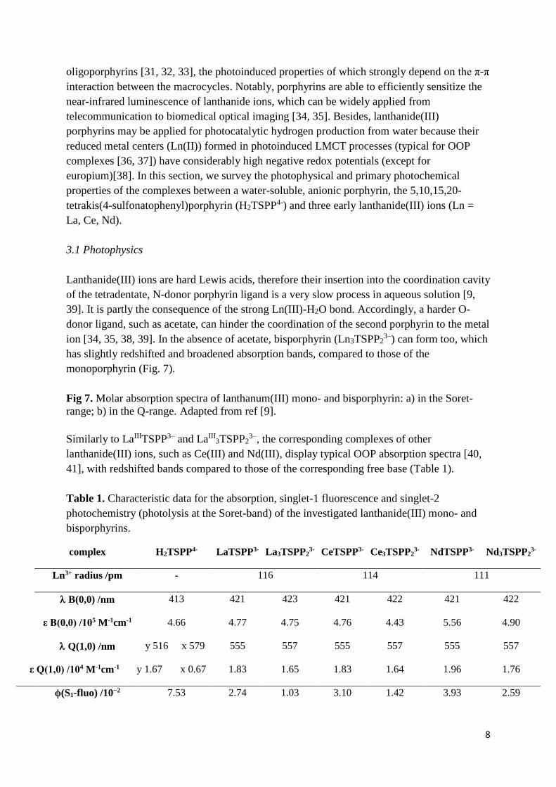
Similarly to LaIIITSPP3– and LaIII3TSPP2
3–, the corresponding complexes of other
lanthanide(III) ions, such as Ce(III) and Nd(III), display typical OOP absorption spectra [40,
41], with redshifted bands compared to those of the corresponding free base (Table 1).
Table 1. Characteristic data for the absorption, singlet-1 fluorescence and singlet-2
photochemistry (photolysis at the Soret-band) of the investigated lanthanide(III) mono- and
bisporphyrins.
complex H2TSPP4- LaTSPP3- La3TSPP23- CeTSPP3- Ce3TSPP2
3- NdTSPP3- Nd3TSPP23-
Ln3+ radius /pm - 116 114 111
B(0,0) /nm 413 421 423 421 422 421 422
ε B(0,0) /105 M-1cm-1 4.66 4.77 4.75 4.76 4.43 5.56 4.90
Q(1,0) /nm y 516 x 579 555 557 555 557 555 557
ε Q(1,0) /104 M-1cm-1 y 1.67 x 0.67 1.83 1.65 1.83 1.64 1.96 1.76
ϕ(S1-fluo) /10−2 7.53 2.74 1.03 3.10 1.42 3.93 2.59

9
ϕ (S1-fluo@B) /10−2 5.62 1.62 0.67 2.06 0.95 3.05 1.71
ϕ(IC S2-S1) 0.746 0.59 0.65 0.66 0.67 0.78 0.66
τ(S1) /ns 10.03 1.99 2.00 1.97 2.00 1.94 1.99
kr(S1) /106 s−1 7.5 13.8 5.1 15.7 7.1 20.2 13.0
knr(S1) /107 s−1 9.2 48.9 49.5 49.2 50.5 49.4 48.8
ϕ (S2-photochem) *
/10−3 0.006 0.76 1.51 1.37 1.98 0.81 2.25
redox% 100% 86% 78% 80% 74% 83% 76%
dissociation% - 10% 12% 14% 11% 12% 12%
transformation% - 4% 10% 6% 15% 5% 12%
ref. [43] [9] [40] [41]
*= ~10-6 M porphyrin, ~10-3 M metal ion, 0.01 M ionic strength adjustor.
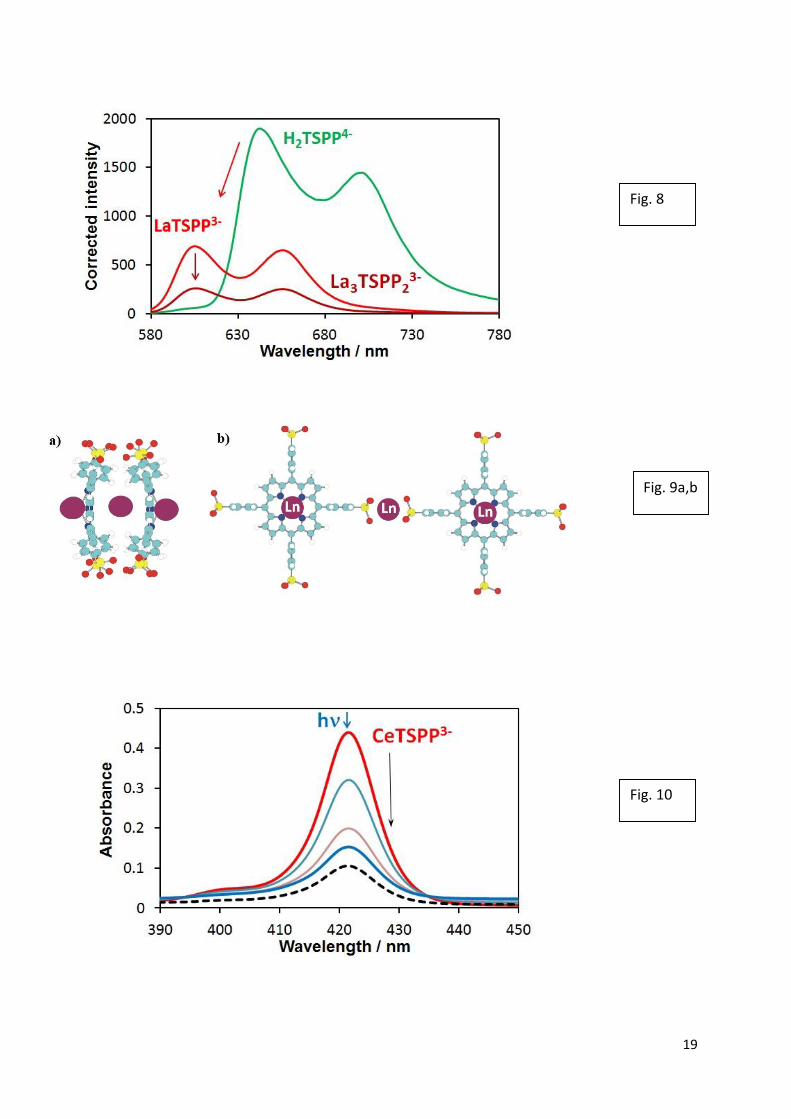
These complexes display characteristic singlet-1 fluorescences, which are blueshifted, less
intense and shorter-lived related to the free-base porphyrin (Fig. 8 and Table 1), due to the
dome distortion.
Fig 8. Singlet-1 fluorescence spectra of lanthanum(III) mono- and bisporphyrin. Adapted
from ref [9].
A paramagnetic metal ion can perturb the molecule orbitals of the macrocycle or can cause
the disappearance of fluorescence by spin-orbit coupling only if it is in the ligand plane.
Hence, the emission properties of the Ce(III) and Nd(III) porphyrins are very similar to those
of the La(III) porphyrins [9, 40, 41]. Besides, the fluorescence of lanthanide(III)
bisporphyrins is much like that of the monoporphyrins, only its quantum efficiency is slightly
lower, probably due to the weak π-π interactions between the two macrocycles. This is a clear
indication of the tail-to-tail dimerization of two metallo-monoporphyrin complexes through a
metal bridge, deviating from the head-to-head connection, i.e., sandwich complexes (Fig. 9).
In the latter case the absorption bands would show much larger redshifts as well
hyperchromicities, and the fluorescence would be much weaker, nearly disappear during the
formation of bisporphyrin) as with the (parallel) head-to-tail dimer of the protonated
porphyrin, (H4TSPP2–)2 [42], and the typical OOP, sandwich-type bisporphyrins of
mercury(II) ion: (parallel) head-to-tail HgII2(TSPP)2
8– and typical head-to-head HgII3(TSPP)2
6–
[11, 43].
Fig 9. Potential structures of 3:2 bisporphyrin: a) head-to-head or b) tail-to-tail.

10
The tendency of the fluorescence quantum yields in Table 1 clearly shows that not the number
of the unpaired electrons, but the size of the metal center is the key factor in this case. The
lanthanide contraction as steric effect results in shorter out-of-plane distances, accordingly,
less dome-distorted structures, which can cause the increase of the emission efficiency.
3.2 Photochemistry
In the lanthanide(III) porphyrins, the out-of-plane location of the metal center promotes two
types of photochemical reactions, which are not characteristic for the in-plane complexes [3,
38]:
1) photoinduced demetalation (without any charge transfer), due to the lability of the
complex; 2) oxidative degradation of the porphyrin ring, initialized by an irreversible,
photochemical LMCT reaction. The reduction of the metal center increases its size (along
with the decrease of its charge density), promoting the split of the coordinative bonds. The
released metal ions of enhanced reduction potential can undergo further redox reactions,
depending on their stability in the given medium. Thus, in aqueous solution, they can produce
hydrogen from water. The oxidized porphyrin, an ionic radical, is a very strong base, which is
immediately protonated, forming a long-lived and relatively strong oxidizer (in oxygen-free
solution τ1/2>>1 ms, E1/2>1 V) [44]. In the absence of appropriate electron donors (such as
alcohols or aldehydes) needed for the regeneration of the porphyrin ligand, an overall four-
electron oxidation takes place, leading to ring-opening and formation of a dioxo-tetrapyrrole
derivative (bilindione) [3, 44]. This transformation (degradation) of the macrocyclic ligand is
manifested in the disappearance of the Soret-band (Fig. 10).
Fig 10. Spectral changes in the range of Soret-bands during the photolysis at the Soret-
maximum (421 nm) (9.2×10⁻⁷ M H2TSPP4−, 10−3 M Ce3+, and 0.01 M acetate, pH ≈ 6, I0(421
nm) = 1.88×10⁻⁶ M photon/s, ℓ = 1 cm) at 0, 4, 10, and 14 min. Dashed line: after one day in
dark. Adapted from refs [38,40].
In the presence of acetate, i.e. during the photolysis of Ln(III) monoporphyrins, the
photoinduced dissociation of the metal ion (without charge transfers) takes place only in 14-
20 % from the total photochemical reactions (Table 1). In the absence of acetate, the
lanthanide(III) mono- and bisporphyrins exist in equilibrium in aqueous system, moreover
free-base ligands may also remain because of the lower stability constants; hence, during the
photolyses, they are simultaneously excited. Beside the photoredox degradations to the
mentioned, open-chain photoproducts and dissociation to the free-base porphyrin,
photoinduced transformation between the mono- and bisporphyrin complexes takes also place
in these systems, affecting the equilibria between the complexes in photostationary state.
Photoinduced dissociation in these systems proved to be reversible; the released free-base
porphyrin converted to the lanthanide(III) complexes again within some days in the dark.
The overall quantum yields of the primary photochemical processes of these OOP complexes,
i.e., the oxidation of the porphyrin ligand, are two orders of magnitude higher than those of
the free-base or the in-plane metalloporphyrins (~10-6 in [3, 43]) as a consequence of the

11
increased irreversibility of the photoinduced charge transfer from the ligand to the metal
center in out-of-plane position.
The overall photochemical quantum yields monotonously increased with the concentration [9,
38, 40]. This phenomenon suggests that a bimolecular excited state (excimer formation), or
dark reactions following the excitations, or the primary photochemical steps involve ground-
state lanthanide(III) porphyrin complexes. This is in accordance with the observation that the
oxidized ligand (radical) reacts further, through several redox steps, forming its final open-
chain bilindion derivative.
Only small differences appeared during the photolysis experiments of lanthanide(III) mono-
and bisporphyrin complexes. This phenomenon may confirm the special type of aggregation
through the peripheral sulfonato substituents (tail-to-tail) because in the case of post-transition
metal ions (head-to-head) the differences are much higher, mainly in the range of Q-bands
[40].
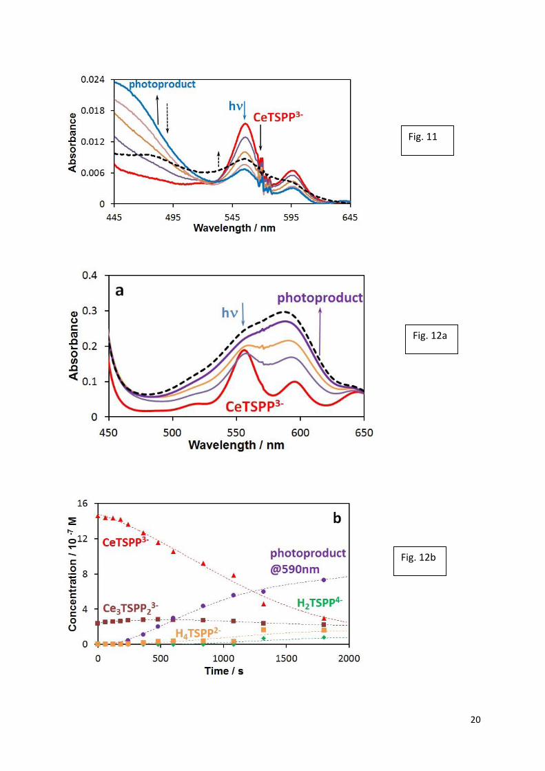
The irradiation wavelength does not significantly influence the overall quantum yield as it
was found for the cerium(III) porphyrins [38, 40]. However, the type of the photoproducts
considerably depends on the excitation energy. During the photolysis at the Soret-band, a
radical type intermediate can be detected (at 450 nm), which disappears in dark (Fig. 11).
Fig 11. Spectral changes in the range of Soret- and Q-bands during the photolysis at the Soret-
maximum (421 nm) at 0, 3, 6, 10, and 14 min (the conditions are the same as at Fig 10).
Dashed line: after one day in dark.. Adapted from refs [38, 40].
However, the photolysis at the Q-bands (555 nm) results in the formation of a new, stable
photoproduct (Fig. 12), which was not observed earlier in the case of the porphyrin complexes
of post-transition metal ions [3,43]. The absorption band at 590 nm may be assigned as a
charge transfer in the complex between cerium(III) and the open-chain, dioxo-tetrapyrrole
derivative (bilindion) [3, 44] formed in the irreversible oxidation of the ligand in LMCT
process.
Fig 12.a) Spectral changes during the photolysis at the Q-maximum (555 nm) of a solution
containing 1.75×10⁻⁶ M H2TSPP4−, 1.84×10⁻³ M Ce3+, and 0.01 M perchlorate (pH ≈ 6,
I0(556 nm) = 6.06×10⁻⁶ M photon/s, ℓ = 1 cm) at 0, 18, 30, and 62 min. Dashed line: after one
day in dark. b) Concentrations vs. irradiation time plots of the light-absorbing species.
Adapted from ref [38].
On the basis of the lanthanide contraction, one can expect a monotonous decrease of the
photochemical activities of the corresponding mono- and bisporphyrin complexes of metal
centers with increasing atomic number, due to the decreasing out-of-plane distance, hindering
the possibility of the photoinduced charge separation following LMCT. However, a small
increase can be observed with these three lanthanide(III) ions, mainly for their bisporphyrin
complexes (Table 1). This tendency suggests that the increasing out-of-plane distance causes
an other, opposite effect too. That may be the hindrance of the electron transfer from the

12
porphyrin ligand to the metal center, i.e. the step before the charge separation. Hence, a
maximum value can be expected upon further increasing the atomic number, followed by a
decreasing section of .
The overall photochemical quantum yield quasi-linearly increases with the concentration of
ionic strength adjustors (acetate, perchlorate) due to the enhanced possibility of the charge
separation following the photoinduced LMCT process, mainly in the case of bisporphyrins as
it was observed for the neodymium(III) complexes [41]. Furthermore, the presence of acetate,
as a potential axial ligand, hinders the photodissociation of the metal center from the
monoporphyrin complex as a consequence of trans effect of the axially coordinated ligand,
strengthening the M-N coordinative bonds.
4. Summary and conclusions
In this paper we summarized the results of investigation of some visible light-induced
photophysical and photochemical features of water-soluble anionic and cationic
metalloporphyrins. The main goal of these studies was to demonstrate the significant
difference between the properties of the kinetically inert in-plane and the labile sitting-atop
(SAT) or out-of-plane (OOP) complexes, due to their essential structural deviation.
As special representatives of the in-plane metalloporphyrins, cationic manganese(III),
cobalt(III), and nickel(II) complexes were studied. The size of these metal centers are
considerably smaller than the ligand cavity, hence they shrink the porphyrin ring, causing its
distortion. Due to this effect, the UV-vis absorption spectra of these complexes deviate from
the other (normal) in-plane metalloporphyrins and show similarities to those of the OOP
types. The photochemical behavior of these hyper porphyrins, however, is characteristic of the
kinetically inert complexes, the excited triplet state of which can be dynamically quenched
with a suitable electron donor (TEOA) to reduce the metal center as in the case of the cationic
Mn(III) and Co(III) porphyrins. Deviating from these complexes, the corresponding Ni(II)
porphyrin forms an associate with this electron donor, preventing the dynamic quenching. But
the long-lived triplet excited state of this associate is oxidatively quenched with
methylviologen (MV2+), realizing a one-step photoinduced electron transfer between the
donor and acceptor. Thus, Ni(II)TMPyP4+ can be considered as a sensitizer in this system,
while in the case of the manganese and cobalt porphyrins electron transmission from the
reduced metal center toward the acceptor, also via outer-sphere mechanism, needs the
absorption of another photon.
From the viewpoint of photoredox behavior, OOP complexes undergo inner-sphere LMCT
reactions, instead of outer-sphere electron transfer. In these cases anionic porphyrin ligands
are much more favorable than the positively charged ones. Lanthanide(III) ions as metal
center of these complexes offered the possibility of tuning the out-of-plane position due to
their contraction as a function of the atomic number. Besides the formation of mono- and
bisporphyrins, they exhibit excitation wavelength-dependent photochemical behavior upon
irradiation at the Soret- or Q-bands.

13
All these results well demonstrate how the size of the metal center determines the
structure and, thus, the photoinduced behavior of the porphyrins complexes, along with the
substituents on the ligand.
Acknowledgments
This work was supported by the Hungarian Scientific Research Fund (OTKA, NN107310),
the Hungarian Government and the European Union (TÁMOP-4.2.2.A-11/1/KONV-2012-
0071), and the Austrian-Hungarian Action Foundation (90öu2).
References
1 .C. Mot, S.A. Syrbu, S.V. Makarov, G. Damian, R. Silaghi-Dumitrescu, Inorg. Chem.
Commun. 18 (2012) 1–3.
2 T.S. Kurtikyan, V.A. Hayrapetyan, M.M. Mehrabyan, P.C. Ford, Inorg. Chem. 53 (2014)
11948-59.
3 O. Horváth, Z. Valicsek, G. Harrach, G. Lendvay, M.A. Fodor, Coord. Chem. Rev. 256
(2012) 1531-1545.
4 K. Ladomenou, M. Natali, E. Iengo, G. Charalampidis, F. Scandola, A.G. Coutsolelos,
Coord. Chem. Rev. 304-305 (2015) 38-54.
5 K. T. Oppelt, E. Wöß, M. Stiftinger, W. Schöfberger, W. Buchberger, G. Knör, Inorg.
Chem. 52 (2013) 11910-11922.
6 O. Horváth, R. Huszánk, Z. Valicsek, G. Lendvay, Coord. Chem. Rev. 250 (2006) 1792-
1803.
7 Z. Valicsek, G. Lendvay, O. Horváth, J. Porph. Phthal. 13 (2009) 910–926.
8 X. Zhang, Y. Chen, Inorg. Chem. Commun. 39 (2014) 79–82.
9 Z. Valicsek, G. Eller, O. Horváth, Dalton Trans. 41 (2012) 13120–13131.
10 M. Gouterman, In Porphyris; Dolphin, D., Ed.; Academic Press: New York, 1978; Vol. III.
11 Z. Valicsek, O. Horváth, Microchem. J. 107 (2013) 47–62.
12 R.D. Shannon, Acta Crystallogr., Sect. A: Found. Crystallogr. 32 (1976) 751–767.
13 K M. Kadish, K.M. Smith, R. Guilard (Eds.) The porphyrin handbook: Inorganic,
organometallic and coordination chemistry, Academic Press, London, 2000.
14 A. Harriman, G. Porter, J. Chem. Soc. Faraday Trans. II. 75 (1979) 1543-1552.
15 K. Kalyanasundaram, M. Neumann-Spallart, J. Phys. Chem. 86 (1982) 5163-5169.
16 M.A. Fodor, O. Horváth, L. Fodor, K. Vazdar, G. Grampp, A. Wankmüller, submitted
17 M.A. Fodor, O. Horváth, L. Fodor, G. Grampp, A. Wankmüller, Inorg. Chem. Commun.
50 (2014) 110-112.
14
18 K. Kalyanasundaram, Inorg. Chem. 23 (1984) 2453-2459.
19 Z. Valicsek, O. Horváth, K. Patonay, J. Photoch. Photobio. A 226 (2011) 23–35.
20 X. He, Y. Zhou, L. Wang, T. Li, M. Zhang, T. Shen, Dyes and Pigments 39 (1998) 173–
182.
21 S.-M. Chen, J. Mol. Catal. A, 138 (1999) 1-13.
22 U. Sehlstedt, S.K. Kim, P. Carter, J. Goodisman, J.F. Vollano, B. Norden, J.C. Dabrowiak,
Biochemistry 33 (1994) 417-426.
23 . I Kikaš, O.Horváth, I. Škorić, J. Mol. Struct. 1034 (2013) 62-68.
24 D. Vuk, I. Kikaš, K. Molčanov, O. Horváth, I. Škorić, J. Mol. Struct. 1063 (2014) 83–91.
25 N. Carnieri, A. Harriman, J. Photochem. 15 (1981) 341-346.
26 K. Takahashi, T. Komura, H. Imanaga, Bull. Chem. Soc. Jpn. 56 (1983) 3203-3207.
27 T. Watanabe, K. Honda, J. Phys. Chem. 86 (1982) 2617-2619.
28 R. F. Pasternack, Inorg. Chem. 15 (1976) 643-646.
29 R.F. Pasternack, E.G. Spiro, M. Teach, JRNC. 36 (1974) 599-606.
30 M.M. Major, M.A. Fodor, Z. Valicsek, L. Fodor, O. Horváth, G. Grampp, A. Wankmüller,
submitted
31 J.W. Buchler, A. Decian, J. Fischer, M. Kihnbotulinski, H. Paulus, R. Weiss, J. Am.
Chem. Soc. 108 (1986) 3652-3659.
32 G. Ricciardi, A. Rosa, E.J. Baerends, S.A.J. van Gisbergen, J. Am. Chem. Soc. 124 (2002)
12319-12334.
33 J. Jiang, M. Bao, L. Rintoul, D.P. Arnold, Coord. Chem. Rev. 250 (2006) 424–448.
34 V. Bulach, F. Sguerra, M.W. Hosseini, Coord. Chem. Rev. 256 (2012) 1468-1478.
35 H. He, Coord. Chem. Rev. 273 (2014) 87-99.
36 F.W. Oliver, C. Thomas, E. Hoffman, D. Hill, T.P.G. Sutter, P. Hambright, S. Haye, A.N.
Thorpe, N. Quoc, A. Harriman, P. Neta, S. Mosseri, Inorg. Chim. Acta 186 (1991) 119-124.
37 H. Kunkely, A. Vogler, Inorg. Chem. Commun. 10 (2007) 479-481.
38 M. Imran, C. Szentgyörgyi, G. Eller, Z. Valicsek, O. Horváth, Inorg. Chem. Commun. 52
(2015) 60-63.
39 M.P. Kiss, M. Imran, C. Szentgyörgyi, Z. Valicsek, O. Horváth, Inorg. Chem. Commun.
48 (2014) 22–25.
40 M. Imran, C. Szentgyörgyi, Z. Valicsek, O. Horváth, RSC Advances, submitted

15
41 M. Imran, Z. Valicsek, O. Horváth, RSC Advances, under preparation
42 D.L. Akins, S. Özcelik, H.R. Zhu, C. Guo, J. Phys. Chem. 100 (1996) 14390-14396.
43 Z. Valicsek, G. Lendvay, O. Horváth, J. Phys. Chem. B. 112 (2008) 14509-14524.
44 P. Neta, M. C. Richoux, A. Harriman, L. R. Milgrom, J.C.S. Faraday Trans. II. 82 (1986)
209-217.

16
Fig. 1
Fig. 2
Fig. 3

17
Fig. 4
Fig. 5
Scheme 1

18
Fig. 6
Fig. 7a
Fig. 7b
19
Fig. 8
Fig. 9a,b
Fig. 10
20
Fig. 11
Fig. 12a
Fig. 12b