visualization and analysis of large three-dimensional
TRANSCRIPT

Visualization and Analysis of LargeThree-Dimensional Hyperspectral Images
Stephen E. Reichenbach*a, Xue Tiana, Robert Lindquista,Qingping Taob, Alex Hendersonc, and John C. Vickermanc
aComputer Science & Engineering Department,University of Nebraska – Lincoln, Lincoln NE 68588-0115, USA;bGC Image, LLC, PO Box 57403, Lincoln NE 68505-7403, USA;
cSurface Analysis Research Centre, Manchester Interdisciplinary Biocentre,University of Manchester, Manchester M1 7DN, UK
ABSTRACTNew technologies for Secondary Ion Mass Spectrometry (SIMS) produce three-dimensional hyperspectral chem-ical images with high spatial resolution and fine mass-spectral precision. SIMS imaging of biological tissues andcells promises to provide an informational basis for important advances in a wide variety of applications, includingcancer treatments. However, the volume and complexity of data pose significant challenges for interactive visual-ization and analysis. This paper describes new methods and tools for computer-based visualization and analysisof SIMS data, including a coding scheme for efficient storage and fast access, interactive interfaces for visualizingand operating on three-dimensional hyperspectral images, and spatio-spectral clustering and classification.
Keywords: hyperspectral image processing, three-dimensional image processing, secondary ion mass spectrom-etry (SIMS)
1. INTRODUCTIONThis paper describes informatic technologies for visualizing and analyzing large, three-dimensional, hyperspectraldatasets generated by a new generation of chemical imaging technologies, such as time-of-flight secondary ion massspectrometry (ToF-SIMS). In ToF-SIMS,1 a beam of primary ions is directed onto a target, eroding moleculesand molecular fragments as neutral species and ions (i.e., secondary ions) from the target surface (as illustratedin Fig. 1). The secondary ions that are eroded from the target surface are electrostatically accelerated to adetector that measures their intensity as a function of flight time — data that can be converted to mass spectra.The primary-ion beam can be directed in a raster pattern to create a mass-spectral image and the raster scanningcan be repeated to generate a three-dimensional mass-spectral image, as illustrated in Fig. 2.
The Ionoptika J105 3D Chemical Imager,2 developed by Professor John C. Vickerman and his research groupat the Manchester Interdisciplinary Biocentre (MIB) in conjunction with Ionoptika (Hampshire UK), combinesseveral advances, including polyatomic primary-ion beams and an advanced buncher for secondary ions thatfacilitates a continuous-beam primary-ion probe. Polyatomic primary-ion beams (e.g., buckminsterfullerene,[C60]+) provide greater secondary-ion yield, more uniformity in the secondary-ion yield, and less damage to thesubstrate of the target than traditional primary-ion beams.3 Greater yield improves the signal-to-noise ratio andsensitivity. Improved uniformity enhances the effective resolution and allows more accurate mapping of chemicalconstituents. With reduced sub-surface degradation as the surface is eroded, subsequent scans across the targetyield more accurate depth profiling to improve three-dimensional chemical imaging. An innovative secondary-ionbuncher shapes the electric field that propels the secondary ions for the time-of-flight mass spectrometer, therebyfocusing the variable-sized, variable-positioned secondary ions. Time focusing obviates the need for pulsing theprimary-ion beam to limit the time range of the secondary ions, which allows quasi-continuous operation of theprimary-ion beam.
*Corresponding author contacts: mailto:[email protected], phone +1(402)472-5007
Visual Information Processing XVIII, edited by ZiaUr Rahman, Stephen E. Reichenbach, Mark Allen Neifeld, Proc. of SPIE Vol. 7341, 734108 ∙ © 2009 SPIE ∙ CCC code: 0277786X/09/$18 ∙ doi: 10.1117/12.820870
Proc. of SPIE Vol. 7341 7341081

secondary ionsto mass spectrometer
sample
'Iprimary ion
Figure 1. In ToF-SIMS, primary ions directed at the tar-get erode secondary ions for analysis by mass spectrometry.The primary ion beam can be directed in a raster patternto create a two-dimensional mass-spectral image. Repeat-ing the raster pattern to drill through the target creates athree-dimensional mass-spectral image.
Figure 2. The three-dimensional mass-spectral data can bevisualized as a three-dimensional data-cube projected ontotwo dimensions. Colorization of each data point can bebased on the total intensity or ion count (TIC), selectedintensity or ion count (SIC), or other spectral properties.(Data from Fletcher et al.2)
The continuous primary-ion beam provides faster analyses and increased spatial resolution. Then, the systemuses a harmonic-field reflectron with the property that the time-of-flight in and out of the reflector dependson mass-to-charge only (and not on their variable energy). This creative design provides high-precision massspectrometry even with continuous operation of the primary-ion beam. The system’s high spatial resolution,fine mass precision, and high-sensitivity surface and depth-profile characterizations of the molecular chemistryof heterogeneous materials, including biological tissues and cells, promise to provide an informational basis forimportant advances in a wide variety of applications, including cancer treatments. However, the volume of dataproduced poses a significant challenge for interactive visualization and analysis.
The J105 ToF-SIMS instrument will produce three-dimensional datasets on the order of 5123 spectra withtens of thousands of ToF channels. In the example datasets presented here, individual mass spectra are sampledin up to 85,000 ToF channels at a rate of 1 ns per 8-bit intensity (a raw data rate of 1 gigabyte per second) andaccumulated in hardware with an Ortec Fastflight-2TM (Oak Ridge, TN). In the example datasets, 200 to 1000raw spectra are accumulated per pixel, but the number may be larger or smaller depending on the application. Ifno more than 256 spectra are accumulated, each accumulated intensity can be represented with a 16-bit unsignedinteger in the raw data file, reducing the data rate to 10 megabytes per second (MB/s). Still, a 128×128, 128-layerimage with 85,000 2-byte ToF channels requires 332 gigabytes (GB) without compression and even a 512×512,one-layer image with 85,000 2-byte ToF channels requires 41GB. Datasets of tens to hundreds of gigabytes cannotbe held in the fast memory of typical computer systems, which creates a bottleneck for interactive visualizationand analysis with general-purpose imaging software. Real-time, interactive, three-dimensional visualization andanalysis requires memory efficient and computationally efficient software.
This paper discusses three fundamental issues for visualization and analysis of large, three-dimensional,hyperspectral data:
• computationally efficient compression for real-time visualization,• three-dimensional hyperspectral interactivity, and• spatio-spectral clustering and classification.
Proc. of SPIE Vol. 7341 7341082

2. COMPRESSION FOR REAL-TIME VISUALIZATIONThis section describes a compression scheme for holding large three-dimensional ToF-SIMS data sets in memoryfor fast visualization. The access pattern is a critical consideration for data compression. The most importantaccess mode for interactive SIMS visualization is retrieving spectra by pixel (i.e., spatial position). Analystsdetermine chemical compositions on the basis of mass spectral characteristics, so mass spectra viewing is funda-mental. Common interactive operations are to view the mass spectrum at a point in the image space indicated bypoint-and-click and to view the mass spectrum summed over a spatial region indicated by drawing. An importantoperation is to generate a classification rule(s) based on mass spectra in two (or more) regions. Analysts alsoview a mass-spectral range (e.g., for a selected ion) across the image space, but such spectral-spatial viewing doesnot require immediate interactivity to the degree required in pixel-oriented spatial-spectral access. Moreover,operations to generate spatial maps from their spectra require pixel-by-pixel access to many or all ToF channels,e.g., to map regions that satisfy a classification rule.
Tretter, Memon, and Bouman4 cite two approaches among methods for lossless compression of hyperspectralimages: predictive coding and reversible transforms, each followed by context modeling and coding. Bothapproaches can be applied either with respect to the spatial dimensions or to the spectral dimension (or both).Predictive coding has been the predominant approach for hyperspectral data. Lossless transform coding methodsfor hyperspectral data are newer and typically require greater computation than lossless predictive methods, butmay achieve greater compression. Given the motivation of interactive visualization and analysis of SIMS data,low computational complexity is more important than optimal compression, so the more traditional approach ofpredictive coding may be better suited. Given the primary need for spatial access in SIMS analysis, each pixelspectrum should be compressed separately.
Reichenbach et al.5 recently described a method that codes individual spectra, consistent with the predomi-nant access mode for SIMS analysis, based on statistical and structural characteristics of SIMS spectra. Unlikesome hyperspectral data generated by remote sensing satellites, for which many hyperspectral compression meth-ods have been developed, SIMS spectra have many zero values and the probability distribution of the intensityvalues is skewed significantly, decreasing rapidly with magnitude. Also, many of the non-zero values are inadjacent ToF channels, forming peaks in the mass spectra. These statistical characteristics can be exploited togive highly compressed data that can be accessed quickly.
The most notable characteristic of SIMS hyperspectral data is that many of the intensity values are zero.This characteristic of the data reflects the fact that some mass-to-charge ratios do not occur physically and thefact that the number of chemical constituents at a sample point of the target limits the number of secondaryions and therefore the number of peaks in each mass spectrum. Another important characteristic of the data isthat the probability distribution of the intensity values decreases with intensity. Many of the non-zero values areequal to one (many of which may be noise but must be coded in a lossless method) and most non-zero values areless than 256. Because of the large number of zero values and the long-tailed skewed probability distributions,least-squares predictors, which are effective for remote sensing data, do not perform well for SIMS data. Anothercharacteristic of the datasets is that ion peaks in the mass spectra may be wider than the ToF channels, so eachmass spectral peak may cause several non-zero values in adjacent ToF channels.
The high probability of zero-values and the clustering of non-zero values suggest that run length encodingmay be used to effectively code the long runs of zeros between non-zero values. Commonly used sparse array rep-resentations of mass spectra (i.e., recording the mass and value for each non-zero value) similarly take advantageof the large number of zeros to efficiently represent MS data. If, instead of the ToF channel index, the differentialof indexes of non-zero-valued channels is used (i.e., the difference between the index of the next channel with anon-zero value and one more than the index of the current non-zero channel), the result is a run length code.
Based on the SIMS data characteristics, the compression method separately codes the ToF differentials andnon-zero intensity values. Because many of the ToF differentials are zero, it is important to code them mostefficiently. Because the compressed data will be decoded for visualization, the approach uses representations ofinteger byte-lengths which do not require computation for decoding — just byte copies. Accordingly, the methoduses 2-bit length codes to record the number of bytes for each ToF differential and zero bytes are used if thedifferential is zero. The length codes (in binary) are: 00 if the differential is zero, with no separate representation
Proc. of SPIE Vol. 7341 7341083
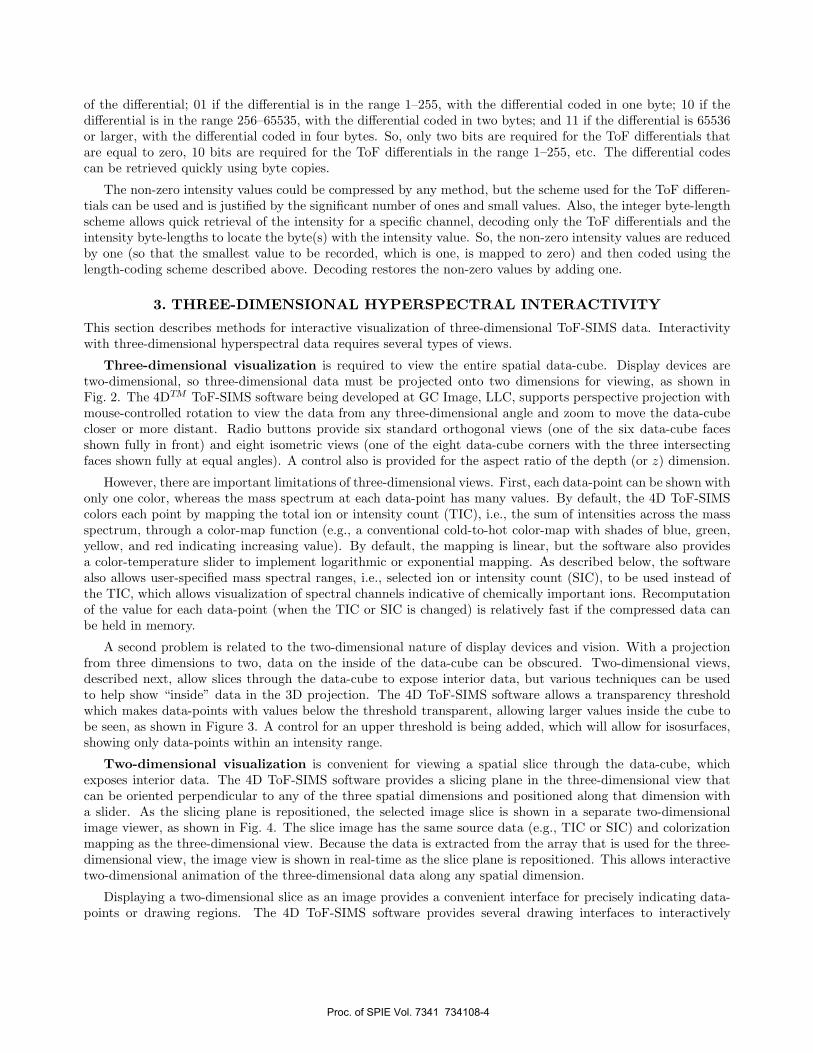
of the differential; 01 if the differential is in the range 1–255, with the differential coded in one byte; 10 if thedifferential is in the range 256–65535, with the differential coded in two bytes; and 11 if the differential is 65536or larger, with the differential coded in four bytes. So, only two bits are required for the ToF differentials thatare equal to zero, 10 bits are required for the ToF differentials in the range 1–255, etc. The differential codescan be retrieved quickly using byte copies.
The non-zero intensity values could be compressed by any method, but the scheme used for the ToF differen-tials can be used and is justified by the significant number of ones and small values. Also, the integer byte-lengthscheme allows quick retrieval of the intensity for a specific channel, decoding only the ToF differentials and theintensity byte-lengths to locate the byte(s) with the intensity value. So, the non-zero intensity values are reducedby one (so that the smallest value to be recorded, which is one, is mapped to zero) and then coded using thelength-coding scheme described above. Decoding restores the non-zero values by adding one.
3. THREE-DIMENSIONAL HYPERSPECTRAL INTERACTIVITYThis section describes methods for interactive visualization of three-dimensional ToF-SIMS data. Interactivitywith three-dimensional hyperspectral data requires several types of views.
Three-dimensional visualization is required to view the entire spatial data-cube. Display devices aretwo-dimensional, so three-dimensional data must be projected onto two dimensions for viewing, as shown inFig. 2. The 4DTM ToF-SIMS software being developed at GC Image, LLC, supports perspective projection withmouse-controlled rotation to view the data from any three-dimensional angle and zoom to move the data-cubecloser or more distant. Radio buttons provide six standard orthogonal views (one of the six data-cube facesshown fully in front) and eight isometric views (one of the eight data-cube corners with the three intersectingfaces shown fully at equal angles). A control also is provided for the aspect ratio of the depth (or z) dimension.
However, there are important limitations of three-dimensional views. First, each data-point can be shown withonly one color, whereas the mass spectrum at each data-point has many values. By default, the 4D ToF-SIMScolors each point by mapping the total ion or intensity count (TIC), i.e., the sum of intensities across the massspectrum, through a color-map function (e.g., a conventional cold-to-hot color-map with shades of blue, green,yellow, and red indicating increasing value). By default, the mapping is linear, but the software also providesa color-temperature slider to implement logarithmic or exponential mapping. As described below, the softwarealso allows user-specified mass spectral ranges, i.e., selected ion or intensity count (SIC), to be used instead ofthe TIC, which allows visualization of spectral channels indicative of chemically important ions. Recomputationof the value for each data-point (when the TIC or SIC is changed) is relatively fast if the compressed data canbe held in memory.
A second problem is related to the two-dimensional nature of display devices and vision. With a projectionfrom three dimensions to two, data on the inside of the data-cube can be obscured. Two-dimensional views,described next, allow slices through the data-cube to expose interior data, but various techniques can be usedto help show “inside” data in the 3D projection. The 4D ToF-SIMS software allows a transparency thresholdwhich makes data-points with values below the threshold transparent, allowing larger values inside the cube tobe seen, as shown in Figure 3. A control for an upper threshold is being added, which will allow for isosurfaces,showing only data-points within an intensity range.
Two-dimensional visualization is convenient for viewing a spatial slice through the data-cube, whichexposes interior data. The 4D ToF-SIMS software provides a slicing plane in the three-dimensional view thatcan be oriented perpendicular to any of the three spatial dimensions and positioned along that dimension witha slider. As the slicing plane is repositioned, the selected image slice is shown in a separate two-dimensionalimage viewer, as shown in Fig. 4. The slice image has the same source data (e.g., TIC or SIC) and colorizationmapping as the three-dimensional view. Because the data is extracted from the array that is used for the three-dimensional view, the image view is shown in real-time as the slice plane is repositioned. This allows interactivetwo-dimensional animation of the three-dimensional data along any spatial dimension.
Displaying a two-dimensional slice as an image provides a convenient interface for precisely indicating data-points or drawing regions. The 4D ToF-SIMS software provides several drawing interfaces to interactively
Proc. of SPIE Vol. 7341 7341084

Figure 3. A threshold on the values shown allows viewingof larger valued data-points inside the data-cube.
Figure 4. As the slice plane (shown in red in the three-dimensional view) is repositioned (with the slider in thelower-right panel), the two-dimensional image is updated.
delineate regions: rectangle (enclosed region, including single-point selection), polygon (enclosed region), free-hand (enclosed region), and scribble. The software allows users to build composite regions using discard (new),addition (union), subtraction, and replace (discard followed by addition). Composite regions are maintained asgeometric aspect functions that can be saved, loaded, edited, and visualized with masks in the two-dimensionaland three-dimensional views.
One-dimensional visualization is especially convenient for showing the mass spectrum of an indicateddata-point or the summed mass spectrum for a selected region. In the 4D ToF-SIMS software, the spectrumviewer displays a spectrum in graphical and tabular formats, as illustrated at toward the bottom-left of thegraphical user interface (GUI) in Fig. 4. The abscissa of the spectrum can be set to ToF, mass-to-charge (m/Q)or nominal mass-to-charge (rounded to whole numbers). ToF-SIMS systems generate hyperspectral data —intensity arrays with tens of thousands of values — so neither display screen-resolution nor visual acuity issufficient to perceive all spectral intensities simultaneously. Therefore, the graphical view of the spectrum allowszooming to show sub-ranges of the spectrum and the tabular view supports scrolling. The tabular view can besorted either by abscissa or intensity (the ordinal) with either increasing or decreasing values.
The graphical view allows interactive delineation of a SIC range by mouse click-and-drag. The SIC can thenbe visualized in the three-dimensional and two-dimensional spatial views with a button click. Generating newSIC visualizations requires summing intensities for the indicated range and may take a few seconds depending onthe size of the data and SIC range. Specific SIC ranges (e.g., that are indicative of various chemically importantions) can saved, loaded, and edited, as well as visualized.
4. SPATIO-SPECTRAL CLUSTERING AND CLASSIFICATIONAnalyses of ToF-SIMS images may entail both spatial and spectral features. For example, it may be necessaryto delineate a spatial feature such as a cell or components within a cell. Similarly, it may be necessary to identifyspectral features, such as a spectral signature indicating the presence of a drug.
The 4D ToF-SIMS software provides several interactive tools for spatial and structural analysis. The previoussection described drawing tools that can be used to delineate spatial regions and examine the summed massspectrum for the region. The Spectral Similarity and Magic Wand tools support semi-automated region selection.
The Spectral Similarity tool selects data points at which the similarity with a reference spectrum is greaterthan a specified threshold. First, the user selects a data point or region to provide a reference spectrum. Then, at
Proc. of SPIE Vol. 7341 7341085

A. Seed in the background. B. Seed in the left bead. C. Seed in the right bead.
Figure 5. The Magic Wand can be used to select proximal and spectrally similar data points for an indicated seed point.(Data from Winograd and Braun.6)
every data point i, the similarity of spectrum si and the reference spectrum r is computed as the cosine betweenthe vectors of the two spectra:
spectralSimilarity(r, si) =r · si
|r| |si|, (1)
which varies from 0 for orthogonal mass spectra to 1 for mass spectra that are identical after normalization. TheAngular Threshold slider allows the user to interactively adjust the threshold for selection, i.e., if its similaritywith the reference spectrum is greater than the threshold then the data point is included. Because the similaritiesfor each pixel are computed only once, the thresholding can be performed in real-time as the slider is adjusted.
The Magic Wand tool selects data points based on both spectral similarity and spatial proximity. First, theuser selects a data point to provide both a reference spectrum and a seed for the selected spatial region. Then,the software iterates a region-growing process. In each cycle of the iteration, data points within a specified spatialdistance of any selected data point (initially just the seed) are tested for similarity with the reference spectrum.Every data point within the specified distance that meets the similarity criterion is added to the selected region.This iterative process repeats each time the region grows, then stops when no more data points nearby anyselected data points are similar enough. Two sliders are provided to parameterize the Magic Wand: the jumpparameter, which specifies the distance from a selected data point at which that the region-growing process caninclude additional data points, and the similarity threshold, which specifies the level of similarity required fornew data points to be added to the selected region. Two types of similarity are allowed: TIC similarity andspectral similarity. Spectral similarity is given in Eq. (1). TIC similarity ranges linearly from 0 for the differencebetween the largest and smallest TIC values in the data to 1 for identical TIC values.
Fig. 5 illustrates use of the Magic Wand on ToF-SIMS data from two 200-µm polystyrene beads coated withdifferent peptide mimics with molecular weights 226 and 547 on a silicon substrate.6 In each image, the regionselected by the Magic Wand is shown as a mauve-colored overlay on the colorized TIC image. In Fig. 5A, theseed point is in the background and the Magic Wand successfully selects much of the background. In Figs. 5Band 5C, the seed point is respectively in the left and right bead and the Magic Wand selects much of the indicatedbead.
The Spectral Clustering tool provides a uniform interface for various clustering algorithms, including k-means,7 hierarchical clustering,8 and spectral clustering.9 Clustering algorithms group objects such that thosein the same group are more similar with one another than with objects in other groups. In the 4D ToF-SIMSsoftware, the normalized mass spectrum (each intensity in the spectrum divided by the largest intensity in thespectrum) is used for assessing the similarity of data points. First, the user selects a subset of data points tobe clustered. Then, the user selects the clustering algorithm and provides its parameters, e.g., some algorithms
Proc. of SPIE Vol. 7341 7341086

rI:...
'-..,f
...;.,
:q4
V
A. TIC. B. Two clusters, k-means with k=2.
C. Three clusters, k-means with k=3. D. Four clusters, k-means with k=4.
Figure 6. Clustering of ToF-SIMS data of coated polystyrene beads.
require the user to specify the number of clusters. Then, the algorithm separates the data points into clustersbased on their spectral similarity (as determined by the algorithm).
Fig. 6 illustrates clustering with the ToF-SIMS data from two polystyrene beads coated with different com-pounds. Fig. 6A shows the colorized TIC image. As can be seen in this image, the background regions belowthe beads and the centers of the beads have smaller TIC values, indicating less intense mass spectra. With onlythe TIC, the data points in the two beads cannot be distinguished from one another nor from the background.Fig. 6B shows k-means clustering with two clusters, i.e., k=2. This clustering is mostly successful in separatingthe two beads (colorized black) from the background (colorized red). Fig. 6C shows k-means clustering with
Proc. of SPIE Vol. 7341 7341087

A. TIC. B. Two clusters, k-means with k=2. C. Three clusters, k-means with k=3.
Figure 7. Clustering of ToF-SIMS data of HeLa cells. (Data from S. Rabbani and J. Fletcher, Surface Analysis ResearchCentre, University of Manchester.)
three clusters (k=3). This clustering also separates the beads (green) from the background and divides thebackground into two clusters, apparently separating the more intense background (black) and the less intensebackground (red). With four clusters (k=4), in Figure 6D, the two beads are well separated (red and yellow)from each other and from the background (cyan and black).
Fig. 7 shows clustering with ToF-SIMS data from HeLa cells (an immortalized cervical cancer cell line widelyused as a standard cell type for a wide range of biological research). The cells were cultured on poly(L-lysine)coated silicon shards for 24 hours at 37◦C with 5% CO2, washed briefly in 0.15 M ammonium formate solutionto remove salt ions, then freeze-dried prior to ToF-SIMS analysis. Typical cell diameter is 20 µm. The data wasacquired using 40 keV C+
60 primary ions scanned to 128×128 pixels over a field of view 88×108 µm2 (a pixeldimension of approximately 0.7×0.8 µm2). The total ion dose used was 3× 10¹⁴ ions cm2. Fig. 7A shows a TICimage. Fig. 7B shows that k-means clustering with two clusters (k=2) separates the cells and background. InFig. 7C, k-means clustering with three clusters (k=3) apparently separates the cell edges and interiors.
Spectral Classification is based on supervised training: two (or more) geometric aspect functions designatedistinct classes of data points, then the classification algorithm assigns class membership to other data pointsbased on mass spectral characteristics. Experiments with the bead data (Fig. 6) compared four classificationalgorithms: C4.5 decision trees,10 soft independent modeling of class analogy (SIMCA),11 principal componentanalysis (PCA)12 with discriminant function analysis (DFA),13 and the most similar neighbor with a probability-based spectrum similarity measure (MSN-PSSM).14 Two data sets were constructed, the first with 100 datapoints from each bead and the second with 50 data points from each bead. With these data sets, leave-one-outcross-validation, which is commonly used in chemometrics, was used for testing.
Overall classification accuracy and Fleiss’ kappa statistic15 are used to quantitatively measure the performanceof the different algorithms. Overall classification accuracy is defined as:
Accuracy =# correctly classified samples
# samples in dataset. (2)
Fleiss’ kappa statistic is a chance corrected measure of agreement between two sets of categorized data thatassesses agreement compared to random-chance levels. In this study, kappa measures the agreement betweensamples’ true labels and samples’ classified labels for different algorithms. A kappa value of 1 means perfectagreement, 0 means agreement expected by chance, and -1 means perfect disagreement. Interpretation of thekappa values is based on Fleiss’ cutoffs:16 kappa values exceeding 0.75 suggest agreement well above chance,values in the range of 0.40 to 0.75 indicate fair levels of agreement above chance, and values below 0.40 areindicative of poor agreement above chance levels. Table 1 shows the overall classification accuracy and Fleiss’kappa statistic for each classification algorithm. Decision trees and PCA with DFA performed best, followed byMSN-PSSM, then SIMCA.
Proc. of SPIE Vol. 7341 7341088

Table 1. Classification results with four classsification methods for two datasets.
Dataset 1 Dataset 2Classifier Accuracy (%) Kappa Accuracy (%) KappaDecision trees 90.00 0.80 93.50 0.87SIMCA 80.00 0.60 83.00 0.66PCA with DFA 91.00 0.82 92.50 0.85MSN-PSSM 89.00 0.78 90.00 0.80
5. CONCLUSIONThis report summarizes new methods and tools for computer-based visualization and analysis of high-resolution,three-dimensional, hyperspectral ToF-SIMS data. The informatics suite includes a coding scheme for efficientstorage and fast access, interactive interfaces for visualizing and operating on three-dimensional hyperspectralimages, and spatio-spectral clustering and classification.
The goal of the work to-date is proof-of-concept and the development of a prototype foundation for futurework. Future work will include continued evaluation of the coding effectiveness as the instrument evolves,interactive spatial operations for 3D drawing such as rotation and extrusion, algorithmic spatial operations suchas dilation and erosion, improved clustering and supervised classification methods, and a general framework forspectral aspects functions such as PCA, ion ratios, etc.
ACKNOWLEDGMENTSThis work was supported by the USA National Science Foundation funding to S. E. Reichenbach (IIS-0431119)and Q. Tao (IIP-0741027) and by the UK Engineering and Physical Sciences Research Council’s ”Collaboratingfor Success through People” funding to John C. Vickerman (EP/FO12985). The authors gratefully acknowledgethe support and data provided by John Fletcher, Sadia Rabbani, and others at the Surface Analysis ResearchCentre of the University of Manchester.
REFERENCES[1] Vickerman, J. C., “ToF-SIMS – an overview,” in [ToF-SIMS: Surface Analysis by Mass Spectrometry ],
Vickerman, J. C. and Briggs, D., eds., 1–40, SurfaceSpectra, Manchester, UK (2001).[2] Fletcher, J. S., Rabbani, S., Henderson, A., Blenkinsopp, P., Thompson, S. P., Lockyer, N. P., and Vicker-
man, J. C., “A new dynamic in mass spectral imaging of single biological cells,” Analytical Chemistry 80(23),9058–9064 (2008).
[3] Fletcher, J. S., Lockyer, N. P., Vaidyanathan, S., and Vickerman, J. C., “TOF-SIMS 3D biomolecular imag-ing of xenopus laevis oocytes using buckminsterfullerene (C60) primary ions,” Analytical Chemistry 79(6),2199–2206 (2007).
[4] Tretter, D., Memon, N., and Bouman, C. A., “Multispectral image compression,” in [Handbook of Imageand Video Processing ], Bovik, A., ed., 539–554, Academic Press, San Diego CA (2000).
[5] Reichenbach, S. E., Henderson, A., Lindquist, R., and Tao, Q., “Efficient encoding and rapid decoding forinteractive visualization of large three-dimensional hyperspectral chemical images,” Rapid Communicationsin Mass Spectrometry 23(9), 1229–1233 (2009).
[6] Winograd, N. and Braun, R. M., “Imaging mass spectrometry and combinatorial chemistry,” Spec-troscopy 16(9), 14–27 (2001).
[7] MacQueen, J. B., “Some methods of classification and analysis of multivariate observations,” in [Proceedingsof the Fifth Berkeley Symposium on Mathematical Statistics and Probability ], 281–297 (1967).
[8] Johnson, S., “Hierarchical clustering schemes,” Psychometrika 32(3), 241–254 (1967).[9] Ng, A. Y., Jordan, M. I., and Weiss, Y., “On spectral clustering: Analysis and an algorithm,” in [Advances
in Neural Information Processing Systems ], 14, 849–856, MIT Press, Cambridge MA (2002).[10] Quinlan, J. R., [C4.5: Programs for Machine Learning ], Morgan Kaufmann (1993).
Proc. of SPIE Vol. 7341 7341089

[11] Wold, S. and Sjostrom, M., “SIMCA: A method for analyzing chemical data in terms of similarity andanalogy,” in [Chemometrics Theory and Application ], American Chemical Society Symposium Series 52,243–282 (1977).
[12] Jolliffe, I. T., [Principal Component Analysis ], Springer, New York NY, second ed. (2002).[13] Krzanowski, W. J., [Principles of multivariate analysis: a user’s perspective ], Oxford University Press, New
York NY, revised ed. (1988).[14] Tian, X., Reichenbach, S. E., Tao, Q., and Henderson, A., “Classification and cluster analysis of complex
time-of-flight secondary ion mass spectrometry for biological samples,” in [International Conference onBioinformatics, Computational, Biology, Genomics and Chemoinformatics ], in press (2009).
[15] Fleiss, J. L., “Measuring nominal scale agreement among many raters,” Psychological Bulletin 76(5), 378–382 (1971).
[16] Fleiss, J. L., [Statistical Methods for Rates and Proportions ], Wiley, New York NY, second ed. (1981).
Proc. of SPIE Vol. 7341 73410810