visualization of fibers at risk for neuronal tract injury in early ms by streamtube diffusion...
TRANSCRIPT

Visualization of Fibers at Risk for Neuronal Tract Injury in Early MS by Streamtube Diffusion Tractography at 3 Tesla
Jack H Simon 1
David E Miller 1
Mark Brown 1
John R Corboy 1
Jeffrey L Bennett 1
Song Zhang 2
David H Laidlaw 2
1University of Colorado Health Sciences Center 2Brown University
Supported by the National MS Society (RG 3307-A-1), GE Medical Systems,and the National Science Foundation (CCR-0086065).

Background – Neuronal Tract Degeneration in Early MS •Axonal injury in inflammatory MS Lesions
•Trapp et al 1998; Ferguson et al 1997•Empty myelin cylinders 2° to and distant from focal lesion
•Bjartmar et al 2001•Major axonal loss (and atrophy) in corpus callosum
•Evangelou et al 2000•Neuronal Tract Degeneration Patterns in Early MS
Transcallosal BandsCorticospinal Tract Degeneration
Bjartmar et al 2001
Simon et al Neurology 2000 Simon et al Neurology 2001

Goals/Question
• Develop a process to identify and then interrogate neuronal fibers that are anatomically related to focal demyelinating lesions
• Can we detect and measure neuronal fiber injury/degeneration in vivo ?

Strategy
• Earliest MS – Identify focal lesions at the time of a CIS
• Identify fibers transiting the focal lesion and reaching a distant structure (corpus callosum)
• Interrogate fibers (in corpus callosum) by MRI distant from focal lesion – MTR, Diffusion tensor (<D>; FA)– Longitudinal MRI follow-up

3T Pulse Parameters • Diffusion Tensor Imaging
– Echo-planar – B-value 1000, 25 gradient directions
• Axial plane - 3 sets of overlapping slices • 5.1 mm thick with 1.7mm shifts• FOV 28 x 28 cm, matrix 160 x160, 2 excitations • TR/TE 6075/69.8-minimum
• Magnetization Transfer Imaging– 3D gradient-echo
• MT pulse – Fermipulse; applied middle 30% of the phase encode steps (SAR) – Pulse width of 8 ms– Peak B1 field of 9.24 μT, flip angle of 670°– 1200 Hz from the center frequency– TR/TE/flip angle 50/2.4(fr)/15° – FOV 22x22, matrix 256 x192, one excitation
• Sagittal plane • 3 mm partitions

YesAt 1 yNo10.69+Optic neuritis26
F
3
YesAt 1m,3mNo2.99+Partial
transverse
myelitis
33
F
2
Yes At 3mNo5.83+Optic neuritis
OCB positive
38
M
1
TreatedNew MR lesions on follow-up?
CDMSBrain
T2 lesion
Volume
(cc)
DISCIS
Presentation
Age
Gender
Patient
OCB = oligoclonal bands DIS= fulfill McDonald criteria for dissemination in spaceCDMS =clinically definite MS based on second attack by 1.5 year follow-upTreated= Treatment with immunomodulatory therapy
Table 1 . Patient Characteristics
Patient Characteristics
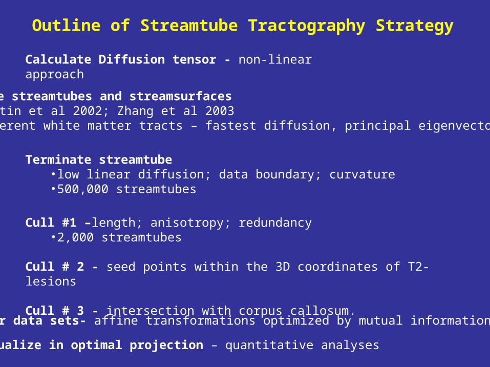
Generate streamtubes and streamsurfaces •Westin et al 2002; Zhang et al 2003•coherent white matter tracts – fastest diffusion, principal eigenvector
Terminate streamtube•low linear diffusion; data boundary; curvature•500,000 streamtubes
Cull #1 –length; anisotropy; redundancy•2,000 streamtubes
Cull # 2 - seed points within the 3D coordinates of T2-lesions
Cull # 3 - intersection with corpus callosum.
Calculate Diffusion tensor - non-linear approach
Register data sets- affine transformations optimized by mutual information algorithm
Outline of Streamtube Tractography Strategy
Visualize in optimal projection – quantitative analyses

Visualization -Streamtubes + Streamsurfaces Segmentation & Registration
AAMI (Analysis Application for Medical Imaging)•David E. Miller - University of Colorado HSC
Visualization Tools •Song Zhang and David Laidlaw - Brown University
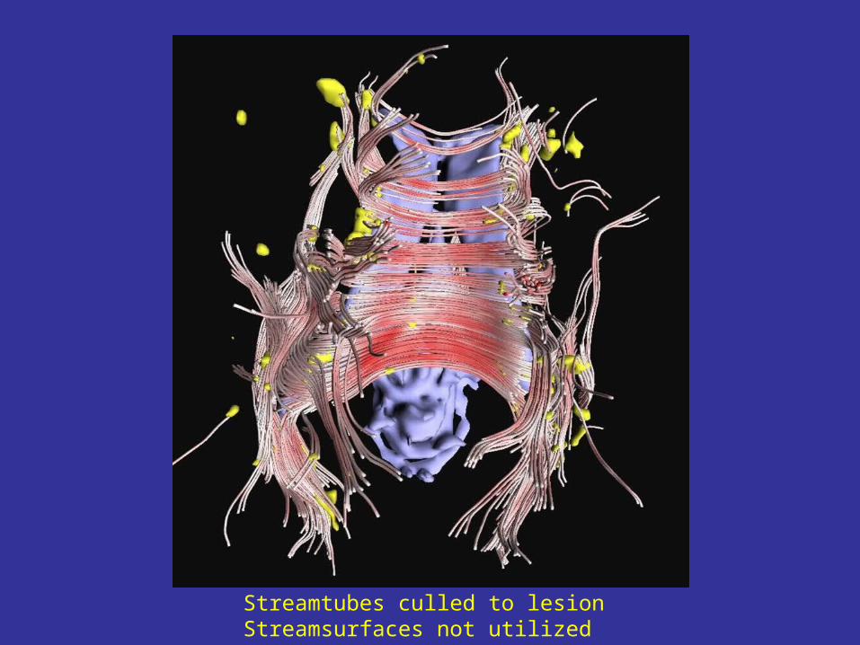
Streamtubes culled to lesionStreamsurfaces not utilized

Streamtubes culled to [lesion + corpus callosum]

Case 1
Streamtubes and Streamsurfaces Culled to Lesion Culled to [lesion + corpus callosum]

Culled to Lesion Culled to [Lesion + Corpus Callosum]
Case 2

Case 2

Culled to [lesion + corpus callosum]
Case 3

Case 1 Registration of culled streamtubes to sagittal representation of corpus callosum
Fibers at Risk (FAR)

Corpus Callosum Area Mean MTR
ID FAR
%AAWM
%
AllCorpus
CallosumNAWMFraction
AAWMFraction
FARFraction
CIS 1 21.0 4.1 48.94 49.06 45.98 50.57
CIS 2 10.0 0 49.55 49.55 49.78
CIS 3 30.0 8.7 48.53 48.55 48.36 50.63
CIS 1 CIS 2 CIS 3

Conclusions
• Viable strategy and methodology to identify a new class of tissue called fibers at risk (FAR)– NAWM– AAWM– FAR
• FAR makes up a formidable percentage of corpus callosum with minimal overlap with AAWM
• Strategy applicable to any neuronal tract• Future studies to determine injury profile over time,
factors associated with more severe neuronal tract injury• Assay of MS neurodegeneration to evaluate treatment ?

Supported by
The National Multiple Sclerosis Society (RG 3307-A-1)GE Medical Systems (3T diffusion tensor MRI)National Science Foundation (CCR-0086065)
University of Colorado 3T Research Instrument (ONDCP)
University of Colorado Brain Imaging Research Laboratory (BIRL) Rebecca Leek and MaryJoel Meyer
BIRL and Brain Imaging Center for Drug Abuse Research Deb Singel