vol. 4, issue 9, september 2015 preparation and … · · 2017-02-10preparation and...
TRANSCRIPT

ISSN(Online) :2319-8753 ISSN (Print) : 2347-6710
International Journal of Innovative Research in Science, Engineering and Technology
(An ISO 3297: 2007 Certified Organization)
Vol. 4, Issue 9, September 2015
Copyright to IJIRSET DOI:10.15680/IJIRSET.2015.0409152 9383
Preparation and Characterization of Nanobioactive Glass Composites for Bone
Tissue Engineering
Pragya Shrivastava1, S.Vijayalakshmi1, Sridhar Dalai1, Pratibha Sharma2, Centre for Research in Nano-Technology and Science (CRNTS), Indian Institute of Technology Bombay (IIT B), Powai,
Mumbai, India1
Dept. Energy Science and Engineering (DESE), Indian Institute of Technology Bombay (IIT B), Powai, Mumbai, India2
ABSTRACT: Bioactive ceramics are emerging as an important bio-material that finds application in the biomedical field for, filling bone defects, dental applications, drug delivery etc.Among the bio-ceramics, nanobioactive Glass (NBG) is known to be a precursor for tissue engineering.In this paper we focus on the preparation of NBG and NBG-polymer composites, their characterization and in vitro bio-activity studies.Sol-gel method was used to prepare the NBG and the composites of NBG with Poly (ethylene glycol)-block-poly (propylene glycol)-block-poly (ethylene glycol) polymer, a tri-block polymer (TBP).No systemic toxicity has been reported for the proposed polymer. Ethanol, acetone and 1,4-dioxanewere used as solvents for makingNBG-Polymer composites.Characterization of the samples prepared was done using electron microscopy, Atomic Emission Spectroscopy, Infrared Spectroscopy and X-Ray Diffraction.The particle size of the NBG, as seen from the FE-SEM was in the range 50 - 200 nm. The chemical composition was determined using ICP-AES after dissolving the NBG samples in hydro fluoric acid (HF).The SiO2, CaO and P2O5 molar ratio was found to be 77:21:3.The XRD analysis of the NBG samples confirmed its amorphous nature.The bio-activity of the NBG samples as well as the polymer composites were tested by soaking the samples in Simulated Body Fluid (SBF) for different intervals of time from 1 – 3 weeks. Hydroxyapatite layer formation was initiated in all the samples within 2 -3 days.The bioactivity and bioresorbability properties of the NBG and NBG-Polymer composites make them suitable for applications on bone tissue engineering. KEYWORDS: Bioactive Glass, Sol-Gel method, Hydroxyapatite, SBF.
I. INTRODUCTION
Bioglass particles both in micron scale and nano scale [1] are found to be highly useful in tissue engineering for hard as well as soft tissues [2], bone regeneration and reconstructive surgeries [3].They also find application as a carrier for biomolecules especially for controlled spot delivery of drugs, proteins, antibiotics, etc [4,5,6].Other than being used directly as bioactive glass [7], polymeric bioactive glass scaffolds [8], hollow/porous glass microspheres [9] and as a coating adjunct [10] over metallic substrates [11] are presently under research for bio-medical applications [12]. The nano-sized bioactive glasses are biocompatible and have advantage of high surface to volume ratio, which helps in the quick release of ionic products into the surrounding fluid promptly and efficiently [13].This high surface area also promotes the surface activation in order to promote osteoblast adhesion which in turn improves bone tissue integration and thereby making it more useful for treating and correcting in vivo bone defects [14]. The NBG possesses considerable strength, which favours their application as bone graft in vivo [15].For drug delivery or tissue engineering applications, a very low concentration of NBG would be required for therapeutic dose. All these advantages make the NBG and NBG composites, suitable for desired applications in biomedical fields.
ISSN(Online) :2319-8753 ISSN (Print) : 2347-6710
International Journal of Innovative Research in Science, Engineering and Technology
(An ISO 3297: 2007 Certified Organization)
Vol. 4, Issue 9, September 2015
Copyright to IJIRSET DOI:10.15680/IJIRSET.2015.0409152 9384
II. MATERIALS AND METHODS
Preparation of NBG) using sol-gel process: About 7.639 g ofcalcium nitrate tetrahydrate dissolved in 120 ml distilled water was added to 9.8 ml Tetra Ethyl Orthosilicate (TEOS) dissolved in 60 ml ethanol, with constant stirring till a clear solution (soln.1) was obtained.The pH of soln. 1 was adjusted to 2 by adding citric acid and was added dropwise, with continuous stirring to a solution of 1.078 g ammonium dibasic phosphate and 15g Polyethylene glycol in 1500 ml deionised water at pH 10-11. The pH of the resulting solution was kept 9-10 by adding ammonia solution.The mixture was stirred for 24hrs, kept for ageing at room temperature for 48hrs, centrifuged at 6500 rpm for 20 min., lyophilized in Liquid N2 for 15min. and finally freeze dried at -55°C for 24h. The NBG thus obtained, was calcined at temperatures 300° C, 500° C, 700° C and 900° C for 2 hrs each [16].The FE SEM images of the calcined samples showed that the optimum calcinations temperature was 700oC, beyond which the particle suffered shrinkage in size due to sintering.Hence NBG samples calcined at 700oC were used for preparing the NBG-TBP composite. Preparation of NBG-Poly(PEG-PPG-PEG) Triblock polymer composite: Composites of NBG with the TBP were prepared by freeze drying method.A 5% solution of TBP in ethanol, 1,4-Dioxane as well as in acetone were prepared. Weighed quantities of the NBG and the polymer solution to make 60 : 40 mixture ofNBG and TBP, was mixed and sonicated for 1h.The mixture was then stirred on a magnetic stirrer till the solvent was evaporated and a semi-solid was obtained.This was then flashfreezed in liquid nitrogen for 15mins and finally freeze dried for 24h at -55 °C.The NBG-TBP composites thus prepared were labelled as NBG-TBP_Ethanol, NBG-TBP_Dioxane and NBG-TBP_Acetone. Preparation of SBF: SBF was prepared by sequentially mixing calculated amounts of concentrated solutions of KCl (59.64 g/l), NaCl (116.88 g/l), NaHCO3 (45.37 g/l), MgSO4.7H2O (49.30 g/l), CaCl2,TRIS(tris-hydroxymethylaminomethane) 121.16 g/l, NaN3 (100 g/l) and KH2PO4 (27.22 g/l) into double-distilled water to prevent precipitation of homogeneously nucleated calcium phosphates or other phases.The pH of human blood plasma is in the range 7.3 to7.4 at 37 °C, therefore pH of SBF was adjusted between 7.75 and 7.85 at 25°C, in order to get pH 7.3 - 7.4 at 37°C. Addition of NaN3 helps to inhibit the growth of bacteria [17, 18]. Characterization: Morphology of NBG and the NBG-Polymer were studied using FE SEM (JEOL JSM-7600F).The bioglass powder was dispersed in acetone by ultra-sonication and was used for FE SEM analysis. The chemical composition of the NBG sample was analysed after dissolving the powder in hydro fluoric acid, using Inductively coupled Plasma –Atomic Emission Spectrometry (ICP-AES), Model ARCOS from M/s. Spectro, Germany. Biocompatibility studies of the samples were done by soaking thin pellets of the samples in SBF and monitoring the growth of hydroxyapatite (HA) over them.Pellets of NBG and the NBG composites were soaked in SBF for3 weeks at 37°C in a water bath.They were removed from the SBF after 4, 7, 15 and 21 days, washed, dried and stored in desiccator.The formation of hydroxyapatite (HA) on the pellets was confirmed by X-ray Diffractogram, scanned in the 2 theta range 5 - 90°, using Cu Kα X-radiation having wavelength 1.54A° on XRD,X’Pert-pro,Pan analytical.The FTIR Nicolet USA, MAGNA 559 instrument was used for further confirmation of the formation of HA on SBF soaking.
III. RESULTS AND DISCUSSION
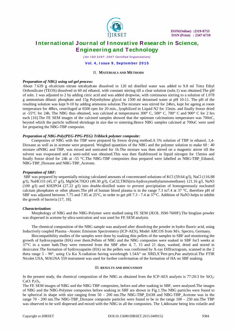
In the present study, the chemical composition of the NBG as obtained from the ICP-AES analysis is 77:20:3 for SiO2: CaO: P2O5. The FE SEM images of NBG and the NBG-TBP composites, before and after soaking in SBF, were analysed.The images of NBG and the NBG-Polymer composites before soaking in SBF are shown in Fig.1.The NBG particles were found to be spherical in shape with size ranging from 50 – 200 nm.The NBG-TBP_EtOH and NBG-TBP_Acetone was in the range 70 - 200 nm.The NBG-TBP_Dioxane composite particles were found to be in the range 100 – 250 nm.The TBP was observed to be well dispersed and mixed with the NBG in all the composites. The 1,4dioxane being less volatile and
ISSN(Online) :2319-8753 ISSN (Print) : 2347-6710
International Journal of Innovative Research in Science, Engineering and Technology
(An ISO 3297: 2007 Certified Organization)
Vol. 4, Issue 9, September 2015
Copyright to IJIRSET DOI:10.15680/IJIRSET.2015.0409152 9385
more viscous compared to acetone or ethanol, agglomerates of TBP coated NBG were formed.Thus the particle size of the NBG-TBP_dioxane was higher than that of NBG-TBP_acetone and NBG-TBP_ethanol.
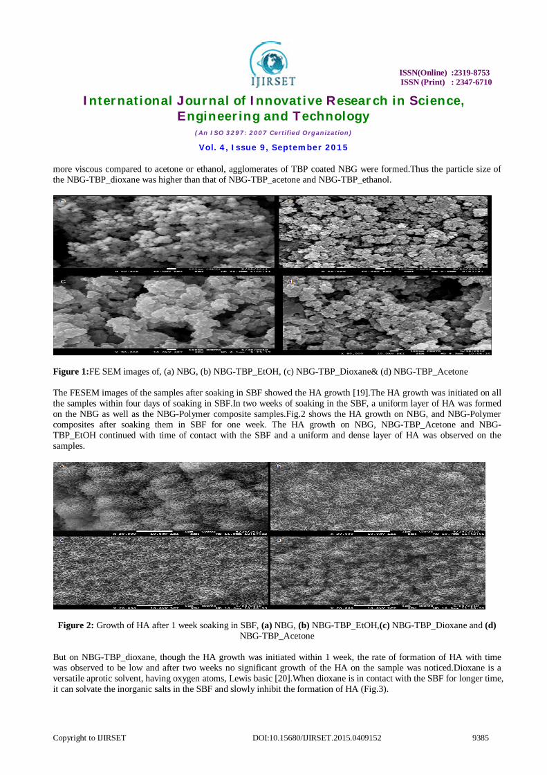
Figure 1:FE SEM images of, (a) NBG, (b) NBG-TBP_EtOH, (c) NBG-TBP_Dioxane& (d) NBG-TBP_Acetone The FESEM images of the samples after soaking in SBF showed the HA growth [19].The HA growth was initiated on all the samples within four days of soaking in SBF.In two weeks of soaking in the SBF, a uniform layer of HA was formed on the NBG as well as the NBG-Polymer composite samples.Fig.2 shows the HA growth on NBG, and NBG-Polymer composites after soaking them in SBF for one week. The HA growth on NBG, NBG-TBP_Acetone and NBG-TBP_EtOH continued with time of contact with the SBF and a uniform and dense layer of HA was observed on the samples.
Figure 2: Growth of HA after 1 week soaking in SBF, (a) NBG, (b) NBG-TBP_EtOH,(c) NBG-TBP_Dioxane and (d) NBG-TBP_Acetone
But on NBG-TBP_dioxane, though the HA growth was initiated within 1 week, the rate of formation of HA with time was observed to be low and after two weeks no significant growth of the HA on the sample was noticed.Dioxane is a versatile aprotic solvent, having oxygen atoms, Lewis basic [20].When dioxane is in contact with the SBF for longer time, it can solvate the inorganic salts in the SBF and slowly inhibit the formation of HA (Fig.3).

ISSN(Online) :2319-8753 ISSN (Print) : 2347-6710
International Journal of Innovative Research in Science, Engineering and Technology
(An ISO 3297: 2007 Certified Organization)
Vol. 4, Issue 9, September 2015
Copyright to IJIRSET DOI:10.15680/IJIRSET.2015.0409152 9386
(a& b) NBG-TBP_EtOH
(c& d) NBG-TBP_Dioxane
(e& f) NBG-TBP_Acetone
Figure 3: Growth of HA after 2 and 3 weeks of soaking in SBF,
The formation of HA on the NBG samples were further confirmed by FTIR Spectra analysis of all the samples.The KBr mixed NBG and the NBG-TBP composites were scanned in the wave number range 4000 to 300 cm-1. FTIR spectra of all the samples are shown in Fig.4 and the corresponding peak assignments are given in Table1.
Figure 4: FTIR spectra of (a) NBG-TBP_EtOH, (b) NBG-TBP_Dioxane and(c) NBG-TBP_Acetone
FTIR spectra of NBG powder as well as the composites showed peaks corresponding to Si-O-Si bending and stretching modes, 444.40, 466.77, 512.13 cm-1. Peak corresponding to Si-O-Ca was at 954cm-1. Characteristic peaks of hydroxyapatite includes the phosphates and the carbonate peaks at 1158cm-1, 978cm-1, 626 cm-1, 574 cm-1, 568 cm-1 and 1377.22 cm-1, 1456.05, 1459.71, 1543.23, 1637.22 cm-1, respectively. Hydroxyl group peak was found to be at 1637 cm-1, while water showed the peak at 3556 cm-1 [21].
Table 1: FTIR Peak assignments for NBG and NBG-Polymer composites
S No. Groups Wave number(cm-1) 1. Si-O-Si and O-Si-O 444.40,466.77,512.13,
2. Si-O-Ca 954
3. Phosphate group 568, 574, 626, 978, 1158,
4. Hydroxyl group 1637
5. Water (Bending) 3556
6. Carbonate ʋ3 1377.22, 1456.05, 1459.71, 1543.23, 1637.22

ISSN(Online) :2319-8753 ISSN (Print) : 2347-6710
International Journal of Innovative Research in Science, Engineering and Technology
(An ISO 3297: 2007 Certified Organization)
Vol. 4, Issue 9, September 2015
Copyright to IJIRSET DOI:10.15680/IJIRSET.2015.0409152 9387
The XRD analysis of the NBG and the NBG-TBP composites showed no sharp peaks except a hump in the 2Ɵ range 18 to 25o.This hump is characteristic of glass.After soaking pellets of NBG and the NBG-TBP composites in SBF, the XRD showed sharp peaks confirming the formation of crystalline salt of HA over the surface. The crystallinity was imparted to the surface of the pellet by calcium and phosphorous present in HA, which is confirmed by the appearance of sharp peaks in the spectra [22]. The hydroxyapatite reported here in XRD has same pattern, similar to the hydroxyapatite standard sample reported in literature, with the JCPDS Reference code: 01-074-0566.It had lattice dimensions of a = b = 0.9418 nm, c = 0.6884 nm and “d” 2.815 at 2Ɵ value ‘31’ which corresponds to hydroxyapatite from the JCPDS database, with the chemical formula Ca10 (PO4)6 (OH)2. The Ca/P ratio in the HA is 1.67 [23]. The NBG polymer composites also showed HA formation over the surface of the composite pellets upon soaking in SBF.The XRD spectra of all the composites are shown in Fig.5.
Figure 5: XRD spectra of composites: (a) NBG-TBP_EtOH(b) NBG-TBP_Dioxane(c)NBG-TBP_Acetone
When all the HA peaks from XRD spectra was analysed by X’Pert-Pro analysis software for the three composites, a difference in HA composition was found.For the NBG-TBP_EtOH composite, HA with chemical formula Ca5(PO4)6(OH) and JCPDS reference code: 01-073-0293 was found corresponding to calcium phosphate hydroxide. The lattice dimensions are; a=b= 9.4320, c= 6.8810; (h, k, l = 2, 1, 1) with d= 2.8168 (Ǻ) at 31.741 2Theta. For NBG-TBP_Acetone, HA with chemical formula Ca10 (PO4)6(OH)2 was found. The JCPDS reference code for it is: 01-072-1243. The lattice dimentions for this are; a=b= 9.430, c= 6.8881; (h, k, l= 2, 1, 1) with d=2.81681 (A°) at 31.741 2Theta. So for both of the above polymers we can say that the HA formed corresponds to the same HA molecule. But for the third composite, NBG-TBP_Dioxane, a slightly different form of the HA was found.It has a chemical formula of Ca8.8(PO4)6(H2O)2 with JCPDS reference code: 01-082-1943 corresponding to calcium phosphate dihydride.The lattice dimensions are; a=b= 9.460, c= 6.8800; (h, k, l= 2, 1, 1) with d= 2.82370 at 2Ɵ value 31.662. Thus we can observe that the solvents also affect the formation of hydroxyapatite giving different forms of HA.Depending upon their chemical structure all the forms have slightly different properties which ultimately affects their application in biomedical field.The hydroxyapatites grown over NBG-TBP_EtOH and NBG-TBP_Acetone have a chemical composition similar to the mineral component of bones, thus they could be used for treating defective sites of bones and joints in vivo, aroused by diseases, accident, trauma or injury [24], where the NBG composites would be implanted onto the defective site.The HA growth will commence at the site and it would support the cellular growth there.Thus new bone would start growing at the defective site. This would lead to bone regeneration and healing of the defect. Thus these composites would prove useful for treating biomedical problems. While the other form of hydroxyapatite i.e. calcium phosphate dihydride, grown over NBG-TBP_Dioxne has different chemical structure and was closer to tissues of dentures.Hence the NBG-TBP_Dioxane could be used to repair the dental problems [25].
IV. CONCLUSION Nanobioactive glass synthesized by sol gel method, had particle size of 50-100nm and are amorphous in nature. Upon incubation in SBF for 1-3 weeks at 37°C, they showed bioactivity by forming hydroxyapatite (HA) layer on their surface. Composites from nBG with this Poly (PEG-PPG-PEG)-Tri block polymer had particle size from 100-300 nm. Composites also form HA layer over their surface upon soaking in SBF under the same conditions.Use of different

ISSN(Online) :2319-8753 ISSN (Print) : 2347-6710
International Journal of Innovative Research in Science, Engineering and Technology
(An ISO 3297: 2007 Certified Organization)
Vol. 4, Issue 9, September 2015
Copyright to IJIRSET DOI:10.15680/IJIRSET.2015.0409152 9388
solvents for preparing the NBG-Polymer composite lead to the formation of different forms of HA, on soaking them in SBF.Thus they also show bioactivity and bioresorbability.
V. ACKNOWLEDGEMENT
The authors thank Centre for Research in Nanotechnology and Science (CRNTS), Sophisticated Analytical Instrumentation Facility (SAIF) of Indian Institute of Technology, Bombay for characterization facility. The authors greatly acknowledge the Council of Scientific and Industrial Research (CSIR)-JRF/SRF fellowship scheme.
REFERENCES
1. Superb K. Misra, Dirk Mohn, Tobias J. Brunner, Wendelin J. Stark, Sheryl E. Philip, Ipsita Roy, VehidSalih, Jonathan C. Knowles, Aldo R. Boccaccini(2008) Comparison of nanoscale and microscale bioactive glass on the properties of P(3HB)/Bioglass® composites. Biomaterials 29:1750-1761.
2. Kai Zhang, Yue Ma, Lorraine F. Francis (2002) Porous polymer/bioactive glass composites for soft-to-hardtissue interfaces. J Biomed Mater Res 61: 551–563.
3. Xin Liu, Mohamed N. Rahaman , Qiang F. Bone regeneration in strong porous bioactive glass (13-93) scaffolds with an oriented microstructure implanted in rat calvarial defects (2013) ActaBiomaterialia 9:4889–4898.
4. S. Ladron de Guevara-Fernandez, C.V. Ragel, M. Vallet-Reg (2003) Bioactive glass-polymer materials for controlled release of ibuprofen. Biomaterials 24:4037–4043.
5. Z.R. Domingues, M.E. Cortes, T.A. Gomes, H.F. Diniz, C.S. Freitas, J.B. Gomes, A.M.C. Faria, R.D Sinisterra (2004) Bioactive glass as a drug delivery system of tetracycline and tetracycline associated with b-cyclodextrin. Biomaterials 25:327–333.
6. Shuyi Li, Lynsa Nguyen, HairongXiong, Meiyao Wang, Tom C.-C. Hu, Jin-Xiong She, Steven M. Serkiz, George G. Wicks,William S. Dynan (2010) Porous-wall hollow glass microspheres as novel potential nanocarriers for biomedical applications. Nanomedicine: Nanotechnology, Biology, and Medicine 6:127–136.
7. Sepulveda P, Jones JR, Hench LL (2001) Characterization of melt-derived 45S5 and sol–gel-derived 58S bioactive glasses. J Biomed Mater Res 58(6):734–40.
8. Aldo R. Boccaccini, Veronique Maquet (2003) Bioresorbable and bioactive polymer/Bioglass composites with tailored pore structure for tissue engineering applications. Composites Science and Technology 63:2417–2429.
9. PragyaShrivastava ,Sridhar Dalai, PrernaSudera, S.Vijayalakshmiand Pratibha Sharma (2014) Hollow Glass Microspheres as potential adjunct with orthopaedic metal implants. Microelectronic Eng. 126:103–106.
10. S. Lopez-Estebana, E. Saiza, S. Fujinob, T. Okuc, K. Suganumac, A.P. Tomsia (2003) Bioactive glass coatings for orthopedic metallic implants: J Eur Ceram Soc.23:2921–2930.
11. Gabriel Marques, Luis Eduardo Marques Padovan, Mariza Akemi Matsumoto, Paulo DomingosRibeiroJúnior, Elisa MattiasSartori, Marcela Claudino (2013) Bone healing in titanium and zirconia implants surface: a pilot study on the rabbit tibia. Rev Sul Bras Odontol. 10(2):110-5.
12. Karen J.L. Burg, Scott Porter, James F. Kellam (2000) Biomaterial developments for bone tissue engineering. Biomaterials 21:2347-2359. 13. Boccaccini AR, Erol M, Stark WJ, Mohn D, Hong Z, Mano JF. Polymer/bioactive glass nanocomposites for biomedical applications. A review
(2010) Compos Sci Technol. 70:1764–76. 14. Gabriela E, Vargas, Luis A,Haro Durand, VanesaCadena,Marcela Romero,Rosa Vera Mesones,Mirza Macˇkovic´, Stefanie Spallek, Erdmann
Spiecker, Aldo R. Boccaccini , Alejandro A. Gorustovich (2013) Effect of nano-sized bioactive glass particles on the angiogenic properties of collagen based composites. J Mater Sci: Mater Med. 24:1261–1269.
15. Mohamed N. Rahaman, Delbert E. Day, B. Sonny Bal, Qiang Fu, Steven B. Jung, Lynda F. Bonewald, Antoni P. Tomsia (2011) Bioactive glass in tissue engineering. ActaBiomaterialia 7:2355–2373.
16. Mathew Peter ,PandianThodiSudheesh Kumar , Nelson SathyBinulal, Shanti V. Nair, Hiroshi Tamura, RangasamyJayakumar (2009) Development of novel a chitin/nanobioactive glass ceramic composite scaffolds for tissue engineering applications. CarbohydrPolym. 78:926–931.
17. Gil FJ, Padrósb A, Maneroa JM, Aparicioa C, Nilssona M, Planella J (2002) Mater SciEng C, 22 (2002), 53-60. 18. Müller L, Müller FA. ActaBiomaterialia, 2 (2006), 181–189. 19. Tadashi Kokubo, Hiroaki Takadama (2006) How useful is SBF in predicting in vivo bone bioactivity?.Biomaterials. 27:2907–2915. 20. http://en.wikipedia.org/wiki/1,4-Dioxane 21. Rehman and W. Bonfield (1997) Characterization of hydroxyapatite and carbonated apatite by photo acoustic FTIR spectroscopy. J Mater Sci:
mater in med. 8:1-4. 22. JOKANOVIC, D. IZVONAR, M.D. DRAMICANIN, B. JOKANOVIC, V. ZIVOJINOVIC, D. MARKOVIC, B.DACIC (2006) THIN FILMS OF SIO2 AND
HYDROXYAPATITE ON TITANIUM DEPOSITED BY SPRAY PYROLYSIS, J. MATER. SCI: MATER. MEDICAL, VOL. 17:539–546. 23. S. Koutsopoulos(2002) Synthesis and characterization of hydroxyapatite crystals: A review study on the analytical methods. J Biomed Matel Res.
62: 600-612. 24. Lutz-Christian Gerhardt and Aldo R. Boccaccini Bioactive Glass and Glass-Ceramic Scaffolds for Bone Tissue Engineering. Materials 2010, 3,
3867-3910; doi: 10.3390/ma3073867. 25. http://en.wikipedia.org/wiki/Dicalcium_phosphate.