volume 12|number 3 article 10 9-1964 refractory hypotension
TRANSCRIPT

Henry Ford Hospital Medical Journal
Volume 12 | Number 3 Article 10
9-1964
Refractory HypotensionJ. Keith Welborn
Joseph L. Ponka
Follow this and additional works at: https://scholarlycommons.henryford.com/hfhmedjournal
Part of the Life Sciences Commons, Medical Specialties Commons, and the Public HealthCommons
This Article is brought to you for free and open access by Henry Ford Health System Scholarly Commons. It has been accepted for inclusion in HenryFord Hospital Medical Journal by an authorized editor of Henry Ford Health System Scholarly Commons. For more information, please [email protected].
Recommended CitationWelborn, J. Keith and Ponka, Joseph L. (1964) "Refractory Hypotension," Henry Ford Hospital Medical Bulletin : Vol. 12 : No. 3 ,365-378.Available at: https://scholarlycommons.henryford.com/hfhmedjournal/vol12/iss3/10

Henry Ford Hosp. Med. Bull. Vol. 12, September, 1964
REFRACTORY HYPOTENSION
J. K E I T H WELBORN, M.D.* AND JOSEPH L . PONKA, M.D.**
PHYSICIANS MANAGE the hypotensive state with great facility in the majority of cases. A large group of patients are seen with obvious lesions which promptly respond to classically prescribed methods of treatment. A smaller group of patients are seen, however, with hypotension refractory to the commonly employed rescusitative measures. These patients represent complex problems in evaluation and management. The mortality in this group has been reported to be in the range of 80 per cent."'̂ '
The term "irreversible shock" (a description of an experimental model borrowed from the animal laboratory) popularly has been applied to human patients as an autonomous diagnosis. The term implies a state of progressive hypotension and "toxemia", the treatment for which thus far has been unsuccessful. Acceptance of the term has led to a diversion of attention from the fundamental pathogenic sequences which initiate and perpetuate hypotension. Although subtle, difficult to recognize, and associated with a high mortality, the physiologic alterations responsible for this state, more appropriately called refractory hypotension, can be treated successfully.
Management of refractory hypotension is one of the most challenging problems the physician sees. We have attempted to review its pathophysiology by reducing complex physiochemical processes to simplest terms so that a logical approach to the problem in a given patient might be made.
HISTORY
Hippocrates recognized the prognosis of patients with multiple traumatic injuries who displayed visible evidence of shock. Ambroise Pare wrote of "Syncope and Heart Failure" in describing the picture of neurogenic shock. James Latta of Edinburgh, 1795, is given credit for the first use of the word shock.
Shock was described as a clinical state in the 16th, 17th and 18th centuries. Glowes 1568, Wiseman 1719, and Garengiot 1723 attributed shock to the presence of some foreign matter in the wound or blood. The Civil War surgeons. Weir Mitchell, Morehouse, and Keen referred to shock as a "reflex disturbance". Billroth described
Associate, Fourth Surgical Division. Chief, Fourth Surgical Division. Henry Ford Hospital, Detroit 2, Michigan.
365

WELBORN AND PONKA
a "molecular disturbance of the brain." Blum referred to reflex cardiac inhibition
due to vagal irritation. Benjamin Travers is credited by Blalock' to have made a
major contribution to the understanding of shock in his time. His book, "An Inquiry
Concerning That Disturbed State of Vital Functions Usually Denominated Constitu
tional Irritation" published in 1820 disagreed with the accepted teachings of his
eminent contemporaries (John Hunter, Broussais, and his teacher. Sir Astley Cooper)
who were advocates of blood letting.
In 1899 Crile published his first book describing what was later called the
Crile-Mummery Hypothesis. In it, shock was ascribed to nervous exhaustion in an
electro-chemical system with two poles, the brain and liver. In shock the electrical
conductivity of the brain was described as decreased and that of the liver increased.
Crile's efforts attempting to block painful stimuli resulted in a significant reduction
of trauma associated with surgical procedures and handling of patients in shock,
although his concept of a primary etiologic mechanism was refuted.
Gray and Parsons, 1912, and Janeway and Ewing, 1914, concluded with ex
perimental data that nerve cell exhaustion played no role in the pathogenesis of shock.
Malcolm, in reports published as early as 1893, demonstrated the constriction
of peripheral vessels in shock and made two important observations based on clinical
rather than experimental work: 1) The danger to vital structures imposed by pro
longed intense vasoconstriction. He advised against the use of vasoconstrictors
therapeutically. 2) The principle of diminution of intravascular fluid volume in shock.
Mann, 1915, discussed the concept of "bleeding volume" (and thus inferentially blood volume). At this time it can be seen that a shift of emphasis from CNS depression to circulating volume loss was taking place.
Porter, 1916, described the importance of the diastolic pressure on prognosis
in shock and in 1917 described fat embolism as a cause of shock (previously
mentioned by Mausell-Moullin in 1894). Guthrie, 1917, discussed the significance
of vasomotor tone in shock.
Henderson, 1917, described decreased tissue metabolism as measured by oxygen
uptake in burn shock and described hypothermia as a result of shock.
Dale and Laidaw in 1910, studied the effect of histamine, peptones, and pressor
animes in shock.
The work of Erianger, Gesell, Gasser, Meek, Cannon, Bayliss, Phemister, and
Parsons added greatly to our knowledge of blood volume changes, replacement methods
and experimental technique.
366

REFRACTORY HYPOTENSION
The contributions of Blalock made as early as 1927 form the foundation for
some of the basic pathophysiology of shock as we know it today. Blalock and
Phemister, 1928, showed the importance of loss of fluid from circulating volume
into extravascular tissue space due to local trauma. They suggested the possibility
of two types of hemorrhage:
1) Outside of the body
2) Into the tissues
Their findings were based on a repeat of work done previously, 1919, by Cannon
and Bayliss, who had erroneusly concluded that increase in the weight of a traumatized
dog hind limb due to local fluid accumulation was insufficient to cause shock.
Blalock and Phemister, 1928, and subsequently Parsons and Phemister, 1930, demon
strated the importance of this phenomenon and showed that the original work failed
to include the areolar tissue of the groin where the fluid had migrated and thus had
not been included in the traumatized limb weight.
Johnson and Blalock, 1931, demonstrated the importance of a time relationship
between "secondary shock" and "histamine shock". Blalock, et al., 1934, demon
strated the importance of rate of blood loss to onset of signs of shock.
Blalock's concept of shock was: "A clinical syndrome, the features of which
are familiar to all, resulting in inadequate blood supply to the tissues of the body,
occurring as a result of underlying disturbances of different systems." With this as
a. basis, he classified shock as: hematogenic, neurogenic, vasogenic, cardiogenic,
and unclassified.'
Gregerson and Gibson in 1937, reported a method for estimation of blood
volume using Evans Blue Dye (T-1824).
More recently, numerous important contributions have been made from the laboratory and clinical work done by Simeone, Fine, Moore, McLean, Lellehei, Altmeier and Thai.
Throughout the history of the study of shock confusion has persisted regarding initiating, accompa.nying and perpetuating factors.
Even more confusion evolves from a discussion of the semantics of the definition of shock. Some deplore the use of the work "shock." However, "shock" carries with it a connotation of a clinical state which is distinct to experienced clinicians. We think it matters little which word is used to denote this state, which has as its common feature decreased tissue perfusion.
367

WELBORN AND PONKA
depends on • • •
® Blood Pressure
® Number of Patent Vessels diameter length
contractil ity
® Constituents 6 Properties of Blood
Figure 1
®Pre-Existing Myocardial Damage ® Coronary Occlusion ® Cor Pulmonale ® Cardiac Tamponade ® Cardiac Contusion ® Diminished Coronary Flow ® Toxic Injury
Hypoxemia Bacterial Toxins Metabolic Toxicity Deficiency of Metabolic Substrates
Figure 2
368

REFRACTORY HYPOTENSION
ETIOLOGY
The final common path in the refractory hypotensive state is persistent deficient tissue perfusion. This is perpetuated by decreased blood flow in the most terminal peripheral vessels. Perfusion of a segment of tissue depends upon: 1) Arterial pressure; 2) the number of patent vessels supplying the tissue, their diameter, length and contractility; and 3) the proper constituents and physical properties of blood. (Figure 1.)
The maintenance of tissue perfusion depends upon three factors: 1) efficiency of the pump; 2) adequcy of the blood volume; and 3) peripheral resistance.
THE PUMP Pump efficiency can be diminished by pre-existing myocardial damage, acute
coronary occlusion, cor pulmonale, cardiac tamponade, contusion, severely diminished coronary artery flow and toxic injury due to hypoxemia from its number of causes, bacterial toxins, metabolic toxins, and deficiency of metabolic substrates necessary for normal muscle contraction. (Figure 2.)
Figure 3 shows the four major categories of pump deficiencies: infarction, tamponade, cor pulmonale, and toxic injury. Familiar with these possibilities, the physician can be thorough and prompt in the evaluation of his patient with shock.
TAMPONADE
COR PUMONALE
TOXIC INJURY
Figure 3 Four major categories of pump deficiencies.
369

WELBORN AND PONKA
Volume ^044 « Plasma Loss
« Hemorrhage Obvious Occult
Intraperitoneal Retroperitoneal
® Sequestration
Obvious Burn
Occult Dehydration Persistent Vomiting Severe Diarrhea Massive Exudation
Peritonitis Pancreatitis Fatal Pneumonitis Intestinal Obstruction
Figure 4
THE VOLUME
Volume loss is commonly seen in shock. Frequently, howeve.-, failure to recognize
occult sources of blood volume deficit result in the refractory state. Hemorrhage from
both obvious and occult sources and sequestration, as is seen in the burn and endotoxin
shock, represent the possibilities for whole blood loss. Plasma volume loss, not always
as apparent as whole blood loss, may be obvious as in the burn, or occult as with
dehydration, persistent vomiting or diarrhea. Exudation losses are frequently greater
than expected in peritonitis, pancreatitis, fatal pneumonitis, and intestinal obstruction.
(Figure 4) .
370

REFRACTORY HYPOTENSION
OBVIOUS ^ HEMORRH^G^^^ \
• • •
OBVIOUS ^ HEMORRH^G^^^ \
OCCULT HEMORRHAGE
PLASMA LOSS
PANCREATITI
INTRAPERiTONt HEMORI^HAGE/
Figure 5
Possible sources of occult plasma loss.
Al l areas of possible volume loss should be considered in evaluating each patient
with shock, since multiple sources of loss commnoly co-exist. In the absence of
obvious hemorrhage, occult sources must be ruled out, such as intraperitoneal, retro
peritoneal, and associated with long bone fractures, the possible sources of occult
plasma loss are represented by the picture of fulminant pancreatitis. (Figure 5).
371

WELBORN AND PONKA
® Direct Vascular Cytotoxins
Chemical Metabolic Immunogenic Radiation Bacterial
® Local or Regional Hypoxemia
® C NS Injury
Medullary Vasomotor Areas Sympathetic Ganglia Embolization
Thrombus Fat
(* Anesthesia Fisure 6
THE RESISTANCE
Deterioration of peripheral resistance occurs frequently, associated with or as
a result of prolonged hypotension. Vascular cytotoxins directly affecting vasomotor
stability fall under the general headings of chemical, metabolic, immunogenic, irradia
tion, and bacterial. Local or regional hypoxemia from toxic or ischemic injury may
be important. Central nervous system injury, particularly to the medullary vasomotor
areas, the sympathetic ganglia and reflex injury associated with intracerebral emboli
zation may precipitate rapid collapse of peripheral resistance. Anesthesia is included
as a separate category because of its iatrogenic nature. (Figure 6).
372

REFRACTORY HYPOTENSION
RESISTANCE f
CAPILLARY
0
I
NJURY
SEQUESTRATION
CNS INJURY
Figure 7
Three major categories of decreased peripheral resistance.
Three major categories of decreased resistance should be considered. Toxic
injury affecting the precapillary and capillary cells and the more subtle causes of
capillary permeability represent direct cellular insult. CNS injury which may be the
result of hypoxia due to trauma, vascular accident, or anesthesia results in reflex
sympathetic block. Sequestration, localized increase in vascular capacity, is encountered
commonly in burns and endotoxin shock. (Figure 7).
THE REFRACTORY STATE
Al l of the factors known to be responsible for the refractory state should be
considered in the evaluation of each patient with hypotension of occult etiology.
Multiple processes may result in a complex physiometabolic interaction which can
be reversed when each is attacked individually. Or, a single factor may be missed
if each is not considered carefully. Advanced age, pre-existing myocardial disease,
occult blood volume deficit, invasive infection, renal failure, electrolyte and acid-base
imbalance are the factors most frequently associated with refractory hypotension.
With the exception of advanced age, each category can, if recognized, be treated
successfully. (Figure 7.)
373

WELBORN AND PONKA
TREATMENT
Successful management of refractory hypotension depends on:
1) Awareness of the multiple factors which can be of importance in the patient at hand.
2) Aggressive steps to ehminate each possible factor.
3) Precise aggressive therapy.
The Pump: A history of myocardial insult, physical signs of diminished cardiac output, the
EKG and central venous pressure monitorinng should rapidly make the efficiency of the pump clear to the alert physician in all but the most unusual circumstances. Digitalization should be accomplished in most cases, particularly in elderly patients.
The Volume:
Experimental as well as clinical data suggest that occult volume deficit is a major etiologic factor in a large number of patients with refractcry hypotension.
Advanced Age
M yocardial Disease
Occult Blood Volume Deficit
Invasive Infection
Renal Failure
Electrolyte Alterations
Acid Base Imbalance Figure 8
374

REFRACTORY HYPOTENSION
Sequestration of red cell and/or plasma volume (depending on the condition) may
diminish the circulating volume. Therefore it is important that peripheral flow be
augmented by transfusion with whole blood. Using the central venous pressure as
a guide, the volume should be increased until C.V.P. reaches 20 to 25 cm. saline.
In some cases this technique will "over transfuse" the patient and result in congestive
signs when the patient regains integrity of peripheral vascular tone. Continued ex
amination of the patient and C.V.P. monitoring supplemented by blood volume deter
minations (which can be relied upon 24 to 48 hours after stabilization) will allow
proper volume adjustments to be made rather accurately.
The Resistance:
Invasive Infection:
Invasive infection may precipitate hypotension as a primary etiology, as seen
commonly with gram negative septice.mia. Or, bacterial endotoxins may produce
severe unrelenting splanchnic vasodilatation having gained entrance to the portal
circulation by means of ischemic intestinal injury resulting from prolonged mesenteric
vasoconstriction due to hypotension of another primary etiology. Thus septic shock
may be primory or secondary. Fine feels that septic shock is the final common path
for shock of any cause, if it lasts long enough.'' Experimental and clinical data
suggest that patients with refractory hypotension should be treated with antibiotics
effective against gram negative organisms in doses regulated by the adequacy of
renal function. Blood and other appropriate cultures should be obtained at the
onset and continually throughout the critical period.
Renal Failure:
Prolonged hypotension, multiple rapid blood transfusions and pump oxygenator
procedures can result in renal failure.'* Anticipatory treatment with Mannitol given
intravenously can in most cases restore failing urinary output, if other therapeutic
measures at the same time are able to maintain adequate renal plasma flow.
Electrolyte Alterations and Acid Base Imbalance:
Acute metabolic acidosis in hypotension can be due to a number of causes
(Figure 9). This may be aggravated by acute or chronic respiratory acidosis. Thus
the use of tracheostomy and mechanical ventillatory assistance may be imperative.
Arterial pOz, p CO2 and pH determinations may quickly clarify a complex problem.
Recently it has been suggested that Hydrocortisone in pharamacologic doses
given intravenously is effective in refractory hypotension due to septicemia.We
have used this regimen with success in a small number of cases.
375

WELBORN AND PONKA
ACUTE METABOLIC ACIDOSIS
ETIOLOGIC FACTORS IN HYPOTENSION
GENERAL: Accumulation of tissue metabolites (Lactates, pyruvates due to anaerobic glycolysis)
BLOOD: Transfusion of old blood Transfusion of large amounts of citrated blood Hyponatremia due to:
Loss of volume Differential loss of sodium
HEPATIC: Injury preventing hepatic clearance of accumulating tissue metabolic acids
RENAL: Decreased Glomerular Filtrate Tubular damage due to:
prolonged hypoxemia precipitation of colloidal hemoglobin
Acid Hematin
PRE-EXISTING METABOLIC DISEASE: Diabetes Mellitus Chronic Glomerulonephritis
Figure 9
DISCUSSION
Evaluation and management of a given patient with refractory hypotension is frequently not as easy as we would make it seem. There are some general rules which can be applied which are quite reliable.
A patient in the operative, or recently postoperative period should be considered to have occult whole blood loss. Any suggestion of respiratory functional impairment in a patient during this period, particularly if he has undergone surgery about the neck or chest wall, should be quickly investigated for tension pneumothorax and treated promptly.'
Patients with peritonitis require in most instances vigorous whole blood, plasma, and fluid replacement. They may also manifest septic shock with httle or no suggestion of the signs or sepsis.
Fractures of the long bones can be a misleading source of occult hemorrhage requiring vigorous volume replacement, particularly if other injuries co-exist.
376
REFRACTORY HYPOTENSION
LEVEL
LEVC-«l,0 CROPS
mi
' LEVEL ,
1500'
5S 0 •*'.
5 3 J 1 J O
10,000- ' 2 6 0 -
J l _ J ,
8 $ 2«0
ISO .000
s S tm
9 ,0 -
SVSTOIIC 8 P P«tS£
2 0 0 -1 8 0 -
( 0 0 -
l»S
• ooo 500 "00
250 I0?5
?429 l»tft
<31 SI 61 t a
Z « IO!Z 4 8 12 4 8 (2 * I i2 MS I NOOK Wf» NOON
{*• ~- ——~ —M^f^^iS •••
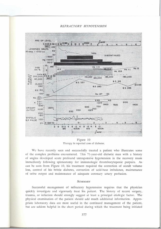
Figure 10 Therapy in reported case of diabetes.
We have recently seen and successfully treated a patient who illustrates some of the complex problems encountered. This 71-year-old diabetic man with a history of angina developed acute profound unresponsive hypotension in the recovery room immediately following splenectomy for immunologic thrombocytopenic purpura. As can be seen from Figure 10, his treatment required the correction of occult volume loss, control of his brittle diabetes, correction of acid-base imbalance, maintenance of urine output and maintenance of adequate coronary artery perfusion.
SUMMARY
Successful management of refractory hypotension requires that the physician quickly investigate and vigorously treat his patient. The history of recent surgery, trauma, or infection should strongly suggest at least a principal etiologic factor. The physical examination of the patient should add much additional information. Appropriate laboratory data are most useful in the continued management of the patient, but are seldom helpful in the short period during which the treatment being initiated
377

WELBORN AND PONKA
will have to be either effective or ineffective. For instance, bacteriologic cultures,
electrolyte and other chemical determinations are not available in most hospitals on
an emergency basis.
Recalling the outline presented above by which the pump, volume and resistance
factors can each be reviewed in a given patient, the physician can institute prompt
aggressive therapy which in more instances will be likely to result in successful
management of otherwise refractory hypotension.
REFERENCES
1. Antoni, R. O., and Ponka, J. L.: The hazard of iatrogenic pneumothorax in certain diagnostic and therapeutic procedures, Surg. Gynec. Obstet. 113:24, 1961.
2. Blalock, A.: Acute circulatory failure as exemplified by shock and hemorrhage, Surg. Gynec. Obstet. 58:551, 1934.
3. Ditzler, J. W., and Eckenhoff, J. E.: A comparison of blood loss and operative time in certain surgical procedures completed with and without controlled hypotension, Ann. Surg. 143:289, 1956.
4 . Fine, J.: Septic shock, JAMA 188:427, 1964.
5. Guyton, A. C, and Crowell, J. W.: Dynamics of the heart in shock. Fed. Proc. 20 (SuppI. 9):51, 1961.
6. Harkins, H. N.: Recent advances in the study and management of traumatic shock. Surgery
9:231, 447, 607, 1941.
7. Hershey, S. G.: Current theories of shock, Anesthesiology 21:303, 1960.
8. McHenry, M. C, and Martin, W. J.: Bacteremic shock due to gram-negative enteric bacilh, Proc. Mayo Clin. 37:162, 1962.
9. McHenry, M. C, Martin, W. J.. Hargraves, M. M., and Baggenstoss, A. H.: Bacteremia in
patients with neoplastic or hematologic disease, Proc. Mayo Clin. 37:43, 1962.
10. Miles, A. A.: Local and systemic factors in shock. Fed. Proc. 20 (SuppI. 9): 141, 1961.
11. Ravdin, I . S., and Ravdin, R. G.: Shock, fluids and electrolytes, 1905-1955, Internal. Abstr. Surg. 100:101, 1955.
12. Reeve, E. B.: Development of knowledge of traumatic shock in man. Fed. Proc. 20 (SuppI. 9): 12, 1961.
13. Schumer, W., and Durrani, K. M. : Study of effects of Noreninephrine on microcirculation of the dog omentum in ohgemic shock, Ann. Surg. 158:982, 1963.
14. Shoemaker, W. C : Recent additions to the knowledge of the cause and management of shock,
Surg. Clin. N. Amer. 42:3, 1962.
15. Shubin, H , and Weil, M. H.: Bacterial shock, JAMA 185:850, 1963.
16. Simeone, F. A.: Some issues in the problem of shock, Fed. Proc. 20 (SuppI. 9): 3, 1961.
17. Smith, D. E.: Radiation injury and shock. Fed. Proc. 20 (SuppI. 9): 158, 1961.
18. Smith, L. L. and Moore, F. D.: Refractory hypotension in man. New Eng. J. Med. 267:733, 1962.
378