volume1 - issue 1 - october 2017 resolution · preconceptional and gentic counselling sessions...
TRANSCRIPT

RESOLUTIONfeel the fetus, heal the fetus
Volume1 - Issue 1 - October 2017
Ÿ Monochorionic twins complications
Ÿ Treating fetal arrhythmias
Ÿ Neonatal resuscitation guidelines
Ÿ Binder phenotype-interesting case series
Ÿ Vaccination during pregnancy - updates
Ÿ Minor markers for aneuploidies
Ÿ Four chamber view abnormalities

FETAL MEDICINE SERVICES:
�
�
�
�
�
�
�
�
�
�
�
�
�
�
TIFFA (anomaly) Scans
NT scans
Growth and Doppler scans
3D/4D imaging
First trimester combined screening
Quadruple test
Amniocentesis
Fetal blood Sampling
Chorionic Villus sampling
Fetal reductions
Fetal therapy
- TTTS surgery, intrauterine blood transfusion, shunts etc
Preconceptional and Gentic counselling sessions
Multiple pregnancy assessments
Fetal care plan for High risk pregnancy
RESOLUTIONFetal Care Team
CONTACT FOR MORE DETAILS / APPOINTMENTS 92980 00015 / 99850 77986 / 98853 48600
CHEER
- Care- Health- Education- Expertise- Research
They Guide us, They Support us They Inspire us, They Teach us...
They make the hospital feel like a family
Dr. M. Indra Shekhar Rao MD, (Ped) DCH, Neo (USA), FIAPDirector Division of Pediatrics and NeonatologyProf. of Pediatrics NMCFormer Prof. Head Dept. of Pediatrics and Medical Supdt. Niloufer Hospital
Dr. Indira Sabapathy MD (Ped) DCH
Dr.Chinmayee RathaMS (ObGyn) MRCOG (UK) FIMSA FICOGLead Consultant – Fetal medicine

Chairman's Message
AnimportanttenetofhealthcareinIndiaiscaringforthepregnantwomen,fetusandnewbornswithcomplete intensive care back up such that theultimate perinatal outcome is optimised. Ourendeavour in Navodaya Hospitals has been todeliver thebestpossibleservices to thisgroupofpatients who will eventually help in shaping thefutureofoursociety.Wearethusworkingintandemtowardsourmottoof“Protectingtheprecious”.
Inthepursuitofexcellenceinperinatalhealthcare,oneoftheimportantdevelopmentsinourunithasbeen the initiationandexpansionof “stateof theart” Fetal medicine services – Navodaya’sResolution Fetal Care Centre headed by Dr.ChinmayeeRathaandherpassion forhersubjectwhichhas led to themottoof theunit– “feel thefetus,healthefetus”.Theunitcaterstoscreening,diagnosisandtreatmentofmanycomplicatedfetalproblems. In addition to minimally invasive fetaldiagnostic procedures to complicated medicaltherapy and perinatal surgical arrangements arebeingregularlydoneinthefetalmedicineunit. Weareproudtoannouncethesuccessfulcasesoffetaltherapy for complicated severe twin – twintransfusionsyndrome in theunitmaking this thefirstcentreinTelanganatoofferafullrangeofFetaltherapyservicesatparwithanyothercentreinthecountry.
Ourmissionnowexpandstoenhancingcareforthefetuswithspecialneedsandimprovingtheoutlookforfamilieswhodecidetocontinuepregnancywithdiagnosed fetal problems. This newsletter is anacademic endeavour of the Resolution Fetal carecentre to spread awareness in the medicalcommunity about recent developments andpracticalmanagementprotocolsforFetalproblems.ItisbeingreleasedontheoccasionofthefirstCMEof the department and I hope it continues as aperiodicactivity.IwishRESOLUTION2017agrandsuccess.
Shri. S Rajendar ReddyChairman, Navodaya Group of institutions, Raichur
01

From the Editor’s desk
Lifebeginsbefore“birth”andpreparationforthis“life”beginsevenbeforethesinglecellstageoftheactual zygote! This “unborn” individual occupiescentrestageinthecareparaphernaliaofthescienceof the “next generation” – Fetal Medicine. Myobsessionwith the fetusand itswellbeing isnowreaching its “teenage” chronologically, and it is asatisfyingfeelingtoseethedreamofafull-fledgedFetal Care team come together with passion,innovationanddetermination–allculminatingina“RESOLUTION”!
Resolutionisaverypowerfulword–anexpressionof determination in purpose and interestingly, inimagingscienceitisameasureofclarity!Thevisionassociated with Resolution encompasses thisdeterminationinstrivingforthebeststandardsinfetal health care with a clarity of purpose,possibilitiesandpotentialswithoutlosingtrackofpracticallimitationsandregulationsandthebasicprincipleofmedicine-Primum Non Nocere (First,donoharm).
ResolutionwantstospreadCHEER (Care, Health, Education, Expertise and Research)inthefieldofFetalMedicineanditsusers.Thisnewsletterisaninitiative of spreading awareness amongst themedical fraternity about the ongoing clinicalactivitiesinthishighlyexcitingfieldofcareforthefetus.Everydaywearedefining someconditionsbetter,understandingmoreofthepathophysiologyof fetal ailments and finding some solutions topersistentproblems–allthisisexhilaratinginitspaceandpotential: andawarenessisthefirststeptoachievingthebenefitsofadvancements.Withacollection of interesting articles on interestingclinicalcaseswehopeitwillmakeforanenrichingreadingexperience.Thisbeingour first issue,wehavetakentheopportunitytointroduceourteamandrangeoffacilitiestoo.
I also take this opportunity to thank all mycolleaguesinObGyn–practicisingacrossthelengthand breadth of the state who have supported uswiththeirfaithandallowedustocontributeinthe
careoftheirpregnantwomenandfoetuses-atrisk.Iamhumbledwith your belief in our abilities andsalute your commitment in looking for the bestsolutionstoyourpatient’sproblems.
We are proud to announce our successful Fetaltherapy programme – Resolution Fetal MedicineCentre, Navodaya Hospitals, the First centre inTelangana to provide a full range of FetalinterventionslikeminimallyInvasiveFetalsurgeryforMonochorionictwincomplicationswithablativeprocedures, Fetal transfusions, Fetal shunts inaddition to the basic diagnostic procedures likeAmniocentesis,BloodsamplingandCVS–the“ABC”of fetal diagnostics.We provide pre conceptionalcare for distressed couples with recurrentpregnancymishapsorgeneticconditions.Wecarefor mothers with severe medical complicationsthroughourdedicatedIntensivecareteamasonlyahealthymothercanprovideahealthyenvironmentforthefetustothrive.WeareayoungunitbutIamproud to announce that we have a successfuleducation and training programme in fetalmedicine and our fellows have been working onresearchprojects,presentedclinicalcasereportsinconferencesandsubmittedsomeoftheinitialworkforpublicationtoo.Wehopetocontinuethejourneyspreading“CHEER”withallyourgoodwishes.
Resolution Fetal Medicine Centre, NavodayaHospitals,isaninstitutiondedicatedtothehealthandsafetyofthefetuswhoisgoingtobethecitizenof tomorrow’s society.The motto of NavodayaHospitalsis“protectingtheprecious”andwhatcanbemoreprecioustoparentsthanthewellbeingoftheir little unborn childwhowill be the focus oftheirhappinesstomorrow.Soweprovidewhatwecall“today’sshareoftomorrow’scare”!
HappyReading!
Dr. Chinmayee RathaMS (ObGyn) MRCOG (UK) FIMSA FICOGLead Consultant Fetal MedicineResolution Fetal Medicine Centre, Navodaya Hospitals, Secunderabad
02

MONOCHORIONIC TWINS complications
Identical twins – Non identical problems!! Challenging solutions
Dr. Chinmayee Ratha
Monochorionic twin fetuses are connected to a singleplacentainthemother’swombandaresusceptibletoaunique complication called Twin to Twin transfusionsyndrome. Twin-twin transfusion syndrome (TTTS) isoneofthemostseriouscomplicationsofmonochorionicmultiple gestations.The cardinalprenatal findingsaremonochorionic placentation with concordant genderand discordant amniotic fluid volumes (maximumverticalpocket:donor<2cmandrecipient>8cm).TTTSisassociatedwithahighriskoffetal/neonatalmortality,especially in previable gestations, and fetuses whosurvive are at risk of severe cardiac, neurologic, anddevelopmentaldisorders.
It is generally estimated that TTTS complicates about15%ofMCpregnancies.TheexactincidenceofTTTSisnot clear since some fetal losses in monochorionicmultifetalgestationsinthefirsthalfofpregnancymayberelated to undiagnosed TTTS and thus the incidencebasedondatafromlivebornsorsonogramsinthesecondhalfofpregnancymaynotbeaccurate.
Placental intertwin vascular anastomoses, especiallyunbalanced AV anatomoses, play a key role in thepathophysiologyofTTTS.Thisleadstoanimbalanceinbloodflowinbothfetusesleadingtocirculatoryoverloadinthe“recipient”fetusandhypovolemiainthe“donor”twin.Thisisultimatelyharmfulandevenfatalforbothfetusesifitprogressesunchecked.
Quinterohasproposed fivestagesofdiseasebasedonfindingsfromtwo-dimensionalultrasoundandDopplervelocimetryintheumbilicalarteryandveinandductusvenosus:
Stage I - Oligohydramnios and polyhydramniossequence,andthebladderofthedonortwinisvisible.Dopplersinbothtwinsarenormal.
Stage II - Oligohydramnios and polyhydramniossequence,butthebladderofthedonorisnotvisualized.Dopplersinbothtwinsarenormal.
Stage III - Oligohydramnios and polyhydramniossequence, nonvisualized bladder, and abnormalDopplers. There is absent/reversed end-diastolicvelocityintheumbilicalartery,reversedflowina-waveoftheductusvenosus,orpulsatileflowintheumbilicalveinineitherfetus.
Stage IV - Oneorbothfetusesshowsignsofhydrops.
Stage V - Oneorbothfetuseshavedied.
TheaddedchallengewiththedeathofonefetusinaMCpairisthatatthetimeofdeaththereisexsanguinationofthe live fetus into the dead fetus and this transienthypotension in the live fetus results in neurologicalinjury in upto 20% of cases. Therefore, the idealmanagement of severe TTTS is to separate thecirculationsof the twinsbeforeanycatastrophiceventhappens.
CASE REPORT
AsecondgravidapreviousLSCS(9monthsback)referredat24weekswithMCDAtwinswith?TTTS–presentingcomplaintwassuddenonset severeuterinedistensionfromthepastweek.
Onexaminationmotherhadsignificantdiscomfort,withdifficulty in breathing due to overdistension of theabdomen.Abdomenwastenseandshinyandclinicallynotmuchcouldbediscernedbypalpation.
USG findings: MCDAtwins,anteriorhighplacenta
T1-severely increased AFI,DVP-18cm, growth767gm(74thcentile),largeurinarybladder,Activefetalmovements, normal umbilical flows and signs ofhyperdynamiccirculationseeninthefetalheart.
T2-severely reduced AFI – no measurable pocket,583gm(16thcentile)non-visualizationoffetalbladder.Umbilical artery flows showed absent and reverseddiastolicflow.
Cervicallengthwas26mm.Internalosclosed.
03

You treat a disease, you win, you lose. You treat a person, I guarantee you, you’ll win, no matter what the outcome.
- Patch Adams
04
RESOLUTION 2017feel the fetus, heal the fetus
Thecouplewereveryanxiousandscaredmoreaboutthehealth of the mother and requested termination ofpregnancyinmaternalinterest!
OUR DILLEMMAS
1. Termination at this gestation is not properlyindicated.
2. Maternaldistressdefinitelywassevereandneededtobeaddressed.
3. Short interconceptional interval with this severeuter ine distension could have obstetr ic complications.
4. Both fetuses were at risk and death of one fetuswouldjeopardisetheoutcomeoftheother.
We discussed the situationwith the parents and as amethod of immediate pal l iat ion we did anamniodrainagefromtherecipient’ssacreleasing3000mlamniotic fluid over 3 hours to avoid suddendecompression.Shewasrelievedimmediately,DVPwasreduced to 9cm and we kept her under observation.However, in 2 days time, the uterine overdistensionreappearedandtheDVPwas16!WehadexplainedthepossibilityofdefinitivetreatmentbydirectinterventionbyeitherLASERorRadiofrequencyablation(RFA)andasitwasalargeanteriorplacenta,theyoptedforRFA.
The purpose of this interventionwas to coagulate theumbilical cordofone twinso that theongoing “TTTS”wasstoppedandthepregnancycouldcontinuewithoneviablefetuswithareasonablygoodprognosis.Ifnothingisdone,bothfetusesareatriskofIUFD!
Detailedparentalcounselingandconsentingwasdone.RiskofPPROM,pretermlaborandIUDofbothfetuseswasexplained.Thecouplewerewillingfortheprocedureasitwouldatleastallowthemtohaveonelivebabyasopposedtolosingbothasaresultofthiscomplication.
Underasepticconditions,theRFAprocedurewasdone
onthedonortwincordat24wks2daysgestation.TheprocedurewentoffuncomplicatedandpostprocedureT1did not show any signs of compromise. She wasdischargedfromhospitalafter3daysfollowingadetailedscan includinganeurosonogramandcerebralDopplerstudyofthesurvivingtwinwhichwasnormal.
WefollowedherupweeklywithMCADopplersandserialgrowthassessmentevery2-3weeks.Shecomplainedofreduced fetal movements at 32 weeks of gestationpersistently foraweekandhad lowerabdominalpainandpreterm labour.AnEmergencyLSCSperformedat32wks3days
Indication: PreviousLSCSwithpretermlabour.
Baby–livebirthofagirlchild,1.8kg,goodcryatbirthAPGARscoresnormal
BabykeptO2andEBM,dischargedonday5,stableatthetimeofdischarge.
Wearehappytosharewithyouthesuccessstoryofthefirst definitive intervention in Telangana for thechallengingproblemofTTTS.
After this case, we have had three more successfuloutcomesandwe thank the revereddoctorswhohavetrusteduswiththecareofthesepatientsandtheparentswho have walked this difficult path with courage andconviction that humbles us. I am overwhelmed withgratitude when the parents voluntarily sent us theirphotographs and permitted us to share themwith thefraternityforincreasingawarenessinthisregard.

The final common pathway of successful fetal care isoptimalneonatalcareallowingasmoothtransitionoflifefromintrauterinetotheextrauterineenvironment.
Dr.N.SaravananDMNeonatologyhasbeeninstrumentalinco-ordinatingnewborncareinthewellequippedNICUatNavodayahospitalsandhashelpedusinpromisingthebestoutcomestoprospectiveparentseveninthemostchallengingsituations.
Fore ward: StateofartNICU18Bedded
Inborn: 12Bedded
Outborn:6bedded
MC/ Step down: 3Bedded
Neonatologist:
Dr.N.Saravanan,DM(Neo)
Dr.VenkatReddy,DNB(Ped)
Dr.Dhyaneshwar,(DCH)
Dr.NitinPatil,(DCH)
DedicatedwelltrainedSTAFF.
Census–2016–2017
Totaladmission–365/23months.
Neurosonogramdone.
5. Surgicalcasesdone - CDH-2 - TEF-3
ComplicatedMeconiumCyst-1
PUV-1 1HPS-2
ARM(colostomy)-1
6. Challenging cases managed - Severe IUGR,Congenital diaphragmatic hernia, TTTS survivors,Binder facies (Chondrodysplasia punctata),TuberousSclerosis(Rhabdomyoma),
Anorectalanomalies,TAPVC(surgeryatKIMS)
Neonates who received fetal treatment for CongenitalHeartBlock,tachyarrhytmias,hypothyroidismandfetalanemia.
Newbornswith familyhistoryofmultipleunexplainedneonataldeathshaveposedadifficultclinicaldilemmabutwithlogicalprotocolswehavebeenabletoprovidereasonableandgoal-orientedcareforallfamilies.
SmallestbirthWeightdischargedsuccessfully-680gms
SmallestGAdischargedsuccessfully-25weeks
NICUNeonatal Intensive Care Support
the back bone of successful fetal care prognosis!!
Navodaya Hospital NICU
05
<1Kg
1-1.2Kg
1.2–1.5Kg
1.5–2Kg
2-2.5Kg
2.5&above
30
23
27
61
73
152
84%Survival
98%Survival
100%Survival
100%Survival
98%Survival
98%Survival
CauseofdeathSepsis
CauseofdeathSepsis
CauseofdeathSepsis,CCHD,Cardiomyopathy,IEM
CauseofdeathsevereBA,PPHN,HIEStageIII
1. HFOV conventional ventilation, surfactant CPAP,ExchangeTransfusion, PDprocedure central lines,(PICC lines),ROP laser, ICDproceduresdone. LEDphototherapyavailable.
2. 24HoursoncallNeonatologist.
3. On call Pediatric Surgeon, Pediatric ENT,Ophthalmologist,PediatricCardiologistavailable.
4. AllinvestigationsdoneinhouseandwithtieupwithAmpathandBabyShield.
Digital Bed Side X-ray, Bed side 2D Echo and

They may forget your name, but they will never forget how you made them feel.
- Maya Angelou
06
RESOLUTION 2017feel the fetus, heal the fetus

Dr. Sahithi Reddy Fellow Fetal Medicine, Resolution Fetal Medicine Centre, Navodaya Hospitals
Aregularlybeatingheartprovidesthenaturalrhythmfora healthy life.When this heart beats irregularly – it iscalledanarrhythmia.Anirregularfetalheartrateot=rfetal arhythmia is a very worrying situation for thetreatingObstetrician.Fetalarrhythmiascomplicate1to2percent of pregnancies and have the potential tocompromisefetalhealth.Theyarecategorizedaccordingtotheirrhythm(irregular,regular)andrate(tachycardia,bradycardia). They are typically detected whenauscultating the fetalheart,whilemonitoring the fetalheart rate (FHR)with external or internal devices, orduringanantenatalultrasoundexamination.
Theconductionsystemofthefetalheartisfunctionallymatureby16weeksofgestation,andproducesaregularrhythmandratebetween110and160beatsperminute(bpm)fortherestofthepregnancy.Fetalarrhythmiasaredefinedbydeviationsfromtheseparameters.
Case 3 : Fetal Congenital Heart Block (Type II)
AG4A3,IVFconceptionwithsingleliveintrauterinefetuscametoourunitwithadiagnosisof“bradycardia”notedduring routine antenatal visit to the Obstetrician. Onexamination we confirmed fetal bradycardia (72 -80bpm) at 18+4weeksof gestation!Maternal pulse ratewas92/min!!
A detailed fetal echocardiography revealed a normallyconnectedheartwithno structural anomalies.CardiacDoppler revealeda firstdegree fetal congenital heartblock. There was a history of three early pregnancymiscarriagesandshewashypothyroidbutwelltreatedforthesame.
Mother was investigated for Anti Ro and Anti LAantibodies and were found to be strongly positive. Amultidisciplinarymeetingwasheldwiththe
rheumatologistandpediatriccardiologistandadecisiontostartsteroidtherapywastaken.WestartedtreatmentwithoralDexamethasoneanditwasreassuringthatthefetus responded well and recovered the heart rate toabout124bpmwithinthefirstweek.Shewasonregularfollowupwithusandthepregnancywascontinuedwithweekly FHR monitoring and titration of thedexamethasone dose in co-ordination with themultidisciplinary team. She required an escalation ofdosearound27weekswhentheheartratewasslowingtoabout100bpmanditappearedtobeprogressingtoseconddegreeheartblock,neverthelessthepregnancywascontinuedinanticipationofbetterfetalmaturity.
At34+3weekspatientcamewithcomplainsofreducedfetalmomentssince1day.CTGshowedabnormaltracewithbradycardia.OnUSGFHRwas51/min.Patientwastaken for Emergency LSCS after a neonatologycounseling.
Neonatology perspective : Asinglelivepretermmalebabyofweight1.97Kgswasdelivered.Babyapneicatbirthrequired3c y c l e s o f B M V b e f o r e spontaneous effortswith heartrateof80/mins.Babywaskeptfully under cardio respiratorymonitoring.Babydiagnosedascongenital heart block type-IIwithdroppedbeats2:1ratio.
X-ray showed cardiomegalywithincreasedvascularmarkings.PediatricCardiologistopinion(Dr.NitinRao)wastakenwhoreassuredusthatnoactiveinterventionifHR>60/min.2DechoonDay1showedmildtomoderatePDAwithgoodbiventricularfunction. OnDay3,Oxygen graduallyweavedoff andtubefeedintroduced.OnDay7,Babywasactivewithaheart rate varying between 90 to 110/min. and was
discharged.
Post Natal follow up: Baby isdoingfineandhealthy.Postnatalecho after 1 month confirmssecond degree block and thebaby has been kept on serialfollowupwithPediatric
Treating Fetal Arrhymiasrestoring the rhythm!
07
08
RESOLUTION 2017feel the fetus, heal the fetus
cardiologist.Theparentshavebeencounselledthatthechild may require a pacemaker sometime in future -perhapsaftera coupleofyearsand theyarepreparedmentallyforthesame.
The mother has been following up with therheumatologistforherownheathcare.
Learning points in this case:
1. Timelyreferralby thealertObstetricianhelped inearlyidentificationoftheproblem.
2. Quickandexactfetaldiagnosisalongwithmaternalantibodystatusconfirmedthepathology
3. Wewereluckythatthepathologywasmodifiablebytreatmentandhydropswasavoided
4. Co ordination between the multidisciplinary careteamandeffectivecounselingoftheparentshelpedinachievingafavorableoutcome.
5. Neonatalcompleteheartblockmaybefollowedandapacemakerinsertionmaybedeferred, initially, ifthebabyisotherwisestable.
Fetal bradycardia : Bradycardia may be due toconsistentlyblockedprematurecontractions,completeheartblockorsinusbradycardia.Heartblockandsinusbradycardia are more likely to be associated withstructuralheartdiseasethantachyarrhythmias.Ashortburst of sinus bradycardia is a normal physiologicfinding, but sustained bradycardia requires furtherevaluation.
When a fetus is diagnosedwith complete heart block,maternalbloodshouldbesampledforanti-Ro/SSAandanti-La/SSBantibodies.Inaddition,acompleteanatomicsurvey should be done to exclude structural heartdisease,particularly formsofheterotaxy.Treatmentofthe fetuswith complete atrioventricular (AV) block isprimarilyexpectant.
For anti-Ro/SSA anti-La/SSB positive women whosefetuses have incomplete heart block (mechanical AVinterval>~150msecorseconddegreeAVblock),ashortcourse of maternal dexamethasone is recommended(Grade 2C). While dexamethasone does not reversecompleteAVblock,itmayamelioratehydrops,whichhasahighmortalityrate.
Prolonged sinus bradycardia is nonreassuring. Causesinclude maternal hypotension, maternal seizures,paracervical block anesthesia, and impaired fetaloxygenation (eg, abruptio placenta, uterine rupture,prolapsed umbilical cord). Itmay also be due to fetalcardiac disease such as heterotaxy and/or long Q-Tsyndrome.
Ifantepartumtreatmentisinitiated,potentialmaternaltoxicitymustbemonitored,andcollaborativecare
including obstetrics, pediatric cardiology and often anadultcardiologistisneeded.
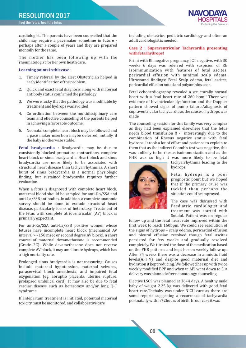
Case 2 : Supraventricular Tachycardia presenting with fetal hydrops!
PrimiwithRhnegativepregnancy,ICTnegative,with30weeks 6 days was referred with suspicion of RhIsoimmunization with features of fetal ascites,pericardial effusion with minimal scalp edema.Ultrasound findings: Fetal Scalp edema, fetal ascites,pericardialeffusionnotedandpolyamniosseen.
Fetal echocardiography revealed a structurally normalheart with a fetal heart rate of 260 bpm!! There wasevidence of biventricular dysfunction and theDopplerpattern showed signs of pump failure.Adiagnosis ofsupraventriculartachycardiaasthecauseofhydropswasmade
Thecounselingsessionforthisfamilywasverycomplexas they had been explained elsewhere that the fetusneeds blood transfusion !! - interestingly due to thecombination of Rhesus negative status with fetalhydrops.IttookalotofeffortandpatiencetoexplaintothemthatastheindirectCoomb’stestwasnegative,thiswasunlikely tobe rhesus isoimmunization and as theFHR was so high it was more likely to be fetal
tachyarrhythmia leading to thehydrops.
Fe ta l hydrops i s a poor prognostic point butwe hopedthat if the primary cause wastackled then perhaps thesituationcouldbeimproved.
The case was discussed withPaediatric cardiologist andtreatment was started withSotalol. Patient was on regular
followupandthe fetalheartrate improvedwithinthefirstweektoreach160bpm.Wecouldseeresolutionofthesignsofhydrops–scalpedema,pericardialeffusionand pleural effusion resolved though fetal ascitespersisted for few weeks and gradually resolvedcompletely.WetitratedthedoseofthemedicationbasedontheFHRpatternsandkeptheronweeklyfollowup.After34weeks therewasadecrease inamniotic fluidlevels(AFI=9) and despite good maternal diet andhydrationitkeptreducing.WefollowedherupwithtwiceweeklymodifiedBPPandwhenteAFIwentdownto5,adeliverywasplannedafterneonatologycounseling.
ElectiveLSCSwasplannedat36+4days.Ahealthymalebaby of weight 2.25 kgwas deliveredwith good fetalheart rate.Thebabywas under NICU care as there aresome reports suggesting a recurrence of tachycardiapostnatallywithin72hoursofbirth.Inourcaseitwas

09
RESOLUTION 2017feel the fetus, heal the fetus
reassuring that thebabycontinued tomaintaina fetalheartrateof90-100/min.Baselineinvestigationswerenormal.Bothmotherandbabyweredischargedonday3.
Themother’s heartratewascloselymonitoredbytheintensivist as she was weaned off the Sotalol.Interestingly she developed bradycardia in the postpartum period – upto 40 bpm. Injection Atropin wasgivenandwasunderICUcare.Thankfullysherecoveredher normal sinus rhythm and has remainedasymptomatictilldate.
Learning points in this case:
1. Everyrhesusnegativepregnancywithhydropsisnotduetoisoimmunization!
2. Detailed fetal echo is a must in any case of fetalhydrops.
3. Fetal hydrops – if not treated can be fatal so ifpossible,appropriatetreatmentsholdbestartedintime
4. Fetal tachycardia can be due to fetal stress orabnormal conduction.While short bursts of sinustachycardia (typically 160 to 200 bpm) can beassociatedwith fetalmovement in late pregnancy,prolonged tachycardia is always abnormal. Sinustachycardia may result from early fetal hypoxia,elevated maternal catecholamine levels due toanxiety or pain, maternal fever, intraamnioticinfection, or maternal administration of beta-adrenergic or vagolytic drugs. Pathologicaltachyarrhythmias include supraventriculartachycardia (SVT), atrial flutter, atrial fibrillation,andventriculartachycardia,withreentrantformsofSVTbeingthemostcommon.
5. Intermittentshortburstsoftachycardia<250bpmare generally well tolerated. For fetuses withintermittentshortburstsoftachycardia<250bpm,expectantmanagementwithclosefollow-upratherthaninterventionissuggested(Grade2C).
6. For preterm fetuseswith sustained SVT or a veryhighheartrateandpulmonaryimmaturity,medicaltreatmentratherthanexpectantmanagementor
deliveryissuggested(Grade1B).
7. For the fetus with sustained SVT, pulmonaryimmaturity and no evidence of hydrops, a trial ofdigoxinisfirst-linetherapy(Grade2C).Weaimformaternaldigoxinlevelsof1to2ng/mL.Ifthefetusdoesnotimproveordeterioratesdespiteadequatematernaldigoxinlevels(1to2ng/mL),wesuggestatrial of other medications, ideally in consultationwithpediatriccardiacelectrophysiologists.
8. Sotalol,flecainide,andamiodaronearesecond-lineagentsfortreatmentofsustainedornearlysustainedtachycardias at rates >220 bpm and pulmonaryimmaturity. Dosesmay need to be higher than innonpregnant women because of increased drugclearanceinpregnancy.
9. For fetuses with sustained SVT and pulmonarymaturity, delivery rather than expectantmanagement(Grade1B)orinuterotherapy(Grade2B)isrecommended.
NeonatalResuscitation Guidelines
10
Approximately 10% of newborns require someassistancetobeginbreathingatbirth.About1%requireextensive resuscitative measures. Although the vastmajority of newly born infants do not requireinterventiontomakethetransitionfromintrauterinetoextrauterinelife,becauseofthelargenumberofbirths,asizable number will require some degree ofresuscitation.
Thefollowingguidelinesareintendedforpractitionersresponsible for resuscitating neonates. They applyprimarily to neonates undergoing transition fromintrauterinetoextrauterinelife.Therecommendationsare also applicable to neonates who have completedperinataltransitionandrequireresuscitationduringthefirstfewweekstomonthsfollowingbirth.Practitionerswhoresuscitateinfantsatbirthoratanytimeduringtheinitial hospital admission should consider followingthese guidelines. The termsnewborn andneonate areintended to apply to any infant during the initialhospitalization.Thetermnewlybornisintendedtoapplyspecificallytoaninfantatthetimeofbirth.
Those newly born infants who do not requireresuscitation can generally be identified by a rapidassessmentofthefollowing4characteristics:
• Wasthebabybornafterafull-termgestation?
• Istheamnioticfluidclearofmeconiumandevidenceofinfection?
• Isthebabybreathingorcrying?
• Doesthebabyhavegoodmuscletone?
Iftheanswertoall4ofthesequestionsis“yes,”thebabydoesnotneedresuscitationandshouldnotbeseparatedfromthemother.Thebabycanbedried,placeddirectlyon the mother’s chest, and covered with dry linen tomaintaintemperature.Observationofbreathing,activity,andcolorshouldbeongoing.
Iftheanswertoanyoftheseassessmentquestionsis“no,”thereisgeneralagreementthattheinfantshouldreceiveone ormore of the following4 categories of action insequence:
A. Initial steps in stabilization (provide warmth,position,clearairway,dry,stimulate,reposition)
B. Ventilation
C. Chestcompressions
D. Administration of epinephrine and/or volumeexpansion
Thedecisiontoprogressfromonecategorytothenextisdeterminedby the simultaneous assessmentof 3 vitalsigns:respirations,heartrate,andcolor.Approximately30secondsisallottedtocompleteeachstep,reevaluate,and decide whether to progress to the next step inFigure. Neonatal Flow Algorithm.
Anticipation of Resuscitation Need
Anticipation,adequatepreparation,accurateevaluation,and prompt initiation of support are critical forsuccessful neonatal resuscitation. At every delivery,there should be at least one person whose primaryresponsibility is thenewlyborn.Either thatpersonorsomeoneelsewhoisimmediatelyavailableshouldhavetheskillsrequiredtoperformacompleteresuscitation,includingendotrachealintubationandadministrationofmedications.With careful consideration of risk factors,themajority of newbornswhowill need resuscitationcanbe identifiedbefore birth. If thepossible need forresuscitationisanticipated,additionalskilledpersonnelshould be recruited and the necessary equipmentprepared.
If a preterm delivery (<37 weeks of gestation) isexpected,specialpreparationswillberequired.Pretermbabieshaveimmaturelungsthatmaybemoredifficulttoventilate and are also more vulnerable to injury bypositive-pressureventilation.Pretermbabiesalsohaveimmaturebloodvessels in thebrain that areprone tohemorrhage; thin skinanda large surfacearea,whichcontributetorapidheatloss;increasedsusceptibilitytoinfection; and increased risk of hypovolemic shockcausedbysmallbloodvolume.
Initial Steps
Theinitialstepsofresuscitationaretoprovidewarmthbyplacingthebabyunderaradiantheatsource,positiontheheadina“sniffing”positiontoopentheairway,cleartheairwaywithabulbsyringeorsuctioncatheter,anddry the baby and stimulate breathing. Recent studieshave examined several aspects of these initial steps.Thesestudiesaresummarizedbelow.
Temperature Control: Verylowbirthweight(<1500g)pretermbabiesarelikelytobecomehypothermicdespitetheuseoftraditionaltechniquesfordecreasingheatloss.

FLOW CHART FOR NEONATAL RESUSCITATION
Approximate Time Birth
-Termgestation?-AmnioticFluidclear?-Breathingorcrying?-Goodmuscletone?
30 sec
No
YesRoutnine Care-Providewarmth-Clearairwayifneeded-Dry-Assesscolor
A-Providewarmth-Positionclearairway(asnecessary)-Dry,stimulate,reposition
Evaluate respiration, heart rate and color
Observational care
Breathing ,HR>100 & pink
Pink
Breathing, HR > 100 but Cyanotic
Apnoic or HR <100
Effective Ventilation,
HR >100 & pink
Persistent cyanosis
Givesupplementary
oxygen30 sec
B Providepositivepressureventilation Postresuscitation care
HR <60 HR >60
-Providepositivepressureventilation-AdministerchestcompressionsC30 sec
D Administerepinephrine and/orvolume*
HR <60
Endotrachealintubationmaybeconsideredatseveralsteps
Figure : Neonatal Flow Algorithm
11
RESOLUTION 2017feel the fetus, heal the fetus

All resuscitation procedures, including endotrachealintubation,chestcompression,andinsertionoflines,canbe performed with these temperature-controllinginterventionsinplace.
Infants born to febrilemothershavebeen reported tohave a higher incidence of perinatal respiratorydepression, neonatal seizures, and cerebral palsy andincreased risk of mortality. Hyperthermia should beavoided(ClassIIb).Thegoalistoachievenormothermiaandavoidiatrogenichyperthermia.
Clearing the Airway of Meconium: Aspiration ofmeconium before delivery, during birth, or duringresuscitation can cause severe aspiration pneumonia.Current recommendations no longer advise routineintrapartum oropharyngeal and nasopharyngealsuctioningfor infantsborntomotherswithmeconiumstainingofamnioticfluid(ClassI).
Periodic Evaluation at 30-Second Intervals
After the immediate postbirth assessment andadministration of initial steps, further resuscitativeeffortsshouldbeguidedbysimultaneousassessmentofrespirations, heart rate, and color. After initialrespiratoryefforts,thenewlyborninfantshouldbeableto establish regular respirations that are sufficient toimprove color and maintain a heart rate >100 bpm.Gasping and apnea indicate the need for assistedventilation.Increasingordecreasingheartratecanalsoprovideevidenceofimprovementordeterioration.
Anewlyborninfantwhoisuncompromisedwillachieveand maintain pink mucous membranes withoutadministrationofsupplementaryoxygen.Acrocyanosis(bluecolorofhandsandfeetalone)isusuallyanormalfinding at birth and is not a reliable indicator ofhypoxemia butmay indicate other conditions, such ascoldstress.Pallorormottlingmaybeasignofdecreasedcardiac output , severe anemia, hypovolemia,hypothermia,oracidosis.
Administration of Oxygen : Thereareconcernsaboutthe potential adverse effects of 100% oxygen onrespiratoryphysiologyandcerebralcirculationandthepotential tissue damage from oxygen free radicals.Converselytherearealsoconcernsabouttissuedamagefrom oxygen deprivation during and after asphyxia.Supplementary oxygen is recommended wheneverpositive-pressure ventilation is indicated forresuscitation;free-flowoxygenshouldbeadministeredtobabieswhoarebreathingbuthavecentral cyanosis(Class Indeterminate). The standard approach toresuscitationistouse100%oxygen.Administrationofavariable concentration of oxygen guided by pulseoximetrymay improve theability toachievenormoxiamore quickly. Concerns about potential oxidant injuryshouldcaution theclinicianabout theuseofexcessiveoxygen,especiallyintheprematureinfant.
Positive-Pressure Ventilation: If the infant remains
apneicorgasping,iftheheartrateremains<100bpm30seconds after administering the initial steps, or if theinfant continues to have persistent central cyanosisdespite administrationof supplementaryoxygen, startpositive-pressureventilation.
Initial Breaths and Assisted Ventilation : In terminfants, initial inflations—either spontaneous orassisted—create a functional residual capacity. Theoptimumpressure,inflationtime,andflowraterequiredtoestablishaneffectivefunctionalresidualcapacityhavenot been determined. Average initial peak inflatingpressuresof30to40cmH2O(inflationtimeundefined)usuallysuccessfullyventilateunresponsiveterminfants.Assistedventilationratesof40to60breathsperminutearecommonlyused,buttherelativeefficacyofvariousrateshasnotbeeninvestigated.
Theprimarymeasureof adequate initial ventilation ispromptimprovementinheartrate.Chestwallmovementshouldbeassessedifheartratedoesnotimprove.Theinitialpeakinflatingpressuresneededarevariableandunpredictableandshouldbeindividualizedtoachieveanincreaseinheartrateand/ormovementofthechestwitheachbreath.
Devices : Effective ventilation can be achievedwith aflow-inflatingbag,aself-inflatingbag,orwithaT-piece.The pop-off valves of self-inflating bags are flow-dependent, and pressures generated may exceed thevalue specified by the manufacturer . Target inflationpressures and long inspiratory times are moreconsistently achieved in mechanical models when T-piecedevicesareusedrather thanbags , although theclinicalimplicationsarenotclear.Toprovidethedesiredpressure,healthcareprovidersneedmoretrainingintheuseofflow-inflatingbagsthanwithself-inflatingbags.Aself-inflatingbag,aflow-inflatingbag,oraT-piececanbeusedtoventilateanewborn(ClassIIb).Laryngealmaskairways(LMAs)thatfitoverthelaryngealinlethavebeenshown tobeeffective forventilatingnewlybornnear-termandfull-terminfants.
Assisted Ventilation of Preterm Infants : Whenventilating preterm infants after birth, excessive chestwall movement may indicate large-volume lunginflations, which should be avoided. Monitoring ofpressuremayhelptoprovideconsistent inflationsandavoidunnecessaryhighpressures(ClassIIb).Ifpositive-pressure ventilation is required, an initial inflationpressure of 20 to 25 cm H2O is adequate for mostpreterm infants (Class Indeterminate). If promptimprovement in heart rate or chest movement is notobtained, higher pressures may be needed. If it isnecessary to continue positive-pressure ventilation,application of PEEP may be beneficial (ClassIndeterminate).Continuouspositiveairwaypressureinspontaneously breathing preterm infants afterresuscitat ion may also be beneficial (Class Indeterminate).
12
RESOLUTION 2017feel the fetus, heal the fetus

Endotracheal Tube Placement
Endotracheal intubation may be indicated at several points during neonatal resuscitation:
• Whentrachealsuctioningformeconiumisrequired
• Ifbag-maskventilationisineffectiveorprolonged
• Whenchestcompressionsareperformed
• Whenendotrachealadministrationofmedicationsisdesired
• For special resuscitation circumstances, such ascongenitaldiaphragmaticherniaor extremely lowbirthweight(<1000g)
Thetimingofendotrachealintubationmayalsodependontheskillandexperienceoftheavailableproviders.
Other clinical indicators of correct endotracheal tubeplacementareevaluationofcondensedhumidifiedgasduringexhalationandthepresenceorabsenceofchestmovement, but these have not been systematicallyevaluated in neonates. Endotracheal tube placementmust be assessed visually during intubation and byconfirmatorymethodsafterintubationiftheheartrateremains lowand isnotrising.Except for intubationtoremove meconium, exhaled CO2 detection is therecommendedmethodofconfirmation(ClassIIa).
Chest Compressions : Chestcompressionsareindicatedfor a heart rate that is <60 bpm despite adequateventilationwithsupplementaryoxygenfor30seconds.Because ventilation is the most effective action inneonatalresuscitationandbecausechestcompressionsarelikelytocompetewitheffectiveventilation,rescuersshouldensurethatassistedventilationisbeingdeliveredoptimallybeforestartingchestcompressions.
Medications : Drugsarerarelyindicatedinresuscitationof the newly born infant. Bradycardia in the newborninfantisusuallytheresultofinadequatelunginflationorprofound hypoxemia, and establishing adequateventilationisthemostimportantsteptocorrectit.Butifthe heart rate remains <60 bpm despite adequateventilationwith100%oxygenandchestcompressions,administrationofepinephrineorvolumeexpansion,orboth, may be indicated. Rarely, buffers, a narcoticantagonist, or vasopressors may be useful afterresuscitation.
Route and Dose of Epinephrine Administration
Past guidelines recommended that initial doses ofepinephrine be given through an endotracheal tubebecausethedosecanbeadministeredmorequicklythanwhenanintravenousroutemustbeestablished.
Volume Expansion
Considervolumeexpansionwhenbloodlossissuspectedor the infant appears to be in shock (pale skin, poorperfusion, weak pulse) and has not respondedadequatelytootherresuscitativemeasures.Anisotoniccrystalloidratherthanalbuministhesolutionofchoicefor volume expansion in the delivery room. The
A compression-relaxation ratiowith a slightly shortercompression than relaxation phase offers theoreticaladvantagesforbloodflowintheveryyounginfant.Also,compressionsandventilationsshouldbecoordinatedtoavoid simultaneous delivery. The chest should bepermitted to fully reexpand during relaxation, but therescuer's thumbs should not leave the chest. Thereshouldbea3:1ratioofcompressionstoventilationswith90 compressions and 30 breaths to achieveapproximately 120 events per minute to maximizeventilationatanachievablerate(ClassIndeterminate).Thus, each event will be allotted approximately ½second, with exhalation occurring during the firstcompressionaftereachventilation.
Respirations,heartrate,andcolorshouldbereassessedabout every 30 seconds, and coordinated chestcompressionsandventilationsshouldcontinueuntilthespontaneousheartrateis>60bpm(ClassIIa).
13
RESOLUTION 2017feel the fetus, heal the fetus
recommendeddoseis10mL/kg,whichmayneedtoberepeated. When resuscitating premature infants, careshouldbe taken toavoidgivingvolumeexpanders toorapidly, because rapid infusionsof largevolumeshavebeenassociatedwithintraventricularhaemorrhage.
Naloxone : Administration of naloxone is notrecommendedaspartofinitialresuscitativeeffortsinthedeliveryroomfornewbornswithrespiratorydepression.If administrationof naloxone is considered, heart rateand color must first be restored by supportingventilation.
Postresuscitation Care : Babies who requireresuscitationareatriskfordeteriorationaftertheirvitalsignshavereturnedtonormal.Onceadequateventilationandcirculationhavebeenestablished,theinfantshouldbemaintained in or transferred to an environment inwhich close monitoring and anticipatory care can beprovided.
Glucose : Lowbloodglucosehasbeenassociatedwithadverseneurologicoutcomeinaneonatalanimalmodelofasphyxiaandresuscitation.
Induced Hypothermia : Inamulticentertrialinvolvingnewbornswith suspectedasphyxia (indicatedbyneedforresuscitationatbirth,metabolicacidosis,andearlyencephalopathy),selectiveheadcooling(34°Cto35°C)was associated with a nonsignificant reduction in theoverallnumberofsurvivorswithseveredisabilityat18months but a significant benefit in the subgroupwithmoderate encephalopathy. Infants with severeelectrographicsuppressionandseizuresdidnotbenefitfrom treatment with modest hypothermia. There isinsufficient data to recommend routine use ofmodestsystemic or selective cerebral hypothermia afterresuscitationof infantswithsuspectedasphyxia(ClassIndeterminate).
Guidelines for Withholding and Discontinuing Resuscitation : Morbidityandmortalityfornewbornsvariesaccordingtoregionandavailabilityofresources. Social science studies indicate that parents desire alarger role in decisions to initiate resuscitation andcontinue life support of severely compromisednewborns. Opinions among neonatal providers varywidely regarding the benefits and disadvantages ofaggressivetherapiesinsuchnewborns,
Withholding Resuscitation : It is possible to identifyconditions associated with high mortality and pooroutcomeinwhichwithholdingresuscitativeeffortsmaybeconsideredreasonable,particularlywhen therehasbeentheopportunityforparentalagreement.
The following guidelines must be interpreted according to current regional outcomes:
• When gestation, birth weight, or congenitalanomaliesareassociatedwithalmostcertainearlydeath and when unacceptably high morbidity is
likelyamongtheraresurvivors,resuscitationisnotindicated(ClassIIa).Examplesmayincludeextremeprematurity (gestational age <23 weeks or birthweight <400 g), anencephaly, and chromosomalabnormalities incompatible with life, such astrisomy13.
• Inconditionsassociatedwithahighrateofsurvivaland acceptable morbidity, resuscitation is nearlyalways indicated (Class IIa). This will generallyinclude babies with gestational age > 25 weeks(unlessthereisevidenceoffetalcompromisesuchasintrauterine infection or hypoxia-ischemia) andthosewithmostcongenitalmalformations.
• Inconditionsassociatedwithuncertainprognosisinwhich survival isborderline, themorbidity rate isrelatively high, and the anticipated burden to thechildishigh,parentaldesiresconcerninginitiationof resuscitation should be supported (ClassIndeterminate).
Discontinuing Resuscitative Efforts : Infantswithoutsignsoflife(noheartbeatandnorespiratoryeffort)after10minutesofresuscitationshoweitherahighmortalityor severe neuro developmental disability. After 10minutes of continuous and adequate resuscitativeefforts,discontinuationofresuscitationmaybejustifiediftherearenosignsoflife.
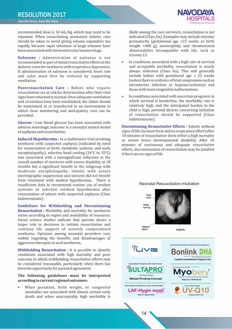
Neonatal Resuscitation-Intubation
14
RESOLUTION 2017feel the fetus, heal the fetus
citrate malate with

Binder Phenotypean interesting case series
15
Dr. Aditi GoundanFellow Fetal Medicine, Resolution Fetal Medicine Centre, Navodaya Hospitals
Wepresentaseriesofcasesreferredtothefetalmedicineunitas“hypoplastic/absentnasalbone”or“abnormallyflatfetalface”Manywereactuallyadvisedtermination!
On detailed TIFFA scan the fetus was found to havegenerally normal growth and structure but the facialprofilerevealedaflatnasalbridge.
Fig: flatnasalbridge
Case 1 :
G2P1L1referredwithabsentnasalboneandtripletestshowinganincreasedriskforT21
She gave history of significant hyperemesis requiringmultiplehospitaladmissions
USG revealed a flat nasal bone consistentwithBinderFacies
Restoftheskeletalsystemwasnormal
Nosoftmarkersfortrisomy21ongeneticsonogram
Amniocentesisdonerevealednormalkaryotype
Couple decided to continue with the pregnancy anddeliveredahealthygirlchild2.8kgattermwithnormaloutcome.
Case 2 :
PrimigravidawithhistoryofMitralValveReplacement
Conceivedwhileonwarfarin(5-6mg/day)
Anomaly scan revealed flattened nasal bridgewith noevidenceofepiphysealstippling
Longboneswerealongthelessercentiles
Counselled regarding warfarin embryopathy andchondrodysplasiapunctata
Decidedtocontinuethepregnancy
Shecontinuedantenatalcareatherprimarycarecentreanddeliveredatterm2.2kgmalebabywithnoapparentproblems
Case 3 :
Primigravidareferredat19weekswithsuspectedspinedeformity
USGfindingsshowedahemivertebraeinthoracicregioninassociationwithBinderFacies
Nosignificantfamilyhistory/medicalhistory
After counselling they opted for termination ofpregnancy
Notwillingforanyprenatalkaryotypetesting

16
RESOLUTION 2017feel the fetus, heal the fetus
Binderfacies,wasfirstdescribedbyBinder1962andhastypicaldysmorphicfeatures-Shortnosewithflatnasalb r idge , Re t rudedmid face ,Absence o f nasa l spine,Shortcolumella,Acute nasolabial angle,Convexupperlip
Itmay be an isolated finding only in the fetal face ormaybepartofageneralizedsystemicabnormality.Bindersyndrome, also known as maxillonasal dysplasia, is avery rare disorder of unknown etiology. It ishypothesizedthatBindersyndromeisthemildestformof chondrodysplasia punctata. Binder syndrome ischaracterized by a shortened nose with an acutenasolabial angle and a convex upper lip . Class IIImalocclusion may be present. An underdevelopedfrontalsinusandcervicospinalabnormalitiesoccurin40to50percentofcases .Thetreatmentconsistsofnasaland maxillary correction, followed by orthodonticrehabilitation.
Case 4 :
RarepresentationofmultiplefetalproblemsalongwithBinderphenotypewithagoodoutcome!
A Primi gravida referred with polyhydramnios at 29weeks. ShewasaknowncaseofPIHonAlphamethylDopa. On examination she had significant physicaldistress due to overdistension of the abdomen, grossbilateral pedal and facial edema, Bp-140/90-150-90mmhg. PIH profile within normal limits and noproteinuria.
USG findings: SLF, AFI46, severe polyamnios, FG 18thcentile,depressednasalbridgewithmidfacialhypoplasia
Amniodrainage was performed and about 2,5litres offluidreleased.Thefluidrefilledinoneweek'stimeandwe did a repeat procedure but also started her onIndomethacinforaweek.
Serial scans revealed severe growth restriction and liquorremainedinthenormallimts.Wecounselledthecoupleabout thepossibilityofraregeneticsyndromesduetothepresenceofmanydisturbingfeaturesinthefetus.
Maternal condition remained stable and fetus wasfollowedupwithweeklyBPP.Therewasreduced fetalmovementsandnonreassuringCTGandanemergencyLSCSperformedat36wks:SevereFGR-LB/1.2kg/fchat36weeksbyLSCS
Whilewe suspected theworst, the baby had a ratheruneventfulstayatNICUnotrequiringanyinterventionfor respiratory distress and themetabolic screenwasnegative. We kept the parents alerted of delayedpresentation of any genetic condition an dtheymaintained regular postnatal follow up including aclinicalgeneticsworkup.
Presentlybabyisdoingfineandhascelebratedherfirstbirthdayfewmonthsback.Wewishthefamilywell. Inthiscasetherewasalotofuncertainityduetotheseverepolyamniosandgrowthrestrictionbuttheparentsweresocommittedtothepregnancythattheykepttheirownmoralehighandhelpedusbyreinforcingourconfidenceduringuncertainmoments.Thiscasewasatremendouslearningexperienceforus,
TAKE HOME MESSAGES:
- Isolated Binder phenotype has a good prognosis-doesnotwarrantTOP
- Lookforstigmataofassociatedconditions
- Possibiltyofallraregeneticsyndromescannotberuledout
- Diagnosticdilemma-Noconfirmatorytests
- Ruleoutanyknowncausesofskeletal/cartilaginousmaldevelopment.
- Deliveryatatertiarycarecentre(fewcasereportsofnewborns with respiratory distress requiringintubation)
- Cosmeticimplications

VaccinationDuring Pregnancy - UPDATES
17
Dr. ShaliniFellow Fetal Medicine, Resolution Fetal Medicine Centre, Navodaya Hospitals
“Protectingtheprecious”pregnancyistheendeavourofevery Obstetrician. One important aspect in thisprotection is to prevent inadvertent infections andvaccinations help in providing that safetymechanism.We have collated some recent recommendationsregardingvaccinationinpregnancywhichwillbeaquick,practicalupdateforallpractitioners
Ideally,womenshouldbevaccinatedagainstpreventablediseases in their environment prior to conceptionaccording to the recommended adult immunizationschedule. In particular, for susceptible women ofchildbearingagewhomaybecomepregnant, ensuringimmunity against measles, mumps, and rubella andvaricella is important since these immunizations arecontraindicated during pregnancy and infection innonimmune pregnant women can adversely affectpregnancyoutcome.
Beforeadministeringanyvaccine,reasonablepracticesinclude asking thewoman if she is pregnant or couldbecomepregnantinthenextfourweeksandcounselingher about the potential risks of vaccination duringpregnancyorjustbeforeconception
Live vaccines are generally avoided during pregnancybecause of a theoretical risk to the fetus. Pregnancyshouldbeavoidedfor28daysfollowingadministrationofalivevaccine.However,whenpregnancyoccurswithinone month of immunization with the live measles,mumps,rubella(MMR)vaccine,varicellavaccine,yellowfevervaccine,ororalpoliovaccine,teratogenesishasnotbeenreported.Therefore,terminationofpregnancyforexposuretothesevaccinesisNOTwarranted.
Pregnant women should minimize their risk ofexposuretoinfectionstowhichtheyaresusceptiblebyavoiding travel to high-risk locations (eg, areaswhereyellow fever is prevalent), assuring that household
members are immunized according to standardimmunizationschedules,andmaintaininggoodhygienicpractices.
During influenza season, we recommend that allpregnant women receive the inactivated influenzavaccineregardlessoftrimesterofpregnancy(Grade1B).Influenzaisparticularlymorbidinpregnantwomenandcanbepreventedbyvaccination.Maternalimmunizationadditionallyprovidespassiveprotectiontotheinfantinthefirstfewmonthsoflife.
Werecommendadministrationofthetetanustoxoid,reduced diphtheria toxoid, and acellular pertussisvaccine(Tdap)toallpregnantwomenineachpregnancybetween27and36weeksofgestation(andpreferablyduringtheearlierpartofthisperiod),evenifthewomanhasaprevioushistoryofpertussisorvaccination(Grade1B). Tdap is given to reduce the risk of maternalpertussis,andthustransmissiontotheinfant,inwhompertussis can be lethal or have significant morbidity.Placentaltransferofmaternalantibodiesmayprovideadegree of passive protection of the infant againstpertussisfortwotosixmonths.
Pregnant women with comorbidities or exposuresthatplacethemathighriskforhepatitisA,hepatitisB,pneumococcal, Haemophilus influenzae b, ormeningococcal infect ions can receive theseimmunizations
If travel to high-risk areas cannot be avoided,inactivated travel vaccines, such as for poliovirus andtyphoidfever,canbeadministeredpriortotravel.Iftherisk of yellow fever exposure is expected to be higherthantheriskofvaccination,theyellowfevervaccine,alive attenuated vaccine, can be administered inconsultation with an infectious disease specialist. Incaseswheretheriskofdiseaseislowbutvaccinationisan international travel requirement, a medical waivercanbeissued.
Human papillomavirus (HPV) vaccination duringpregnancy is typically avoided. However, women whoinadvertentlyreceiveHPVvaccineduringpregnancycanbe reassured that there is no evidence of adversepregnancyorfetaloutcomeswithvaccination.Theymayresumetheserieswhenpostpartum.
Likeotherlivevaccines,MMRandvaricellavaccinesaregenerallyavoidedduringpregnancy.Fornonimmune

18
RESOLUTION 2017feel the fetus, heal the fetus
women, MMR and varicella vaccines are givenpostpartum, ideally prior to discharge. MMR andvaricella vaccines can be given safely to postpartumwomen who are breastfeeding and to the children ofpregnant women, since the virus is not transmittedthrough breast milk or casual contact. Tdap is givenpostpartum to women who did not receive it duringpregnancy.
Update onTetanus, Diptheria and Pertusis:
RevisedAdvisoryCommitteeonImmunizationpractisesguideline recommend that health care providesadministeradoseofTdapduringpregnancy,irrespectiveofwhethershereceivedearlierornot.Tomaximizethematernal antibody response and passive antibodytransferandlevelsinthenew-born,optimaltimingforTdapadministrationisbetween27weeksand36weeksofgestation.Althoughitmaybegivenatanytimeduringpregnancy.
ThereisnoevidenceofadversefetaleffectsfromTdapvaccination in a pregnant women with an inactivatedvirusorbacterialvaccinesortoxidsandsoissafe.Ifthewomen is not administered Tdap during pregnancy itshouldbeadministeredimmediatelypostpartumtothemotherinordertoreduceriskoftransmissiontothenewborn.
Additionally,otherfamilymembersandcaregiveralsoshouldreceiveTdapasrecommended.
Thereisnoevidenceofanyvaccineassociatedwithanincreased risk of autism and adverse effects due toexposure to traces ofmercury containingpreservativethimerosal.
Pregnantwomenwho live in geographic regionswithepidemicsofPertusisshouldbe immunizesassoonasfeasibly possible. No re-vaccination is recommended laterinsamepregnancyifshehasalreadyreceivedthevaccineinfirstorsecondtrimester.
Update on Influenza vaccine in Pregnancy:
Influenza vaccination is an essential element of pre-conception,prenatalandpostpartumcare.Aspregnantwomen are at increased risk of serious illness due toseasonalandpandemicinfluenza.Womenwhoareorwillbepregnantduringinfluenzaseasonshouldreceiveaninactivatedinfluenzavaccineassoonasitisavailable.
Mostreportsofincreasedincidenceofseasonalinfluenzarelated morbidity have focused on increased hospitaladmissions for respiratory illness during influenzaseason. In addition to risks associated with seasonalinfluenza morbidity and mortality increased amongpregnantwomenpreviously.Specially2009-2010H1N1influenzapandemic.
Theinactivatedinfluenzavaccinecanbegivenatanytimeduring pregnancy. Live attenuated influenza vaccineavailable as intranasal is NOT RECOMMENDED forpregnantwomenbutsafeforpostpartumperiod.
Currentlytrivalentorquadrivalentvaccinesmaybeused.Newerquadrivalentinfluenzavaccines(containingTwoAandTwoBinfluenzastrains)havebeenmanufacturedand are beginning to be used nationally among allpopulations.
Passivelyacquiredantibodiesfrommaternalcirculationbywayof transplacental transmission is currently thebest prevention strategy for newborns because thevaccineisnotapprovedforuseininfantsyoungerthan6months.Hencematernalinfluenzaimmunizationofferspreventionofdiseasesinwomenandtheirnewbornsandiscriticallyimportantcomponentofprenatalcare.
Vaccine
MMR
TDap
Varicella
HepatitisB
Influenza
Y
Y
Y
Y
Y
BeforePregnancy
-
Y
-
Y
Y
DuringPregnancy
Y
Y
Y
Y
Y
AfterPregnancy
Pregnancy Vaccinations At a Glance:

Minor Markersfor chromosomal problems on ultrasound
– confusion to clarity
Ready Reckoner for practice
19
Themorewelearnaboutthings,weendupdiscerningthe finest minutiae in the subject and sometimes theinformationexplosionleadstoconfusioninevitably.TheminormarkersforchromosomalproblemsinthefetusareonesuchissuewhichperplextheregularObstetricianaswellasthepatientalongwiththereportingimagingspecialistIfyoudon’treportthem,that’snotfair–ifyoureport–theyleadtounwarrantedanxiety!!Bothoptionsseemunacceptableandhereonemustremembertheoldadage“littlelearningisdangerous”.Thesolutiontotheenigmaofminormarkersiscompletecomprehensionoftheissueratherthanalocalised,myopicapproach.Thefollowing paragraphs are designed to help you movefromconfusiontowardsclarityinthissubject.
What are “minor markers”?
Theultrasound“minormarkers”arevariationsinnormalanatomythat,withtheexceptionofrelationshiptofetalaneuploidies,may be clinically insignificant. Common examples are Echogenic intracardiac foci(EIF), Absent nasal bone, Nuchal edema etc. Whileeachofthemarkers has a fair sensitivity for the detection ofDown syndrome, isolatedmarkers occur frequently inthenormal,euploidfetus.
What is the significance of seeing one marker in a routine anomaly scan?
Withtheadventofhighresolutionmachinesandbettertechniqueofscanning,wearepickingupmoreandmoreof these “minor markers” on routine ultrasound.Mentioning the presence of one marker and itsassociationwithDownSyndromeorotheraneuploidiesincreasesanxietyofthepregnantwomanandherfamily.Itisthusimportanttounderstandthatmostofthetimes“isolated markers” could be benign associations ornormalvariants,althoughthechancesofaneuploidywilldependonthe“likelihoodratio”ofthatmarker.
Likelihoodratio=Theincidenceofamarkerintrisomy21pregnancies
Theincidenceofthemarkerinchromosomallynormalpregnancies
Points to remember :
1. Ifyouseeanyoneminormarkeronascan–pleaselook forallothers .Absenceofothermarkerswillalter the importance of the indexmarker and thefinalriskforchromosomalproblemswillchange.
2. Itisimportantthustounderstandthatjustlikethepresence of a marker increases the risk ofaneuploidies, theabsenceofthemarkerdecreasesthe same and the final risk of aneuploidies is acombination of positive and negative likelihoodratiosofallmarkers.
3. In the absence of any markers, a proper geneticsonogramcanreducetheriskofDownSyndromeby7.7fold.

4. Markerslikeabsentnasalbone,ventriculomegaly ,nuchaledemaandaberrantrightsubclavianarteryare strong markers and even when present inisolation,canincreasetheriskofDownsyndrome3-6fold.
5. Othermarkers are rather equivocalwhen seen inisolation eg. The likelihood ratio of ISOLATEDechogenicintracardiacfocusisonly0.98–soactuallyit does NOT increase the a priori risk of DownSyndrome.
Analysisofminormarkers isonlypartof aSCREENINGprocess for fetal aneuploidies .Presenceofsuchmarkersindicatesneedforfurther testing for DIAGNOSIS of fetalchromosomalstatus.NOMAJOROBSTETRICDECISION should be taken based on theMERE PRESENCE OF MINOR MARKER foraneuploidy.
MUST READ DOCUMENT FOR ALL
AgathokleousM1,ChaveevaP,PoonLC,KosinskiP,NicolaidesKH.Meta-analysisofsecond-trimestermarkersfortrisomy21.UltrasoundObstetGynecol.2013Mar;41(3):247-61
20
RESOLUTION 2017feel the fetus, heal the fetus
DEPARTMENT OF CRITICAL CARE
TheDepartmentofCriticalCareatNavodayaHospitalsisalevelIIIcentrewithanextensivenetworkofIntensiveCareUnitsthatprovideresponsivecareforthemanagementofallcriticallyillpatients.Thedepartmentisstaffedbycriticalcarespecialistswhooffercareroundtheclock,andhaveaccesstothebestdiagnosticandradiologicalservices.Intensivecareorcriticalcareisaspecialtythatcaresforthosewhosufferfromlife-threateningillness,injuryoracutedisorders.
TheDepartmentofCriticalcareisheadedbyDr.VijayBhaskar,theseniormostpulmonologistinAndhraPradeshandTelangana.HisteamofdedicatedintensivistsworksroundtheclocktoprovideutmostqualitycaretothepatientsassistedbytheLab&RadiologyServices.Itisnomeanfeatthatthehospitalhasbeenmakinganameforitselfinthefewdayssinceitstartedoperations.

FETAL ECHOFour chamber view abnormalities
21
Dr. Chinmayee RathaMS (ObGyn) MRCOG (UK) FIMSA FICOGLead Consultant – Fetal medicineNavodaya Hospitals, Hyderabad
Theevaluationofthefour-chamberviewisoneofthemostimportantstepsofthebasicfetalechocardiography.Reviewofliteratureonfetalechocardiographyhadseveralauthorsagreeing that the four-chamber viewof the fetal heart isreportedtobethemostusefulviewfordetectingcongenitalheart defects.1,2This articles is about the potentialpossibilitiesinan“abnormal”four-chamberview.
Before labelling any view as “abnormal” a thoroughunderstanding of what is “normal” is of paramountimportance. The normal four-chamber view of the fetalheart is assessed on a transverse section (axial plane)throughthefetalthorax.Acompleteassessmentofthisviewinvolvescheckingallthefollowingpoints:
• situs:establishheartontheleftside,samesideasfetalstomach
• axis: cardiac apex normally points to the left, at anangleof45o+/-20o
• heartsize:shouldoccupyapproximately1/3rdofthethoracicarea
• heartrate:normal120-160bpm
• atrialchambers:similarinsize,withforamenovaleflapopeningtotheleftatrium.
• ventricularchambers:themorphologicrightventricleshouldbelocatedimmediatelybehindthesternumandischaracterizedbythepresenceofthemoderatorband
• interventricularandinteratrialsepta
• atrioventricular valves: tricuspid valve (right heart)septal leaflet inserts into the septum more anteriorthan themitral valve (left heart) - the normal valveoffset.
Any deviation from the expected normalcy in the four-chamberviewraisesasuspicionofacongenitalheartdefect.In a study done by Fernandez et al3the possibility ofdetectingapproximately64%ofcongenitalheartdefectsbyanalysingthefour-chamberviewwasputforward.
Abnormalities of situs and heart rate would be detectedwhentheexaminationisextendedtobeyondtheclassical4-chamberview.Inthisarticlewewilllimitourselvestothe
possibledetectionofabnormalitiesinthe4-chamberview.
VENTRICULAR SEPTAL DEFECT
A ventricular septal defect (VSD) is an opening in theventricular septum, leading to a hemodynamicc ommun i c a t i on b e tween t h e l e f t a nd r i gh t ventricles.Ventricular septal defects and can be locatedanywhere in the interventricularseptumandare found inabout1in1000livebirths.
Theyvaryinsizeandcanbesingleormultiple.Thedefectinthe ventricular septum has bright margins – mildhyperechogenicityisseenattheedgesoftheVSDasshownintheFig2.ThereisusuallynofloworminimalflowacrosstheVSDinfetallifebecausethepressuredifferencebetweentherightandleftsideofthefetalheartisnotsignificant.IftheVSDislarge,theremightbefreemixingofbloodacrossthedefect.Onlymoderateorlargedefectsareusuallyseeninthe fetus, especially since routinescansareperformedatfirst and second trimester. The defects are normallycategorised according to their position when visualisedfrom the right ventricular aspect. The categories areperimembranous, doubly committed subarterial andmuscular.
Ventricular septal defects are one of the commonestabnormalities detectedduring routine fetal scanning andmay be isolated or associated with extracardiac andchromosomalabnormalities,mainlytrisomy18and21.Infetuses with VSDs without other sonographic defects ormarkers of aneuploidy the incidence of chromosomalabnormalitiesislow.
About 70% of prenatally diagnosed VSDs are eitherasymptomaticorclosespontaneouslywithinthefirstyearsoflifeanddonotrequireanytreatment.However,ifneeded,surgicalclosureofVSDcanbeaccomplishedataverylow
Fig1:Normalfour-chamberview

mortality(<5%)withagoodlong-termoutlook.
Fig2:Ventricularseptaldefect
ATRIAL SEPTAL DEFECT
Thenormalfourchamberviewshowsboththeatriawithanormalopeningcalledtheforamenovale.Itisthusdifficulttodiagnoseatrialseptaldefectsunlesscarefulevaluationisdone.ASDsarebestvisualizedonsubcostalfour-chamberviewoftheheart.
ASDsareclassifiedintoseptumprimum,septumsecundum,sinus venosus, and coronary sinusdefects based on theiranatomiclocations.
Septumsecundumdefectisthemostcommon,accountingforabout80%ofallASDs.ThediagnosisofanisolatedASDisvery difficult and can easily be missed in the fetus.OstiumsecundumASD'sappearas a larger thanexpectedarea of dropout in the central portion of the septumsecundum (in the vicinity of the foramen ovale) or as adeficientforaminalflap(septumprimum)thatfailstocovertheentire foramenovale.Therewill stillbevisible tissuebetweenthedefectandthemitral/tricuspidvalveleafletsandthebackwalloftheatria.
SeptumprimumASDiscalledpartialAVSDandisdetectablebythedefectintheseptumprimumregionandthelinearinsertion of the atrioventricular valves.Ostium primumASD'sresultintheabsenceofthelowerportionoftheatrialseptum(justabovetheatrioventricularvalves).Theseptalleafletofthetricuspidvalveandanteriorleafletofthemitralvalveinsertatthesamelevel(normallythetricuspidvalveinsertsslightlymoreapicalthantheanteriorleafletofthemitralvalve).
Septum primum ASD is commonly associated withaneuploidies,suchastrisomy21.Partialanomalousvenousconnectionisassociatedwith10%to15%ofASDIIand80%to90%ofthesinusvenosus–superiorvenacava–typeASD.Coronary sinus defects are commonly associated withpersistentleftsuperiorvenacava.
Fig3:typesofASD
ATRIOVENTRICULAR SEPTAL DEFECT
In the four-chamberview, the lossofoff-settingof theatrioventricular valves indicates a commonatrioventricularvalve;thereisasinglevalveopeningintobothventricularchambers.
Initscompleteformthereisaholeinthewallbetweentheatriaandaholeintheinterventricularseptumandonecommonvalvebetween the twoatriaand the twoventricles.
Inthepartialformstheremaynotbeaholebetweentheventriclesorthemitralandtricuspidvalvesmaynotbejoined together, but either or both valves may beincompetent.
Becauseofthehighpressureintheleftventricle,bloodisforcedthroughthegapintheseptumwhentheventriclecontracts, thus increasing the pressure in the rightventricle.Thiscausespulmonaryhypertension.
An AVSD is themost common congenital heart defectfoundinchildrenwithDown'sSyndrome,accountingfor50%ofthetotal.Thisdefectisfoundinabout1in5,000livebirths.
AVSD seen in the setting of normal atrial situs iscommonly associated with extracardiac defects andchromosomal anomalies, mainly trisomies 21 and 18,found inmore than 80%of cases. In cases of isolatedAVSD,survivalafterpostnatalsurgeryismorethan95%andlong-termoutcomeisgood.
Fig4:AVSD
22
RESOLUTION 2017feel the fetus, heal the fetus

Fig5:PathophysioloyofAVSD
HYPOPLASTIC LEFT HEART
Hypoplasticleftheartsyndrome(HLHSisaconditionwherethe left sided chambers and outfow tract appears to bepoorlydeveloped.Thisconditionisseenin1in10,000livebirths.InHLHSthereisacombinationofsevereobstructivelesions,includingatresiaorseverestenosisofthemitralandaorticvalves,associatedwithhypoplasiaoftheleftventricle.In the four-chamberview, theatrioventricularvalvedoesnotopenontheleftsideandthereisnodemonstrableflowfromtheleftatriumtotheleftventricleonpulsed-waveorcolourDoppler.Theleftventricleappearssmall,echogenicorslit-likewithpoorcontractility.AfurtherelementoftheHLHSishypoplasiaoftheascendingaortaandaorticarch.Theaorta is receiving lessblood flowthannormaland itconsequently appears small by comparison with a largepulmonaryartery.
If left untreated, hypoplastic left heart syndrome is fatal. TreatmentofHLHSalwaysrequiressurgery–multistagedprocedures(Norwoodprocedureinstage1andthenGlennshunt and the Fontan procedure). Survival rates for thestaged repair continue to rise as surgery techniques andcare after surgery improve. In centres with extensiveexperience in HLHSmanagement, survival after the firststageismorethan75%.
Thechild'soutcomeaftersurgerydependsonthesizeandfunctionoftherightventricle.
Fig6:Hypoplasticleftheartwithcolourflowshowingpoorfillingofleftsidedchambers
Fig7:Hypoplasticrightventriclewithatretictricuspidvalve
HYPOPLASTIC RIGHT HEART
HypoplasticRightHeartSyndrome (HRHS) is a conditionwithunderdevelopmentoftherightsidestructuresoftheheart. This malformation involves the pulmonary valveatresiawhichhasnotformed,averysmallrightventricle,a
small tricuspid valve and a small hypoplastic pulmonaryartery.
Astheventriclehasfailedtogrowanddeveloptheventriclesmuscle structure is poor, so additional problems areencountered as the heart attempts to pumpblood to thepulmonary valve for transfer to the lungs. The properamount of blood pumped from the right atrium is notsufficient and this causes the blood to be not pumpedefficientlytothelungs.
Thisconditionisrarerascomparedtothehypoplasticleftheart syndrome and postnatal treatment again warrantsmulti staged surgery. In early neonatal period a Blalock-Taussig Shunt Surgery is usually performed followed instagesbytheGlennshuntandtheFontanprocedure.
EBSTEIN’S ANOMALY
Ebstein’s anomaly is found in about 1 in 20,000 livebirths.Therightatriumisgrosslyenlarged.Thetricuspidvalve ismoreapicallyplaced thanusual.Typically, theoffsetbetweenthemitralandtricuspidvalvesisaround8mm.ThereisregurgitationacrossthetricuspidvalveoncolourDoppler.Thisisaprogressivedisorder.
Significant cardiomegaly may cause secondary lungcompressionandconsequenthypoplasiaaswellasfetalhydropsandsubsequentdeath.Pulmonarystenosisorfunctionalatresiaisoftenseenasconcomitantlesions.
Prognosisdependsontheseverityofthemalformation.Severe casesmay result in intrauterine death. On theotherhand,mildormoderateformsofEbstein’sanomalycanbeasymptomaticandsurgicaltreatmentmaynotbenecessary.
Fig8:Ebstein’sanomaly
23
RESOLUTION 2017feel the fetus, heal the fetus

DYSPLASTIC TRICUSPID VALVE
In this condition, thepositionof the tricuspid valve isnormalbuttheleafletsaredysplastic.Thetricuspidvalveis incompetent, demonstrated by colour Dopplershowingtricuspidregurgitation,andtherightatriumisdilated.incontrasttoEbstein’sanomaly,thereisnormaloffsetting of the atrioventricular valves. This defect isfoundinlessthan1in20,000livebirthsandiscommonlyassociated with chromosomal anomalies, mainlyTrisomy18.
Significant cardiomegaly may cause secondary lungcompression and consequent hypoplasia. Severe casescanresultinfetalhydropsandsubsequentdeath
If cardiac anomaly is isolated, prognosis is guarded inthose cases where there is a severe tricuspid valveregurgitationandcardiomegaly.
The above examplesweredemonstrating the inherentcardiacdisordersseenasabnormalitiesinthestandardfour-chamberviewofthefeatlheart.Insomesituations,thefourchamberviewitselfmaynotbeabnormalbutinanattempttogetthestandardviews,onemaynoticethattheoverallsizeoftheheartmayappeartobestrikinglydifferentthusdirectingthediagnosisofsomeanomalies.
Fig9:enlargedrightatriumduetodysplasticTV
ABNORMALLY SMALL HEART
Thisisacaseofhighairwayobstructionwhereboththefetallungsareechogenic,bulkyandappeartocompresstheheartinthemidline.
The cardiothoracic ratio is significantly altered.Thisheart appears abnormally small but that is due to theeffectofexternalcompressionfromthemassivelungs.
The prognosis is extremely guarded in such cases asusuallytheyarefollowedbydevelopmentofhydropsduetothepressureontheheartcausingfailure.
UnlessEXITproceduresandintensiveneonatalsupportis available, some caseswhichmay reach the stage ofviabilityarelethalpostnatally.
Abnormalitiesinthefourchamberviewofthefetalheartarethusimportantindiagnosingbothcardiacandextracardiacmalformationsinthefetus.
Fig:heartappearingsmallduetoexternalcompression
REFERENCES
1. TegnanderE,Eik-NesSH,LinkerDT. Incorporatingthe four-chamber view of the fetal heart into thesecond trimester routine fetal examination.UltrasoundObstetGynecol1994;4:24–28.
2. Comstock CH.What to expect from routinemidtrimester screening for congenital heartdisease.SeminPerinatol.2000Oct;24(5):331-42.
3. Fernandez CO, Ramaciotti C, Martin LB et-al. Thefour-chamber view and its sensitivity in detectingcongenital heart defects. Cardiology. 1998;90 (3):202-6.
4. Sharland GK1, Chita SK, Allan LD.Tricuspid valvedysplasiaordisplacement in intrauterine life.JAmCollCardiol.1991Mar15;17(4):944-9.
24
RESOLUTION 2017feel the fetus, heal the fetus
DrAlfredKratochwilworkingwithetheA-scanonapregnantpatientinthemid1960s**

NAVODAYA CARE TEAM
You can do what i cannot, i can do what you cannot, together we can do great things.- Mother Teresa

NAVODAYA GROUP OF HOSPITALS
www.navodayahospita ls .com
Telangana ChowrastaMahabubnagarT : +91 92460 80808
Paradise CircleSecunderabad T : +91 40 2789 3366 / 2781 3366
Harsha Towers, Karkhana Road, Sec'badT : +91 92920 00013 040 6648 7817
Beside Samanarasimha Reddy Garden
Champapet, HyderabadT : +91 40 6429 1999 / 2407 9199
Vijayapuri ColonyDr. A. S. Rao Nagar, Hyd.
T : +91 92460 12129
Near Nehru StadiumBidar, KarnatakaT : +91 084 8222 0055