we recommended you to study histology & anatomy of ...ksumsc.com/download_center/2nd/2) gnt...
TRANSCRIPT
1
Lecture
No.4
“Be The Best Version Of You”
Text
Only in Females’ slide
Only in Males’ slides
Important
Numbers
Doctor notes
Notes and explanation
We recommended you to study Histology & Anatomy of pancreas first.
Physiology of the pancreas
Objectives:
1-Functional Anatomy.
2-Major components of pancreatic juice and their physiologic roles.
3-Cellular mechanisms of bicarbonate secretion.
4-Cellular mechanisms of enzyme secretion.
5-Activation of pancreatic enzymes.
6-Hormonal & neural regulation of pancreatic secretion.
7-Potentiation of the secretory response.
8- Pancreatic acini.
2
Functional Anatomy & Histology of the Pancreas
3
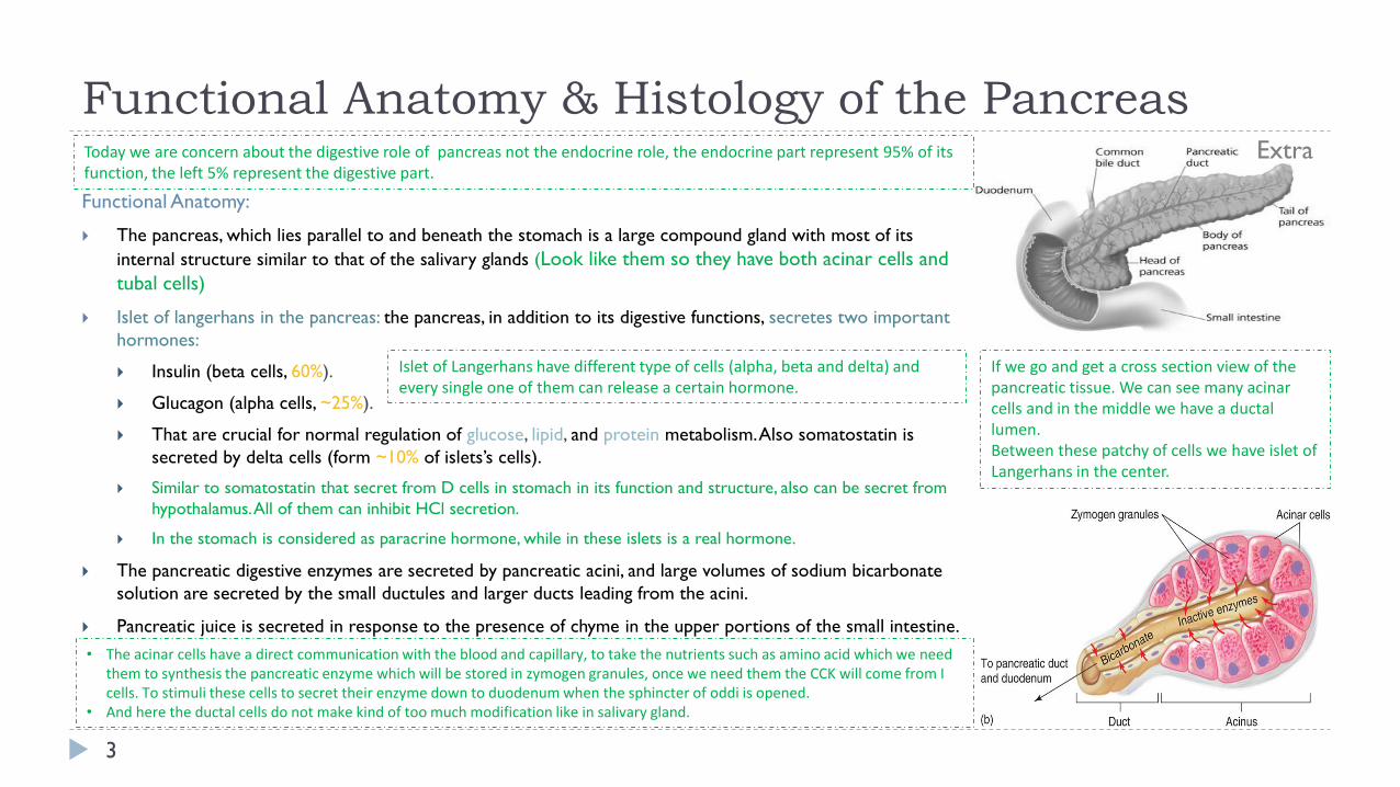
Functional Anatomy:
The pancreas, which lies parallel to and beneath the stomach is a large compound gland with most of its
internal structure similar to that of the salivary glands (Look like them so they have both acinar cells and
tubal cells)
Islet of langerhans in the pancreas: the pancreas, in addition to its digestive functions, secretes two important
hormones:
Insulin (beta cells, 60%).
Glucagon (alpha cells, ~25%).
That are crucial for normal regulation of glucose, lipid, and protein metabolism. Also somatostatin is
secreted by delta cells (form ~10% of islets’s cells).
Similar to somatostatin that secret from D cells in stomach in its function and structure, also can be secret from
hypothalamus. All of them can inhibit HCl secretion.
In the stomach is considered as paracrine hormone, while in these islets is a real hormone.
The pancreatic digestive enzymes are secreted by pancreatic acini, and large volumes of sodium bicarbonate
solution are secreted by the small ductules and larger ducts leading from the acini.
Pancreatic juice is secreted in response to the presence of chyme in the upper portions of the small intestine.
ExtraToday we are concern about the digestive role of pancreas not the endocrine role, the endocrine part represent 95% of its function, the left 5% represent the digestive part.
Islet of Langerhans have different type of cells (alpha, beta and delta) and every single one of them can release a certain hormone.
If we go and get a cross section view of the pancreatic tissue. We can see many acinar cells and in the middle we have a ductal lumen.Between these patchy of cells we have islet of Langerhans in the center.
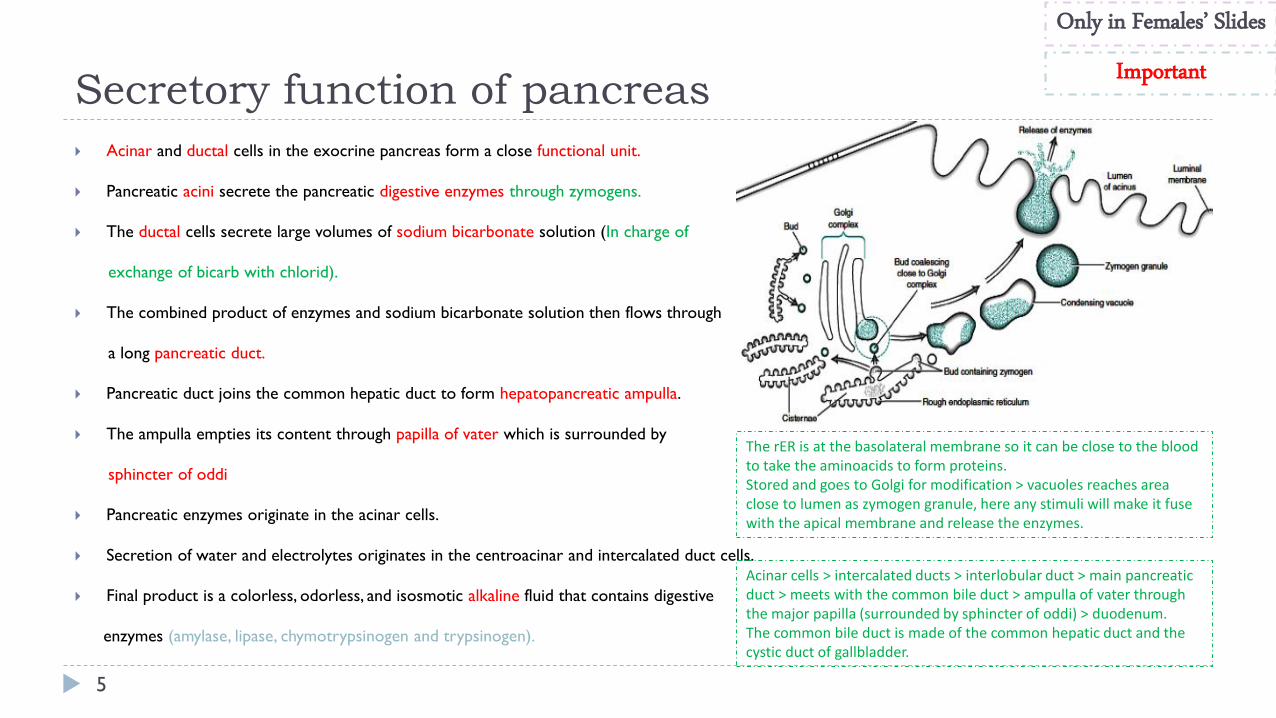
• The acinar cells have a direct communication with the blood and capillary, to take the nutrients such as amino acid which we need them to synthesis the pancreatic enzyme which will be stored in zymogen granules, once we need them the CCK will come from I cells. To stimuli these cells to secret their enzyme down to duodenum when the sphincter of oddi is opened.
• And here the ductal cells do not make kind of too much modification like in salivary gland.
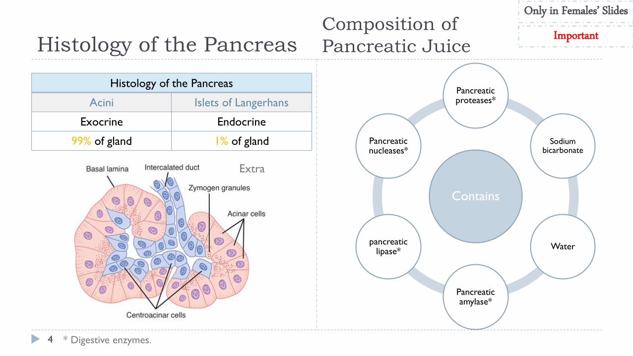
Histology of the Pancreas
4
Composition of
Pancreatic Juice
Contains
Pancreatic proteases*
Sodium bicarbonate
Water
Pancreatic amylase*
pancreatic lipase*
Pancreatic nucleases*
* Digestive enzymes.
Histology of the Pancreas
Acini Islets of Langerhans
Exocrine Endocrine
99% of gland 1% of gland
Extra
Only in Females’ Slides
Important
Secretory function of pancreas
5
Acinar and ductal cells in the exocrine pancreas form a close functional unit.
Pancreatic acini secrete the pancreatic digestive enzymes through zymogens.
The ductal cells secrete large volumes of sodium bicarbonate solution (In charge of
exchange of bicarb with chlorid).
The combined product of enzymes and sodium bicarbonate solution then flows through
a long pancreatic duct.
Pancreatic duct joins the common hepatic duct to form hepatopancreatic ampulla.
The ampulla empties its content through papilla of vater which is surrounded by
sphincter of oddi
Pancreatic enzymes originate in the acinar cells.
Secretion of water and electrolytes originates in the centroacinar and intercalated duct cells.
Final product is a colorless, odorless, and isosmotic alkaline fluid that contains digestive
enzymes (amylase, lipase, chymotrypsinogen and trypsinogen).
Only in Females’ Slides
Important
Acinar cells > intercalated ducts > interlobular duct > main pancreatic duct > meets with the common bile duct > ampulla of vater through the major papilla (surrounded by sphincter of oddi) > duodenum.The common bile duct is made of the common hepatic duct and the cystic duct of gallbladder.
The rER is at the basolateral membrane so it can be close to the blood to take the aminoacids to form proteins.Stored and goes to Golgi for modification > vacuoles reaches area close to lumen as zymogen granule, here any stimuli will make it fuse with the apical membrane and release the enzymes.
Pancreatic Secretion
6
The major functions of pancreatic secretion:
The main function To neutralize the acids in the duodenal chyme to optimum range (ph= 7.0-8.0) (ph=7.6-9.0) for activity of pancreatic enzymes.
To prevent damage to duodenal mucosa by acid & pepsin (Only this enzyme need acidic medium to be activated, the other enzyme need alkaline solution).
The chyme is very acidic, so it needs to be neutralized by a basic fluid (up to 9 PH) so it doesn’t cause any damage.
To produce enzymes involved in the digestion of dietary carbohydrate, fat, and protein.
Pancreatic secretions contain many enzymes for digesting proteins, carbohydrates(starch), and fats(Lipids), and large quantities of HCO3 ions.
Only in M
ales’ Slides
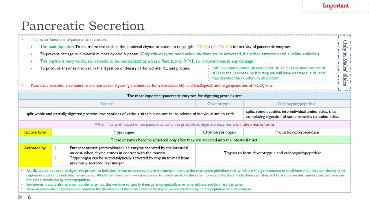
The most important pancreatic enzymes for digesting proteins are:
Trypsin Chymotrypsin Carboxypolypeptidase
split whole and partially digested proteins into peptides of various sizes but do not cause release of individual amino acids.splits some peptides into individual amino acids, thus
completing digestion of some proteins to amino acids.
When first synthesized in the pancreatic cells, the proteolytic digestive enzymes are in the inactive forms
Inactive form Trypsinogen Chymotrypsinogen Procarboxypolypeptidase
These enzymes become activated only after they are secreted into the intestinal tract.
Activated by 1. Enteropeptidase (enterokinase), an enzyme secreted by the intestinal
mucosa when chyme comes in contact with the mucosa.
2. Trypsinogen can be autocatalytically activated by trypsin formed from
previously secreted trypsinogen.
Trypsin to form chymotrypsin and carboxypolypeptidase.
• Acually we do not need to digest the protein to individual amino acids completly in the luemen, because the entrocyte(epitheilum cells which are lining the mucosa of small intestine), they can absorp di-tri
peptide in addition to individual amino acids. All of them have their own transporter to take them from the lumen to entrocyte, And inside these cells they will broken down into amino acids before enter
the blood circulation by enteropeptidase.
• Sometimes it could also as brush border enzymes. But we have to specify them as Enteropeptidase or enterokinase and both are the same.
• Now all pancreatic enzymes are activated in the duodenum of the small intestine by trypsin which activated by Enteropeptidase or enterokinase.
Important
Both liver and duodenum can secret HCO3, but the main source of HCO3 is the Pancreas. So if it stop we will have decrease in PH and may develop the duodenum ulceration.
Cont.
7
Secretion of trypsin inhibitor prevents digestion of the pancreas
itself.
Proteolytic enzymes of the pancreatic juice do not become activated
until after they have been secreted into the intestine because the
trypsin and the other enzymes would digest the pancreas itself.
The same cells that secrete proteolytic enzymes into the acini of the
pancreas secrete another substance called trypsin inhibitor, which is
formed in the cytoplasm of the glandular cells, and it prevents
activation of trypsin both inside the secretory cells and in the acini
and ducts of the pancreas.
Because trypsin activates the other pancreatic proteolytic enzymes,
therefore trypsin inhibitor prevents activation of the other enzymes
as well.
The trypsin inhibitor will be degeaded once it is secreted out from
the pancrease into intestin. The alpha amylase which is secreted from
pancrease is more potent than the lingual one.
Only in Females’ Slides
Pancreatic enzymes are secreted in inactive form: trypsinogen, chymotrypsinogen, procarboxypolypeptidase.
They will be activated by enterokinase enzyme in small intestine to their active forms.
The acinar cells that secrete the enzymes secretes trypsin inhibitor which prevent activation of trypsin inside acini and ducts.
When a duct is blocked the trypsin inhibitor can not inhibit activation of accumulated enzymes which will be activated and digest the pancreas in few hours.
Enterokinase is an enzyme that is secreted by brush border of small intestine and activate pancreatic enzymes.
Trypsin inhibitor is secreted by acinar cells to prevent activation of the enzymes inside the cells, in the acini and in the ducts.
In the stomach, HCL is what activates the enzymes.
Here, we have trypsin inhibitors and enterokinase to control the activation.
Because as soon as trypsin is activated, this will lead to activation of all other enzymes.
What protect pancreas from digestion by its enzymes?
Cont.
8
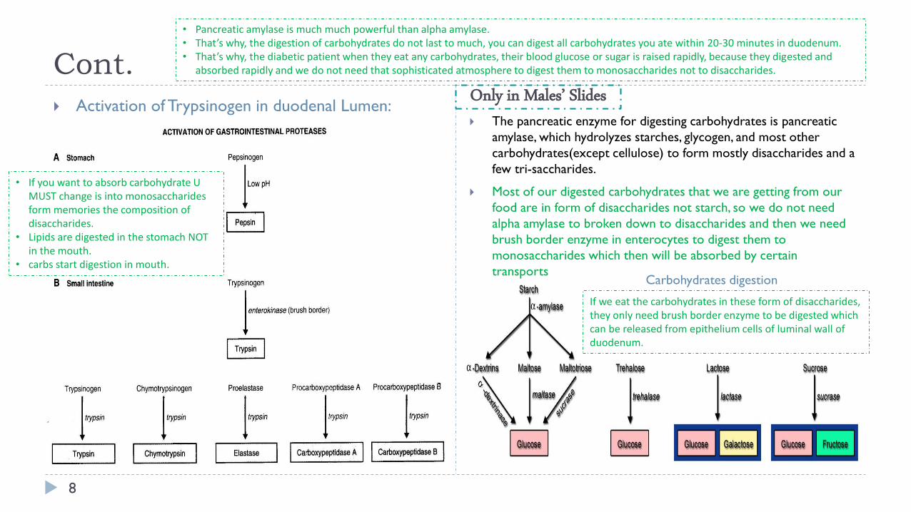
Activation of Trypsinogen in duodenal Lumen: The pancreatic enzyme for digesting carbohydrates is pancreatic
amylase, which hydrolyzes starches, glycogen, and most other
carbohydrates(except cellulose) to form mostly disaccharides and a
few tri-saccharides.
Most of our digested carbohydrates that we are getting from our
food are in form of disaccharides not starch, so we do not need
alpha amylase to broken down to disaccharides and then we need
brush border enzyme in enterocytes to digest them to
monosaccharides which then will be absorbed by certain
transports
Only in Males’ Slides
• Pancreatic amylase is much much powerful than alpha amylase.• That’s why, the digestion of carbohydrates do not last to much, you can digest all carbohydrates you ate within 20-30 minutes in duodenum.• That’s why, the diabetic patient when they eat any carbohydrates, their blood glucose or sugar is raised rapidly, because they digested and
absorbed rapidly and we do not need that sophisticated atmosphere to digest them to monosaccharides not to disaccharides.
Carbohydrates digestion
If we eat the carbohydrates in these form of disaccharides, they only need brush border enzyme to be digested which can be released from epithelium cells of luminal wall of duodenum.
• If you want to absorb carbohydrate U MUST change is into monosaccharides form memories the composition of disaccharides.
• Lipids are digested in the stomach NOT in the mouth.
• carbs start digestion in mouth.
Cont.
9
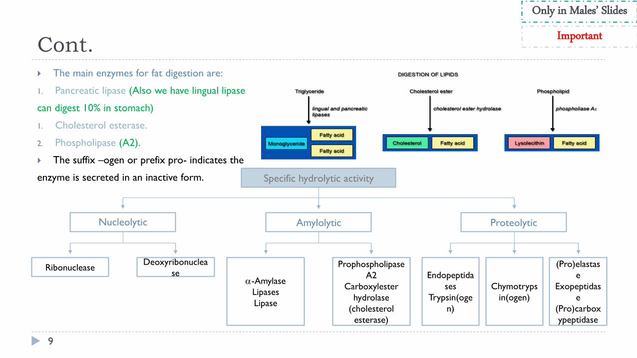
The main enzymes for fat digestion are:
1. Pancreatic lipase (Also we have lingual lipase
can digest 10% in stomach)
1. Cholesterol esterase.
2. Phospholipase (A2).
The suffix –ogen or prefix pro- indicates the
enzyme is secreted in an inactive form. Specific hydrolytic activity
Nucleolytic Amylolytic Proteolytic
Deoxyribonuclea
seRibonuclease Prophospholipase
A2
Carboxylester
hydrolase
(cholesterol
esterase)
-Amylase
Lipases
Lipase
Endopeptida
ses
Trypsin(oge
n)
Chymotryps
in(ogen)
(Pro)elastas
e
Exopeptidas
e
(Pro)carbox
ypeptidase
Only in Males’ Slides
Important
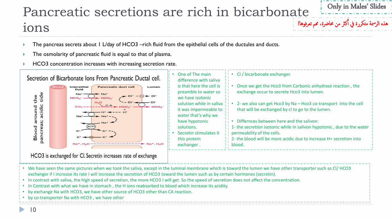
Pancreatic secretions are rich in bicarbonate
ions
10
The pancreas secrets about 1 L/day of HCO3 –rich fluid from the epithelial cells of the ductules and ducts.
The osmolarity of pancreatic fluid is equal to that of plasma.
HCO3 concentration increases with increasing secretion rate.
Only in Males’ Slides
• We have seen the same pictures when we took the saliva, except in the luminal membrane which is toward the lumen we have other transporter such as Cl/ HCO3 exchanger if I increase its rate I will increase the secretion of HCO3 toward the lumen such as by certain hormones (secretin).
• In contrast with saliva, the high speed of secretion, the more HCO3 I will get. So the speed of secretion does not affect the concentration.• In Contrast with what we have in stomach , the H ions reabsorbed to blood which increase its acidity.• by exchange Na with HCO3, we have other source of HCO3 other than CA reaction. • by co-transporter Na with HCO3 , we have other
• One of The main difference with saliva is that here the cell is preamble to water so we have isotonic solution while in saliva it was impermeable to water that’s why we have hypotonic solutions.
• Secretin stimulates it this protein exchanger .
• Cl / bicarbonate exchanger.
• Once we get the Hco3 from Carbonic anhydrase reaction , the exchange occur to secrete Hco3 into lumen.
• 2- we also can get Hco3 by Na – Hco3 co-transport into the cell that will be exchanged by cl to go to the lumen.
• Differnces between here and the salivon: 1- the secretion isotonic while in salivon hypotonic , due to the water permeability of the cells.2- the blood will be more acidic due to increase H+ secretion into blood.
!وهاهذه الرمسة متكررة يف أ كرث من حمارضة، همم تعرف
Mechanism of HCO3- Secretion
11
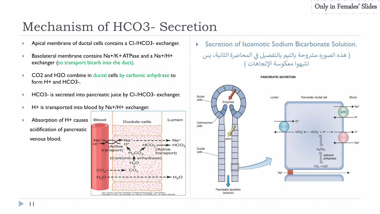
Apical membrane of ductal cells contains a Cl-/HCO3- exchanger.
Basolateral membrane contains Na+/K+ ATPase and a Na+/H+
exchanger (to transport bicarb into the duct).
CO2 and H2O combine in ductal cells by carbonic anhydrase to
form H+ and HCO3-.
HCO3- is secreted into pancreatic juice by Cl-/HCO3- exchanger.
H+ is transported into blood by Na+/H+ exchanger.
Absorption of H+ causes
acidification of pancreatic
venous blood.
Secretion of Isosmotic Sodium Bicarbonate Solution.
وحة الصورههذه ) ة الثانية، ببالتيممشر ي المحاضرس بالتفصيل فر
(اإلتجاهاتانتبهوا معكوسة
Only in Females’ Slides
Pancreatic secretion is under neural and hormonal
control
12
Control of pancreatic secretion:
1. Acinar cells (enzymatic secretion)
o Receptors for CCK and muscarinic receptors for ACh
o CCK is most important stimulant.
(i cells) secrete CCK in presence of amino acids and fatty
acids in intestinal lumen.
o ACh also stimulates enzyme secretion.
2. Ductal cells (aqueous secretion of HCO3-)
o Receptors for CCK, ACh, and secretin.
o Secretin (from S cells of duodenum) is major stimulant
Secreted in response to H+ in intestine.
o Effects of secretin are potentiated by both CCK and ACh.
o Secretin travels to blood to pancreatic ducts to release
Bicarb and water.
Parasympathetic stimulation (through ach on acinar cells) results in an increase in enzyme secretion-fluid and HCO3.
Secretin tends to stimulate a hco3 rich secretion by activating ductal cells.
Cck stimulates a marked increase in enzyme secretion by stimulating the acinar cells.
Pancreatic secretion normally results from the combined effects of the multiple basic stimuli, not from one alone (potentiate each other).
It means that pancreatic secretion will be produced due to the stimuli from (CCK, secretin, ach) they stimuli TOGETHER AT THE SAME TIME, to potentiate and to increase the amount of the secretion , so the amount of pancreatic secretion will be much higher if these hormones will stimuli at the same time rather than each one individual.
All of these work together to release too much HCO3 secretion.
Only in Males’ Slides Only in Females’ Slides
Stimuli for pancreatic secretion
13
Only in Males’ Slides
Stimuli for pancreatic secretion
Acetylcholine Cholecystokinin Secretin
Released from the
parasympathetic vagus nerve
endings and from other
cholinergic nerves in the enteric
nervous system.
Secreted by the duodenal and upper
jejunal mucosa (i cells) when food
enters the small intestine.
Also secreted by the duodenal and jejunal mucosa (s cells) when highly
acidic chyme enters the small intestine.
Stimulate the acinar cells of the pancreas, causing production of large
quantities of pancreatic digestive enzymes, but relatively small quantities of
water and electrolytes to go with the enzymes.
• In contrast to the first two basic stimuli, stimulates secretion of
large quantities of h2o and nahco3 solution by the
pancreatic ductal epithelium.
• Secretin stimulates secretion of copious quantities of bicarbonate
ions—neutralization of acidic stomach chyme.
• Secretin is present in an inactive form, prosecretin (in s cells in the
mucosa of the duodenum and jejunum).
• When acid chyme with ph less than 4.5-5.0 enters the duodenum
from the stomach, it causes duodenal mucosal release and activation
of secretin, which is then absorbed into the blood.
• Secretin causes the pancreas to secrete large quantities of fluid
containing a high concentration of hco3 (up to 145 meq/l = ~5x
normal) but a low concentration of cl-.
HCl + NaHco3 → Nacl + H2co3
H2CO3 dissociates into CO2 and H2O
Cont.
14
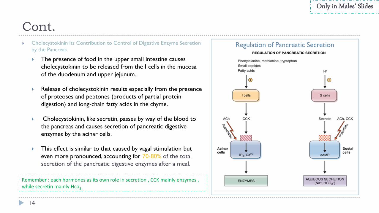
Cholecystokinin Its Contribution to Control of Digestive Enzyme Secretion
by the Pancreas.
The presence of food in the upper small intestine causes
cholecystokinin to be released from the I cells in the mucosa
of the duodenum and upper jejunum.
Release of cholecystokinin results especially from the presence
of proteoses and peptones (products of partial protein
digestion) and long-chain fatty acids in the chyme.
Cholecystokinin, like secretin, passes by way of the blood to
the pancreas and causes secretion of pancreatic digestive
enzymes by the acinar cells.
This effect is similar to that caused by vagal stimulation but
even more pronounced, accounting for 70-80% of the total
secretion of the pancreatic digestive enzymes after a meal.
Only in Males’ Slides
Remember : each hormones as its own role in secretion , CCK mainly enzymes , while secretin mainly Hco3.
Multiplicative or potentiation effects of
different pancreatic secretion stimuli
15
When all different stimuli of pancreatic secretion (acetylcholine, cholecystokinin, and secretin) occur at once, then the total
secretion is far greater than the sum of the secretions caused by each stimulus separately. The stimuli are said to “multiply” or
“potentiate” one another.
Usually, pancreatic secretions are the result of multiple stimuli rather than one stimulus.
Only in Males’ Slides
Pancreatic secretion is phasic
Phase stimulus Mediators In general
Cephalic phase Smell, taste, chewing and
swallowing
Release of Ach • From CNS Through vagus nerve.
• 20% of pancreatic enzymes.
Gastric phase
Protein, gastric distention
Vago-vagal reflex
• The stomach is distended, so it will send
impulses to the brain to prepare the
pancreas to release some of its content.
• Through vagus nerve
• 5-10% of pancreatic enzymes
Intestinal phase
The most important
one Acid in chyme, fatty acids Secretin, CCK and vago-vagal reflex
• For digestion and protection.
• Through hormonal stimulation (secretin &
CCK).
• 70-75% of pancreatic enzymes & fluid.
ة الاالاة مرشوحة بشلك تفصييل يف احملارض phasesال
Cont.
16
Only in Females’ Slides
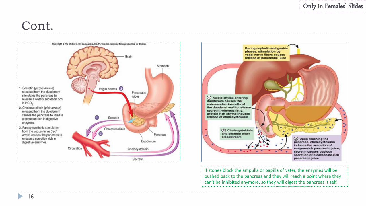
If stones block the ampulla or papilla of vater, the enzymes will be pushed back to the pancreas and they will reach a point where they can’t be inhibited anymore, so they will digest the pancreas it self.
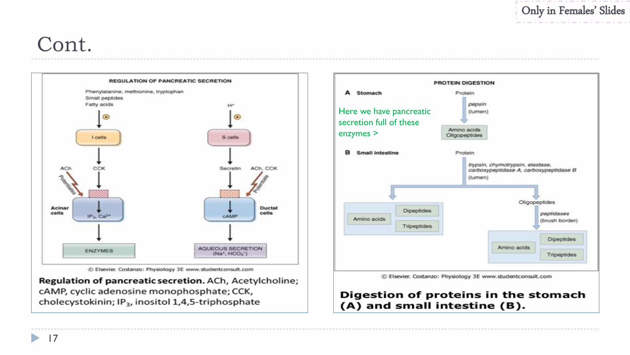
Cont.
17
Only in Females’ Slides
Here we have pancreatic
secretion full of these
enzymes >
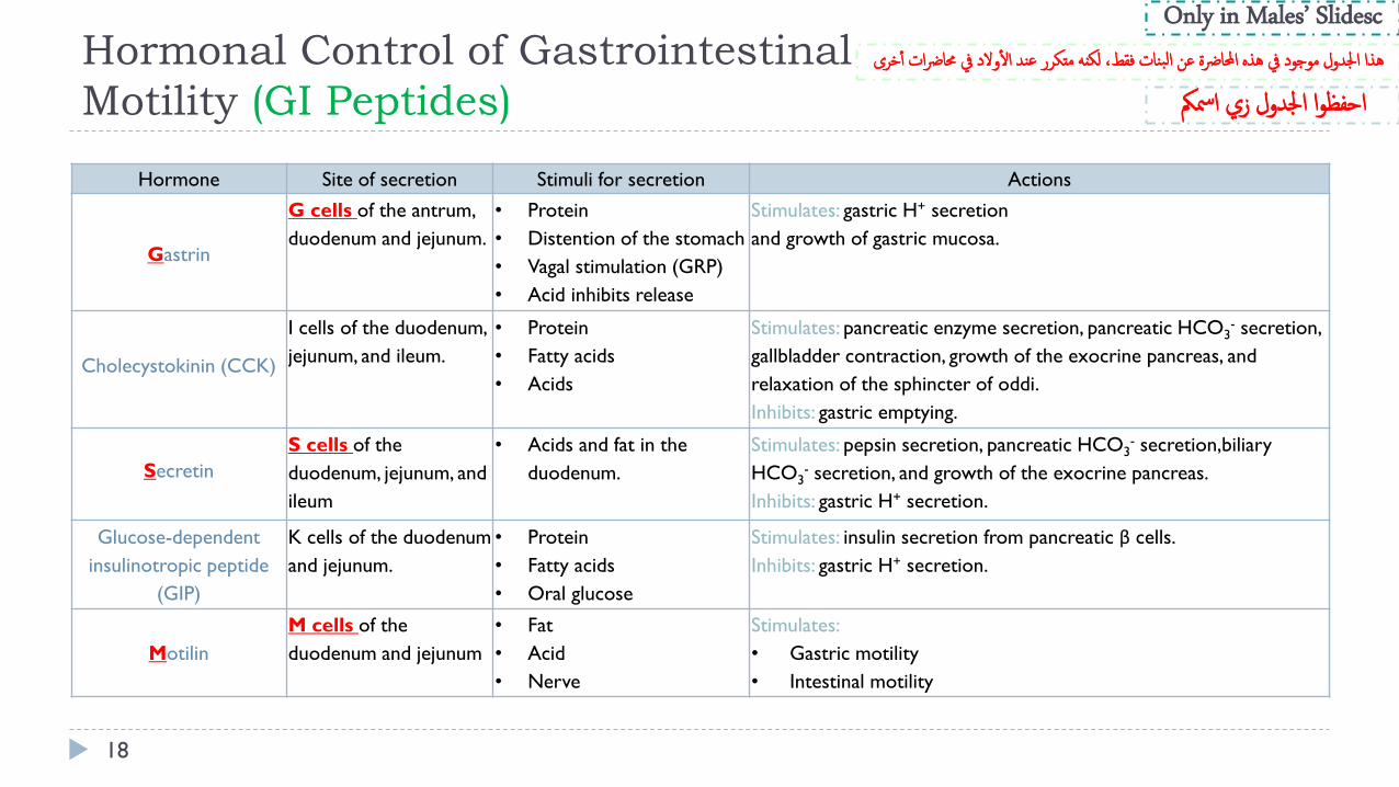
Hormonal Control of Gastrointestinal
Motility (GI Peptides)
18
Hormone Site of secretion Stimuli for secretion Actions
Gastrin
G cells of the antrum,
duodenum and jejunum.
• Protein
• Distention of the stomach
• Vagal stimulation (GRP)
• Acid inhibits release
Stimulates: gastric H+ secretion
and growth of gastric mucosa.
Cholecystokinin (CCK)
I cells of the duodenum,
jejunum, and ileum.
• Protein
• Fatty acids
• Acids
Stimulates: pancreatic enzyme secretion, pancreatic HCO3- secretion,
gallbladder contraction, growth of the exocrine pancreas, and
relaxation of the sphincter of oddi.
Inhibits: gastric emptying.
Secretin
S cells of the
duodenum, jejunum, and
ileum
• Acids and fat in the
duodenum.
Stimulates: pepsin secretion, pancreatic HCO3- secretion,biliary
HCO3- secretion, and growth of the exocrine pancreas.
Inhibits: gastric H+ secretion.
Glucose-dependent
insulinotropic peptide
(GIP)
K cells of the duodenum
and jejunum.
• Protein
• Fatty acids
• Oral glucose
Stimulates: insulin secretion from pancreatic β cells.
Inhibits: gastric H+ secretion.
Motilin
M cells of the
duodenum and jejunum
• Fat
• Acid
• Nerve
Stimulates:
• Gastric motility
• Intestinal motility
Only in Males’ Slidesc
ىهذا اجلدول موجود يف هذه احملارضة عن البنات فقط، لكنه متكرر عند ال والد يف حمارضات أ خر
احفظوا اجلدول زي امسمك
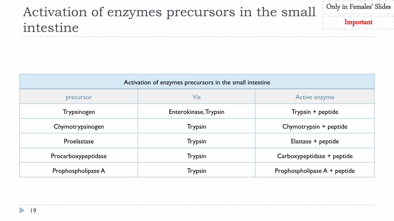
Activation of enzymes precursors in the small
intestine
19
Only in Females’ Slides
Important
Activation of enzymes precursors in the small intestine
precursor Via Active enzyme
Trypsinogen Enterokinase, Trypsin Trypsin + peptide
Chymotrypsinogen Trypsin Chymotrypsin + peptide
Proelastase Trypsin Elastase + peptide
Procarboxypeptidase Trypsin Carboxypeptidase + peptide
Prophospholipase A Trypsin Prophospholipase A + peptide
Thank you!
The Physiology 436 Team:
20
.اعمل لترسم بسمة، اعمل لتمسح دمعة، اعمل و أنت تعلم أن هللا ال يضيع أجر من أحسن عمال
Females Members:
Anwar Alajmi
Deena Alnowaiser
Amal Alshaibi
Rawan Alqahtani
Rana Barasian
Males Members:
Fouad Bahgat
Team Leaders: Laila Mathkour
Mohammad Alayed
Contact us:
References: • 2017-2018 Dr. Hana Alzamel’s Lecture.
• 2017-2018 Dr. Mohammed Al Zoghaibi’s Lecture.
• Guyton and Hall Textbook of Medical Physiology (Thirteenth Edition.)
ي إليه ياي استودعتك ما حفظت وما قرأت وما فهمت، فرده لي وقت حاجت
.من ال تضيع عنده الودائعاللهم انر