why trace and delay conditioning are sometimes (but not
TRANSCRIPT

Author's personal copy
www.elsevier.com/locate/brainres
Available online at www.sciencedirect.com
Research Report
Why trace and delay conditioning are sometimes (but notalways) hippocampal dependent: A computational model
Ahmed A. Moustafaa,b,n, Ella Wufongb, Richard J. Servatiusa,c, Kevin C.H. Panga,c,Mark A. Gluckd, Catherine E. Myersa,c,e
aDepartment of Veterans Affairs, New Jersey Health Care System, East Orange, NJ, USAbSchool of Social Sciences and Psychology & Marcs Institute for Brain and Behaviour, University of Western Sydney,Sydney, NSW 2751, AustraliacStress & Motivated Behavior Institute, New Jersey Medical School, University of Medicine and Dentistry of New Jersey,Newark, NJ, USAdCenter for Molecular and Behavioural Neuroscience, Rutgers University-Newark, Newark, NJ, USAeDepartment of Psychology, Rutgers University-Newark, Newark, NJ, USA
a r t i c l e i n f o
Article history:
Accepted 15 November 2012
Available online 23 November 2012
Keywords:
Computational model
Hippocampus
Fear and eyeblink conditioning
Interstimulus interval (ISI)
Hippocampal lesion (HL)
Trace conditioning
Short vs. long delay conditioning
Associative learning
a b s t r a c t
A recurrent-network model provides a unified account of the hippocampal region in
mediating the representation of temporal information in classical eyeblink conditioning.
Much empirical research is consistent with a general conclusion that delay conditioning
(in which the conditioned stimulus CS and unconditioned stimulus US overlap and co-
terminate) is independent of the hippocampal system, while trace conditioning (in which
the CS terminates before US onset) depends on the hippocampus. However, recent studies
show that, under some circumstances, delay conditioning can be hippocampal-dependent
and trace conditioning can be spared following hippocampal lesion. Here, we present an
extension of our prior trial-level models of hippocampal function and stimulus represen-
tation that can explain these findings within a unified framework. Specifically, the current
model includes adaptive recurrent collateral connections that aid in the representation of
intra-trial temporal information. With this model, as in our prior models, we argue that the
hippocampus is not specialized for conditioned response timing, but rather is a general-
purpose system that learns to predict the next state of all stimuli given the current state of
variables encoded by activity in recurrent collaterals. As such, the model correctly predicts
that hippocampal involvement in classical conditioning should be critical not only when
there is an intervening trace interval, but also when there is a long delay between CS onset
and US onset. Our model simulates empirical data from many variants of classical
conditioning, including delay and trace paradigms in which the length of the CS, the
inter-stimulus interval, or the trace interval is varied. Finally, we discuss model limitations,
future directions, and several novel empirical predictions of this temporal processing
model of hippocampal function and learning.
Published by Elsevier B.V.
0006-8993/$ - see front matter Published by Elsevier B.V.http://dx.doi.org/10.1016/j.brainres.2012.11.020
nCorresponding author. New Jersey Health Care System, Department of Veterans Affairs, Locked bag 1797, Sydney, NSW 2751, Australia.Fax: þ61 973 353 1272.
E-mail addresses: [email protected], [email protected] (A.A. Moustafa).
b r a i n r e s e a r c h 1 4 9 3 ( 2 0 1 3 ) 4 8 – 6 7

Author's personal copy
1. Introduction
Classical conditioning, in which a cue (the conditionedstimulus or CS) is paired with a reflex-evoking unconditionedstimulus (US) until the CS comes to produce an anticipatoryresponse (the conditioned response or CR) has proven auseful testbed for examining the psychological principlesand neurobiological substrates of learning. Under manycircumstances, delay conditioning, in which the CS and USoverlap and co-terminate, is spared or even mildly facilitatedfollowing hippocampal damage (e.g., Berger et al., 1983;Gabrieli et al., 1995; Ito et al., 2005, 2006; Schmaltz andTheios, 1972); conversely, under many conditions, hippocam-pal lesion disrupts trace conditioning, in which CS offsetoccurs before US onset, producing a temporal gap known asthe trace interval (e.g., Beylin et al., 1999; McGlinchey-Berrothet al., 1997; Solomon et al., 1986; Weisz et al., 1980). This hasled some researchers to assume that the hippocampus playsa more important role in trace than in delay conditioning(Beylin et al., 1999; McGlinchey-Berroth et al., 1997; Solomonet al., 1986)—as we discuss below, this assumption ignoresempirical findings on the role of the hippocampus in delayconditioning. Along the same lines, some studies of humansand non-human animals use trace conditioning as a canoni-cal task to demonstrate evidence of hippocampal dysfunctionin transgenic models, healthy aging, and pharmacologicalmodels (Brown et al., 2010; Disterhoft et al., 1999).
However, there are strong reasons to challenge this delay/trace dichotomy. First, it has long been known that thehippocampus shows learning-related changes during acqui-sition of the delay CR in intact animals and humans. Thesechanges include the development of responses by somehippocampal pyramidal neurons that precede the behavioraleyeblink CR and mirror its form (Berger et al., 1976, 1983;Berger and Thompson, 1978; Green and Arenos, 2007;Thompson et al., 1980). Initially, these responses occur inthe US period, but increases in the CS period occur at aboutthe time that behavioral CRs appear, and decline withcontinued training (for review, see Christian and Thompson,2003). Similar hippocampal activity occurs in rabbits giventrace conditioning (Weiss et al., 1996) but not in rabbits givenunpaired CS/US trials (Solomon et al., 1986).
Functional imaging studies in humans show a similarpattern of learning-related activity in the hippocampus dur-ing delay eyeblink conditioning to those observed in animals(Blaxton et al., 1996; Cheng et al., 2008; Knight et al., 2004Stein and Helmstetter, 2004; Logan and Grafton, 1995;Schreurs and Alkon, 2001). Thus, these data suggest that –even if the hippocampus is not necessary for acquisition of adelay CR – it nevertheless normally plays a role. Thischallenges the simple view of delay conditioning as hippo-campal-independent, and begs a more nuanced view of thedifference between brain substrates that are sufficient tomediate a learned response, versus those that are normallyinvolved.
Second, under many conditions, delay conditioning isspared or slightly enhanced following hippocampal lesion.Specifically, the ability of hippocampal lesioned animals toacquire a delay eyeblink CR depends on the length of the CS
interval. Specifically, while hippocampal-lesioned rats canacquire an eyeblink CR when the delay between CS onset andUS onset is short, they are impaired when the delay islengthened (Beylin et al., 2001). Thus, short-delay condition-ing is spared by hippocampal lesion, but long-delay condi-tioning is not. Further, disruption of the hippocampus, viaelectrical stimuli or pharmacological intervention, can retardacquisition of even a short-delay CR (Kaneko and Thompson,1997; Sakamoto et al., 2005; Salafia et al., 1979, 1977; Solomonand Gottfried, 1981; Solomon et al., 1983). Together, theseresults document that delay conditioning is not alwaysspared following hippocampal lesion or disruption.
Third, although trace conditioning is often disrupted byhippocampal lesion, this is not always the case. For example,Thompson et al. (1980) speculated that the hippocampusmight be involved in trace conditioning, to bridge the tem-poral gap between CS and US. Solomon et al. (1986) presentedan early study showing that dorsal hippocampal lesionsdisrupted trace eyeblink conditioning in rabbits, by decreas-ing the number of CRs. However, other studies followed thatreported no trace conditioning impairment in hippocampal-lesioned animals (James et al., 1987; Port et al., 1986).
Another factor affecting the hippocampal-dependence oftrace conditioning may be differences in the trace intervalused. In studies where the trace interval has been explicitlyvaried, a deficit in trace conditioning appears only for longtrace intervals. Thus, for example, hippocampal-lesionedrabbits are impaired on eyeblink CR acquisition with a long(500 ms) but not a short (100 ms CS or 300 ms) trace interval(Moyer et al., 1990). There may also be interactions betweenCS duration, and trace interval: Steinmetz and colleagues(Walker and Steinmetz, 2008) found that hippocampal-lesioned rats were impaired relative to controls on acquisi-tion of an eyeblink CR when the CS duration was 50 ms andthe trace interval was 500 ms, but not when the CS durationwas 500 ms and the trace interval was 50 ms. In addition,Shors and colleagues (Beylin et al., 2001) showed that –although hippocampal-lesioned rats were impaired at bothtrace and long-delay eyeblink conditioning – once thelesioned animals had acquired a long-delay CR, they couldthen learn and perform the trace CR. Together, these resultsdocument that, at least under some circumstances, subjectswith hippocampal lesion can acquire a trace CR as well asmatched controls.
In summary, while the idea that trace conditioning ishippocampal-dependent whereas delay conditioning ishippocampal-independent provides a useful rule of thumb,it is not sufficient to adequately address the full range ofexisting data. An additional complication involves the inter-stimulus interval (ISI). When the ISI is short, responsesystems (such as the brainstem and cerebellum for eyeblinkconditioning) can successfully learn CS–US associations andproduce a well-timed CR. When the ISI is longer, the hippo-campus helps to bridge the temporal gap between CS and US,facilitating production of a well-timed CR. Thus, in the caseof eyeblink conditioning, both short-delay and short-traceparadigms can be acquired without impairment byhippocampal-lesioned animals; however, both long-delayand long-trace paradigms are disrupted following hippocam-pal lesion.
b r a i n r e s e a r c h 1 4 9 3 ( 2 0 1 3 ) 4 8 – 6 7 49
Author's personal copy
Consistent with this view, although infant rats (withimmature hippocampus) can acquire short-delay condition-ing, they are impaired at both long-delay and trace condition-ing, which emerge in parallel during later development(Barnet and Hunt, 2005; Ivkovich et al., 2000). Thus, thesedata all suggest that in addition to the presence of a traceinterval, the duration between CS onset and US arrivaldetermines whether hippocampal mediation is required, asis the case in short- and long-delay conditioning.
Interestingly, when Hoehler and Thompson (1980) firstspeculated that trace conditioning might be hippocampaldependent, they did so based on their studies of ISI manip-ulations in eyeblink conditioning, and on their findings thatthe hippocampus appeared to be involved in forming atemporal map of the learned behavioral response to be made,allowing for the CR to be accurately timed even when the ISIis beyond the timing parameters that are ‘‘optimal’’ for thebasic associative substrate. Thus, for example, the ‘‘optimal’’parameters for eyeblink conditioning (operationalized interms of acquisition speed) may be a few hundred millise-conds and may reflect temporal processing mechanisms inother structures such as the cerebellum.
As discussed by Christian and Thompson (2003), optimaltemporal parameters for learning in the cerebellum are50–200 ms between CS and US presentation. Most traceconditioning studies on animals use a trace interval of500 ms to induce hippocampal involvement in task learning.Optimal parameters for fear conditioning tend to be an orderof magnitude longer (usually41 s, see for example, Bevinsand Ayres, 1995), and may reflect temporal processingmechanisms in the amygdala. But in fear conditioning, justas in eyeblink conditioning, hippocampal lesions affect traceconditioning as a function of the trace interval, so thathippocampal lesions impair expression of contextual fearconditioning with long but not short trace intervals(Chowdhury et al., 2005; Pang et al., 2010). In other words,we argue that the hippocampus plays a similar role in botheyeblink and fear conditioning, but temporal differences inoptimal ISI length for eyeblink and fear conditioning acquisi-tions are, respectively related to processing in the cerebellumand amygdala.
Extending this principle beyond classical conditioning,other brain systems mediate other behavioral responses,and each may have an operating window of temporal delaysthat can be spanned; in each case, these brain systems alonemay be capable of mediating learning with sufficiently shortdelays, but as the delay is lengthened, hippocampal media-tion becomes critical. This basic idea is consistent with a largenumber of theories of hippocampal-region function (e.g.,Hoehler and Thompson, 1980; Rawlins, 1985; Wallensteinet al., 1998) and finds broad support from a range of prepara-tions. For example, in delayed non-matching-to-place in aneight-arm radial maze, rats with hippocampal lesion canlearn under short delays, but are impaired when the delayperiod is extended (Lee and Kesner, 2003). Similarly, indelayed non-match to sample (DNMS), primates with lesionslimited to the hippocampus (sparing nearby medial temporalareas) can learn the non-matching task as well as controls,but show increasing impairments as the delay betweensample and response is lengthened (Zola-Morgan and Squire,
1986). In each case, the important factor determining hippo-campal dependence is not presence or absence of a stimulus-free gap, but rather the length of the interval across whichinformation must be maintained before responding.
Again, similar findings have also been reported in humanstudies. For example, humans with bilateral hippocampaldamage are impaired relative to healthy controls on temporaland spatial estimation at long, but not short, delays (Kesnerand Hopkins, 2001). Patients with medial temporal lobe andhippocampal lesions also perform much better on relationalmemory tasks when the interval between learning andmemory test is short than long. Specifically, Squire andcolleagues (Jeneson et al., 2010) found that patients withmedial temporal lobe damage do not show impairment inperforming relational learning tasks when the delay betweenlearning and test is short (1 s). Similarly, Ryan and Cohen(2004) have tested amnesic patients on a relational memorytask using both short and long delay periods. They havefound that patients are impaired only for the long-intervalcondition.
Thus, a conceptualization of eyeblink conditioning inwhich the length of the ISI, in addition to the presence of atrace interval, determines hippocampal dependence wouldappear to help integrate our understanding of the eyeblinkconditioning literature with that of other preparations.
In this paper, we present an extension of our prior trial-level computational models of hippocampal function andstimulus representation. Our models assumed that the hip-pocampal region interacted with other brain systems, such ascortex and cerebellum, during associative learning, specifi-cally by forming new stimulus representations that providedinformation about stimulus–stimulus and contextual regula-rities (Gluck and Myers, 1993, 2001 Myers and Gluck, 1994;Moustafa et al., 2009). These models were correctly able toaccount for the effects of hippocampal lesion and disruptionon various trial-level phenomena such as acquisition, dis-crimination, latent inhibition, and contextual shift effects.A later extension which modeled the effects of cholinergicmanipulations by altering the hippocampal region learningrate was correctly able to address the effects of cholinergicagonists and antagonists on classical conditioning (Myerset al., 1996, 1998; Moustafa et al., 2010). However, these earliermodels simulated trial-level information only, meaning thatthey could simulate whether a CR is given on a particulartrial, but could not address within-trial events, such as therelative timing of CS and US onset. As such, these earliertrial-level models could not address the differences betweendelay and trace conditioning, nor the effects of manipulatingthe length of the ISI or trace interval. The need to addressthese aspects of the empirical data partially motivates thecurrent work.
Our new model simulates performance in various delayand trace eyeblink conditioning data within a unified frame-work. Specifically, the current model includes adaptive recur-rent collateral connections that aid in the representation ofintra-trial temporal information. With this model, as in ourprior models, we argue that the hippocampus is a general-purpose system that learns to predict the next state of allstimuli given the current state of variables encoded byactivity in recurrent collaterals. As such, the model correctly
b r a i n r e s e a r c h 1 4 9 3 ( 2 0 1 3 ) 4 8 – 6 750

Author's personal copy
predicts that hippocampal involvement in associative learn-ing, including classical conditioning, should be most criticalnot only when there is an intervening trace interval, but alsowhen there is a long delay between CS onset and US onset,as in short-delay vs. long-delay conditioning.
1.1. Modeling
Fig. 1 shows a schematic diagram of the current model, whichbuilds off our prior models of hippocampal-region processesin classical conditioning. Like our earlier models (Gluck andMyers, 1993, 2001; Moustafa et al., 2009), the present modelconceives of the hippocampal region (Fig. 1, green) as apredictive autoencoder, which learns to predict the next stateof the world given current inputs. In the process, thehippocampal-region network forms new stimulus represen-tations in its internal layer that compress (or make moresimilar) the representations of co-occurring inputs whiledifferentiating (making less similar) the representations ofinputs that make different predictions about future eventssuch as US arrival. In other words, the essential function ofthe hippocampus in our model is monitoring environmentalregularities and using prior experience and current inputs topredict what (out of all possible events) is likely to happennext. Classical conditioning is a good example of predictionprocesses because the most salient event – the US arrival –can be predicted with high accuracy by learning the CS andthe ISI. In our model, the hippocampal network learns notonly whether a particular CS will be followed by a US, butwhen this will occur.
Also as in our prior models, the hippocampal-region net-work communicates with a second motor network (Fig. 1,blue), which is assumed to represent some of aspects corticaland cerebellar substrates of motor learning. The motor out-put network is modeled as a single adaptive node that learnsto map from weighted inputs specifying the presence of CSs,as well as contextual or background stimuli and an efferentcopy of the CR. The activities of the hippocampal-regionnetwork hidden layer units are also provided as inputs tothe motor output network, allowing the motor output net-work to incorporate the adaptive representations formed inthe hippocampal-region network into its own ongoing learn-ing. The output from the motor response network representsthe behavioral CR. The difference between this output (CR)and the US constitutes an error signal that can be used totrain the connection weights in the motor response network,using an error correction rule such as the least-mean squaresor LMS rule (Widrow and Hoff, 1960); full details of thelearning rule and other model details are provided inSection 4.
The major differences between this model and the priormodels are (1) the consideration of each trial not as a discreteevent, but as a series of timepoints, (2) the addition ofrecurrent pathways within the hippocampal-region andmotor output networks. We discuss each of these pointsbelow; full simulation details are provided in Section 4.
First, to simulate within-trial events, each trial is dividedinto a number of timesteps, which represent small timeintervals within a conditioning trial (e.g., 50 ms). Typically,contextual inputs are present during the entire trial, and are
present alone during the first several timesteps of a trial; thenone or more CSs may be presented for a specified number oftimesteps; the US, when present, appears for a single time-step. The US may overlap with the CS (as in delay condition-ing) or may occur after CS cessation (as in trace conditioning).A further series of context-alone presentations ends the trial(simulating the intertrial interval or ITI). Fig. 2 providesschematic illustrations of some example paradigms that wesimulate in our model.
A second difference between the prior and current modelsis that Fig. 1 includes recurrent connections within thehippocampal and motor output models. Specifically, thehippocampal region network includes recurrent connectionswithin the internal layer, while the motor output networkcontains a feedback CR pathway, carrying informationregarding the current state of the CR. Provision of feedbackwithin the hippocampal network allowed the activation of
Current state at time t (CS and contextual inputs,
as well as US and prior CR)
Output Prediction of next state (t+1)
t-1
Motor outputNetwork
Hippocampal-Region Network
t-1
t-1
CS and contextual inputs
at time t
t-1
Fig. 1 – The hippocampal model which includes recurrentconnections within the motor and hippocampal networks,as well as processing of within-trial events. In the motornetwork, CS inputs project through modifiable weights tothe output node, which in turn project to motor areas thatdrive the behavioral response (eyeblink CR). The predictionerror module receives excitatory US projections andinhibitory CR projections, and provides the response error(US–CR) as a ‘‘teaching signal’’ to the motor network. Thehippocampal-region network receives inputs detailing thecurrent state of all inputs at time t, including presence orabsence of CSs, contextual cues, US, and CR. Thehippocampal-reigon network learns to produce outputs thatpredict the state of all inputs at the next timestep tþ1; in theprocess it forms new stimulus representations in itsinternal node layer that are sensitive to stimulus co-occurrence and association with the US. In the intact model,these new representations also provided as input to themotor network, which can then map from them to newbehavioral responses. Arrows represent weightedconnections; filled circles¼ inhibitory connections. (Forinterpretation of the references to color in this figure legend,the reader is referred to the web version of this article.)
b r a i n r e s e a r c h 1 4 9 3 ( 2 0 1 3 ) 4 8 – 6 7 51

Author's personal copy
internal layer nodes at any time step to be a function ofexternal (CS and contextual) input and also of the adaptiverepresentation of input from a previous timestep. Becausethe weights on these recurrent connections are adaptive, it ispossible for a sequence of activation patterns to be stored inthe network that ‘‘buffers’’ input information over severaltimesteps. Importantly, this buffering function is not pre-wired into the network, but emerges dynamically as a resultof training and learning. Anatomical studies support theexistence of such recurrent loops in the hippocampal region,particularly hippocampal subfield CA3 (Amaral et al., 1990;Amaral and Witter, 1989) as well as the dentate gyrus (Amaralet al., 2007). Prior models of the hippocampus have alsoincluded recurrent connections to simulate conditioningtasks (see for example, Rodriguez and Levy, 2001).
In this model as in our prior models, hippocampal lesion issimulated by disabling learning in the hippocampal-regionnetwork, in which case the motor output network can stilllearn new responses by modifying weights from the CS andcontextual inputs, but no new adaptive stimulus representa-tions are formed in the hippocampal region (Gluck andMyers, 1993; Myers and Gluck, 1994). In addition, and also
as in our prior models, the effects of cholinergic agonists andantagonists are simulated by raising (agonist) or lowering(antagonist) learning rates in the hippocampal-region net-work (Myers et al., 1996, 1998; Moustafa et al., 2010).
2. Results
The recurrent model of Fig. 1 successfully simulates the basicfindings usually interpreted as evidence that trace condition-ing is hippocampal-dependent but delay conditioning ishippocampal-independent . Fig. 3A shows that, for a shortISI (ISI¼4), delay conditioning (Fig. 2A) is acquired by theintact system more quickly than trace conditioning; this isconsistent with empirical data (Beylin et al., 2001). As men-tioned above, the role of the hippocampus in our model ispredicting next state of stimuli. Accordingly, the more unpre-dictable the CS (i.e., if CS is not always present at sometimesteps including the trace interval as in trace condition-ing), the more difficult the prediction problem becomes, andthe longer it takes to learn to predict the right US at the right
CSUS
ISI=4
CSUS
ISI=8
CSUS
ISI=4
TI=2
CSUS
ISI=1
Fig. 2 – Schematic illustration of stimulus events during a single trial of conditioning. (A) Delay conditioning (ISI¼4): On eachtrial several context-alone presentations are given (not shown), followed by CS onset (left dashed line); the CS remainspresent for five timesteps. The US appears for a single timestep (US onset marked by right dashed line) and co-terminateswith the CS. Additional context-alone presentations complete the trial (not shown). (B) Trace conditioning (ISI¼4) is similar,except that the CS is present for only two timesteps, producing a two-timestep trace interval (TI¼2) before US arrival.(C) Long-delay conditioning (ISI¼8) is similar to short-delay conditioning except that the CS is present for 8 timesteps beforethe US appears; CS and US co-terminate. (D) Short-trace conditioning in which the US appears on the next timestep after CSoffset. Abbreviation, ISI, interstimulus interval; TI, trace interval.
Mea
n m
odel
out
put
00.20.40.60.8
1
Intact Model
Delay
Trace
Mea
n m
odel
out
put
00.20.40.60.8
1
0 100 200 300 400 500 600 700 800 900 1000 0 100 200 300 400 500 600 700 800 900 1000
Hippocampal Lesioned Model
Delay
Trace
Fig. 3 – Simulation results of delay and trace conditioning depicted in Fig. 2A and B. (A) For a given short ISI (here, ISI¼4), theintact model acquires a delay CR more quickly than a trace CR. (B) Under the same parameters, the lesioned model can learna delay CR but is severely impaired at acquiring the trace CR. In this figure, and in subsequent figures depicting learningcurves, the mean model output is defined as the output of the motor network at timestep t#1, where t is the time of USarrival on that trial; results shown are averaged over 5 simulation runs; bars represent standard error of the mean. (Results –not shown – are similar if CRs are scored as the peak output during the time interval between CS onset and US onset.) In allsubsequent figures, HL refers to hippocampal-lesioned model.
b r a i n r e s e a r c h 1 4 9 3 ( 2 0 1 3 ) 4 8 – 6 752

Author's personal copy
time. This explains why trace conditioning generally takeslonger to acquire than delay conditioning.
Interestingly, mathematical models and empirical data alsoshow the existence of an ‘‘Aha!’’ moment during learning byhuman subjects, defined as an abrupt increase in perfor-mance (Bower, 1961; Trabasso and Bower, 1968). Rats andrabbits also show evidence of an ‘‘aha’’ effect, manifest asabrupt increases in conditioned eyeblink responses duringboth delay and trace conditioning (Gallistel et al., 2004). Weargue that the abrupt jump in performance in our modeloccurs because early in training the internal layer of thehippocampal module learns to represent the CS duringall timesteps of the ISI. Once this process is complete,there is a fairly quick process of mapping from theserepresentations to behavioral output, resulting in an abruptincrease in performance.
Fig. 3B shows that, given ISI¼4, delay conditioning in thelesioned model is comparable to that in the intact model, butthat trace conditioning is severely impaired; again, this isconsistent with empirical data (Berger et al., 1983; Beylinet al., 1999; Gabrieli et al., 1995; Ito et al., 2005, 2006;McGlinchey-Berroth et al., 1997; Schmaltz and Theios, 1972;Solomon et al., 1986; Weisz et al., 1980). Without the hippo-campal region network, the model output network alonecannot perform trace conditioning since it cannot forminternal-layer node representations as described above tospan CS-free intervals in trace conditioning. We obtainedsimilar results when the hippocampal network was left in
place but hippocampal plasticity was blocked, which is inagreement with empirical data from Sakamoto et al. whofound that the administration of NMDA blockers to hippo-campus in mice abolished trace but not delay conditioning.
However, the simple learning curves of Fig. 3 mask severaladditional points: first, both delay and trace conditioning inthe intact model are affected by ISI; second, the pattern ofimpaired and spared learning in the lesioned model reflectsnot only the presence or absence of a trace interval, but alsothe ISI. In each case, the effects of ISI manipulation in themodel parallel those observed empirically. Below, we presentsimulation results, along with discussion of the relevantempirical data, for (1) delay conditioning at a variety ofinter-stimulus intervals (ISIs) and (2) trace conditioning withshort and without trace intervals.
2.1. Delay conditioning
We first present simulation results for delay conditioningwith short and long ISI, in the intact and lesioned models.
2.1.1. Delay conditioning: Short ISIIn delay conditioning with a short ISI (ISI¼4), both the intactand lesioned models can learn a CR within a few hundredtrials, as shown in Fig. 3. Fig. 4A shows the same data, withintact and hippocampal lesioned model data presentedtogether for comparison, and also shows the absence of
ISI=4 - Hippocampal Trace
0
0.2
0.4
0.6
0.8
1
Within-Trial Events
50 Trials300 Trials
ISI=4
0
0.2
0.4
0.6
0.8
1
Trials
Intact, CRHL, CRIntact, pre-CSHL, pre-CS
Mea
n m
odel
out
put
Short-delay conditioning
outp
ut
00.20.40.60.8
1
. . . . . . c c c c * . . . . .
0 50 100 150 200 250 300
. . . c c c c * . . . .
outp
ut
Within-trial Events
ISI=4- Hippocampal hidden layer trace
Fig. 4 – Short-delay (ISI¼4) conditioning in the model. (A) Both intact and lesioned model can learn the eyeblink (EB) CR(CR, solid lines) while maintaining low background responding measured at the timestep before CS onset (pre-CS, dashedlines). (B) Activity of the hippocampal-region output node learning to predict the next state of the US in the intact model.(C) Individual responses of two representative hippocampal network hidden units during the last conditioning trial, one(light blue) which responds at CS onset and continues to respond throughout the CS period, and one (dark blue) whichresponds during the CS period, close to the time of expected US arrival. The x-axis indicates within-trial events:.¼contextonly (no CS or US present on that timestep); c¼CS present; u¼US present; !¼both CS and US present; HL, hippocampallesion; Hipp; hippocampus. (For interpretation of the references to color in this figure legend, the reader is referred to the webversion of this article.)
b r a i n r e s e a r c h 1 4 9 3 ( 2 0 1 3 ) 4 8 – 6 7 53

Author's personal copy
model output during the pre-CS period of each trial (onetimestep before CS onset). This is consistent with the largebody of evidence documenting that hippocampal lesion doesnot impair acquisition of a delay eyeblink CR in rabbits(Schmaltz and Theios, 1972), rats (Christiansen and Schmajuk,1992), or humans (Gabrieli, et al., 1995).
However, the recurrent model allows analysis not only oflearning across trials, as in Fig. 4A, but also consideration ofwithin-trial events, including the shape of the CR. Empiricaldata have also documented learning-related changes inhippocampal neuronal activity during delay eyeblink condi-tioning. Specifically, as discussed above, hippocampal neu-rons of the hippocampus show activity during conditioningtrials that is similar in form to the learned response. This‘‘hippocampal model’’ of the behavioral response tends topeak slightly earlier than the behavioral CR (e.g., Berger andThompson, 1978). Fig. 4B shows activity pattern of the outputnode in the hippocampal-region network that is trained topredict the next state of the US. A similar pattern to thatshown in Fig. 4B develops in the hippocampal-region outputnode that learns to predict the CR (not shown); otherhippocampal-region output nodes respond to CS onset, whilestill others respond to neither the CS nor the US. This isconsistent with empirical data showing that, although somehippocampal CA1 pyramidal neurons show the ‘‘hippocampalmodel’’ of the CR, other neurons respond to the CS or do notshow CS-evoked changes in activity (e.g., Berger andThompson, 1978).
To better explain how the hippocampus participates inlearning delay conditioning, Fig. 4C shows the activity of
two hippocampal hidden nodes which increase their activityduring the ISI, but at different time steps after CS onset.Other hippocampal hidden units (not shown) have otherresponse profiles, some for example stay on or off duringthe entire experiment, and these do not contribute to modelperformance (for similar results, see Rodriguez and Levy,2001). This variation in responses among hippocampal net-work hidden nodes is similar to the neural activity of thehippocampus, where different neurons respond at differentpoints within a trial; together, the set of hippocampal net-work hidden units is sufficient to represent events and time-steps spanning the trial duration.
2.1.2. Delay conditioning: Long ISIFig. 5A shows learning curves for the intact and lesionedmodel in a long-delay paradigm, in which CS duration is 9timesteps (ISI¼8). Under these parameters, the hippocampallesion model does not reach the same level of responding asthe intact model. Empirical data likewise suggest that,although hippocampal-lesioned animals can learn short-delay eyeblink CRs, they are impaired at long-delay condi-tioning (Beylin et al., 2001; Port et al., 1985). Here also, thehippocampal network’s prediction of the US is very similar to,but precedes, the behavioral response (Fig. 5B). As mentionedabove, the role of the hippocampus in US prediction alsoexplains behavioral differences in short and long-delay con-ditioning. In our model, the more unpredictable the CS (i.e.,the more distal in time the CS is from the US as in long-delayconditioning), the more difficult the prediction problem
ISI=8
0
0.2
0.4
0.6
0.8
1
Trials
IntactHLIntact, pre-CSHL, pre-CS
ISI=8 - Hippocampal Trace
00.20.40.60.8
1
Within-Trial Events
50 Trials300 Trials
Long-delay conditioning
Mea
n m
odel
out
put
outp
ut
00.20.40.60.8
1
0 50 100 150 200 250 300
. . . . c c c c c c c c * . . . . . . . c c c c c c c c * . . . . .
outp
ut
Within-trial Events
ISI=8 - Hippocampal hidden layer trace
Fig. 5 – Long-delay (ISI¼8) conditioning in the model. (A) As in Fig. 4, both the intact and HL model can learn long-delaycondition but at a lower rate. (B) Hippocampal prediction of the US in the intact model that produced the responses in (A).(C) Individual responses of three representative hippocampal network hidden units during the last conditioning trial,showing that different units respond to CS onset (purple), later in the CS period (blue), or to predicted US arrival (green).(For interpretation of the references to color in this figure legend, the reader is referred to the web version of this article.)
b r a i n r e s e a r c h 1 4 9 3 ( 2 0 1 3 ) 4 8 – 6 754
Author's personal copy
becomes, and the longer it takes to learn to predict the rightUS at the right time, as in long-delay conditioning.
We also found that the hippocampal-lesioned model ismore likely to produce CRs at the wrong time during the ISIinterval. To better understand the hippocampus’s role inlong-delay conditioning, Fig. 5C shows the activity of severalrepresentative hippocampal hidden nodes. As in the simula-tion of delay conditioning (Fig. 4C), some hippocampal unitsincrease their activity after CS onset (e.g., Fig. 5C, purple line),while others show activity later during the ISI. Thehippocampal-lesioned model does not form such traces,and thus shows impairment performing long-delay condi-tioning tasks.
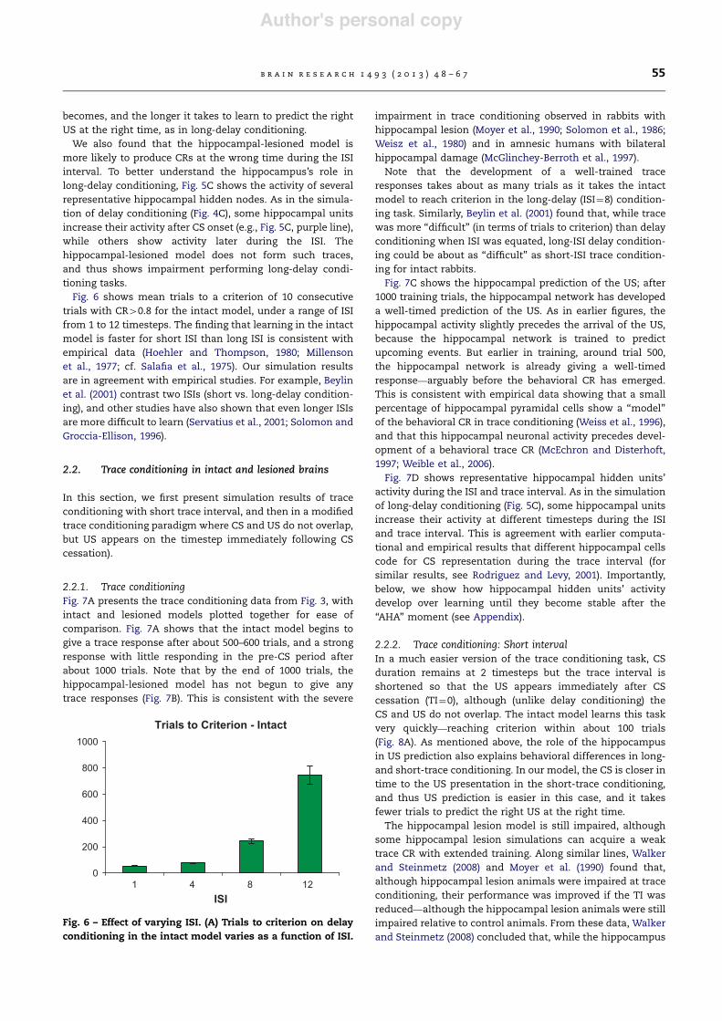
Fig. 6 shows mean trials to a criterion of 10 consecutivetrials with CR40.8 for the intact model, under a range of ISIfrom 1 to 12 timesteps. The finding that learning in the intactmodel is faster for short ISI than long ISI is consistent withempirical data (Hoehler and Thompson, 1980; Millensonet al., 1977; cf. Salafia et al., 1975). Our simulation resultsare in agreement with empirical studies. For example, Beylinet al. (2001) contrast two ISIs (short vs. long-delay condition-ing), and other studies have also shown that even longer ISIsare more difficult to learn (Servatius et al., 2001; Solomon andGroccia-Ellison, 1996).
2.2. Trace conditioning in intact and lesioned brains
In this section, we first present simulation results of traceconditioning with short trace interval, and then in a modifiedtrace conditioning paradigm where CS and US do not overlap,but US appears on the timestep immediately following CScessation).
2.2.1. Trace conditioningFig. 7A presents the trace conditioning data from Fig. 3, withintact and lesioned models plotted together for ease ofcomparison. Fig. 7A shows that the intact model begins togive a trace response after about 500–600 trials, and a strongresponse with little responding in the pre-CS period afterabout 1000 trials. Note that by the end of 1000 trials, thehippocampal-lesioned model has not begun to give anytrace responses (Fig. 7B). This is consistent with the severe
impairment in trace conditioning observed in rabbits withhippocampal lesion (Moyer et al., 1990; Solomon et al., 1986;Weisz et al., 1980) and in amnesic humans with bilateralhippocampal damage (McGlinchey-Berroth et al., 1997).
Note that the development of a well-trained traceresponses takes about as many trials as it takes the intactmodel to reach criterion in the long-delay (ISI¼8) condition-ing task. Similarly, Beylin et al. (2001) found that, while tracewas more ‘‘difficult’’ (in terms of trials to criterion) than delayconditioning when ISI was equated, long-ISI delay condition-ing could be about as ‘‘difficult’’ as short-ISI trace condition-ing for intact rabbits.
Fig. 7C shows the hippocampal prediction of the US; after1000 training trials, the hippocampal network has developeda well-timed prediction of the US. As in earlier figures, thehippocampal activity slightly precedes the arrival of the US,because the hippocampal network is trained to predictupcoming events. But earlier in training, around trial 500,the hippocampal network is already giving a well-timedresponse—arguably before the behavioral CR has emerged.This is consistent with empirical data showing that a smallpercentage of hippocampal pyramidal cells show a ‘‘model’’of the behavioral CR in trace conditioning (Weiss et al., 1996),and that this hippocampal neuronal activity precedes devel-opment of a behavioral trace CR (McEchron and Disterhoft,1997; Weible et al., 2006).
Fig. 7D shows representative hippocampal hidden units’activity during the ISI and trace interval. As in the simulationof long-delay conditioning (Fig. 5C), some hippocampal unitsincrease their activity at different timesteps during the ISIand trace interval. This is agreement with earlier computa-tional and empirical results that different hippocampal cellscode for CS representation during the trace interval (forsimilar results, see Rodriguez and Levy, 2001). Importantly,below, we show how hippocampal hidden units’ activitydevelop over learning until they become stable after the‘‘AHA’’ moment (see Appendix).
2.2.2. Trace conditioning: Short intervalIn a much easier version of the trace conditioning task, CSduration remains at 2 timesteps but the trace interval isshortened so that the US appears immediately after CScessation (TI¼0), although (unlike delay conditioning) theCS and US do not overlap. The intact model learns this taskvery quickly—reaching criterion within about 100 trials(Fig. 8A). As mentioned above, the role of the hippocampusin US prediction also explains behavioral differences in long-and short-trace conditioning. In our model, the CS is closer intime to the US presentation in the short-trace conditioning,and thus US prediction is easier in this case, and it takesfewer trials to predict the right US at the right time.
The hippocampal lesion model is still impaired, althoughsome hippocampal lesion simulations can acquire a weaktrace CR with extended training. Along similar lines, Walkerand Steinmetz (2008) and Moyer et al. (1990) found that,although hippocampal lesion animals were impaired at traceconditioning, their performance was improved if the TI wasreduced—although the hippocampal lesion animals were stillimpaired relative to control animals. From these data, Walkerand Steinmetz (2008) concluded that, while the hippocampus
Trials to Criterion - Intact
0
200
400
600
800
1000
1 4 8 12ISI
Fig. 6 – Effect of varying ISI. (A) Trials to criterion on delayconditioning in the intact model varies as a function of ISI.
b r a i n r e s e a r c h 1 4 9 3 ( 2 0 1 3 ) 4 8 – 6 7 55

Author's personal copy
is important for trace conditioning, the relative lengths of CSand trace interval are also important.
2.3. Pharmacological manipulations
Finally, we have simulated the effects of cholinergic agentson delay and trace conditioning. First, we have also foundthat reducing hippocampal plasticity in the model (bydecreasing learning rate values) slightly impairs delay con-ditioning but severely impairs trace conditioning (Fig. 9A),similar to the results of cholinergic antagonists on eyeblinkconditioning discussed above.
In addition, we simulated the effects of low- and high-doseof cholinergic agonists on delay and trace conditioning by
increasing hippocampal plasticity in the model (throughmanipulation of learning rate parameter in the hippocampalmodule). In empirical studies, mild doses of cholinergic ago-nists slightly enhance delay conditioning in aged rabbits(Woodruff-Pak and Santos, 2000; Woodruff-Pak et al., 2001),and significantly enhance performance in trace eyeblink con-ditioning (Simon et al., 2004); the model shows the sameeffects (Fig. 9B). Our simulation results also suggest thatenhancing learning abilities in healthy animals using choliner-gic agonists may improve more difficult tasks, such as traceconditioning (Simon et al., 2004). Finally, administration of alarge dose of cholinergic agonists mildly impairs delay con-ditioning, but severely impairs trace conditioning in the model(Fig. 9C). This is a new prediction of the model, and it is
Fig. 7 – Trace conditioning paradigm with ISI¼4 and TI¼2. (A, B) Across-trials responding in the intact and HL model given100 training trials (left) and in the HL model given 10,000 training trials, right). (C) Activity of the hippocampal-region outputnode learning to predict the next state of the US in the intact model. (D) Individual responses of three representativehippocampal network hidden units during the last conditioning trial. As in long-delay conditioning, some nodes respondto CS onset (blue), others later in the CS period (purple), and some peak at the time of expected US arrival (brown).(For interpretation of the references to color in this figure legend, the reader is referred to the web version of this article.)
Trace (ISI=1)
0
0.2
0.4
0.6
0.8
1
0003000200010
Trials
Intact: CRIntact: pre-CSHL: CRHL: pre-CS
Mea
n m
odel
out
put
Fig. 8 – The ‘‘simplest’’ version of trace conditioning in the model, in which US onset occurs just after CS cessation (TI¼0);see Fig. 6D for task description. (A) The intact model learns this task quickly; the HL model is impaired, but learns to producesome CRs after extended training.
b r a i n r e s e a r c h 1 4 9 3 ( 2 0 1 3 ) 4 8 – 6 756

Author's personal copy
plausible since other experimental studies have shown thatthe administration of large doses of cholinergic agonists toanimals interferes with the performance of hippocampal-based tasks (Dumery et al., 1988; Ennaceur and Meliani, 1992).
3. Discussion
Here, we have presented a computational model of thehippocampal region and its role in stimulus representation,that includes the ability to simulate within-trial events, andthus to address not only trial-level data regarding whether abehavioral response is emitted, but within-trial data regard-ing the timing of that response. Applied to classical eyeblinkconditioning, the model is correctly able to account for thefindings that, in intact animals, learning is slower as the ISIincreases (also see Servatius et al., 2001), that for a given ISIdelay CRs are acquired faster than trace CRs, and that some(but not all) neurons in the hippocampus show activitypatterns that predict and precede the form of the behavioralCR. Our model is also in agreement with studies showing thatthe hippocampus participates in appetitive (Flesher et al.,2011; Seager et al., 1999) and fear (Esclassan et al., 2009) traceconditioning.
The model also addresses data from hippocampal-lesionedanimals including impairments of trace and delay condition-ing that vary as a function of ISI; thus, both trace and long-delay conditioning are impaired in the lesioned model, butboth delay and trace conditioning are spared if the ISI issufficiently short. Finally, our current computational modelshows how the hippocampus role in stimulus predictionexplains the effects of hippocampal lesion, and cholinergicagonists and antagonists on delay and trace conditioning.To our knowledge, this is the first model to simulate these
various delay and trace conditioning results, from both intactand lesioned animals, using one single framework.
In sum, our model not only simulates behavioral differ-ences between delay and trace conditioning, but alsoaddresses a large body of empirical data on the effects ofmanipulating ISI on conditioning. Along the same lines, ourmodel shows that the same computational theory of hippo-campal function – that is, prediction of future state of theenvironment – explains performance in various conditioningstudies. Third, unlike prior models of delay and trace con-ditioning, our model also shows how hippocampal lesion(and thus inability to correctly predict US timing) interfereswith some conditioning tasks (e.g., trace conditioning), butnot others (e.g., delay conditioning). Our model also inagreement with data showing that long-delay conditioningis acquired more slowly than short-delay conditioning usinga fear conditioning paradigm (Barnet and Hunt, 2005).
3.1. Computational role of the hippocampus in USprediction learning and pattern separation
The hippocampus in our model has two related functions:one is predicting upcoming events (in the output layer) butthe other is developing new stimulus representations in thehidden layer that allow this prediction. These functions areinterrelated. In other words, if the model is presented withtwo sets of stimuli that predict the same outcome, throughlearning, they will have largely similar representations in thehidden layer of the hippocampus. On the other hand, if themodel is presented with two sets of stimuli that predictdifferent outcomes, they will have different representationin the hidden layer of the hippocampus. This latter process isalso known as pattern separation or differentiation. Since themodel is presented with relevant stimuli (including CSs, and
Trials Trials
Trials Trials
Mea
n m
odel
out
put
0
0.2
0.4
0.6
0.8
1
Low ACh Agonist ModelDelay
Trace
Mea
n m
odel
out
put
0
0.2
0.4
0.6
0.8
1High ACh Agonist Model
Delay
Trace
Trials
Mea
n m
odel
out
put
0
0.2
0.4
0.6
0.8
1
0 100 200 300 400 500 600 7 00 800 900 1000 0 100 200 300 400 500 600 700 800 900 1000
0 100 200 300 400 500 600 700 800 900 1000
ACh Antagonist Model
Delay
Trace
Fig. 9 – Simulation results show that (A) cholinergic antagonists mildly affect delay conditioning but severely impair traceconditioning. In contrast, (B) mild doses of cholinergic agonists slightly enhance delay conditioning and significantlyenhance performance in trace eyeblink conditioning, while (C) large doses of cholinergic agonists mildly impair delayconditioning, but severely impair trace conditioning.
b r a i n r e s e a r c h 1 4 9 3 ( 2 0 1 3 ) 4 8 – 6 7 57

Author's personal copy
USs) and irrelevant stimuli (including contextual informationsuch as smell/shape of the cage or testing room, which arenot related to current study being that our focus is on thedifference between delay and trace conditioning), the modellearns to form separate representations of context and otherstimuli. Not only that, but the model also learns to formrepresentation of the CS in the hidden layer of the hippo-campus during the trace interval of trace conditioning tasks.Weight modification (i.e., learning) occurs in connections inthe hippocampus that help form separate representation ofCS and context. Learning takes place in all weights inthe hippocampal module, as we explain in Section 4.2.This mechanism explains how the hippocampus participatesin US prediction, pattern separation, and learning to repre-sent stimuli that are not perceptually present as in traceconditioning.
Importantly, the role of the hippocampus in US predictionlearning also explains behavioral differences between long-and short-trace conditioning. In our model, the CS is closer intime to the US presentation in the short-trace conditioning,and thus US prediction is easier in this case, and it takesfewer trials to learn to predict the US at the right time than inlong-trace conditioning. The same analysis also applies toshort- and long-delay conditioning. To summarize, the mainfunction of the hippocampus in our model is US prediction,which then explain its role in learning to represent inputinformation in the hippocampus, which in turn explains thehippocampal role in conditioning tasks.
3.2. Comparison to other hippocampal modelsof conditioning
The conception of the hippocampal region as a general-purpose prediction device, learning to map from currentinputs to expectation of future events, and helping to spantemporal gaps between stimulus and desired response, hasgained a great deal of currency in the last decade, asempirical data have emerged suggesting that hippocampal‘‘place cells’’ encode not only spatial location but also ‘‘loca-tion’’ in temporal sequences (e.g., Dragoi and Buzsaki, 2006;Johnson and Redish, 2007; Lehn et al., 2009; Pastalkova et al.,2008). This is not the first computational model to instantiatesuch a function; other computational models have alsofocused on putative hippocampal-region roles in temporalsequence learning or short-term buffering of information, atvarious levels of biological and empirical detail (Hasselmoet al., 2000; Hazy et al., 2007, 2006; Howe and Levy, 2007; Levy,1996; Wiebe et al., 1997), and it is entirely possible that someor all of these earlier models could also address the delay,trace, and ISI results which have been simulated here. Todate, only a subset of such existing models of hippocampalfunction has been explicitly applied to address classicalconditioning. Of this small set, most existing models haveassumed some degree of ‘‘hard-wired’’ connections that allowthe hippocampal region to perform temporal processing.
For example, some models have assumed that temporalinformation is provided to the hippocampus from externalsources. For example, Schmajuk and colleagues proposed amodel of hippocampal-region function based on the atten-tional models of Pearce and Hall (1980) in which the
hippocampus computes the aggregate prediction of reinforce-ment – i.e., the expectation of US arrival based on all availablecues (e.g., CSs) – and the difference between this aggregateprediction and the actual US is then used to compute theassociability of cues contributing to this prediction (also seeSchmajuk and Labar, 2007; Schmajuk and Moore, 1988). Thesemodels assumed that each CS had a memory trace, which wasmaximal when the CS was present, and then decayed back tobaseline. This trace could be associated with a subsequent US,even if the CS and US did not overlap, and thus the modelcould perform trace conditioning. Similar assumptions of anexplicit CS trace that occurs outside the hippocampus aremade in other models (Buhusi and Schmajuk, 1996; Schmajukand DiCarlo, 1992; Schmajuk and Moore, 1989).
Other models, often known as ‘‘tapped delay line’’ models,have suggested that the CS trace could arise in the hippo-campus, with different dentate gyrus cells responding atdifferent delays to a CS (Grossberg and Merrill, 1996; Ludviget al., 2009; Zipser, 1986), generating a spectral representationof the CS; a CS could be associated with a US at a specific ISI(with or without a trace interval) by adjusting the weightsfrom the cell that responded to the CS with the correct delay.Such models generally require one cell to represent eachpossible CS at each possible delay, which leads to a problemof combinatorial explosion. Further, the biological plausibilityof models invoking tapped delay lines is weakened by the factthat cellular recordings do not show any obvious CS storagewithin the hippocampus (Rodriguez and Levy, 2001) and that,although hippocampal units display CS-related activity dur-ing the trace interval in eyeblink conditioning, this activityshifts across training so that, in a well-trained animal,neuronal activity tends to model the time-course of thebehavioral CR (e.g.,Solomon et al., 1986).
In contrast to models that assume tapped delay lines orother explicit representations of a CS trace, the current modelfollows in a different tradition of prior models suggesting thatthe hippocampus’s ability to maintain stimulus traces acrossshort delays is not hardwired but adaptive. These modelsassume that the high degree of internal recurrency in thehippocampus, particularly within field CA3, could allow thehippocampus to store sequences of neural activity by forminga reverbatory memory (Levy and Sederberg, 1997; Rodriguezand Levy, 2001; Wallenstein and Hasselmo, 1997a, 1997b;Wiebe et al., 1997; Yamazaki and Tanaka, 2005). Networkmodels with recurrent connections can adaptively learn tobuffer information across a stimulus-free interval withoutrequiring a multitude of hardwired delay lines (Levy, 1989).A few prior recurrent network models have been explicitlyapplied to classical conditioning and to trace conditioning inparticular (e.g., Howe and Levy, 2007; Rodriguez and Levy,2001; Yamazaki and Tanaka, 2005).
For example, Rodriguez and Levy (2001) have considered abiologically-based model of CA3 in which a CS input excites asubset of cells, which in turn excite other cells at a shortdelay, and so on until a final group of cells representing theUS is excited at the correct temporal distance from CS onset(see also Levy and Sederberg, 1997). This model has the virtuethat its ability to span a CS-free interval is learned, ratherthan hardwired into the network via tapped delay lines. Howeand Levy (2007) subsequently showed that such a model
b r a i n r e s e a r c h 1 4 9 3 ( 2 0 1 3 ) 4 8 – 6 758

Author's personal copy
could correctly predict data showing that various subpopula-tions of hippocampal neurons are activated by the CS, by theUS, or during the trace interval, as well as data showing thatthe emergence of neuronal activity that accurately predictsUS onset occurs suddenly after a period of training in rabbits.However, this model does not consider representationalchanges in the hippocampus, nor does it interact with amotor output module, and so it does not directly generateCRs, nor can it simulate hippocampal lesion data.
Similar to several of these prior models, the current modelincludes a recurrent network as a model of the hippocampalregion, in which different subsets of hippocampal cellsmaintain representation of CS information during the ISI indelay and trace eyeblink conditioning. This is supported byan empirical study by McEchron and Disterhoft (1997), whorecorded from the CA1 of the hippocampus in rabbits duringtrace eyeblink conditioning; results showed that differenthippocampal neurons maintains representation of CS atdifferent timesteps during the trace interval. As similar totrace conditioning, other neurophysiological studies haveshown that different hippocampal neurons are activated atdifferent timesteps during task performance, including spa-tial navigation (Pastalkova et al., 2008) and sequence learning(MacDonald et al., 2011).
3.3. Model limitations
In turn, the current model also suffers from some limitations.First, it is a simple connectionist model, with abstract nodesthat do not simulate the biophysical properties of neurons. Assimilar to the Rodriguez and Levy model, Itskov et al. (2011)show how a recurrent hippocampal network and hippocam-pal cells can maintain events over time. While the Rodriguezand Levy model is applied to trace conditioning data, theItskov model is applied to spatial navigation tasks. In contrastto Levy’s and Itskov’s models, the abstraction of our modelallows us to simulate a large number of behavioral data thatwere not simulated by Levy’s or other models of the hippo-campus. Nevertheless, future models should address howphysiologically detailed models of the hippocampus (as inthe model of Rodriguez and Levy, 2001) can simulate perfor-mance in a large number of behavioral studies, includingconditioning data (as in our model).
Furthermore, our model considers the hippocampal regionas a single functional system, without considering the ana-tomical and functional differences of different subregions.However, empirical data strongly suggest that some puta-tively hippocampal-dependent representational processesdepend more on entorhinal cortex than on the hippocampusproper (Allen et al., 2002; Coutureau et al., 2002; Jarrard, 1993;Shohamy et al., 2000). Other experiments have shown thatlesion to CA1, but not to CA3, interferes with performingpaired associates tasks that include temporal delay (Kesneret al., 2005). Consistent with this, prior modeling work hasshown that some aspects ascribed by our model to thehippocampal region as a whole could emerge naturally fromthe anatomy and physiology, including redundancy compres-sion in the entorhinal cortex (Myers et al., 1995) and patternseparation in the dentate gyrus (Myers and Scharfman, 2009,2010). The pattern separation function of the hippocampus in
our model is also much in line with the conjunctive encodingfunction proposed in O’Reilly’s models (O’Reilly and Norman,2002; O’Reilly and Rudy, 2001).
Future empirical work should determine whether traceconditioning similarly depends primarily on one or more ofthe hippocampal subregions. In particular, Czerniawski et al.(2009) have suggested that ventral, but not dorsal, hippocam-pal lesions impair trace conditioning, which might meaneither that the ventral hippocampus is more important fortrace conditioning than the dorsal hippocampus, or mightmerely reflect the relative importance for trace conditioningof inputs that preferentially target the ventral hippocampus.This view is, however, challenged by other physiologicalexperimental studies, which argue that dorsal CA1 neuronsare more active than ventral CA1 neurons during traceconditioning (Weible et al., 2006). Future modeling work couldexplore these possibilities.
Another limitation of model is not addressing the differ-ential roles of ventral vs. dorsal hippocampus in conditioning(nor simulating subregions of the hippocampus includingseptum, CA1, CA3, and dentate gyrus). Importantly, there isno consensus in the literature in the role of dorsal vs. ventralhippocampus in conditioning, and it is not clear how bothinteract during acquisition and performance. For example,Burman et al. (2006) argue that dorsal (septal) hippocampus isimportant for acquisition, while the ventral hippocampus isimportant for expression of fear responses (for similarresults, also see Kjelstrup et al., 2002). Interestingly,Gonzalez-Pardo et al. (2012) found opposite results: dorsalhippocampus being important for expression of fearresponses, and ventral hippocampus for acquisition! Unlikethe Burman et al. results, Czerniawski et al. (2012) found thatboth ventral and dorsal hippocampi are required for theacquisition of trace conditioning. On the other hand, Wanget al. (2012) found that dorsal hippocampus is important forcontextual fear conditioning, and that dorsal or ventralhippocampus is sufficient for subsequent conditioning in adifferent context, while Kenney et al. (2012) suggest thatthere is a competition between dorsal and ventral hippocam-pus on control over behavior in contextual conditioningtasks. Future modeling work, which takes into accountdifferences in connectivity patterns and cell types in thedorsal vs. ventral hippocampus, might be able to helpreconcile some of these conflicting results.
Another limitation of our model is the finding that traceconditioning recruits additional brain areas, such as thesupplementary motor area (Knight et al., 2004) and anteriorcingulate cortex (Han et al., 2003), which are not simulated inour model. The function of these brain areas in traceconditioning is perhaps related to the short-term encodingof conditioned stimuli during the trace interval. It is possiblethat the recurrent connection in our model perhaps corre-sponds to hippocampal interactions with other cortical areasresponsible for maintaining information during trace inter-vals, though future computational modeling work shouldaddress this point more explicitly.
The medial prefrontal cortex (mPFC) is also important fortrace conditioning. For example, lesion of mPFC impairsacquisition of long-interval trace but not short-interval traceor delay eyeblink CRs in the rabbit (McLaughlin et al., 2002),
b r a i n r e s e a r c h 1 4 9 3 ( 2 0 1 3 ) 4 8 – 6 7 59

Author's personal copy
and also impairs extinction of previously-learned trace eye-blink CRs (Weible et al., 2007). It has been suggested that themPFC plays a role in contextual-dependent suppression oflearned responses (Milad et al., 2007; Morgan and LeDoux,1995; Resstel et al., 2008), and medial prefrontal cortex maybe important in suppressing a response to the CS during atrace interval and/or helping to suppress responses to con-textual stimuli that are present at the time of US arrival. Animportant goal for future research will be considering theinteraction between the hippocampus and mPFC in classicalconditioning. Similarly, the model does not simulate thefunctionality of the rubro-trigeminal pathway which hasbeen demonstrated to inhibit activity in the inferior olivefollowing stimulation of the magnocellular red nucleus whichpresumably plays an important role in eyeblink conditioning(Weiss et al., 1990).
The current model focuses on the hippocampal region’srole in associative learning; other hippocampal-dependentprocesses, such as declarative (consciously-mediated) mem-ory, are beyond the scope of the model, but may also play arole in trace conditioning. Several studies have now shownthat, in human trace eyeblink conditioning, participants whoself-report becoming aware of the stimulus contingenciesearly in the conditioning session emit more CRs later in thesession than participants who report becoming aware later inthe session or not at all (Clark et al., 2002; Clark and Squire,1998). There was no such interaction between awareness andconditioning under delay contingencies (Manns et al., 2001),suggesting that awareness in delay conditioning could per-haps be epiphenomenal. Similarly, in a two-cue discrimina-tion task, awareness of stimulus contingencies wasassociated with emergence of differential eyeblink CRs undertrace but not delay conditioning (Clark and Squire, 1998).A possible conclusion to be drawn from these data is thattrace but not delay conditioning requires conscious aware-ness, probably mediated by hippocampal declarative memorysystems (Clark et al., 2002), at least in humans.
While the data correlating awareness and trace conditioningare robust, there are at least three reasons to be cautious inassuming a causative link. First, although eyeblink condition-ing appears to share very similar substrates across speciesfrom rodents to rabbits to primates (including humans), it isunclear whether conscious awareness is required in non-human animals, or how such awareness might be assessed.Second, as noted by LaBar and Disterhoft (1998), patients withamygdala lesions show disrupted conditioning with spareddeclarative knowledge of the stimulus contingencies (Becharaet al., 1995), while partial medial temporal lobe damage thatspares declarative memory for stimulus contingencies can beinsufficient to support development of conditioning (Daumet al., 1991). These data suggest that awareness per se is notsufficient for conditioning to occur. Third, as mentioned above,although humans and other animals with bilateral hippocam-pal damage can acquire delay CRs as quickly as controls, thislearning is not necessarily ‘‘normal’’—for example, there maybe ill-timed short-latency CRs (e.g., Christiansen andSchmajuk, 1992; Clark et al., 2002) and impairment at long-delay conditioning (Beylin et al., 2001).
A final important limitation of the current model is that itignores consolidation processes (McGaugh, 2000). Although
the hippocampus is important for acquisition of trace eye-blink CRs, it apparently is not the final site of memorystorage, because trace CRs are abolished in rabbits givenbilateral hippocampal lesion one day, but not one month,after trace conditioning (Kim et al., 1995). Apparently, thehippocampus either functions as a temporary memory store,or else supports the gradual formation of trace eyeblinkassociations elsewhere, so that eventually the memories arestored outside the hippocampal region and can survivehippocampal lesion. This final storage site is a matter ofdebate, but may involve association cortex; in the case ofeyeblink conditioning, the cerebellum is also a possibility.The current model could be expanded to explore thesepossibilities as further empirical data emerge to constrainthe model. It is also worth noting that memories can beconsolidated, and even strengthened, during sleep (Walkerand Stickgold, 2006). In this context, it is interesting to notethat sleep deprivation may have a particularly detrimentaleffect on learning that involves temporal information (suchas recency judgments) and motor sequence learning, andthat sleep deprivation is associated with decreases in hippo-campal nerve growth factor (Walker and Stickgold, 2006).Since any plasticity or consolidation that occurs during sleephappens (by definition) in the absence of external sensorystimuli (and thus, during a stimulus-free trace interval), therelationship between trace conditioning and hippocampalactivity during sleep may prove profitable for further explora-tions using both empirical techniques and computationalmodels.
3.4. Future directions
One important question is, how does the role of the hippo-campus in prediction explains its role in long-term memory?In our earlier work, we have addressed the relationshipbetween the hippocampus role in both episodic long-termmemory and classical conditioning (see for example, Glucket al., 2003; Meeter et al., 2005). We conceptualize long-termmemory as binding of information in one single unit. Forexample, one’s memory of visiting a certain place with somefriends at certain time is the binding of the where, who, andwhen together. The relationship between the hippocampusrole in US prediction and long-term memory is ratherindirect. First, we have previously shown that the role ofthe hippocampus in US prediction can support associativebinding of information, as in contextual conditioning, inwhich subjects learn to associate contextual and cue infor-mation (see for example, Gluck and Myers, 1993; Myers andGluck, 1994) and associative learning in humans (Moustafaet al., 2010): while learning to predict the US, the hippocam-pal module’s internal layer learns associative properties ofinput stimuli. Accordingly, long-term memory (which is fastbinding of information, see for example, Meeter et al., 2005) isa special case of associative learning. Similar modelingstudies have also addressed relationships between episodicmemory and associative learning (see for example, thecomputational model of Li et al., 2005).
Experimental studies have shown that the hippocampus isinvolved in the reactivation of recently acquired memories,particularly during slow-wave sleep. This reactivation
b r a i n r e s e a r c h 1 4 9 3 ( 2 0 1 3 ) 4 8 – 6 760

Author's personal copy
consequently leads to the stabilization of memories (Marshalland Born, 2007). It was also shown that hippocampus-mediated declarative-memory improves after a night’s sleep(Ellenbogen et al., 2006). Moreover, additional experimentalfindings suggest that the hippocampus sends vast amountsof information to the cortex only during slow-wave sleep(Montgomery et al., 2008) and that following sleep depriva-tion, activity in the medial temporal lobes is decreased andits memory capacity reduced (Drummond et al., 2000; Gaiset al., 2007; Takashima et al., 2006). Such findings weretheorized as supporting the role of the hippocampus as afast episodic learning system which, during short-wave sleep,gradually transfers its content to the more robust memorystorage located in the cortex (McClelland et al., 1995). In sum,it was found that sleep affects memory consolidation bymodulating various learning mechanisms and that theselearning mechanisms are preferentially affected by differentstages of sleep. Following earlier modeling studies on sleep(Hinton et al., 1995; Norman et al., 2005), we also argue thatsleep enhances offline learning with input stimuli occurs.According to our modeling framework, offline learning duringsleep should enhance binding of information in the hippo-campus, and thus improved episodic memory. Althoughexplicit simulation of sleep is beyond the scope of the currentpaper, future modeling work could address data showing howsleep deprivation impairs conditioning tasks (Hagewoudet al., 2011).
In addition, we are currently designing a computationalmodel of the interactions of the hippocampal region, mPFC,and the amygdala in fear conditioning, in which thehippocampal-region network plays a similar function to itsrole in eyeblink conditioning (as shown here). In other words,we argue that the hippocampal-region is essential for learn-ing stimulus prediction, a process essential to learningvarious forms of conditioning paradigms.
In sum, our model provides a unified account on the role ofthe hippocampus in delay and trace conditioning suggestingthat the hippocampal region is a general-purpose predictiondevice, with the ability to help other brain substrates spantemporal gaps between stimulus arrival and desired time ofbehavioral response; this role is critical when the temporalgap is longer than can be bridged by the brain substratesmediating the behavioral response, whether or not there is anaccompanying trace interval.
4. Experimental procedures
The recurrent hippocampal model was implemented inobjective-Cþþ using the Xcode 3.0 applications developmentsuite for Macintosh OS 10.5.
4.1. External inputs and timing consideration
Each trial is divided into a number of timesteps; for most ofthe simulations presented here, there are 30 timesteps pertrial. At each timestep t, external inputs consist of a 18-element vector ½x1ðtÞ. . .x18ðtÞ' detailing the presence (1.0) orabsence (0.0) of 18 possible cues. For the experimentsreported here, the first three elements of the vector are
considered as CSs (CS1, CS2, CS3) and the remaining 15elements are considered as contextual elements. As in priormodels (Moustafa et al., 2009; Myers and Gluck, 1994), CSs areusually presented phasically, with discrete onset and cessa-tion during each trial, while contextual cues are presentedtonically throughout the trial and across trials, but otherwisethere is no special treatment given to CSs vs. contextual cuesin the model.
For the first several timesteps of each trial, only contextualcues are present. One or more CSs may then be presented fora fixed number of timesteps, and the US may be presented fora single timestep to co-terminate with the CS (simulatingdelay conditioning) or a few timesteps after CS cessation (tosimulate trace conditioning). The remaining timesteps in thetrial are additional context-alone presentations. As in empiri-cal studies, the time of CS onset is varied pseudorandomlyacross trials so that CS onset cannot be predicted simply bypassage of time from last CS presentation. Fig. 2 showsschematic examples within-trial events during a single trialof short-delay, trace, and long-delay paradigms.
At the start of a simulation run, the model is given 100 trials(each consisting of 30 timesteps) of exposure to the context(s)alone. All results shown are averaged across 5 simulation runs,except figures depicting within-trial data, which each show datafrom one individual, representative, simulation run.
4.2. Hippocampal-region network
As in prior models (Gluck and Myers, 1993; Moustafa et al.,2009), the hippocampal region is implemented as an predic-tive autoencoder network. At each timestep t, the networkreceives 20 inputs detailing the state of the world, includingthe 18 CS and context inputs ½x1ðtÞ. . .x18ðtÞ' (via entorhinalcortex), an efferent copy of the CR from the previous timestep(x19ðtÞ ¼CR t#1ð Þ), and the US at the present timestepx20ðtÞ ¼USðtÞ.
The hidden layer includes 10 nodes j, each fully connectedto the inputs, and computing activation as:
yj tð Þ ¼ fX
i
xiðtÞwijðtÞ þX
j0yj0 t#1ð Þwj0 jðtÞ þ yj
2
4
3
5 ð1Þ
for all input layer nodes i and hidden layer nodes j’. yj is nodej’s bias, which is treated as a weight from an input that isalways active.
The hidden layer units also project to the motor cortex(possibly via waystations in cortex, thalamus, or brainstem),providing the hippocampal-mediated adaptive representa-tions as a secondary set of inputs that can be used to driveCRs. This projection is not assumed to be instantaneous;rather the hippocampal hidden layer activations at time t#1are provided as input to the motor network at time t.
Finally, the output layer includes 20 nodes k that are fullyconnected to the hidden layer nodes j and compute activation as:
yk tð Þ ¼ fX
j
yjðtÞwjkðtÞ þ yk
2
4
3
5 ð2Þ
The resulting array yk(t) is a prediction of the next state ofthe external inputs xi(tþ1). The difference between the pre-dicted and actual inputs is used as an error signal to drive
b r a i n r e s e a r c h 1 4 9 3 ( 2 0 1 3 ) 4 8 – 6 7 61

Author's personal copy
plasticity in the hippocampal network, with each hippocam-pal output-layer node k updating its weights as:
wjk tþ 1ð Þ ¼wjkðtÞ þ DwjkðtÞ
DwjkðtÞ ¼ b( @kðtÞ ( yiðtÞ þ a( Dwjk t#1ð Þ@kðtÞ ¼ xkðtð Þ#ykðtÞÞ ( f 0 ykðt
! "Þ ð3Þ
for all hidden-layer nodes j, where f0(x)¼x(1#x), a is amomentum term set to 0.9 and b is a learning rate parameterset to 0.5 if US(t)¼1, and to 0.05 otherwise. Each hidden-layernode j then updates its weights as:
wij tþ 1ð Þ ¼wijðtÞ þ DwijðtÞDwijðtÞ ¼ b( @jðtÞ ( xiðtÞ þ a( Dwij t#1ð Þ
@jðtÞ ¼X
k
@kðtÞ (wjkðtÞÞ ( f 0ðyj tð Þ
!ð4Þ
for all input-layer nodes i. All weights in the cortical andhippocampal networks (including biases) are initialized fromthe uniform distribution U(#0.3,þ0.3) at the start of eachsimulation run.
4.3. Motor output network
The motor output network is responsible for the generationof motor responses during conditioning. It is implemented asa single adaptive node, receiving inputs representing CSs,contextual stimuli, an efferent copy of the previous CR, andthe activations of the hippocampal-region network hiddenlayer nodes from the previous timestep. The output of thisnode at time t, which is assumed to drive the behavioral CR,
Fig. 10 – Activity of hippocampal hidden units at time step before US presentation during trace conditioning, showingprediction of US during learning. The first plot shows activity of the output of the Motor network. The other 10 figures showactivity of the hippocampal hidden units during trace conditioning. The ‘‘AHA’’ moment here is roughly at trial 500 (first plot,top left). Some hidden unit also show increase of activity around the ‘‘AHA’’ moment (see in particular, hidden units #2,3,4,6,8,9); they activities become stable after the AHA moment, and are not altered by overtraining the network.Abbreviation: hh¼hippocampal hidden unit.
b r a i n r e s e a r c h 1 4 9 3 ( 2 0 1 3 ) 4 8 – 6 762
Author's personal copy
is computed as:
CRðtÞ ¼ fX
i
xiðtÞwikðtÞ þX
j
yj t#1ð ÞwjkðtÞ þ CR t#1ð ÞwCR tð Þ
0
@
1
A
ð5Þ
for all inputs i and all hippocampal hidden layer nodes j. Notethat, because both the hippocampal inputs yj and the efferentcopy of the CR are from the previous timestep (t#1), the inputlayer is not getting any information as input about whether aUS has arrived on the current timestep t. The w are modifi-able weights on each of the inputs, and f(x)¼1/(1þe#x)
Weights in the motor response network are updated pro-portional to output error d(t)¼US(t)#CR(t):
wi tþ 1ð Þ ¼wiðtÞ þ b( @ðtÞ ( CSiðtÞ
wj tþ 1ð Þ ¼wjðtÞ þ b( @ðtÞ ( yjðtÞwCR tþ 1ð Þ ¼wCRðtÞ þ b( @ðtÞ ( CRðtÞ ð6Þ
for each input i and hippocampal hidden node j. b is alearning rate parameter; if US(t)¼1, b¼0.05; else b¼0.005.All weights w are initialized to 0 at the start of asimulation run.
In the model, hippocampal-region damage is simulated bydeleting the hippocampal-region network. In this case, themotor output network still receives (and can modify weightsfrom) inputs representing CS and context, as well as theefferent copy of the CR. Lastly, as in prior work (Myers et al.,1996, 1998; Moustafa et al., 2010), we simulate the effects ofcholinergic antagonists by decreasing the learning rate para-meter in the hippocampal-region network, and cholinergicagonists by increasing hippocampal-region learning rate.
Acknowledgments
This work was partially supported by the NSF/NIH Collabora-tive Research in Computational Neuroscience (CRCNS) pro-gram and by NIAAA (RO1 AA018737-01).
Appendix. The development of stableactivation in the hippocampus during traceconditioning
In this section, we explain how the activation of hidden layernodes o of the hippocampal module develops during traceconditioning. Fig. 10 shows several plots showing the activityof the 10 hidden nodes of the hippocampus along with themotor output node during trace conditioning. Each of theseplots shows the activity of each node at time step t-1 only,where t is defined as the time step where US presentationoccurs. The plots show that there are clear correlationsbetween the activities of nodes in the hidden layer and themotor output node. Importantly, in some of the hippocampalhidden nodes, activity stabilizes after the ‘‘AHA’’ moment.Overtraining the model did not alter the activation of hiddenunits, suggesting they become stable after the AHA moment.
r e f e r e n c e s
Allen, M.T., Chelius, L., Masand, V., Gluck, M.A., Myers, C.E.,Schnirman, G., 2002. A comparison of latent inhibition andlearned irrelevance pre-exposure effects in rabbit and humaneyeblink conditioning. Integr. Physiol. Behav. Sci. 37 (3), 188–214.
Amaral, D.G., Ishizuka, N., Claiborne, B., 1990. Neurons, numbersand the hippocampal network. Prog. Brain Res. 83, 1–11.
Amaral, D.G., Scharfman, H.E., Lavenex, P., 2007. The dentategyrus: fundamental neuroanatomical organization (dentategyrus for dummies). Prog. Brain Res. 163, 3–22.
Amaral, D.G., Witter, M.P., 1989. The three-dimensional organiza-tion of the hippocampal formation: a review of anatomicaldata. Neuroscience 31 (3), 571–591.
Barnet, R.C., Hunt, P.S., 2005. Trace and long-delay fear condition-ing in the developing rat. Learn. Behav. 33 (4), 437–443.
Bechara, A., Tranel, D., Damasio, H., Adolphs, R., Rockland, C.,Damasio, A.R., 1995. Double dissociation of conditioning anddeclarative knowledge relative to the amygdala and hippo-campus in humans. Science 269 (5227), 1115–1118.
Berger, T.W., Alger, B., Thompson, R.F., 1976. Neuronal substrateof classical conditioning in the hippocampus. Science 192(4238), 483–485.
Berger, T.W., Rinaldi, P.C., Weisz, D.J., Thompson, R.F., 1983.Single-unit analysis of different hippocampal cell types dur-ing classical conditioning of rabbit nictitating membraneresponse. J. Neurophysiol. 50 (5), 1197–1219.
Berger, T.W., Thompson, R.F., 1978. Neuronal plasticity in thelimbic system during classical conditioning of the rabbitnictitating membrane response. I. The hippocampus. BrainRes. 145 (2), 323–346.
Bevins, R.A., Ayres, A.M., 1995. One-trial context fear conditioningas a function of the interstimulus interval. Anim. Learn.Behav. 23 (4), 400–410.
Beylin, A., Gandhi, C.C., Wood, G.E., Talk, A.C., Matzel, L.D., Shors,T.J., 2001. The role of the hippocampus in trace conditioning:temporal discontinuity or task difficulty?. Neurobiol. Learn.Mem. 76 (3), 447–461.
Beylin, A., Talk, A., Gandhi, C., Wood, G., Matzel, L., & Shors, T.(1999). Acquisition but not Performance of Trace EyeblinkConditioning is Dependent on the Hippocampus. Paper Pre-sented at the Abstracts of the Society for NeuroscienceAnnual Meeting.
Blaxton, T.A., Zeffiro, T.A., Gabrieli, J.D., Bookheimer, S.Y., Carrillo,M.C., Theodore, W.H., et al., 1996. Functional mapping ofhuman learning: a positron emission tomography activationstudy of eyeblink conditioning. J. Neurosci. 16 (12), 4032–4040.
Bower, G.H., 1961. Application of a model to paired-associatelearning. Psychometrika 26, 255–280.
Brown, K.L., Comalli, D.M., De Biasi, M., Woodruff-Pak, D.S., 2010.Trace eyeblink conditioning is impaired in alpha7 but not inbeta2 nicotinic acetylcholine receptor knockout mice. Front.Behav. Neurosci. 4, 166.
Buhusi, C.V., Schmajuk, N.A., 1996. Attention, configuration, andhippocampal function. Hippocampus 6 (6), 621–642.
Burman, M.A., Starr, M.J., Gewirtz, J.C., 2006. Dissociable effects ofhippocampus lesions on expression of fear and trace fearconditioning memories in rats. Hippocampus 16 (2), 103–113.
Cheng, D.T., Disterhoft, J.F., Power, J.M., Ellis, D.A., Desmond, J.E.,2008. Neural substrates underlying human delay and trace eye-blink conditioning. Proc. Nat. Acad. Sci. USA 105 (23), 8108–8113.
Chowdhury, N., Quinn, J.J., Fanselow, M.S., 2005. Dorsal hippocampusinvolvement in trace fear conditioning with long, but not short,trace intervals in mice. Behav. Neurosci. 119 (5), 1396–1402.
Christian, K.M., Thompson, R.F., 2003. Neural substrates of eye-blink conditioning: acquisition and retention. Learn. Mem. 10(6), 427–455.
b r a i n r e s e a r c h 1 4 9 3 ( 2 0 1 3 ) 4 8 – 6 7 63
Author's personal copy
Christiansen, B.A., Schmajuk, N.A., 1992. Hippocampectomy dis-rupts the topography of the rat eyeblink response duringacquisition and extinction of classical conditioning. BrainRes. 595 (2), 206–214.
Clark, R.E., Manns, J.R., Squire, L.R., 2002. Classical conditioning,awareness, and brain systems. Trends Cogn. Sci. 6 (12),524–531.
Clark, R.E., Squire, L.R., 1998. Classical conditioning and brainsystems: the role of awareness. Science 280 (5360), 77–81.
Coutureau, E., Killcross, A.S., Good, M., Marshall, V.J., Ward-Robinson, J., Honey, R.C., 2002. Acquired equivalence anddistinctiveness of cues: II. Neural manipulations and theirimplications. J. Exp. Psychol. Anim. Behav. Process 28 (4),388–396.
Czerniawski, J., Ree, F., Chia, C., Otto, T., 2012. Dorsal versusventral hippocampal contributions to trace and contextualconditioning: differential effects of regionally selective nmdareceptor antagonism on acquisition and expression. Hippo-campus 22 (7), 1528–1539.
Czerniawski, J., Yoon, T., Otto, T., 2009. Dissociating space andtrace in dorsal and ventral hippocampus. Hippocampus 19 (1),20–32.
Daum, I., Channon, S., Polkey, C.E., Gray, J.A., 1991. Classicalconditioning after temporal lobe lesions in man: impairmentin conditional discrimination. Behav. Neurosci. 105 (3),396–408.
Disterhoft, J.F., Kronforst-Collins, M., Oh, M.M., Power, J.M., Pre-ston, A.R., Weiss, C., 1999. Cholinergic facilitation of traceeyeblink conditioning in aging rabbits. Life Sci. 64 (6-7),541–548.
Dragoi, G., Buzsaki, G., 2006. Temporal encoding of placesequences by hippocampal cell assemblies. Neuron 50 (1),145–157.
Drummond, S.P., Brown, G.G., Gillin, J.C., Stricker, J.L., Wong, E.C.,Buxton, R.B., 2000. Altered brain response to verbal learningfollowing sleep deprivation. Nature 403 (6770), 655–657.
Dumery, V., Derer, P., Blozovski, D., 1988. Enhancement of passiveavoidance learning through small doses of intra-amygdaloidphysostigmine in the young rat. Its relation to the develop-ment of acetylcholinesterase. Dev. Psychobiol. 21 (6), 553–565.
Ellenbogen, J.M., Hulbert, J.C., Stickgold, R., Dinges, D.F.,Thompson-Schill, S.L., 2006. Interfering with theories of sleepand memory: sleep, declarative memory, and associativeinterference. Curr. Biol. 16 (13), 1290–1294.
Ennaceur, A., Meliani, K., 1992. Effects of physostigmine andscopolamine on rats’ performances in object-recognition andradial-maze tests. Psychopharmacology (Berl) 109 (3), 321–330.
Esclassan, F., Coutureau, E., Di Scala, G., Marchand, A.R., 2009.A cholinergic-dependent role for the entorhinal cortex in tracefear conditioning. J. Neurosci. 29 (25), 8087–8093.
Flesher, M.M., Butt, A.E., Kinney-Hurd, B.L., 2011. Differentialacetylcholine release in the prefrontal cortex and hippocam-pus during pavlovian trace and delay conditioning. Neurobiol.Learn. Mem. 96 (2), 181–191.
Gabrieli, J.D., McGlinchey-Berroth, R., Carrillo, M.C., Gluck, M.A.,Cermak, L.S., Disterhoft, J.F., 1995. Intact delay-eyeblink clas-sical conditioning in amnesia. Behav. Neurosci. 109 (5),819–827.
Gais, S., Albouy, G., Boly, M., Dang-Vu, T.T., Darsaud, A., Des-seilles, M., et al., 2007. Sleep transforms the cerebral trace ofdeclarative memories. Proc. Nat. Acad. Sci. USA 104 (47),18778–18783.
Gallistel, C.R., Fairhurst, S., Balsam, P., 2004. The learning curve:implications of a quantitative analysis. Proc. Nat. Acad. Sci.USA 101 (36), 13124–13131.
Gluck, M.A., Meeter, M., Myers, C.E., 2003. Computational modelsof the hippocampal region: linking incremental learning andepisodic memory. Trends Cogn. Sci. 7 (6), 269–276.
Gluck, M.A., Myers, C.E., 1993. Hippocampal mediation of stimu-lus representation: a computational theory. Hippocampus 3(4), 491–516.
Gluck, M.A., Myers, C.E., 2001. Gateway to Memory: An Introduc-tion to Neural Network Modeling of the Hippocampus andLearning. MIT Press, Cambridge, MA.
Gonzalez-Pardo, H., Conejo, N.M., Lana, G., Arias, J.L., 2012.Different brain networks underlying the acquisition andexpression of contextual fear conditioning: a metabolic map-ping study. Neuroscience 202, 234–242.
Green, J.T., Arenos, J.D., 2007. Hippocampal and cerebellar single-unit activity during delay and trace eyeblink conditioning inthe rat. Neurobiol. Learn. Mem. 87 (2), 269–284.
Grossberg, S., Merrill, J.W.L., 1996. The hippocampus and cere-bellum in adaptively timed learning, recognition, and move-ment. J. Cogn. Neurosci. 8, 257–277.
Hagewoud, R., Bultsma, L.J., Barf, R.P., Koolhaas, J.M., Meerlo, P.,2011. Sleep deprivation impairs contextual fear conditioningand attenuates subsequent behavioural, endocrine and neu-ronal responses. J. Sleep Res. 20 (2), 259–266.
Han, C.J., O’Tuathaigh, C.M., van Trigt, L., Quinn, J.J., Fanselow,M.S., Mongeau, R., et al., 2003. Trace but not delay fearconditioning requires attention and the anterior cingulatecortex. Proc. Nat. Acad. Sci. USA 100 (22), 13087–13092.
Hasselmo, M.E., Fransen, E., Dickson, C., Alonso, A.A., 2000.Computational modeling of entorhinal cortex. Ann. N.Y. Acad.Sci. 911, 418–446.
Hazy, T.E., Frank, M.J., O’Reilly, R.C., 2007. Towards an executivewithout a homunculus: computational models of the prefron-tal cortex/basal ganglia system. Philos. Trans. R. Soc. London,Ser. B.
Hazy, T.E., Frank, M.J., O’Reilly, R.C., 2006. Banishing the homun-culus: making working memory work. Neuroscience 139 (1),105–118.
Hinton, G.E., Dayan, P., Frey, B.J., Neal, R.M., 1995. The ‘‘wake-sleep’’ algorithm for unsupervised neural networks. Science268 (5214), 1158–1161.
Hoehler, F.K., Thompson, R.F., 1980. Effect of the interstimulus(CS–UCS) interval on hippocampal unit activity during classi-cal conditioning of the nictitating membrane response of therabbit (Oryctolagus cuniculus). J. Comp. Physiol. Psychol. 94 (2),201–215.
Howe, A.G., Levy, W.B., 2007. A hippocampal model predicts afluctuating phase transition when learning certain trace con-ditioning paradigms. Cogn. Neurodyn. 1 (2), 143–155.
Ito, R., Everitt, B.J., Robbins, T.W., 2005. The hippocampus andappetitive Pavlovian conditioning: effects of excitotoxic hip-pocampal lesions on conditioned locomotor activity andautoshaping. Hippocampus 15 (6), 713–721.
Ito, R., Robbins, T.W., McNaughton, B.L., Everitt, B.J., 2006. Selec-tive excitotoxic lesions of the hippocampus and basolateralamygdala have dissociable effects on appetitive cue and placeconditioning based on path integration in a novel Y-mazeprocedure. Eur. J. Neurosci. 23 (11), 3071–3080.
Itskov, V., Curto, C., Pastalkova, E., Buzsaki, G., 2011. Cell assemblysequences arising from spike threshold adaptation keep trackof time in the hippocampus. J. Neurosci. 31 (8), 2828–2834.
Ivkovich, D., Paczkowski, C.M., Stanton, M.E., 2000. Ontogeny ofdelay versus trace eyeblink conditioning in the rat. Dev.Psychobiol. 36 (2), 148–160.
James, G.O., Hardiman, M.J., Yeo, C.H., 1987. Hippocampal lesionsand trace conditioning in the rabbit. Behav. Brain Res. 23 (2),109–116.
Jarrard, L.E., 1993. On the role of the hippocampus in learning andmemory in the rat. Behav. Neural. Biol. 60 (1), 9–26.
Jeneson, A., Mauldin, K.N., Squire, L.R., 2010. Intact workingmemory for relational information after medial temporal lobedamage. J. Neurosci. 30 (41), 13624–13629.
b r a i n r e s e a r c h 1 4 9 3 ( 2 0 1 3 ) 4 8 – 6 764

Author's personal copy
Johnson, A., Redish, A.D., 2007. Neural ensembles in CA3 tran-siently encode paths forward of the animal at a decisionpoint. J. Neurosci. 27 (45), 12176–12189.
Kaneko, T., Thompson, R.F., 1997. Disruption of trace conditioning ofthe nictitating membrane response in rabbits by central choli-nergic blockade. Psychopharmacology (Berl) 131 (2), 161–166.
Kenney, J.W., Raybuck, J.D., Gould, T.J., 2012. Nicotinic receptors inthe dorsal and ventral hippocampus differentially modulatecontextual fear conditioning. Hippocampus.
Kesner, R.P., Hopkins, R.O., 2001. Short-term memory for durationand distance in humans: role of the hippocampus. Neuropsy-chology 15 (1), 58–68.
Kesner, R.P., Hunsaker, M.R., Gilbert, P.E., 2005. The role of CA1 inthe acquisition of an object-trace-odor paired associate task.Behav. Neurosci. 119 (3), 781–786.
Kim, J.J., Clark, R.E., Thompson, R.F., 1995. Hippocampectomyimpairs the memory of recently, but not remotely, acquiredtrace eyeblink conditioned responses. Behav. Neurosci. 109 (2),195–203.
Kjelstrup, K.G., Tuvnes, F.A., Steffenach, H.A., Murison, R., Moser,E.I., Moser, M.B., 2002. Reduced fear expression after lesions ofthe ventral hippocampus. Proc. Nat. Acad. Sci. USA 99 (16),10825–10830.
Knight, D.C., Cheng, D.T., Smith, C.N., Stein, E.A., Helmstetter, F.J.,2004. Neural substrates mediating human delay and trace fearconditioning. J. Neurosci. 24 (1), 218–228.
LaBar, K.S., Disterhoft, J.F., 1998. Conditioning, awareness, and thehippocampus. Hippocampus 8 (6), 620–626.
Lee, I., Kesner, R.P., 2003. Differential roles of dorsal hippocampalsubregions in spatial working memory with short versusintermediate delay. Behav. Neurosci. 117 (5), 1044–1053.
Lehn, H., Steffenach, H.A., van Strien, N.M., Veltman, D.J., Witter,M.P., Haberg, A.K., 2009. A specific role of the human hippo-campus in recall of temporal sequences. J. Neurosci. 29 (11),3475–3484.
Levy, W.B., 1996. A sequence predicting CA3 is a flexible associa-tor that learns and uses context to solve hippocampal-liketasks. Hippocampus 6 (6), 579–590.
Levy, W.B., Sederberg, P. (1997). A neural network model ofhippocampally mediated trace conditioning. In: Paper Pre-sented at the Proceedings of the IEEE International Conferenceon Neural Networks.
Levy, W.B., 1989. A computational approach to hippocampalfunction. R.D.H. a. G.H. Bower (Ed.), Computational Modelsof Learning in Simple Neural Systems. Academic Press, NewYork, NY. pp. 243–305.
Li, S.C., Naveh-Benjamin, M., Lindenberger, U., 2005. Agingneuromodulation impairs associative binding: a neurocompu-tational account. Psychol. Sci. 16 (6), 445–450.
Logan, C.G., Grafton, S.T., 1995. Functional anatomy of humaneyeblink conditioning determined with regional cerebralglucose metabolism and positron-emission tomography. Proc.Nat. Acad. Sci. USA 92 (16), 7500–7504.
Ludvig, E., Sutton, R.S., Verbeek, E., Kehoe, E.J. (2009). A computa-tional model of hippocampal function in trace onditioning.In: Paper Presented at the Advances in Neural InformationProcessing Systems Vancouver, BC.
MacDonald, C.J., Lepage, K.Q., Eden, U.T., Eichenbaum, H., 2011.Hippocampal ‘‘time cells’’ bridge the gap in memory fordiscontiguous events. Neuron 71 (4), 737–749.
Manns, J.R., Clark, R.E., Squire, L., 2001. Single-cue delay eyeblinkconditioning is unrelated to awareness. Cogn. Affect. Behav.Neurosci. 1 (2), 192–198.
Marshall, L., Born, J., 2007. The contribution of sleep to hippocampus-dependent memory consolidation. Trends Cogn. Sci. 11 (10),442–450.
McClelland, J.L., McNaughton, B.L., O’Reilly, R.C., 1995. Why there arecomplementary learning systems in the hippocampus and
neocortex: insights from the successes and failures of connec-tionist models of learning and memory. Psychol. Rev. 102 (3),419–457.
McEchron, M.D., Disterhoft, J.F., 1997. Sequence of single neuronchanges in CA1 hippocampus of rabbits during acquisition oftrace eyeblink conditioned responses. J. Neurophysiol. 78 (2),1030–1044.
McGaugh, J.L., 2000. Memory—a century of consolidation. Science287 (5451), 248–251.
McGlinchey-Berroth, R., Carrillo, M.C., Gabrieli, J.D., Brawn, C.M.,Disterhoft, J.F., 1997. Impaired trace eyeblink conditioning inbilateral, medial-temporal lobe amnesia. Behav. Neurosci. 111(5), 873–882.
McLaughlin, J., Skaggs, H., Churchwell, J., Powell, D.A., 2002.Medial prefrontal cortex and pavlovian conditioning: traceversus delay conditioning. Behav. Neurosci. 116 (1), 37–47.
Meeter, M., Myers, C.E., Gluck, M.A., 2005. Integrating incrementallearning and episodic memory models of the hippocampalregion. Psychol. Rev. 112 (3), 560–585.
Milad, M.R., Wright, C.I., Orr, S.P., Pitman, R.K., Quirk, G.J., Rauch,S.L., 2007. Recall of fear extinction in humans activates theventromedial prefrontal cortex and hippocampus in concert.Biol. Psychiatry 62 (5), 446–454.
Millenson, J., Kehoe, E.J., Gormezano, I., 1977. Classical condition-ing of the rabbit’s nictitating membrane response under fixedand mixed CS–US intervals. Learn. Motiv. 8, 351–366.
Montgomery, S.M., Sirota, A., Buzsaki, G., 2008. Theta and gammacoordination of hippocampal networks during waking andrapid eye movement sleep. J. Neurosci. 28 (26), 6731–6741.
Morgan, M.A., LeDoux, J.E., 1995. Differential contribution ofdorsal and ventral medial prefrontal cortex to the acquisitionand extinction of conditioned fear in rats. Behav. Neurosci.109 (4), 681–688.
Moustafa, A.A., Keri, S., Herzallah, M.M., Myers, C.E., Gluck, M.A.,2010. A neural model of hippocampal-striatal interactions inassociative learning and transfer generalization in various neu-rological and psychiatric patients. Brain Cogn. 74 (2), 132–144.
Moustafa, A.A., Myers, C.E., Gluck, M.A., 2009. A neurocomputa-tional model of classical conditioning phenomena: a putativerole for the hippocampal region in associative learning. BrainRes. 1276, 180–195.
Moyer Jr., J.R., Deyo, R.A., Disterhoft, J.F., 1990. Hippocampectomydisrupts trace eye-blink conditioning in rabbits. Behav. Neu-rosci. 104 (2), 243–252.
Myers, Gluck, M.A., 1994. Context, conditioning, and hippocam-pal rerepresentation in animal learning. Behav. Neurosci. 108(5), 835–847.
Myers, C.E., Gluck, M.A., Granger, R., 1995. Dissociation of hippo-campal and entorhinal function in associative learning:a computational approach. Psychobiology 23 (2), 116–138.
Myers, C.E., Ermita, B.R., Harris, K., Hasselmo, M., Solomon, P.,Gluck, M.A., 1996. A computational model of cholinergicdisruption of septohippocampal activity in classical eyeblinkconditioning. Neurobiol. Learn Mem. 66 (1), 51–66.
Myers, C., Ermita, B., Hasselmo, M., Gluck, M.A., 1998. Furtherimplications of a computational model of septohippocampalcholinergic modulation in eyeblink conditioning. Psychobiol-ogy 26 (1), 1–20.
Myers, C.E., Scharfman, H.E., 2009. A role for hilar cells in patternseparation in the dentate gyrus: a computational approach.Hippocampus 19 (4), 321–337.
Myers, C.E., Scharfman, H.E., 2010. Pattern separation in the dentategyrus: a role for the CA3 backprojection. Hippocampus.
Norman, K.A., Newman, E.L., Perotte, A.J., 2005. Methods forreducing interference in the complementary learning systemsmodel: oscillating inhibition and autonomous memoryrehearsal. Neural. Networks 18 (9), 1212–1228.
b r a i n r e s e a r c h 1 4 9 3 ( 2 0 1 3 ) 4 8 – 6 7 65

Author's personal copy
O’Reilly, R.C., Norman, K.A., 2002. Hippocampal and neocorticalcontributions to memory: advances in the complemen-tary learning systems framework. Trends Cogn. Sci. 6 (12),505–510.
O’Reilly, R.C., Rudy, J.W., 2001. Conjunctive representations inlearning and memory: principles of cortical and hippocampalfunction. Psychol. Rev. 108 (2), 311–345.
Pang, M.H., Kim, N.S., Kim, I.H., Kim, H., Kim, H.T., Choi, J.S., 2010.Cholinergic transmission in the dorsal hippocampus modu-lates trace but not delay fear conditioning. Neurobiol. Learn.Mem. 94 (2), 206–213.
Pastalkova, E., Itskov, V., Amarasingham, A., Buzsaki, G., 2008.Internally generated cell assembly sequences in the rathippocampus. Science 321 (5894), 1322–1327.
Pearce, J.M., Hall, G., 1980. A model for Pavlovian learning:variations in the effectiveness of conditioned but not ofunconditioned stimuli. Psychol. Rev. 87 (6), 532–552.
Port, R.L., Mikhail, A.A., Patterson, M.M., 1985. Differential effectsof hippocampectomy on classically conditioned rabbit nicti-tating membrane response related to interstimulus interval.Behav. Neurosci. 99 (2), 200–208.
Port, R.L., Romano, A.G., Steinmetz, J.E., Mikhail, A.A., Patterson,M.M., 1986. Retention and acquisition of classical trace con-ditioned responses by rabbits with hippocampal lesions.Behav. Neurosci. 100 (5), 745–752.
Rawlins, J., 1985. Associations across time: the hippocampus as atemporary memory store. Behav. Brain Sci. 8, 479–496.
Resstel, L.B., Joca, S.R., Correa, F.M., Guimaraes, F.S., 2008. Effectsof reversible inactivation of the dorsal hippocampus on thebehavioral and cardiovascular responses to an aversive con-ditioned context. Behav. Pharmacol. 19 (2), 137–144.
Rodriguez, P., Levy, W.B., 2001. A model of hippocampal activity intrace conditioning: where’s the trace?. Behav. Neurosci. 115(6), 1224–1238.
Ryan, J.D., Cohen, N.J., 2004. Processing and short-term retentionof relational information in amnesia. Neuropsychologia 42 (4),497–511.
Sakamoto, T., Takatsuki, K., Kawahara, S., Kirino, Y., Niki, H.,Mishina, M., 2005. Role of hippocampal NMDA receptors intrace eyeblink conditioning. Brain Res. 1039 (1-2), 130–136.
Salafia, W.R., Chiaia, N.L., Ramirez, J.J., 1979. Retardation of rabbitnictitating membrane conditioning by subseizure electricalstimulation of hippocampus. Physiol. Behav. 22 (3), 451–455.
Salafia, W.R., Romano, A.G., Tynan, T., Host, K.C., 1977. Disruptionof rabbit (Oryctolagus cuniculus) nictitating membrane condi-tioning by posttrial electrical stimulation of hippocampus.Physiol. Behav. 18 (2), 207–212.
Salafia, W.R., Terry, W.S., Daston, A.P., 1975. Conditioning of therabbit (Oryctolagus cuniculus) nictitating membrane responseas a function of trials per session, ISI, and ITI. Bull. Psychon.Soc. 6, 505–508.
Schmajuk, DiCarlo, J.J., 1992. Stimulus configuration, classical con-ditioning, and hippocampal function. Psychol. Rev. 99 (2), 268–305.
Schmajuk, Larrauri, Labar, 2007. Reinstatement of conditionedfear and the hippocampus: an attentional-associative model.Behav. Brain Res. 177 (2), 242–253.
Schmajuk, Moore, J., 1988. The hippocampus and the classicallyconditioned nictitating membrane response: a real-timeattentional-associative model. Psychobiology 16, 20–35.
Schmajuk, Moore, J.W., 1989. Effects of hippocampal manipula-tions on the classically conditioned nictitating membraneresponse: simulations by an attentional-associative model.Behav. Brain Res. 32 (2), 173–189.
Schmaltz, L.W., Theios, J., 1972. Acquisition and extinction of aclassically conditioned response in hippocampectomized rab-bits (Oryctolagus cuniculus). J. Comp. Physiol. Psychol. 79 (2),328–333.
Schreurs, B.G., Alkon, D.L., 2001. Imaging learning and memory:classical conditioning. Anat. Rec. 265 (6), 257–273.
Seager, M.A., Asaka, Y., Berry, S.D., 1999. Scopolamine disruptionof behavioral and hippocampal responses in appetitive traceclassical conditioning. Behav. Brain Res. 100 (1-2), 143–151.
Servatius, R.J., Brennan, F.X., Beck, K.D., Beldowicz, D., Coyle-DiNorcia, K., 2001. Stress facilitates acquisition of the classi-cally conditioned eyeblink response at both long and shortinterstimulus intervals. Learn. Motiv. 32, 178–192.
Shohamy, D., Allen, M.T., Gluck, M.A., 2000. Dissociating entorh-inal and hippocampal involvement in latent inhibition. Behav.Neurosci. 114 (5), 867–874.
Simon, B.B., Knuckley, B., Powell, D.A., 2004. Galantamine facil-itates acquisition of a trace-conditioned eyeblink response inhealthy, young rabbits. Learn. Mem. 11 (1), 116–122.
Solomon, P.R., Gottfried, K.E., 1981. The septohippocampal choli-nergic system and classical conditioning of the rabbit’s nicti-tating membrane response. J. Comp. Physiol. Psychol. 95 (2),322–330.
Solomon, P.R., Groccia-Ellison, M.E., 1996. Classic conditioning inaged rabbits: delay, trace, and long-delay conditioning. Behav.Neurosci. 110 (3), 427–435.
Solomon, P.R., Solomon, S.D., Schaaf, E.V., Perry, H.E., 1983.Altered activity in the hippocampus is more detrimental toclassical conditioning than removing the structure. Science220 (4594), 329–331.
Solomon, P.R., Vander Schaaf, E.R., Thompson, R.F., Weisz, D.J.,1986. Hippocampus and trace conditioning of the rabbit’sclassically conditioned nictitating membrane response.Behav. Neurosci. 100 (5), 729–744.
Takashima, A., Petersson, K.M., Rutters, F., Tendolkar, I., Jensen,O., Zwarts, M.J., et al., 2006. Declarative memory consolidationin humans: a prospective functional magnetic resonanceimaging study. Proc. Natl. Acad. Sci. USA 103 (3), 756–761.
Thompson, R.F., Berger, R., Berry, S., Hoehler, F., Kettner, R., Weisz,D.J., 1980. Hippocampal substrate of classical conditioning.Physiol. Psychol. 8 (2), 262–279.
Trabasso, T., Bower, G.H., 1968. Attention in Learning: Theory andResearch. John Wiley & Sons, New York.
Walker, Steinmetz, J.E., 2008. Hippocampal lesions in rats differ-entially affect long- and short-trace eyeblink conditioning.Physiol. Behav. 93 (3), 570–578.
Walker, Stickgold, R., 2006. Sleep, memory, and plasticity. Annu.Rev. Psychol. 57, 139–166.
Wallenstein, G.V., Hasselmo, M.E., 1997a. Functional transitionsbetween epileptiform-like activity and associative memory inhippocampal region CA3. Brain Res. Bull. 43 (5), 485–493.
Wallenstein, G.V., Hasselmo, M.E., 1997b. GABAergic modulationof hippocampal population activity: sequence learning, placefield development, and the phase precession effect. J. Neuro-physiol. 78 (1), 393–408.
Wallenstein, G., Eichenbaum, H., Hasselmo, M.E., 1998. Thehippocampus as an associator of discontiguous events. TrendsNeurosci. 21, 317–323.
Wang, S.H., Finnie, P.S., Hardt, O., Nader, K. (2012). Dorsalhippocampus is necessary for novel learning but sufficientfor subsequent similar learning. Hippocampus.
Weible, A.P., O’Reilly, J.A., Weiss, C., Disterhoft, J.F., 2006.Comparisons of dorsal and ventral hippocampus cornuammonis region 1 pyramidal neuron activity during traceeye-blink conditioning in the rabbit. Neuroscience 141 (3),1123–1137.
Weible, A.P., Weiss, C., Disterhoft, J.F., 2007. Connections of thecaudal anterior cingulate cortex in rabbit: neural circuitryparticipating in the acquisition of trace eyeblink conditioning.Neuroscience 145 (1), 288–302.
b r a i n r e s e a r c h 1 4 9 3 ( 2 0 1 3 ) 4 8 – 6 766

Author's personal copy
Weiss, C., Houk, J.C., Gibson, A.R., 1990. Inhibition of sensoryresponses of cat inferior olive neurons produced by stimula-tion of red nucleus. J. Neurophysiol. 64 (4), 1170–1185.
Weiss, C., Kronforst-Collins, M.A., Disterhoft, J.F., 1996. Activity ofhippocampal pyramidal neurons during trace eyeblink con-ditioning. Hippocampus 6 (2), 192–209.
Weisz, D.J., Solomon, P.R., Thompson, R.F. (1980). The hippocam-pus appears necessary for trace conditioning. In: Paper Pre-sented at the Bulletin of Psychonomic Society Abstracts.
Widrow, B., Hoff, M.E., Jr. 1960. Adaptive switching circuits. Paperpresented at the IRE WESCON Convention Record, Part 4, IRE,New York. pp. 96–104.
Wiebe, S.P., Staubli, U.V., Ambros-Ingerson, J., 1997. Short-termreverberant memory model of hippocampal field CA3. Hippo-campus 7 (6), 656–665.
Woodruff-Pak, D.S., Santos, I.S., 2000. Nicotinic modulationin an animal model of a form of associative learningimpaired in Alzheimer’s disease. Behav. Brain Res. 113 (1-2),11–19.
Woodruff-Pak, D.S., Vogel 3rd, R.W., Wenk, G.L., 2001. Galanta-mine: effect on nicotinic receptor binding, acetylcholinester-ase inhibition, and learning. Proc. Nat. Acad. Sci. USA 98 (4),2089–2094.
Yamazaki, T., Tanaka, S., 2005. A neural network model for traceconditioning. Int. J. Neural. Syst. 15 (1–2), 23–30.
Zipser, D., 1986. A model of hippocampal learning during classicalconditioning. Behav. Neurosci. 100 (5), 764–776.
Zola-Morgan, S., Squire, L.R., 1986. Memory impairment in mon-keys following lesions limited to the hippocampus. Behav.Neurosci. 100 (2), 155–160.
b r a i n r e s e a r c h 1 4 9 3 ( 2 0 1 3 ) 4 8 – 6 7 67