williams & wilkins, 2004:1659-1702. ve
TRANSCRIPT

Section XV: Organization and Implementation of Infection Control Programs1672
insic risk-adjusted rates of adverse outcomes. Failure to do sowill certainly make interhospital comparisons meaningless orFrom: Horan TC, Gaynes RP. Surveillance of nosocomialinfections. In:Hospital Epidemiology and Infection Control,3rd ed., Mayhall CG, editor. Philadelphia:Lippincott Williams & Wilkins, 2004:1659-1702.ven misleading (27).
APPENDIX A-1. CDC DEFINITIONS OFNOSOCOMIAL INFECTIONS [EXCLUDINGPNEUMONIA (SEE APPENDIX A-2)]
Listing of Major and Specific Site Codes andDescriptions
UTI Urinary Tract InfectionSUTI Symptomatic urinary tract infectionASB Asymptomatic bacteriuriaOUTI Other infections of the urinary tract
SSI Surgical Site InfectionSKIN Superficial incisional site, except after
CBGB1
SKNC After CBGB, report SKNC for superficialincisional infection at chest incision site
SKNL After CBGB, report SKNL for superficialincisional infection at leg (donor) site
ST Deep incisional surgical site infection,except after CBGB
STC After CBGB, report STC for deepincisional surgical site infection at chestincision site
STL After CBGB, report STL for deepincisional surgical site infection at leg(donor) site
Organ/Space Surgical Site InfectionIndicate specific site:BONE, BRST, CARD, DISC, EAR,EMET, ENDO, EYE, GIT, IAB, IC, JNT,LUNG, MED, MEN, ORAL, OREP,OUTI, SA, SINU, UR, VASC, VCUP.
PNEU Pneumonia (See Appendix A:2)PNU 1PNU 2PNU 3
BSI Bloodstream InfectionLCBI Laboratory-confirmed bloodstream infectionCSEP Clinical sepsis
BJ Bone and Joint InfectionBONE OsteomyelitisJNT Joint or bursaDISC Disc space
CNS Central Nervous System InfectionIC Intracranial infectionMEN Meningitis or ventriculitisSA Spinal abscess without meningitis
1 CBGB, coronary artery bypass graft with both chest and donor site inci-sions.
CVS Cardiovascular System InfectionVASC Arterial or venous infectionENDO EndocarditisCARD Myocarditis or pericarditisMED Mediastinitis
EENT Eye, Ear, Nose, Throat, or Mouth InfectionCONJ ConjunctivitisEYE Eye Other than conjunctivitisEAR Ear MastoidORAL Oral Cavity (mouth, tongue, or gums)SINU SinusitisUR Upper respiratory tract, pharyngitis,
laryngitis, epiglottitisGI Gastrointestinal System Infection
GE GastroenteritisGIT Gastrointestinal (GI) tractHEP HepatitisIAB Intraabdominal, not specified elsewhereNEC Necrotizing enterocolitis
LRI Lower Respiratory Tract Infection, Other ThanPneumoniaBRON Bronchitis, tracheobronchitis, tracheitis,
without evidence of pneumoniaLUNG Other infections of the lower respiratory
tractREPR Reproductive Tract Infection
EMET EndometritisEPIS EpisiotomyVCUF Vaginal cuffOREP Other infections of the male or female
reproductive tractSST Skin and Soft Tissue Infection
SKIN SkinST Soft tissueDECU Decubitus ulcerBURN BurnBRST Breast abscess or mastitisUMB OmphalitisPUST Infant pustulosisCIRC Newborn circumcision
SYS Systemic InfectionDI Disseminated infection
Definitions of Infection Sites
INFECTION SITE: Symptomatic urinary tract infectionCODE: UTI-SUTIDEFINITION: A symptomatic urinary tract infection mustmeet at least one of the following criteria:Criterion 1: Patient has at least one of the following signs or
symptoms with no other recognized cause: fever(.388C), urgency, frequency, dysuria, or su-prapubic tendernessandpatient has a positive urine culture, that is, $105
microorganisms per cm3 of urine with no morethan two species of microorganisms.
Criterion 2: Patient has at least two of the following signs or

Chapter 94: Surveillance of Nosocomial Infections 1673
symptoms with no other recognized cause: fever(.388C), urgency, frequency, dysuria, or su-prapubic tendernessandat least one of the following:a. Positive dipstick for leukocyte esterase and/or
nitrateb. Pyuria (urine specimen with $10 WBC/mm3
or $3 WBC/high power field of unspun urine)c. Organisms seen on Gram stain of unspun urined. At least two urine cultures with repeated isola-
tion of the same uropathogen (gram-negativebacteria or S. saprophyticus) with $102 colo-nies/mL in nonvoided specimens
e. #105 colonies/mL of a single uropathogen(gram-negative bacteria or S. saprophyticus) ina patient being treated with an effective antimi-crobial agent for a urinary tract infection
f. Physician diagnosis of a urinary tract infectiong. Physician institutes appropriate therapy for a
urinary tract infectionCriterion 3: Patient #1 year of age has at least one of the fol-
lowing signs or symptoms with no other recog-nized cause: fever (.388C), hypothermia(,378C), apnea, bradycardia, dysuria, lethargy, orvomitingandpatient has a positive urine culture, that is, $105
microorganisms per cm3 of urine with no morethan two species of microorganisms.
Criterion 4: Patient #1 year of age has at least one of the fol-lowing signs or symptoms with no other recog-nized cause: fever (.388C), hypothermia(,378C), apnea, bradycardia, dysuria, lethargy, orvomitingandat least one of the following:a. Positive dipstick for leukocyte esterase and/or
nitrateb. Pyuria (urine specimen with $10 WBC/mm3
or $3 WBC/high power field of unspun urine)c. Organisms seen on Gram stain of unspun urined. At least two urine cultures with repeated isola-
tion of the same uropathogen (gram-negativebacteria or S. saprophyticus) with $102 colo-nies/mL in nonvoided specimens
e. #105 colonies/mL of a single uropathogen(gram-negative bacteria or S. saprophyticus) ina patient being treated with an effective antimi-crobial agent for a urinary tract infection
f. Physician diagnosis of a urinary tract infectiong. Physician institutes appropriate therapy for a
urinary tract infection
COMMENTS:
j A positive culture of a urinary catheter tip is not an acceptablelaboratory test to diagnose a urinary tract infection.
j Urine cultures must be obtained using appropriate technique,such as clean catch collection or catheterization.
j In infants, a urine culture should be obtained by bladdercatheterization or suprapubic aspiration; a positive urine cul-ture from a bag specimen is unreliable and should be con-firmed by a specimen aseptically obtained by catheterizationor suprapubic aspiration.
INFECTION SITE: Asymptomatic bacteriuriaCODE: UTI-ASBDEFINITION: An asymptomatic bacteriuria must meet at leastone of the following criteria:Criterion 1: Patient has had an indwelling urinary catheter
within 7 days before the cultureandpatient has a positive urine culture, that is, $105
microorganisms per cm3 of urine with no morethan two species of microorganismsandpatient has no fever (.388C), urgency, frequency,dysuria, or suprapubic tenderness.Criterion 2: Patient has not had an indwelling uri-nary catheter within 7 days before the first positivecultureandpatient has had at least two positive urine cultures,that is, $105 microorganisms per cm3 of urinewith repeated isolation of the same microorganismand no more than two species of microorganismsandpatient has no fever (.388C), urgency, frequency,dysuria, or suprapubic tenderness.
COMMENTS:
j A positive culture of a urinary catheter tip is not an acceptablelaboratory test to diagnose bacteriuria.
j Urine cultures must be obtained using appropriate technique,such as clean catch collection or catheterization.
INFECTION SITE: Other infections of the urinary tract (kid-ney, ureter, bladder, urethra, or tissues surrounding the retroper-itoneal or perinephric spaces)CODE: SUTI-OUTIDEFINITION: Other infections of the urinary tract must meetat least one of the following criteria:Criterion 1 Patient has organisms isolated from culture of
fluid (other than urine) or tissue from affected site.Criterion 2: Patient has an abscess or other evi-dence of infection seen on direct examination,during a surgical operation, or during a histopath-ologic examination.
Criterion 3: Patient has at least two of the following signs orsymptoms with no other recognized cause: fever(.388C), localized pain, or localized tendernessat the involved siteandat least one of the following:a. Purulent drainage from affected site

Section XV: Organization and Implementation of Infection Control Programs1674
b. Organisms cultured from blood that are com-patible with suspected site of infection
c. Radiographic evidence of infection, for exam-ple, abnormal ultrasound, computed tomogra-phy (CT), magnetic resonance imaging (MRI),or radiolabel scan (gallium, technetium)
d. Physician diagnosis of infection of the kidney,ureter, bladder, urethra, or tissues surroundingthe retroperitoneal or perinephric space
e. Physician institutes appropriate therapy for aninfection of the kidney, ureter, bladder, ure-thra, or tissues surrounding the retroperitonealor perinephric space
Criterion 4: Patient #1 year of age has at least one of thefollowing signs or symptoms with no other recog-nized cause: fever (.388C), hypothermia(,378C), apnea, bradycardia, lethargy, or vom-itingandat least one of the following:a. Purulent drainage from affected siteb. Organisms cultured from blood that are com-
patible with suspected site of infectionc. Radiographic evidence of infection, for exam-
ple, abnormal ultrasound, CT, MRI, or ra-diolabel scan (gallium, technetium)
d. Physician diagnosis of infection of the kidney,ureter, bladder, urethra, or tissues surroundingthe retroperitoneal or perinephric space
e. Physician institutes appropriate therapy for aninfection of the kidney, ureter, bladder, ure-thra, or tissues surrounding the retroperitonealor perinephric space
REPORTING INSTRUCTION:
j Report infections following circumcision in newborns as SST-CIRC.
INFECTION SITE: Surgical site infection (superficial inci-sional)CODE: SSI-(SKIN) except following the NNIS operative proce-dure, CBGB. For CBGBa only, if infection is at chest site, useSKNC (skin-chest) or if at leg (donor) site, use SKNL (skin-leg)DEFINITION: A superficial SSI must meet the following cri-teria:Infection occurs within 30 days after the operative procedureandinvolves only skin and subcutaneous tissue of the incisionandpatient has at least one of the following:
a. Purulent drainage from the superficial incisionb. Organisms isolated from an aseptically ob-
a CBGB, coronary artery bypass graft with both chest and donor site inci-sions.
tained culture of fluid or tissue from the super-ficial incision
c. At least one of the following signs or symptomsof infection: pain or tenderness, localized swell-ing, redness, or heat, and superficial incision isdeliberately opened by surgeon, unless incisionis culture-negative
d. Diagnosis of superficial incisional SSI by thesurgeon or attending physician
REPORTING INSTRUCTIONS:
j Do not report a stitch abscess (minimal inflammation anddischarge confined to the points of suture penetration) as aninfection.
j Do not report a localized stab wound infection as SSI, insteadreport as skin or soft tissue infection, depending on its depth.
j Report infection of the circumcision site in newborns as SST-CIRC. Circumcision is not an NNIS operative procedure.
j Report infection of the episiotomy site as REPR-EPIS. Episi-otomy is not an NNIS operative procedure.
j Report infected burn wound as SST-BURN.j If the incisional site infection involves or extends into the
fascial and muscle layers, report as a deep incisional SSI.j Classify infection that involves both superficial and deep inci-
sion sites as deep incisional SSI.j Report culture specimen from superficial incisions as ID (inci-
sional drainage).
INFECTION SITE: Surgical site infection (deep incisional)CODE: SSI-[ST (soft tissue)] except following the NNIS opera-tive procedure, CBGB. For CBGB only, if infection is at chestsite, use STC (soft tissue-chest) or if at leg (donor) site, use STL(soft tissue-leg)DEFINITION: A deep incisional SSI must meet the followingcriteria:Infection occurs within 30 days after the operative procedure ifno implantb is left in place or within 1 year if implant is in placeand the infection appears to be related to the operative procedureandinvolves deep soft tissues (e.g., fascial and muscle layers) of theincisionandpatient has at least one of the following:
a. Purulent drainage from the deep incision butnot from the organ/space component of thesurgical site
b. A deep incision spontaneously dehisces or isdeliberately opened by a surgeon when the pa-tient has at least one of the following signs orsymptoms: fever (.388C) or localized pain ortenderness, unless incision is culture-negative
c. An abscess or other evidence of infection in-volving the deep incision is found on direct
b A nonhuman-derived implantable foreign body (e.g., prosthetic heartvalve, nonhuman vascular graft, mechanical heart, or hip prosthesis) thatis permanently placed in a patient during surgery.

Chapter 94: Surveillance of Nosocomial Infections 1675
examination, during reoperation, or by histo-pathologic or radiologic examination
d. Diagnosis of a deep incisional SSI by a surgeonor attending physician
REPORTING INSTRUCTIONS:
j Classify infection that involves both superficial and deep inci-sion sites as deep incisional SSI.
j Report culture specimen from deep incisions as ID.
INFECTION SITE: Surgical site infection (organ/space)CODE: SSI-(Specific site of organ/space)DEFINITION: An organ/space SSI involves any part of thebody, excluding the skin incision, fascia, or muscle layers, thatis opened or manipulated during the operative procedure. Spe-cific sites are assigned to organ/space SSI to further identify thelocation of the infection. Listed later are the specific sites thatmust be used to differentiate organ/space SSI. An example isappendectomy with subsequent subdiaphragmatic abscess,which would be reported as an organ/space SSI at the intraab-dominal specific site (SSI-IAB).An organ/space SSI must meet the following criteria:Infection occurs within 30 days after the operative procedure ifno implantb is left in place or within 1 year if implant is in placeand the infection appears to be related to the operative procedureandinfection involves any part of the body, excluding the skin inci-sion, fascia, or muscle layers, that is opened or manipulatedduring the operative procedureandpatient has at least one of the following:
a. Purulent drainage from a drain that is placedthrough a stab wound into the organ/space
b. Organisms isolated from an aseptically ob-tained culture of fluid or tissue in the organ/space
c. An abscess or other evidence of infection in-volving the organ/space that is found on directexamination, during reoperation, or by histo-pathologic or radiologic examination
d. Diagnosis of an organ/space SSI by a surgeonor attending physician
REPORTING INSTRUCTIONS:
j Occasionally, an organ/space infection drains through the in-cision. Such infection generally does not involve reoperationand is considered a complication of the incision. Therefore,it is classified as a deep incisional SSI.
j Report culture specimen from organ/space as DD (deepdrainage).
The following are specific sites of an organ/space SSI:
Code Site
BONE OsteomyelitisBRST Breast abscess or mastitisCARD Myocarditis or pericarditisDISC Disc spaceEAR Ear, mastoid
EMET EndometritisENDO EndocarditisEYE Eye, other than conjunctivitisGIT GI tractIAB Intraabdominal, not specified elsewhereIC Intracranial, brain abscess or duraJNT Joint or bursaLUNG Other infections of the lower respiratory tractMED MediastinitisMEN Meningitis or ventriculitisORAL Oral cavity (mouth, tongue, or gums)OREP Other male or femaleOUTI Other infections of the urinary tractSA Spinal abscess without meningitisSINU SinusitisUR Upper respiratory tractVASC Arterial or venous infectionVCUF Vaginal cuff
INFECTION SITE: Pneumonia (See Appendix A-2)INFECTION SITE: Laboratory-confirmed bloodstream infec-tionCODE: BSI-LCBIDEFINITION: Laboratory-confirmed bloodstream infectionmust meet at least one of the following criteria:Criterion 1: Patient has a recognized pathogen cultured from
one or more blood culturesandorganism cultured from blood is not related to aninfection at another site.
Criterion 2: Patient has at least one of the following signs orsymptoms: fever (.388C), chills, or hypotensionandat least one of the following:a. Common skin contaminant (e.g., diphthe-
roids, Bacillus sp., Propionibacterium sp., coag-ulase-negative staphylococci, or micrococci) iscultured from two or more blood culturesdrawn on separate occasions
b. Common skin contaminant (e.g., diphthe-roids, Bacillus sp., Propionibacterium sp., coag-ulase-negative staphylococci, or micrococci) iscultured from at least one blood culture froma patient with an intravascular line, and thephysician institutes appropriate antimicrobialtherapy
c. Positive antigen test on blood (e.g., Haemophi-lus influenzae, Streptococcus pneumoniae, Neisse-ria meningitidis, or group B Streptococcus)
andsigns and symptoms and positive laboratory results are not relatedto an infection at another site.Criterion 3: Patient #1 year of age has at least one of the fol-
lowing signs or symptoms: fever (.388C), hypo-thermia (,378C), apnea, or bradycardiaandat least one of the following:a. Common skin contaminant (e.g., diphthe-

Section XV: Organization and Implementation of Infection Control Programs1676
roids, Bacillus sp., Propionibacterium sp., coag-ulase-negative staphylococci, or micrococci) iscultured from two or more blood culturesdrawn on separate occasions
b. Common skin contaminant (e.g., diphthe-roids, Bacillus sp., Propionibacterium sp., coag-ulase-negative staphylococci, or micrococci) iscultured from at least one blood culture froma patient with an intravascular line, and physi-cian institutes appropriate antimicrobialtherapy
c. Positive antigen test on blood (e.g., H. influen-zae, S. pneumoniae, N. meningitidis, or groupB Streptococcus)andsigns and symptoms and positive laboratory re-sults are not related to an infection at anothersite.
REPORTING INSTRUCTIONS:
j Report purulent phlebitis confirmed with a positive semi-quantitative culture of a catheter tip, but with either negativeor no blood culture, as CVS-VASC.
j Report organisms cultured from blood as BSI-LCBI when noother site of infection is evident.
j Pseudobacteremias are not nosocomial infections.
INFECTION SITE: Clinical sepsisCODE: BSI-CSEPDEFINITION: Clinical sepsis must meet at least one of thefollowing criteria:Criterion 1: Patient has at least one of the following clinical
signs or symptoms with no other recognized cause:fever (.388C), hypotension (systolic pressure #
90 mm Hg), or oliguria (,20 cm3/hr)andblood culture not done or no organisms or antigendetected in bloodandno apparent infection at another siteandphysician institutes treatment for sepsis.Criterion 2: Patient #1 year of age has at leastone of the following clinical signs or symptomswith no other recognized cause: fever (.388C),hypothermia (,378C), apnea, or bradycardiaandblood culture not done or no organisms or antigendetected in bloodandno apparent infection at another siteandphysician institutes treatment for sepsis.
REPORTING INSTRUCTION:
j Report culture-positive infections of the bloodstream as BSI-LCBI.
INFECTION SITE: OsteomyelitisCODE: BJ-BONEDEFINITION: Osteomyelitis must meet at least one of the fol-lowing criteria:Criterion 1: Patient has organisms cultured from bone.
Criterion 2: Patient has evidence of osteomyelitison direct examination of the bone during a surgi-cal operation or histopathologic examination.Criterion 3: Patient has at least two of the follow-ing signs or symptoms with no other recognizedcause: fever (.388C), localized swelling, tender-ness, heat, or drainage at suspected site of boneinfectionandat least one of the following:a. Organisms cultured from bloodb. Positive blood antigen test (e.g., H. influenzae,
S. pneumoniae)c. Radiographic evidence of infection, for exam-
ple, abnormal findings on x-ray, CT, MRI, ra-diolabeled scan (gallium, technetium, etc.)
INFECTION SITE: Joint or bursaCODE: BJ-JNTDEFINITION: Joint or bursa infections must meet at least oneof the following criteria:Criterion 1: Patient has organisms cultured from joint fluid or
synovial biopsy.Criterion 2: Patient has evidence of joint or bursa infection
seen during a surgical operation or histopathologicexamination.
Criterion 3: Patient has at least two of the following signs orsymptoms with no other recognized cause: jointpain, swelling, tenderness, heat, evidence of effu-sion or limitation of motionandat least one of the following:a. Organisms and white blood cells seen on Gram
stain of joint fluidb. Positive antigen test on blood, urine, or joint
fluidc. Cellular profile and chemistries of joint fluid
compatible with infection and not explained byan underlying rheumatologic disorder
d. Radiographic evidence of infection, for exam-ple, abnormal findings on x-ray, CT, MRI, ra-diolabel scan (gallium, technetium, etc.)
INFECTION SITE: Disc spaceCODE: BJ-DISCDEFINITION: Vertebral disc space infection must meet at leastone of the following criteria:Criterion 1: Patient has organisms cultured from vertebral disc
space tissue obtained during a surgical operationor needle aspiration.
Criterion 2: Patient has evidence of vertebral disc space infec-tion seen during a surgical operation or histopath-ologic examination.
Criterion 3: Patient has fever (.388C) with no other recog-

Chapter 94: Surveillance of Nosocomial Infections 1677
nized cause or pain at the involved vertebral discspaceandradiographic evidence of infection, e.g., abnormalfindings on x-ray, CT, MRI, radiolabel scan withgallium or technetium.Criterion 4: Patient has fever (.388C) with noother recognized cause and pain at the involvedvertebral disc spaceandpositive antigen test on blood or urine (e.g., H.influenzae, S. pneumoniae, N. meningitidis, orgroup B Streptococcus)
INFECTION SITE: Intracranial infection (brain abscess, subd-ural or epidural infection, encephalitis)CODE: CNS-ICDEFINITION: Intracranial infection must meet at least one ofthe following criteria:Criterion 1: Patient has organisms cultured from brain tissue
or dura.Criterion 2: Patient has an abscess or evidence of intracranial
infection seen during a surgical operation or histo-pathologic examination.
Criterion 3: Patient has at least two of the following signs orsymptoms with no other recognized cause: head-ache, dizziness, fever (.388C), localizing neuro-logic signs, changing level of consciousness, orconfusionandif diagnosis is made antemortem, physician insti-tutes appropriate antimicrobial therapyandat least one of the following:a. Organisms seen on microscopic examination
of brain or abscess tissue obtained by needleaspiration or by biopsy during a surgical opera-tion or autopsy
b. Positive antigen test on blood or urinec. Radiographic evidence of infection, for exam-
ple, abnormal findings on ultrasound, CT,MRI, radionuclide brain scan, or arteriogram
d. Diagnostic single antibody titer (IgM) or four-fold increase in paired sera (IgG) for pathogen
Criterion 4: Patient #1 year of age has at least two of thefollowing signs or symptoms with no otherrecognized cause: fever (.388C), hypothermia(,378C), apnea, bradycardia, localizing neuro-logic signs, or changing level of consciousnessandif diagnosis is made antemortem, physician insti-tutes appropriate antimicrobial therapyandat least one of the following:a. Organisms seen on microscopic examination
of brain or abscess tissue obtained by needleaspiration or by biopsy during a surgical opera-tion or autopsy
b. Positive antigen test on blood or urine
c. Radiographic evidence of infection, for exam-ple, abnormal findings on ultrasound CT,MRI, radionuclide brain scan, or arteriogram
d. Diagnostic single antibody titer (IgM) or four-fold increase in paired sera (IgG) for pathogen
REPORTING INSTRUCTION:
j If meningitis and a brain abscess are present together, reportthe infection as IC.
INFECTION SITE: Meningitis or ventriculitisCODE: CNS-MENDEFINITION: Meningitis or ventriculitis must meet at leastone of the following criteria:Criterion 1: Patient has organisms cultured from cerebrospinal
fluid (CSF).Criterion 2: Patient has at least one of the following signs of
symptoms with no other recognized cause: fever(.388C), headache, stiff neck, meningeal signs,cranial nerve signs, or irritabilityandif diagnosis is made antemortem, physician insti-tutes appropriate antimicrobial therapyandat least one of the following:a. Increased white cells, elevated protein and/or
decreased glucose in CSFb. Organisms seen on Gram stain of CSFc. Organisms cultured from bloodd. Positive antigen test of CSF, blood, or urinee. Diagnostic single antibody titer (IgM) or four-
fold increase in paired sera (IgG) for pathogenCriterion 3: Patient #1 year of age has at least one of the fol-
lowing signs or symptoms with no other recog-nized cause: fever (.388C), hypothermia(,378C), apnea, bradycardia, stiff neck, menin-geal signs, cranial nerve signs, or irritabilityandif diagnosis is made antemortem, physician insti-tutes appropriate antimicrobial therapyandat least one of the following:a. Positive CSF examination with increased white
cells, elevated protein, and/or decreased glucoseb. Positive Gram stain of CSFc. Organisms cultured from bloodd. Positive antigen test of CSF, blood, or urinee. Diagnostic single antibody titer (IgM) or four-
fold increase in paired sera (IgG) for pathogen
REPORTING INSTRUCTIONS:
j Report meningitis in the newborn as nosocomial unless thereis compelling evidence indicating the meningitis was acquiredtransplacentally.
j Report CSF shunt infection as SSI-MEN if it occurs #1 yearof placement; if later, report as CNS-MEN.
j Report meningoencephalitis as MEN.j Report spinal abscess with meningitis as MEN.

Section XV: Organization and Implementation of Infection Control Programs1678
INFECTION SITE: Spinal abscess without meningitisCODE: CNS-SADEFINITION: An abscess of the spinal epidural or subduralspace, without involvement of the CSF or adjacent bone struc-tures, must meet at least one of the following criteria:Criterion 1: Patient has organisms cultured from abscess in the
spinal epidural or subdural space.Criterion 2: Patient has an abscess in the spinal epidural or
subdural space seen during a surgical operation orat autopsy of evidence of an abscess seen duringa histopathologic examination.
Criterion 3: Patient has at least one of the following signs orsymptoms with no other recognized cause: fever(.388C), back pain, focal tenderness, radiculitis,paraparesis, or paraplegiaandif diagnosis is made antemortem, physician insti-tutes appropriate antimicrobial therapyandat least one of the following:a. Organisms cultured from bloodb. Radiographic evidence of a spinal abscess, for
example, abnormal findings on myelography,ultrasound, CT, MRI, or other scans (gallium,technetium, etc.)
REPORTING INSTRUCTION:
j Report spinal abscess with meningitis as MEN.
INFECTION SITE: Arterial or venous infectionCODE: CVS-VASCDEFINITION: Arterial or venous infection must meet at leastone of the following criteria:Criterion 1: Patient has organisms cultured from arteries or
veins removed during a surgical operationandblood culture not done or no organisms culturedfrom blood.Criterion 2: Patient has evidence of arterial or ve-nous infection seen during a surgical operation orhistopathologic examination.
Criterion 3: Patient has at least one of the following signs orsymptoms with no other recognized cause: fever(.388C), pain, erythema, or heat at involved vas-cular sizeandmore than 15 colonies cultured from intravascularcannula tip using semiquantitative culture methodandblood culture not done or no organisms culturedfrom blood.Criterion 4: Patient has purulent drainage at in-volved vascular siteandblood culture not done or no organisms culturedfrom blood.Criterion 5: Patient #1 year of age has at leastone of the following signs or symptoms with no
other recognized cause: fever (.388C), hypother-mia (,378C), apnea, bradycardia, lethargy, orpain, erythema, or heat at involved vascular siteandmore than 15 colonies cultured from intravascularcannula tip using semiquantitative culture methodandblood culture not done or no organisms culturedfrom blood.
REPORTING INSTRUCTIONS:
j Report infections of an arteriovenous graft, shunt, or fistulaor intravascular cannulation site without organisms culturedfrom blood as CVS-VASC.
j Report intravascular infections with organisms cultured fromthe blood as BSI-LCBI.
INFECTION SITE: Endocarditis involving either a natural orprosthetic heart valveCODE: CVS-ENDODEFINITION: Endocarditis of a natural or prosthetic heartvalve must meet at least one of the following criteria:Criterion 1: Patient has organisms cultured from valve or vege-
tation.Criterion 2: Patient has two or more of the following signs or
symptoms with no other recognized cause: fever(.388C), new or changing murmur, embolic phe-nomena, skin manifestations (i.e., petechiae, splin-ter hemorrhages, painful subcutaneous nodules),congestive heart failure, or cardiac conduction ab-normalityandif diagnosis is made antemortem, physician insti-tutes appropriate antimicrobial therapyandat least one of the following:a. Organisms cultured from two or more blood
culturesb. Organisms seen on Gram stain of valve when
culture is negative or not donec. Valvular vegetation seen during a surgical oper-
ation or autopsyd. Positive antigen test on blood or urine (e.g.,
H. influenzae, S. pneumoniae, N. meningitidis,or group B Streptococcus)
e. Evidence of new vegetation seen on echocar-diogram
Criterion 3: Patient #1 year of age has two or more of thefollowing signs or symptoms with no other recog-nized cause fever (.388C), hypothermia(,378C), apnea, bradycardia, new or changingmurmur, embolic phenomena skin manifestations(i.e., petechiae, splinter hemorrhages, painful sub-cutaneous nodules), congestive heart failure, orcardiac conduction abnormalityandif diagnosis is made antemortem, physician insti-tutes appropriate antimicrobial therapy
Chapter 94: Surveillance of Nosocomial Infections 1679
andat least one of the following:a. Organisms cultured from two or more blood
culturesb. Organisms seen on Gram stain of valve when
culture is negative or not donec. Valvular vegetation seen during a surgical oper-
ation or autopsyd. Positive antigen test on blood or urine (e.g.,
H. influenzae, S. pneumoniae, N. meningitidis,or group B Streptococcus)
e. Evidence of new vegetation seen on echocar-diogram
INFECTION SITE: Myocarditis or pericarditisCODE: CVS-CARDDEFINITION: Myocarditis or pericarditis must meet at leastone of the following criteria:Criterion 1: Patient has organisms cultured from pericardial
tissue or fluid obtained by needle aspiration orduring a surgical operation.
Criterion 2: Patient has at least two of the following signs orsymptoms with no other recognized cause: fever(.388C), chest pain, paradoxical pulse, or in-creased heart sizeandat least one of the following:a. Abnormal electrocardiogram (ECG) consistent
with myocarditis or pericarditisb. Positive antigen test on blood (e.g., H. influen-
zae, S. pneumoniae)c. Evidence of myocarditis or pericarditis on his-
tologic examination of heart tissued. Fourfold rise in type-specific antibody with or
without isolation of virus from pharynx or fecese. Pericardial effusion identified by echocardio-
gram, CT, MRI, or angiographyCriterion 3: Patient #1 year of age has at least two of the fol-
lowing signs of symptoms with no other recog-nized cause: fever (.388C), hypothermia(,378C), apnea, bradycardia, paradoxical pulse,or increased heart sizeandat least one of the following:a. Abnormal ECG consistent with myocarditis or
pericarditisb. Positive antigen test on blood (e.g., H. influen-
zae, S. pneumoniae)c. Histologic examination of heart tissue shows
evidence of myocarditis or pericarditisd. Fourfold rise in type-specific antibody with or
without isolation of virus from pharynx or fecese. Pericardial effusion identified by echocardio-
gram, CT, MRI, or angiographyCOMMENT:
j Most cases of postcardiac surgery or postmyocardial infarctionpericarditis are not infectious.
INFECTION SITE: MediastinitisCODE: CVS-MED
DEFINITION: Mediastinitis must meet at least one of the fol-lowing criteria:Criterion 1: Patient has organisms cultured from mediastinal
tissue or fluid obtained during a surgical operationor needle aspiration.
Criterion 2: Patient has evidence of mediastinitis seen during asurgical operation of histopathologic examination.
Criterion 3: Patient has at least one of the following signs orsymptoms with no other recognized cause: fever(.388C), chest pain, or sternal instabilityandat least one of the following:a. Purulent discharge from mediastinal areab. Organisms cultured from blood or discharge
from mediastinal areac. Mediastinal widening on x-ray
Criterion 4: Patient #1 year of age has at least one of the fol-lowing signs or symptoms with no other recog-nized cause: fever (.388C), hypothermia(,378C), apnea, bradycardia, or sternal instabilityandat least one of the following:a. Purulent discharge from mediastinal areab. Organisms cultured from blood or discharge
from mediastinal areac. Mediastinal widening on x-ray
REPORTING INSTRUCTION:
j Report mediastinitis following cardiac surgery that is ac-companied by osteomyelitis as SSI-MED rather than SSI-BONE.
INFECTION SITE: ConjunctivitisCODE: EENT-CONJDEFINITION: Conjunctivitis must meet at least one of thefollowing criteria:Criterion 1: Patient has pathogens cultured from purulent exu-
date obtained from the conjunctiva or contiguoustissues, such as eyelid, cornea, meibomian glands,or lacrimal glands.Criterion 2: Patient has pain or redness of con-junctiva or around eyeandat least one of the following:a. WBCs and organisms seen on Gram stain of
exudateb. Purulent exudatec. Positive antigen test [e.g., enzyme-linked im-
munosorbent assay (ELISA) or immunofluo-rescence (IF) for Chlamydia trachomatis, herpessimplex virus, adenovirus) on exudate or con-junctival scraping
d. Multinucleated giant cells seen on microscopicexamination of conjunctival exudate or scrap-ings
e. Positive viral culturef. Diagnostic single antibody titer (IgM) or four-
fold increase in paired sera (IgG) for pathogen

Section XV: Organization and Implementation of Infection Control Programs1680
REPORTING INSTRUCTIONS:
j Report other infections of the eye as EYE.j Do not report chemical conjunctivitis caused by silver nitrate
(AgNO3) as a nosocomial infection.j Do not report conjunctivitis that occurs as a part of a more
widely disseminated viral illness (e.g., measles, chickenpox, ora URI).
INFECTION SITE: Eye, other than conjunctivitisCODE: EENT-EYEDEFINITION: An infection of the eye, other than conjunctivi-tis, must meet at least one of the following criteria:Criterion 1: Patient has organisms cultured from anterior or
posterior chamber of vitreous fluid.Criterion 2: Patient has at least two of the follow-ing signs or symptoms with no other recognizedcause: eye pain, visual disturbance, or hypopyonandat least one of the following:a. Physician’s diagnosis of an eye infectionb. Positive antigen test on blood (e.g., H. influen-
zae, S. pneumoniae)c. Organisms cultured from blood
INFECTION SITE: Ear, mastoidCODE: EENT-EARDEFINITION: Ear and mastoid infections must meet the fol-lowing applicable criteria:Otitis externa must meet at least one of the following criteria:Criterion 1: Patient has pathogens cultured from purulent
drainage from ear canal.Criterion 2: Patient has at least one of the follow-ing signs or symptoms with no other recognizedcause: fever (.388C), pain, redness, or drainagefrom ear canalandorganisms seen on Gram stain of purulentdrainage.
Otitis media must meet at least one of the following criteria:Criterion 1: Patient has organisms cultured from fluid from
middle ear obtained by tympanocentesis or at sur-gical operation.
Criterion 2: Patient has at least two of the following signs orsymptoms with no other recognized cause: fever(.388C) pain in the eardrum, inflammation, re-traction or decreased mobility of eardrum, or fluidbehind eardrum.
Otitis interna must meet at least one of the following criteria:Criterion 1: Patient has organisms cultured from fluid from
inner ear obtained at surgical operation.Criterion 2: Patient has a physician’s diagnosis of inner ear
infection.Mastoiditis must meet at least one of the following criteria:Criterion 1: Patient has organisms cultured from purulent
drainage from mastoid.Criterion 2: Patient has at least two of the following signs or
symptoms with no other recognized cause: fever(.388C), pain, tenderness, erythema, headache,or facial paralysis
andat least one of the following:a. Organisms seen on Gram stain of purulent ma-
terial from mastoidb. Positive antigen test on blood
INFECTION SITE: Oral cavity (mouth, tongue, or gums)CODE: EENT-ORALDEFINITION: Oral cavity infections must meet at least one ofthe following criteria:Criterion 1: Patient has organisms cultured from purulent ma-
terial from tissues of oral cavity.Criterion 2: Patient has an abscess or other evidence of oral
cavity infection seen on direct examination, dur-ing a surgical operation, or during a histopatho-logic examination.
Criterion 3: Patient has at least one of the following signs orsymptoms with no other recognized cause: abscess,ulceration, or raised white patches on inflamedmucosa, or plaques on oral mucosaandat least one of the following:a. Organisms seen on Gram stainb. Positive potassium hydroxide (KOH) stainc. Multinucleated giant cells seen on microscopic
examination of mucosal scrapingsd. Positive antigen test on oral secretionse. Diagnostic single antibody titer (IgM) or four-
fold increase in paired sera (IgG) for pathogenf. Physician diagnosis of infection and treatment
with topical or oral antifungal therapy
REPORTING INSTRUCTION:
j Report nosocomial primary herpes simplex infections of theoral cavity as ORAL; recurrent herpes infections are not noso-comial.
INFECTION SITE: SinusitisCODE: EENT-SINUDEFINITION: Sinusitis must meet at least one of the followingcriteria:Criterion 1: Patient has organisms cultured from purulent ma-
terial obtained from sinus cavity.Criterion 2: Patient has at least one of the following signs or
symptoms with no other recognized cause: fever(.388C), pain or tenderness over the involvedsinus, headache, purulent exudate, or nasal ob-structionandat least one of the following:a. Positive transilluminationb. Positive radiographic examination
INFECTION SITE: Upper respiratory tract, pharyngitis, laryn-gitis, epiglottitisCODE: EENT-URDEFINITION: Upper respiratory tract infections must meet atleast one the following criteria:Criterion 1: Patient has at least two of the following signs or

Chapter 94: Surveillance of Nosocomial Infections 1681
symptoms with no other recognized cause: fever(.388C), erythema of pharynx, sore throat,cough, hoarseness, of purulent exudate in throatandat least one of the following:a. Organisms cultured from the specific siteb. Organisms cultured from bloodc. Positive antigen test on blood or respiratory
secretionsd. Diagnostic single antibody titer (IgM) or four-
fold increase in paired sera (IgG) for pathogene. Physician’s diagnosis of an upper respiratory
infectionCriterion 2: Patient has an abscess seen on direct examination,
during a surgical operation, or during a histopath-ologic examination.
Criterion 3: Patient #1 year of age has at least two of the fol-lowing signs or symptoms with no other recog-nized cause: fever (.388C), hypothermia(,378C), apnea, bradycardia, nasal discharge, orpurulent exudate in throatandat least one of the following:a. Organisms cultured from the specific siteb. Organisms cultured from bloodc. Positive antigen test on blood or respiratory
secretionsd. Diagnostic single antibody titer (IgM) or four-
fold increase in paired sera (IgG) for pathogene. Physician’s diagnosis of an upper respiratory
infectionINFECTION SITE: GastroenteritisCODE: GI-GEDEFINITION: Gastroenteritis must meet at least one of thefollowing criteria:Criterion 1: Patient has an acute onset of diarrhea (liquid stools
for more than 12 hours) with or without vomitingor fever (.388C) and no likely noninfectiouscause (e.g., diagnostic tests, therapeutic regimen,acute exacerbation of a chronic condition, or psy-chologic stress).
Criterion 2: Patient has at least two of the following signs orsymptoms with no other recognized cause: nausea,vomiting, abdominal pain, or headacheandat least one of the following:a. An enteric pathogen is cultured from stool or
rectal swabb. An enteric pathogen is detected by routine or
electron microscopyc. An enteric pathogen is detected by antigen or
antibody assay on blood or fecesd. Evidence of an enteric pathogen is detected by
cytopathic changes in tissue culture (toxinassay)
e. Diagnostic single antibody titer (IgM) or four-fold increase in paired sera (IgG) for pathogen
INFECTION SITE: GI tract (esophagus, stomach, small and
large bowel, and rectum) excluding gastroenteritis and appendi-citisCODE: GI-GITDEFINITION: Gastrointestinal tract infections, excluding gas-troenteritis and appendicitis, must meet at least one of the fol-lowing criteria:Criterion 1: Patient has an abscess or other evidence of infec-
tion seen during a surgical operation or histopath-ologic examination.
Criterion 2: Patient has at least two of the following signs orsymptoms with no other recognized cause andcompatible with infection of the organ or tissueinvolved: fever (.388C), nausea, vomiting, ab-dominal pain, or tendernessandat least one of the following:a. Organisms cultured from drainage or tissue ob-
tained during a surgical operation or endoscopyor from a surgically placed drain
b. Organisms seen on Gram or KOH stain ormultinucleated giant cells seen on microscopicexamination of drainage or tissue obtained dur-ing a surgical operation or endoscopy or froma surgically placed drain
c. Organisms cultured from bloodd. Evidence of pathologic findings on radiologic
examinatione. Evidence of pathologic findings on endoscopic
examination (e.g., Candida esophagitis or proc-titis)
INFECTION SITE: HepatitisCODE: GI-HEPDEFINITION: Hepatitis must meet the following criterion:Patient has at least two of the following signs or symptoms withno other recognized cause: fever (.388C), anorexia, nausea,vomiting, abdominal pain, jaundice, or history of transfusionwithin the previous 3 monthsandat least one of the following:
a. Positive antigen or antibody test for hepatitisA, hepatitis B, hepatitis C, or delta hepatitis
b. Abnormal liver function tests (e.g., elevated ala-nine/aspartate aminotransferases, bilirubin)
c. Cytomegalovirus detected in urine or oropha-ryngeal secretions
REPORTING INSTRUCTIONS:
j Do not report hepatitis or jaundice of noninfectious origin(alpha-1 antitrypsin deficiency, etc.).
j Do not report hepatitis or jaundice that results from exposureto hepatotoxins (alcoholic or acetaminophen-induced hepati-tis, etc.).
j Do not report hepatitis or jaundice that results from biliaryobstruction (cholecystitis).
INFECTION SITE: Intraabdominal, including gallbladder, bileducts, liver (excluding viral hepatitis), spleen, pancreas, perito-

Section XV: Organization and Implementation of Infection Control Programs1682
neum, subphrenic or subdiaphragmatic space, or other intraab-dominal tissue or area not specified elsewhereCODE: GI-IABDEFINITION: Intraabdominal infections must meet at leastone of the following criteria:Criterion 1: Patient has organisms cultured from purulent ma-
terial from intraabdominal space obtained duringa surgical operation or needle aspiration.
Criterion 2: Patient has abscess or other evidence of intraab-dominal infection seen during a surgical operationor histopathologic examination.
Criterion 3: Patient has at least two of the following signs orsymptoms with no other recognized cause: fever(.388C), nausea, vomiting, abdominal pain, orjaundiceandat least one of the following:a. Organisms cultured from drainage from surgi-
cally placed drain (e.g., closed suction drainagesystem, open drain, T-tube drain)
b. Organisms seen on Gram stain of drainage ortissue obtained during surgical operation orneedle aspiration
c. Organisms cultured from blood and radio-graphic evidence of infection, for example, ab-normal findings on ultrasound, CT, MRI, orradiolabel scans (gallium, technetium, etc.) oron abdominal x-ray
REPORTING INSTRUCTION:
j Do not report pancreatitis (an inflammatory syndrome charac-terized by abdominal pain, nausea, and vomiting associatedwith high serum levels of pancreatic enzymes) unless it isdetermined to be infectious in origin.
INFECTION SITE: Necrotizing enterocolitisCODE: GI-NECDEFINITION: Necrotizing enterocolitis in infants must meetthe following criteria:Infant has at least two of the following signs or symptoms withno other recognized cause: vomiting, abdominal distention, orprefeeding residualsandpersistent microscopic or gross blood in stoolsandat least one of the following abdominal radiographic abnormali-ties:
a. Pneumoperitoneumb. Pneumatosis intestinalisc. Unchanging ‘‘rigid’’ loops of small bowel
INFECTION SITE: Bronchitis, tracheobronchitis, bronchio-litis, tracheitis, without evidence of pneumoniaCODE: LRI-BRONDEFINITION: Tracheobronchial infections must meet at leastone of the following criteria:Criterion 1: Patient has no clinical or radiographic evidence of
pneumoniaand
patient has at least two of the following signs orsymptoms with no other recognized cause: fever(.388C), cough, new or increased sputum pro-duction, rhonchi, wheezingandat least one of the following:a. Positive culture obtained by deep tracheal aspi-
rate or bronchoscopyb. Positive antigen test on respiratory secretions
Criterion 2: Patient #1 year of age has no clinical or radio-graphic evidence of pneumoniaandpatient has at least two of the following signs orsymptoms with no other recognized cause: fever(.388C), cough, new or increased sputum pro-duction, rhonchi, wheezing, respiratory distress,apnea, or bradycardiaandat least one of the following:a. Organisms cultured from material obtained by
deep tracheal aspirate or bronchoscopyb. Positive antigen test on respiratory secretionsc. Diagnostic single antibody titer (IgM) or four-
fold increase in paired sera (IgG) for pathogen
REPORTING INSTRUCTION:
j Do not report chronic bronchitis in a patient with chroniclung disease as an infection unless there is evidence of an acutesecondary infection, manifested by change in organism.
INFECTION SITE: Other infections of the lower respiratorytractCODE: LRI-LUNGDEFINITION: Other infections of the lower respiratory tractmust meet at least one of the following criteria:Criterion 1: Patient has organisms seen on smear or cultured
from lung tissue or fluid, including pleural fluid.Criterion 2: Patient has a lung abscess or empyema seen during
a surgical operation or histopathologic examina-tion.
Criterion 3: Patient has an abscess cavity seen on radiographicexamination of lung.
REPORTING INSTRUCTIONS:
j Report concurrent lower respiratory tract infection and pneu-monia with the same organism(s) as PNEU.
j Report lung abscess or empyema without pneumonia asLUNG.
INFECTION SITE: EndometritisCODE: REPR-EMETDEFINITION: Endometritis must meet at least one of the fol-lowing criteria:Criterion 1: Patient has organisms cultured from fluid or tissue
from endometrium obtained during surgical oper-ation, by needle aspiration, or by brush biopsy.
Criterion 2: Patient has at least two of the following signs orsymptoms with no other recognized cause: fever

Chapter 94: Surveillance of Nosocomial Infections 1683
(.388C), abdominal pain, uterine tenderness, orpurulent drainage from uterus.
REPORTING INSTRUCTION:
j Report postpartum endometritis as a nosocomial infectionunless the amniotic fluid is infected at the time of admissionor the patient was admitted 48 hours after rupture of themembrane.
INFECTION SITE: EpisiotomyCODE: REPR-EPISDEFINITION: Episiotomy infections must meet at least one ofthe following criteria:Criterion 1: Postvaginal delivery patient has purulent drainage
from the episiotomy.Criterion 2: Postvaginal delivery patient has an episiotomy ab-
scess.
REPORTING INSTRUCTION:
j Episiotomy is not a NNIS operative procedure; do not reportas an SSI.
INFECTION SITE: Vaginal cuffCODE: REPR-VCUFDEFINITION: Vaginal cuff infections must meet at least oneof the following criteria:Criterion 1: Posthysterectomy patient has purulent drainage
from the vaginal cuff.Criterion 2: Posthysterectomy patient has an abscess at the vag-
inal cuff.Criterion 3: Posthysterectomy patient has pathogens cultured
from fluid or tissue obtained from the vaginal cuff.
REPORTING INSTRUCTION:
j Most vaginal cuff infections are SSI-VCUF.j Report only late onset (.30 days after hysterectomy) VCUF
as REPR-VCUF.
INFECTION SITE: Other infections of the male or female re-productive tract (epididymis, testes, prostate, vagina, ovaries,uterus, or other deep pelvic tissues, excluding endometritis orvaginal cuff infections)CODE: REPR-OREPDEFINITION: Other infections of the male or female reproduc-tive tract must meet at least one of the following criteria:Criterion 1: Patient has organisms cultured from tissue or fluid
from affected site.Criterion 2: Patient has an abscess or other evidence of infec-
tion of affected site seen during a surgical opera-tion or histopathologic examination.
Criterion 3: Patient has two of the following signs or symptomswith no other recognized cause: fever (.388C),nausea, vomiting, pain, tenderness, or dysuriaandat least one of the following:a. Organisms cultured from bloodb. Diagnosis by physician
REPORTING INSTRUCTIONS:
j Report endometritis as EMET.j Report vaginal cuff infections as VCUF.
INFECTION SITE: SkinCODE: SST-SKINDEFINITION: Skin infections must meet at least one of thefollowing criteria:Criterion 1: Patient has purulent drainage, pustules, vesicles,
or boils.Criterion 2: Patient has at least two of the following signs or
symptoms with no other recognized cause: painor tenderness, localized swelling, redness, or heatandat least one of the following:a. Organisms cultured from aspirate or drainage
from affected site; if organisms are normal skinflora (e.g., coagulase negative staphylococci,micrococci, diphtheroids) they must be a pureculture
b. Organisms cultured from bloodc. Positive antigen test performed on infected tis-
sue or blood (e.g., herpes simplex, varicella zos-ter, H. influenzae, N. meningitidis)
d. Multinucleated giant cells seen on microscopicexamination of affected tissue
e. Diagnostic single antibody titer (IgM) or four-fold increase in paired sera (IgG) for pathogen
COMMENT:
j Nosocomial skin infections may be the result of exposure toa variety of procedures performed in the hospital. Superficialincisional infections after surgery are identified separately asSSI-SKIN unless the operative procedure is a CBGB. If thechest incision site after a CBGB becomes infected, the specificsite is denoted SKNC; if the donor site becomes infected, thespecific site is denoted SKNL. Other skin infections associatedwith important exposures are identified with their own sitesand are listed in the section on reporting instructions.
REPORTING INSTRUCTIONS:
j Report omphalitis in infants as UMB.j Report infections of the circumcision site in newborns as
CIRC.j Report pustules in infants as PUST.j Report infected decubitus ulcers as DECU.j Report infected burns as BURN.j Report breast abscesses or mastitis as BRST.
INFECTION SITE: Soft tissue (necrotizing fascitis, infectiousgangrene, necrotizing cellulitis, infectious myositis, lymphadeni-tis, or lymphangitis)CODE: SST-STDEFINITION: Soft tissue infections must meet at least one ofthe following criteria:Criterion 1: Patient has organisms cultured from tissue or
drainage from affected site.Criterion 2: Patient has purulent drainage at affected site.Criterion 3: Patient has an abscess or other evidence of infec-

Section XV: Organization and Implementation of Infection Control Programs1684
tion seen during a surgical operation or histopath-ologic examination.
Criterion 4: Patient has at least two of the following signs ofsymptoms at the affected site with no other recog-nized cause: localized pain or tenderness, redness,swelling, or heatandat least one of the following:a. Organisms cultured from bloodb. Positive antigen test performed on blood or
urine (e.g., H. influenzae, S. pneumoniae, N.meningitidis, group B Streptococcus, Candidasp.)
c. Diagnostic single antibody titer (IgM) or four-fold increase in paired sera (IgG) for pathogen
REPORTING INSTRUCTIONS:
j Report surgical site infections that involve both the skin anddeep soft tissue (at or beneath the fascial or muscle layer) asSSI-ST (soft tissue) unless the operative procedure is a CBGB.For CBGB, if skin and deep soft tissue at the chest incisionsite become infected, the specific site is STC and if skin anddeep soft tissue at the donor site become infected, the specificsite is STL.
j Report infected decubitus ulcers as DECU.j Report infection of deep pelvic tissues as OREP.
INFECTION SITE: Decubitus ulcer, including both superficialand deep infectionsCODE: SST-DECUDEFINITION: Decubitus ulcer infections must meet the fol-lowing criterion:Patient has at least two of the following signs or symptoms withno other recognized cause: redness, tenderness, or swelling ofdecubitus wound edgesandat least one of the following:
a. Organisms cultured from properly collectedfluid or tissue (see later)
b. Organisms cultured from bloodCOMMENTS:
j Purulent drainage alone is not sufficient evidence of an infec-tion.
j Organisms cultured from the surface of a decubitus ulcer arenot sufficient evidence that the ulcer is infected. A properlycollected specimen from a decubitus ulcer involves needleaspiration of fluid or biopsy of tissue from the ulcer margin.
INFECTION SITE: BurnCODE: SST-BURNDEFINITION: Burn infections must meet one of the followingcriteria:Criterion 1: Patient has a change in burn wound appearance
or character, such as rapid eschar separation; darkbrown, black, or violaceous discoloration of thechar; or edema at wound marginandhistologic examination of burn biopsy shows inva-sion of organisms into adjacent viable tissue.
Criterion 2: Patient has a change in burn wound appearanceor character, such as rapid eschar separation; darkbrown, black, or violaceous discoloration of theeschar; or edema at wound marginandat least one of the following:a. Organisms cultured from blood in the absence
of other identifiable infectionb. Isolation of herpes simplex virus, histologic
identification of inclusions by light or electronmicroscopy or visualization of viral particles byelectron microscopy in biopsies or lesion scrap-ings
Criterion 3: Patient with a burn has at least two of the followingsigns or symptoms with no other recognized cause:fever (.388C) or hypothermia (,368C), hypo-tension, oliguria (,20 cm3/hr), hyperglycemia atpreviously tolerated level of dietary carbohydrate,or mental confusionandat least one of the following:a. Histologic examination of burn biopsy shows
invasion of organisms into adjacent viabletissue
b. Organisms cultured from bloodc. Isolation of herpes simplex virus, histologic
identification of inclusions by light or electronmicroscopy, or visualization of viral particleselectron microscopy in biopsies or lesion scrap-ings
COMMENTS:
j Purulence alone at the burn wound site is not adequate forthe diagnosis of burn infection; such purulence may reflectincomplete wound care.
j Fever alone in a burn patient is not adequate for the diagnosisof a burn infection because fever may be the result of tissuetrauma or the patient may have an infection at another site.
j Surgeons in Regional Burn Centers who take care of burnpatients exclusively, may require Criterion 1 for diagnosisburn infection.
j Hospitals with Regional Burn Centers may further divideburn infections into the following: burn wound site, burngraft site, burn donor site, burn donor site-cadaver; the NNISsystem, however, will code all of these as BURN.
INFECTION SITE: Breast abscess or mastitisCODE: SST-BRSTDEFINITION: A breast abscess or mastitis must meet at leastone of the following criteria:Criterion 1: Patient has a positive culture of affected breast
tissue or fluid obtained by incision and drainageor needle aspiration.
Criterion 2: Patient has a breast abscess or other evidence ofinfection seen during a surgical operation or histo-pathologic examination.
Criterion 3: Patient has fever (.388C) and local inflammationof the breastandphysician’s diagnosis of breast abscess.

Chapter 94: Surveillance of Nosocomial Infections 1685
COMMENT:
j Breast abscesses occur most frequently after childbirth. Thosethat occur within 7 days after childbirth should be considerednosocomial.
INFECTION SITE: OmphalitisCODE: SST-UMBDEFINITION: Omphalitis in a newborn (#30 days old) mustmeet at least one of the following criteria:Criterion 1: Patient has erythema and/or serous drainage from
umbilicusandat least one of the following:a. Organisms cultured from drainage or needle
aspirateb. Organisms cultured from blood.
Criterion 2: Patient has both erythema and purulence at theumbilicus.
REPORTING INSTRUCTIONS:
j Report infection of the umbilical artery or vein related toumbilical catheterization as CVS-VASC if blood culture isnegative or not done.
j Report as nosocomial if infection occurs in a newborn within7 days of hospital discharge.
INFECTION SITE: Infant pustulosisCODE: SST-PUSTDEFINITION: Pustulosis in an infant (#12 months old) mustmeet at least one of the following criteria:Criterion 1: Infant has one or more pustules
andphysician diagnosis of skin infection.
Criterion 2: Infant has one or more pustulesandphysician institutes appropriate antimicrobialtherapy.
REPORTING INSTRUCTIONS:
j Do not report erythema toxicum and noninfectious causes ofpustulosis.
j Report as nosocomial if pustulosis occurs in an infant within7 days of hospital discharge.
INFECTION SITE: Newborn circumcisionCODE: SST-CIRCDEFINITION: Circumcision infection in a newborn (#30 daysold) must meet at least one of the following criteria:Criterion 1: Newborn has purulent drainage from circumci-
sion site.Criterion 2: Newborn has at least one of the following signs
or symptoms with no other recognized cause atcircumcision site: erythema, swelling, or tender-nessandpathogen cultured from circumcision site.
Criterion 3: Newborn has at least one of the following signsor symptoms with no other recognized cause at
circumcision site: erythema, swelling, or tender-nessandskin contaminant (coagulase-negative staphylo-cocci, diphtheroids, Bacillus sp., or micrococci) iscultured from circumcision siteandphysician diagnosis of infection or physician insti-tutes appropriate therapy.
REPORTING INSTRUCTION:
j Newborn circumcision is not an NNIS operative procedure;do not report as an SSI.
INFECTION SITE: Disseminated infectionCODE: SYS-DIDEFINITION: Disseminated infection is infection involvingmultiple organs or systems, without an apparent single site ofinfection, usually of viral origin, and with signs or symptomswith no other recognized cause and compatible with infectiousinvolvement of multiple organs or systems.
REPORTING INSTRUCTIONS:
j This code should be used primarily for viral infections involv-ing multiple organ systems (e.g., measles, mumps, rubella,varicella, erythema infectiosum). These infections often canbe identified by clinical criteria alone. Do not use this codefor nosocomial infections with multiple metastatic sites, suchas with bacterial endocarditis; only the primary site of theseinfections should be reported.
j Do not report fever of unknown origin (FUO) as DI-SYS.j Report neonatal ‘‘sepsis’’ as BSI-CSEP.j Report viral exanthems or rash illness as DI-SYS.
APPENDIX A-2. CRITERIA FOR DEFININGNOSOCOMIAL PNEUMONIA
General Comments Applicable to AllPneumonia Specific Site Criteria
1. Physician’s diagnosis of pneumonia alone is not an acceptablecriterion for nosocomial pneumonia.
2. Although specific criteria are included for infants and chil-dren, pediatric patients may meet any of the other pneumoniaspecific site criteria.
3. Ventilator-associated pneumonia (i.e., pneumonia in personswho had a device to assist or control respiration continuouslythrough a tracheostomy or by endotracheal intubation withinthe 48-hour period before the onset of infection) should beso designated when reporting pneumonia data.
4. When assessing a patient for presence of pneumonia, it isimportant to distinguish between changes in clinical statusresulting from other conditions such as myocardial infarc-tion, pulmonary embolism, respiratory distress syndrome, at-electasis, malignancy, chronic obstructive pulmonary disease,hyaline membrane disease, bronchopulmonary dysplasia, andso forth. Also, care must be taken when assessing intubatedpatients to distinguish between tracheal colonization, upper

Section XV: Organization and Implementation of Infection Control Programs1686
respiratory tract infections (e.g., tracheobronchitis), and earlyonset pneumonia. Finally, it should be recognized that itmay be difficult to determine nosocomial pneumonia in theelderly, infants, and immunocompromised patients becausesuch conditions may mask typical signs or symptoms associ-ated with pneumonia. Alternate specific criteria for the el-derly, infants and immunocompromised patients have beenincluded in this definition of nosocomial pneumonia.
5. Nosocomial pneumonia can be characterized by its onset:early or late. Early onset pneumonia occurs during the first4 days of hospitalization and is often caused by Moraxellacatarrhalis, H. influenzae, and S. pneumoniae. Causativeagents of late onset pneumonia are frequently gram-negativebacilli or Staphylococcus aureus, including methicillin-resistantS. aureus. Viruses (e.g., influenza A and B or respiratory syn-cytial virus) can cause early and late onset nosocomial pneu-monia, whereas yeasts, fungi, legionellae, and Pneumocystiscarinii are usually pathogens of late onset pneumonia.
6. Pneumonia resulting from gross aspiration (e.g., in the settingof intubation in the emergency room or operating room) isconsidered nosocomial if it meets any specific criteria andwas not clearly present or incubating at the time of admissionto the hospital.
7. Multiple episodes of nosocomial pneumonia may occur incritically ill patients with lengthy hospital stays. When deter-mining whether to report multiple episodes of nosocomialpneumonia in a single patient, look for evidence of resolutionof the initial infection. The addition of or change in pathogenalone is not indicative of a new episode of pneumonia. Thecombination of new signs and symptoms and radiographicevidence or other diagnostic testing is required.
8. Positive Gram stain for bacteria and positive KOH mountfor elastin fibers and/or fungal hyphae from appropriatelycollected sputum specimens are important clues that pointtoward the etiology of the infection. However, sputum sam-
ples are frequently contaminated with airway colonizers and,therefore, must be interpreted cautiously. In particular, Can-dida is commonly seen on stain but infrequently causes noso-comial pneumonia.
Abbreviations
BAL—bronchoalveolar lavageEIA—enzyme immunoassayFAMA—fluorescent-antibody staining of membrane antigenIFA—immunofluorescent antibodyLRT—lower respiratory tractPCR—polymerase chain reactionPMN—polymorphonuclear leukocyteRIA—radioimmunoassay
Reporting Instructions
j There is a hierarchy of specific site categories within the majorsite pneumonia. Even if a patient meets criteria for more thanone specific site, report only one:• If a patient meets criteria for both PNU1 and PNU2, report
PNU2.• If a patient meets criteria for both PNU2 and PNU3, report
PNU3.• If a patient meets criteria for both PNU1 and PNU3, report
PNU3.j Report concurrent lower respiratory tract infection (e.g., ab-
scess or empyema) and pneumonia with the same organism(s)as pneumonia.
j Report lung abscess or empyema without pneumonia asLUNG.
j Report acute bronchitis, tracheitis, tracheobronchitis, orbronchiolitis without pneumonia as BRON.

Chapter 94: Surveillance of Nosocomial Infections 1687
APPENDIX A-2. PNEUMONIA ALGORITHMS
Major Site: Pneumonia (PNEU)
Site-Specific Algorithms for Clinically Defined Pneumonia (PNU1)
Radiology Signs/symptoms/laboratory Code
Two or more serial chestradiographs with at least oneof the following1,2:New or progressiveand persistent infiltrateConsolidationCavitationPneumatoceles, in infants#1 year old
NOTE: In patients withoutunderlying pulmonary orcardiac disease (e.g.,respiratory distress syndrome,bronchopulmonary dysplasia,pulmonary edema, or chronicobstructive pulmonarydisease), one definitive chestradiograph is acceptable1.
PNU1FOR ANY PATIENT, at least one of the following:• Fever (.38°C or .100.4°F) with no other recognized cause• Leukopenia (,4,000 WBC/mm3) or leukocytosis ($12,000 WBC/mm3)• For adults $70 years old, altered mental status with no other recognized cause
andAt least two of the following:• New onset of purulent sputum3, or change in character of sputum4, or increased respiratory
secretions, or increased suctioning requirements• New onset or worsening cough, or dyspnea, or tachypnea5
• Rales6 or bronchial breath sounds• Worsening gas exchange (e.g., O2 desaturations [e.g., PaO2/FiO2 #240]7, increased oxygen
requirements, or increased ventilation demand)
ALTERNATE CRITERIA FOR INFANT #1 YEAR OLD:Worsening gas exchange (e.g., O2 desaturations, increased oxygen requirements, or increased
ventilator demand)andat least three of the following:• Temperature instability with no other recognized cause• Leukopenia (,4,000 WBC/mm3)
or leukocytosis ($15,000 WBC/mm3) and left shift ($10% band forms)• New onset of purulent sputum3, or change in character of sputum4, or increased respiratory
secretions, or increased suctioning requirements• Apnea, tachypnea5, nasal flaring with retraction of chest wall, or grunting• Wheezing, rales6, or rhonchi• Cough• Bradycardia (,100 beats/min) or tachycardia (.170 beats/min)
ALTERNATE CRITERIA FOR CHILD .1 OR #12 YEARS OLD, at least three of the following:• Fever (.38.4°C or .101.1°F) or hypothermia (,37°C or ,97.7°F) with no other recognized
cause• Leukopenia (,4,000 WBC/mm3) or leukocytosis ($15,000 WBC/mm3)• New onset of purulent sputum3, or change in character of sputum4, or increased respiratory
secretions, or increased suctioning requirements• New onset or worsening cough or dyspnea, apnea, or tachypnea5
• Rales6 or bronchial breath sounds• Worsening gas exchange (e.g., O2 desaturations [e.g., pulse oximetry ,94%], increased
oxygen requirements, or increased ventilation demand)
••
••
•
Major Site: Pneumonia (PNEU)
Specific Site Algorithms for Pneumonia with Common Bacterial or Filamentous Fungal Pathogens and Specific Laboratory Findings (PNU2)
Radiology Signs/symptoms Laboratory Code
Two or more serial chestradiographs with at least oneof the following1,2:
• New or progressiveand persistent infiltrate
• Consolidation• Cavitation
NOTE: In patients withoutunderlying pulmonary orcardiac disease (e.g.,respiratory distress syndrome,bronchopulmonary dysplasia,pulmonary edema, or chronicobstructive pulmonarydisease), one definitive chestradiograph is acceptable1.
PNU2At least one of the following:• Fever (.38°C or .100.4°F) with no other
recognized cause• Leukopenia (,4,000 WBC/mm3) or leukocytosis
($12,000 WBC/mm3)• For adults $70 years old, altered mental status
with no other recognized causeandAt least one of the following:• New onset of purulent sputum3, or change in
character of sputum4, or increased respiratorysecretions, or increased suctioning requirements
• New onset or worsening cough, or dyspnea, ortachypnea5
• Rales6 or bronchial breath sounds• Worsening gas exchange (e.g., O2 desaturations
[e.g., PaO2/FiO2 #240]7, increased oxygenrequirements, or increased ventilation demand)
At least one of the following:• Positive growth in blood culture8 not related
to another source of infection• Positive growth in culture of pleural fluid• Positive quantitative culture9 from minimally
contaminated LRT specimen (e.g., BAL orprotected specimen brushing)
• $5% BAL-obtained cells contain intracellularbacteria on direct microscopic exam (e.g.,Gram stain)
• Histopathologic exam shows at least one ofthe following evidences of pneumonia:Abscess formation or foci of consolidationwith intense PMN accumulation inbronchioles and alveoli
Positive quantitative culture9 of lungparenchyma
Evidence of lung parenchyma invasion byfungal hyphae or pseudohyphae

Section XV: Organization and Implementation of Infection Control Programs1688
Major Site: Pneumonia (PNEU)
Specific Site Algorithms for Pneumonia with Viral, Legionella, Chlamydia, Mycoplasma, and Other Uncommon Pathogens and Specific Laboratory
Findings (PNU2)
Radiology Signs/symptoms Laboratory Code
Two or more serial chest
radiographs with at least
one of the following1,2:
• New or progressive
and persistent infiltrate
• Consolidation
• Cavitation
NOTE: In patients without
underlying pulmonary or
cardiac disease (e.g.,
respiratory distress syndrome,
bronchopulmonary dysplasia,
pulmonary edema, or chronic
obstructive pulmonary
disease), one definitive chest
radiograph is acceptable1.
PNU2At least one of the following:
• Fever (.38°C or .100.4°F) with no other
recognized cause
• Leukopenia (,4,000 WBC/mm3) or leukocytosis
($12,000 WBC/mm3)
• For adults $70 years old, altered mental status
with no other recognized cause
and
At least one of the following:
• New onset of purulent sputum3, or change in
character of sputum4, or increased respiratory
secretions, or increased suctioning requirements
• New onset or worsening cough, dyspnea, or
tachypnea5
• Rales6 or bronchial breath sounds
• Worsening gas exchange (e.g., O2 desaturations
[e.g., PaO2/FiO2 #240]7, increased oxygen
requirements, or increased ventilation demand)
At least one of the following10–12:
• Positive culture of virus or Chlamydia from
respiratory secretions
• Positive detection of viral antigen or
antibody from respiratory secretions (e.g.,
EIA, FAMA, shell vial assay, PCR)
• Fourfold rise in paired sera (IgG) for
pathogen (e.g., influenza viruses, Chlamydia)
• Positive PCR for Chlamydia or Mycoplasma
• Positive micro-IF test for Chlamydia
• Positive culture or visualization by micro-IF of
Legionella spp. from respiratory secretions or
tissue
• Detection of Legionella pneumophila
serogroup 1 antigens in urine by RIA or EIA
• Fourfold rise in L. pneumophila serogroup 1
antibody titer to $1 : 128 in paired acute and
convalescent sera by indirect IFA
Major Site: Pneumonia (PNEU)
Specific Site Algorithm for Pneumonia in Immunocompromised Patients (PNU3)
Radiology Signs/symptoms Laboratory Code
1. Occasionally, in nonventilated patients, the diagnosis of nosocomial pneumonia may be quite clear on the basis of symptoms, signs, and a single defini-tive chest radiograph. However, in patients with pulmonary or cardiac disease (e.g., interstitial lung disease or congestive heart failure), the diagnosis ofpneumonia may be particularly difficult. Other noninfectious conditions (e.g., pulmonary edema from decompensated congestive heart failure) maysimulate the presentation of pneumonia. In these more difficult cases, serial chest radiographs must be examined to help separate infectious from non-infectious pulmonary processes. To help confirm difficult cases, it may be useful to review radiographs on the day of diagnosis, 3 days prior to the diag-nosis, and on days 2 and 7 after the diagnosis. Pneumonia may have rapid onset and progression but does not resolve quickly. Radiographic changes ofpneumonia persist for several weeks. As a result, rapid radiograph resolution suggests that the patient does not have pneumonia but rather a noninfec-tious process such as atelectasis or congestive heart failure.
2. Note that there are many ways of describing the radiographic appearance of pneumonia. Examples include, but are not limited to, air-space disease, fo-cal opacification, and patchy areas of increased density. Although perhaps not specifically delineated as pneumonia by the radiologist, in the appropri-ate clinical setting these alternative descriptive wordings should be seriously considered as potentially positive findings.
3. Purulent sputum is defined as secretions from the lungs, bronchi, or trachea that contain $25 neutrophils and #10 squamous epithelial cells per lowpower field (3100). If your laboratory reports these data qualitatively (e.g., many WBCs or few squames), be sure their descriptors match this definitionof purulent sputum. This laboratory confirmation is required because written clinical descriptions of purulence are highly variable.
4. A single notation of either purulent sputum or change in character of the sputum is not meaningful; repeated notations over a 24-hour period wouldbe more indicative of the onset of an infectious process. Change in character of sputum refers to the color, consistency, odor, and quantity.
Two or more serial chest
radiographs with at least one
of the following1,2:
• New or progressive and
persistent infiltrate
• Consolidation
• Cavitation
NOTE: In patients without
underlying pulmonary or
cardiac disease (e.g.,
respiratory distress syndrome,
bronchopulmonary dysplasia,
pulmonary edema, or chronic
obstructive pulmonary
disease), one definitive chest
radiograph is acceptable1.
PNU3Patient who is immunocompromised13 has at least
one of the following:
• Fever (.38°C or .100.4°F) with no other
recognized cause
• For adults $70 years old, altered mental status
with no other recognized cause
• New onset of purulent sputum3, or change in
character of sputum4, or increased respiratory
secretions, or increased suctioning requirements
• New onset or worsening cough, or dyspnea, or
tachypnea5
• Rales6 or bronchial breath sounds
• Worsening gas exchange (e.g., O2 desaturations
[e.g., PaO2/FiO2 #240]7, increased oxygen
requirements, or increased ventilation demand)
• Hemoptysis
• Pleuritic chest pain
At least one of the following:
• Matching positive blood and sputum cultures
with Candida spp.14,15
• Evidence of fungi or Pneumocytis carinii from
minimally contaminated LRT specimen (e.g.,
BAL or protected specimen brushing) from
one of the following:
– Direct microscopic exam
– Positive culture of fungi
Any of the following from:
LABORATORY CRITERIA DEFINED UNDER PNU2

Chapter 94: Surveillance of Nosocomial Infections 1689
Footnotes Continued.5. In adults, tachypnea is defined as respiration rate .25 breaths per minute. Tachypnea is defined as .75 breaths per minute in premature infants born at
,37 weeks’ gestation and until the 40th week; .60 breaths per minute in patients ,2 months old; .50 breaths per minute in patients 2–12 months old;and .30 breaths per minute in children .1 year old.
6. Rales may be described as crackles.7. This measure of arterial oxygenation is defined as the ratio of the arterial tension (PaO2) to the inspiratory fraction of oxygen (FiO2).8. Care must be taken to determine the etiology of pneumonia in a patient with positive blood cultures and radiographic evidence of pneumonia, espe-
cially if the patient has invasive devices in place such as intravascular lines or an indwelling urinary catheter. In general, in an immunocompetent pa-tient, blood cultures positive for coagulase negative staphylococci, common skin contaminants, and yeasts will not be the etiologic agent of the pneu-monia.
9. Refer to Table A-2.1 for threshold values of bacteria from cultured specimens. An endotracheal aspirate is not a minimally contaminated specimen.Therefore, an endotracheal aspirate does not meet the laboratory criteria.
10. Once laboratory-confirmed cases of pneumonia due to respiratory syncytial virus (RSV), adenovirus, or influenza virus have been identified in a hospital,clinician’s presumptive diagnosis of these pathogens in subsequent cases with similar clinical signs and symptoms is an acceptable criterion for presenceof nosocomial infection.
11. Scant or watery sputum is commonly seen in adults with pneumonia due to viruses and Mycoplasma although sometimes the sputum may be mucopuru-lent. In infants, pneumonia due to RSV or influenza yields copious sputum. Patients, except premature infants, with viral or mycoplasmal pneumoniamay exhibit few signs or symptoms, even when significant infiltrates are present on radiographic exam.
12. Few bacteria may be seen on stains of respiratory secretions from patients with pneumonia due to Legionella spp, Mycoplasma, or viruses.13. Immunocompromised patients include those with neutropenia (absolute neutrophil count ,500/mm3), leukemia, lymphoma, HIV with CD4 count ,200,
or splenectomy; those who are in their transplant hospital stay; and those who are on cytotoxic chemotherapy, high dose steroids, or other immunosup-pressives daily for .2 weeks [e.g., .40mg of prednisone or its equivalent (.160mg hydrocortisone, .32mg methylprednisolone, .6mg dexamethasone,.200mg cortisone)].
14. Blood and sputum specimens must be collected within 48 hours of each other.15. Semiquantitative or nonquantitative cultures of sputum obtained by deep cough, induction, aspiration, or lavage are acceptable. If quantitative culture
results are available, refer to algorithms that include such specific laboratory findings.
TABLE A-2.1. THRESHOLD VALUES FOR CULTURED SPECIMENS USED IN THEDIAGNOSIS OF PNEUMONIA
Specimen Collection/Technique Values Comment
Lung parenchyma $104 CFU/g tissue 1
Bronchoscopically (B) obtained specimens
Bronchoalveolar lavage (B-BAL) $104 CFU/mL
Protected BAL (B-PBAL) $104 CFU/mL
Protected specimen brushing (B-PSB) $103 CFU/mL
Nonbronchoscopically (NB) obtained (blind) specimens
NB-BAL $104 CFU/mL
NB-PSB $103 CFU/mL
1, open-lung biopsy specimens and immediate postmortem specimens obtained by transthoracic or trans-bronchial biopsy; CFU, colony-forming units; g, gram; mL, milliliter.

Potential Healthcare Acquired Infection Measures Acronym Measure Numerator(s) Denominator(s) Source(s)
CAUTI Catheter-associated urinary tract infections
1. Number of cases 2. Number of cases 3. Number of cases 4. Number of cases 5. Number of cases
1. Pts with a urinary cath. 2. Pt-days for pts with a urinary cath. 3. ICU pts with a urinary cath. 4. ICU pt-days for pts with a urinary cath.5. Urinary catheter days
1. CMS* 2.3.4.5. NHSN
CLABI Central line-associated bloodstream infections
1. Number of cases 2. Number of cases 3. Number of cases 4. Number of cases 5. Number of cases
1. Pts with a central line 2. Pt-days for pts with a central line 3. ICU pts with a central line 4. ICU pt-days for pts with a central line 5. Central line days
1. CMS* 2.3. McKibben 4.5. NHSN, IHI, NQF
CLIP Central line insertion practices 1. Proper hand hygiene used 2. Maximal sterile barriers were used 3. Skin antiseptic used 4. Optimal site selection 5. Daily review of necessity 6. Compliance with all five elements
1. Pts with a central line 2. Pts with a central line 3. Pts with a central line 4. Pts with a central line 5. Pts with a central line 6. Pts with a central line
1. IHI 2. McKibben, IHI 3. McKibben, IHI 4. IHI 5. IHI 6. IHI, NQF
SSI Surgical site infections 1. Number of cases for a specific type of high-volume surgery
2. Mediastinitis in CABG pts 3. Number of cases in “clean” surgery
pts.4. Number of superficial SSIs 5. Number of deep-incision SSIs 6. Number of organ/space SSIs 7. Total number of deep-incision and
organ/space SSIs
1. Pts receiving a specific type of high-volume surgery
2. Pts receiving CABG surgery 3. “Clean” surgery pts 4. Pts receiving NHSN procedures 5. Pts receiving NHSN procedures 6. Pts receiving NHSN procedures 7. Pts receiving NHSN procedures
1. McKibben 2. CMS* 3. IHI 4. NHSN 5. NHSN 6. NHSN 7. NQF
SAMP Surgical antimicrobial prophylaxis (AMP)
1. Pt received AMP within 1 hr of incision
2. Pt received recommended AMP 3. AMP discontinued within 24 hrs of
surgery end time 4. Appropriate hair removal 5. Post-op glucose control 6. Post-op normothermia
1. Surgical pts 2. Surgical pts 3. Surgical pts 4. Surgical pts 5. Major cardiac surgery pts 6. Colorectal surgery pts
1. McKibben, IHI, SCIP** 2. McKibben, IHI, SCIP** 3. McKibben, IHI, SCIP** 4. IHI, NHQM, NQF 5. IHI, NHQM, NQF 6. IHI, NHQM
VAP Ventilator-associated pneumonia 1. Number of cases 2. Number of cases 3. Number of cases
1. Pts receiving mechanical ventilation 2. Pt-days for pts receiving mechanical
ventilation3. Ventilator days
1.2.3. NHSN, IHI

* - Refers to FY 2008 Inpatient Prospective Payment System final rule
MVP Mechanical ventilation practices 1. Elevate head of bed 30-45 degrees 2. Assess readiness to extubate daily 3. PUD prophylaxis 4. DVT prophylaxis 5. Compliance with all four elements
1. Pts receiving mechanical ventilation 2. Pts receiving mechanical ventilation 3. Pts receiving mechanical ventilation 4. Pts receiving mechanical ventilation 5. Pts receiving mechanical ventilation
1. IHI 2. IHI 3. IHI 4. IHI 5. IHI, NQF
FLUVAX Influenza vaccination given to eligible patients or staff
1. Pt vaccinations 2. Staff vaccinations
1. Eligible pts 2. Eligible staff
1. McKibben 2. McKibben, NQF
MRSA Methicillin-resistant Staphylococcus aureus infections
1. Number of cases 2. Number of cases
1. Admissions 2. Pt-days
1. IHI 2. IHI
MTP MRSA transmission precautions 1. Proper hand hygiene used 2. Active surveillance of pts 3. Contact precautions used 4. Proper decontamination performed
1. Admissions 2. Admissions 3. Infected and colonized pts 4. Infected and colonized pts
1. IHI 2. IHI 3. IHI 4. IHI
** - Also HQA, NQF, and NHQM (the Leapfrog Group endorses the NQF measures).
Other acronyms:
AMP: anti-microbial prophylaxis CABG: coronary artery bypass graft CMS: Centers for Medicare and Medicaid Services DVT: deep vein thrombosis ICU: intensive care unit IHI: Institute for Healthcare Improvement HQA: Hospital Quality Alliance Pt: patient PUD: peptic ulcer disease NQF: National Quality Forum NHQM: National Hospital Quality Measures NHSN: National Healthcare Safety Network SCIP: Surgical Care Improvement Project

Cascade Healthcare Community (CHC)
Health Care Acquired Infections Advisory Committee
November 13, 2007
The vision to publicly report quality data
Local media relentless
Reporting 2 year old data
Lack of educational component
Discussions started about one year ago
o Pam Steinke, VP Quality of CHC
o Jim Diegel, CEO of CHC
o Board of Directors
Transparency, openness, honesty, integrity – CHC goals
Approach is to over-report, rather than under-report
Decision to collaborate with local media
Started meeting with local media in March and April 2007
Commitment made to local media
Meet quarterly
Provide current information and statistics
Choosing processes and outcomes to report:
Potentially publicly reported
Evidence based
Data currently being tracked
Published first quality reports on external website in June 2007 -- www.scmc.org
R:\HAI\Committee\Agendas\Meeting materials\November\HCAIAC 111307_ CHC public reporting.doc 1

A Message About Quality from James A. Diegel, FACHE,
President/CEO Cascade Healthcare Community “To improve the health of those we serve in a spirit of love and compassion” is the mission of
Cascade Healthcare Community. We accomplish this through the dedication of our physicians and
caregivers and their commitment to compassion, excellence, relationships, customer service,
sanctuary, and stewardship. CHC is an excellent, award-winning healthcare system that leads the
nation in a number of areas of quality. Yet, like any complex organization, we can be even better.
This Web site is one avenue for CHC’s “transparency commitment” to keep the
public informed about what we do well and also where we need to improve. We will use the best
data we have available and focus whenever possible on standard national metrics that provide
regional and national comparisons. This website will be under constant construction as we add more
and more information, update data periodically, and refine our reports to best meet your needs.
What does “transparency” mean?It means having the courage to be open and honest about what we do and how we do it. It means celebrating publicly both our large and smaller successes, even if we might sometimes appear boastful. It means acknowledging where we need to improve and having clear action plans to do it. It means letting our community inside our quality improvement efforts and seeking their input on
solutions. It means fostering an internal culture that strikes a balance between rewarding courage and demanding accountability.
Why is transparency important?Consumers deserve to know if they are receiving a high-quality product or service. In addition to the ethical imperative of providing quality information, it can help consumers decide where to spend their money. CHC believes healthcare consumers should not be an exception, although historically they have been considered so. Undoubtedly, measuring key aspects of quality such as safety,
effectiveness, reliability, and efficiency is more difficult in healthcare than other industries. There are many uncontrollable factors that make it difficult to compare hospitals apples-to-apples, such as patient differences and vast disparities in federal funding across the United States. Nevertheless, transparency is a journey that is in everyone’s best interest and we must use the best metrics available currently, with the trust that accuracy will improve with practice.
Hospitals are interested in transparency for another important reason: it helps us know which
hospitals are the best, and, more importantly, what we can learn from them. A culture of courage
includes always trying to be better by seeking out the best and replicating their successes. At CHC,
we coined the phrase “Be the Benchmark” to reflect this constant striving for top-tier performance.
What will our transparency look like to you?You will see more data on a regular basis about CHC’s quality, customer service, and charges as compared to other hospitals in Oregon and nationally. Will we be opening up our internal records completely? Of course not. Patient privacy will be our first priority, so we will release aggregate statistics only, making it impossible to identify an individual patient. Also, even though CHC is not-
for-profit, healthcare can be a ferociously competitive industry. In such an environment, we must maintain responsible business acumen.
What do we need from you, our community?First, we need your trust. CHC has always been a not-for-profit organization and healthcare above all is a “caring” profession, so you can assume good intentions on our part. Second, we need your
R:\HAI\Committee\Agendas\Meeting materials\November\HCAIAC 111307_ CHC public reporting.doc 2

understanding that healthcare will never be perfect. However, this will not deter us from pursuing perfection and that is what you should expect from us. Third, we need you to be an advocate for quality healthcare. Educate yourself about standards of good care by visiting websites of major
quality organizations such as the Institute for Healthcare Improvement, National Quality Forum, Agency for Healthcare Research and Quality, Department of Health and Human Services, and the Joint Commission.
When you visit a doctor or come to our hospitals, don’t be embarrassed to ask about quality and
safety practices. This can be as simple as “I’ve read that there are some basic things that nurses
and doctors can do to keep patients from getting an infection during their visit. What do you do
here?” We will be able to tell you. Fourth, we need you to take good care of yourself. Exercise
regularly, eat well-balanced meals, and don’t smoke. Get regular check-ups with your doctor and
follow their advice for keeping healthy. Responsible self-care is the first and most important step in
preventing overuse of the American healthcare system.
Cascade Healthcare Community’s commitment to you—our patients, our customers, our
community—is simply this: In a spirit of love and compassion, we will do our best to improve your
health.
*********************************************************************************
*********************************************************************************
R:\HAI\Committee\Agendas\Meeting materials\November\HCAIAC 111307_ CHC public reporting.doc 3

Quarterly Community Update September 15th, 2007
At Cascade Healthcare Community (CHC) we have a “transparency
commitment” to keep the public informed about what we do well and also where we need to
improve. This Web site is one avenue we use to keep that commitment to our patients and their
families. We will use the best data we have available and focus whenever possible on standard
national metrics that provide regional and national comparisons. This Web site will be under
constant construction as we add more and more information, update data periodically and refine our
reports to best meet your needs.
So, we encourage you to check back often for the latest in quality information from CHC.
What’s new this quarter?
The summary scorecard (below) provides a quick gauge of how CHC compares nationally, if
we are improving and if we have met our lofty “stretch targets”—since we do not aim for
average quality.
A quality report for the St. Charles Regional Cancer Treatment Center shows our
performance on the four national cancer quality measures endorsed recently by the
National Quality Forum.
Patient satisfaction data from our outpatient surgery centers and our emergency room.
Updated quality data for heart attack, heart failure, pneumonia and surgical care sites. We
also changed the format of these reports so we now report the same time period for both
hospitals and show the overall average across all measures.
Summary Performance Scorecard
(updated September 2007):
Exceeding national
average?
Improving (or at
top)?
Meeting our STRETCH
target?
Area
(click area to view report)
Bend Redmond Bend Redmond Bend Redmond
QUALITY AND PATIENT SAFETY
HEART ATTACK CARE
HEART FAILURE CARE
PNEUMONIA CARE
R:\HAI\Committee\Agendas\Meeting materials\November\HCAIAC 111307_ CHC public reporting.doc 4

SURGICAL CARE
CANCER CARE
INFECTION PREVENTION
CUSTOMER SERVICE
INPATIENT SATISFACTION
OUTPATIENT SURGERY
SATISFACTION
ER PATIENT
SATISFACTION
* Regional treatment program based in Bend.
“Stretch” target defined as being in top 10th percentile
*********************************************************************************
*********************************************************************************
R:\HAI\Committee\Agendas\Meeting materials\November\HCAIAC 111307_ CHC public reporting.doc 5

Infection Control Department Quality Overview
Process measures –
Outcomes measures –
Decision made early on to only provide annual report…
o doesn’t match up well with other data being reported in Surgical Care report
Data originates from Infection Control Department’s data base
CDC definitions used to determine hospital acquired infections (with additional input
by epidemiologist
No risk stratification performed
Organism stratification done, but not reported publicly
St. Charles Medical Center – Bend
o Targeted surveillance
o Ventilator Associated Pneumonias
o C. difficile infections
o Non-peripheral IV infections – including PICC line infections, over the wire
infections, MD inserted catheters, CVC combined, NICU line infections
o Surgical Site Infections
Look at all SSI’s by service (only publicly report SCIP measure
procedures)
Rely on culture results and/or readmissions to identify SSI’s after
discharge
Cultures processed for 3 county region
# of SSI’s may be higher than reported
Considering F/U phone calls with CABG patients
St. Charles Medical Center – Redmond
o Total house surveillance
o Ventilator and non-ventilator associated pneumonias
o All IV infections
o Surgical site
Look at all by service (only publicly report SCIP measure procedures)
Rely on culture results and/or readmissions to identify SSI’s after
discharge
Cultures processed for 3 county region
# of SSI’s may be higher than reported
o C.difficile
o UTI
Difficult to get accurate # catheter days
R:\HAI\Committee\Agendas\Meeting materials\November\HCAIAC 111307_ CHC public reporting.doc 6

CHC Quality ReportingInformation about Infection Control
The risk of wound infection after surgery can significantly be reduced by
making sure patients get the right medications at the right time on the day of their surgery.
Quality and Safety
Cascade Community Healthcare is committed to providing safe, high quality care. Measuring how we
provide patient care allows us to evaluate and improve care. We measure care that experts agree is the
best treatment for each condition.
CHC Quality ReportingInfection Control Quality Report
The risk of wound infection after surgery can significantly be reduced by making sure patients get the right medications at the right time on the day of their surgery.
HOSPITAL ACQUIRED INFECTIONS
St. Charles-BEND St. Charles-REDMOND COMPARISON
GROUPS Procedure Procedures
(2006)
Infections
(2006)
Rate
per
1000
Change
from
2005
Procedures
(2006)
Infections
(2006)
Rate
per
1000
Change
from
2005
Oregon U.S. Top
10%
Coron. Artery
Bypass Graft
(CABG)
198 1 5.05 -9.51 0 n/a n/a n/a n/a n/a n/a
Total Hip
Replacement
306 3 9.80 2.79 46 0 0.00 0.00 n/a n/a n/a
Total Knee
Replacement
529 3 5.67 -0.97 57 0 0.00 0.00 n/a n/a n/a
Hysterectomy 365 5 13.70 7.34 60 0 0.00 0.00 n/a n/a n/a
Central Lines
(Central Venous
Catheters)
11,534
(lines days)
19 1.65 -0.17 n/a 1 n/a n/a n/a n/a n/a
AVERAGE ACROSS ALL MEASURES
7.17 -0.10 0.00 0.00
*******************************************************************************
*******************************************************************************
R:\HAI\Committee\Agendas\Meeting materials\November\HCAIAC 111307_ CHC public reporting.doc 7

CHC Quality ReportingInformation about
Surgical Care Improvements
Hospitals can improve surgical care and reduce the risk of wound infection
after surgery by providing the right medicines at the right time on the day of surgery.
There are also steps that you, as a patient, can take to make sure the surgery is as safe as possible.
For example, your doctor or nurse can tell you how to wash with an antibiotic soap the day before
surgery. You can also give your doctor or nurse a list of all your medications, including vitamins,
herbal medicines, and over-the-counter medications. You should also tell your doctor or nurse about
any allergies and bad reactions to anesthesia.
Sometimes patients get an infection after surgery, even if the hospital took steps to prevent it. Here
are signs to look out for:
The surgical wound is red, hot, and swollen.
You have a fever of over 100 degrees after you go home.
A smelly or yellow/green fluid is coming out of the wound.
Your pain is increasing even though you are taking pain medication.
Call your doctor or local hospital immediately if you have any of these signs.
More information about surgery
“THINKING ABOUT SURGERY?”
ST. CHARLES SURGERY INFO CENTER
AMERICAN LUNG ASSOCIATION
THE NATIONAL LIBRARY OF MEDICINE AND THE NATIONAL INSTITUTES OF
HEALTH
Information from WWW.HOSPITALCOMPARE.GOV.
*******************************************************************************
*******************************************************************************
R:\HAI\Committee\Agendas\Meeting materials\November\HCAIAC 111307_ CHC public reporting.doc 8

Surgical Care and Surgical Infection Prevention Quality Report
Getting an antibiotic within one hour before surgery reduces the risk of wound infections. Hospitals
should check to make sure surgery patients get antibiotics at the right time.
It is important for hospitals to stop giving preventative antibiotics within 24 hours after surgery to
avoid side effects and other problems associated with antibiotic use. For certain surgeries, however,
antibiotics may be needed for a longer time.
# = too few patients to report a rate.
SURGICAL CARE
St. Charles-BEND St. Charles-REDMOND COMPARISONS
(Oct 2005-Sept 2006) Measure
4Q
2006
1Q 2007
(N/D)Change
4Q
2006
1Q 2007
(N/D)Change Oregon U.S. Top 10%
Percent
of Surgery
Patients Who
Received
Preventative
Antibiotic(s) One
Hour Before
Incision
83% 87% (183/211) 3.7% 91% 94% (46/49) 2.9% 71% 77% 95%
Percent
of Surgery
Patients who
Received the
Appropriate
Preventative
Antibiotic(s) for
Their Surgery
97% 97% (205/212) -0.3% 91% 94% (46/49) 2.9% 89% 90% 100%
Percent
of Surgery
Patients Whose
Preventative
Antibiotic(s) are
Stopped Within 24
hours After
Surgery
83% 84% (136/162) 1.0% 75% 76% (31/41) 0.6% 65% 72% 95%
Surgery Patients
with
Recommended
Venous
Thromboembolism
Prophylaxis
Ordered
90% 85% (147/173) -5.0% 94% 80% (37/46) -13.6% n/a n/a n/a
Surgery Patients
Who Received
Appropriate
Venous
Thromboembolism
Prophylaxis Within
24 Hours Prior to
90% 80% (139/173) -9.7% 94% 70% (32/46) -24.4% n/a n/a n/a
R:\HAI\Committee\Agendas\Meeting materials\November\HCAIAC 111307_ CHC public reporting.doc 9
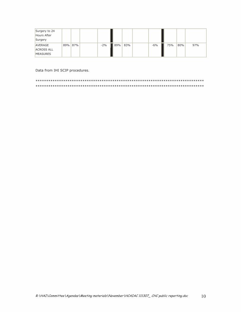
Surgery to 24
Hours After
Surgery
AVERAGE
ACROSS ALL
MEASURES
89% 87% -2% 89% 83% -6% 75% 80% 97%
Data from IHI SCIP procedures.
*******************************************************************************
*******************************************************************************
R:\HAI\Committee\Agendas\Meeting materials\November\HCAIAC 111307_ CHC public reporting.doc 10

Barriers / Challenges
Resources to get data
o 2.5 FTE’s to gather CMS data (abstraction, collection of core measures)
o inpatient only
o unknown when we will gather outpatient data
o How to educate public in a way that’s understandable to broad audience
one size doesn’t fit all
Infection Control department
o 1.5 FTE’s
o 80% of time spent gathering data
o No automated system – everything is manual
o Lag time from submission of data to CMS reports
o Lack of epidemiology software program with our current EMR system
CHC hospitals serve unique populations and treat varied conditions and illnesses
CHC hospitals offer different services, specialties and expertise
Government data may be two or three years old… doesn’t mean it’s wrong, but much
may be changed since it was reported
o Public can’t really compare apples to apples
Ideas / discussion ongoing:
Surveillance cultures –
o Which patients?
o Which sites?
o Who pays?
o Hospital wide versus unit specific versus procedure specific?
Report specific organisms –
o MRSA
o VRE
o C. Difficile
o Others
Public reaction to external reporting:
Media (attached)
Written letters
Inquiries to IC nurses and other caregivers
Physician offices
R:\HAI\Committee\Agendas\Meeting materials\November\HCAIAC 111307_ CHC public reporting.doc 11

R:\HAI\Committee\Agendas\Meeting materials\November\HCAIAC 111307_ CHC public reporting.doc 12
Future state:
Develop quality portals under CHC quality reporting on website
o www.cascadehealthcare.org
o St. Charles Medical Center – Bend (SCMC-B)
o St. Charles Medical Center – Redmond (SCMC-R)
o Pioneer Memorial Hospital – Prineville (PMH)
o Mountain View Hospital – Madras (MVH)
January / February 2008
o Initiate public launch to actively promote the new sites through the media
and our own marketing tools, including FOCUS magazine