xplorr - Фонд промышленных...
TRANSCRIPT
0901592
HORIBAJOBIN YVON
у а п с П ? а т а г ^ / 7 е А ^
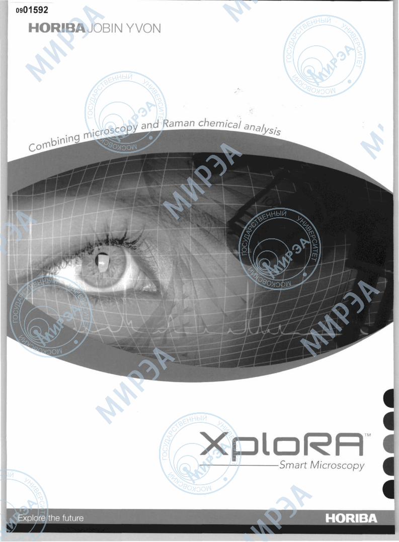
XploRR TM
Smart Microscopy
HORIBA
With more than three decades of experience in the field of Raman spectroscopy, HORIBA Jobin Yvon introduces a revolutionary and easy to use Raman microscope. The new XploRA™ completes the LabRAM family bringing routine Raman microscopy to your lab.
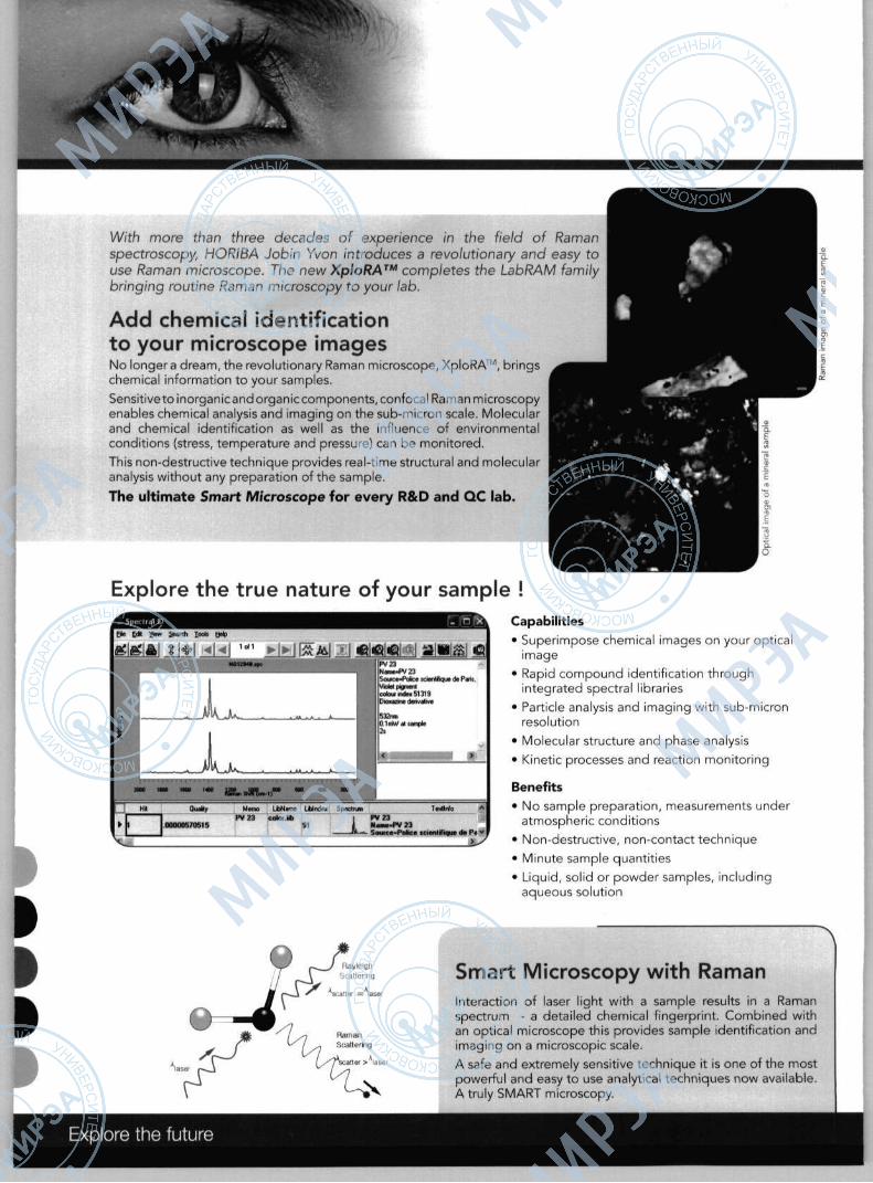
Add chemical identification to your microscope images No longer a dream, the revolutionary Raman microscope, XploRA™, brings chemical information to your samples.
Sensitive to inorganic and organic components, confocal Raman microscopy enables chemical analysis and imaging on the sub-micron scale. Molecular and chemical identification as well as the influence of environmental conditions (stress, temperature and pressure) can be monitored.
This non-destructive technique provides real-time structural and molecular analysis without any preparation of the sample.
The ul t imate Smart Microscope for every R & D and Q C lab.
Explore the true nature of your sample !
£ Ml C i P S a i i s «3 PV23 N«i»JV23 SourccPobcc KMntfiqut (N Pens. V»oW допет cotowndwSniS Окяшгшк deftvative
EL-к Quttty
0 0 0 0 0 5 / 0 5 1 5
Memo P V 2 3
LtbN*ne ЬЫгккх Spectrum TtxHnfo **• c o i n lib 1 PV 23
91 ) N M M - P V 23 Л.«- Souica»Pofcce tcienlriique de Pa v
V _ _xs
Capabilities • Superimpose chemical images on your optical
image • Rapid compound identification through
integrated spectral libraries • Particle analysis and imaging with sub-micron
resolution • Molecular structure and phase analysis • Kinetic processes and reaction monitoring
Benefits • No sample preparation, measurements under
atmospheric conditions • Non-destructive, non-contact technique • Minute sample quantities • Liquid, solid or powder samples, including
aqueous solution
a t t e r > laser
Smart Microscopy with Raman Interaction of laser light with a sample results in a Raman spectrum - a detailed chemical fingerprint. Combined with an optical microscope this provides sample identification and imaging on a microscopic scale. A safe and extremely sensitive technique it is one of the most powerful and easy to use analytical techniques now available. A truly SMART microscopy.
Explore the future

FEATURES Get to full speed at once with LabSpec+ The XploRA™ comes with LabSpec+, an intuitive interface based on the award-winning LabSpec software suite. It features a guided operation wizard (GO!) with on-screen hints taking you through the analysis, to create perfect conditions - no fuss, no guess work - just simple analysis every time. User defined templates or "analysis recipes" can be created and recalled at the touch of a button for those routine analyses or experiments.
To make your analysis faster and easier, LabSpec+ features: • Analysis recipes - pre-defined and custom templates for common samples (solids, liquids, films, etc) • AutoCAL - auto-calibration and self-validation function for reliable and validated results everytime • SPECTRAL database - for fast chemical identification (optional)
Technical specifications Spatial resolution
Spectral resolution
Spectral range
Laser excitation (up to three internal lasers)
Spectrograph
Detector
Laser power control
Peripherals • Automated XYZ mapping with autofocus • Light microscopy - dark f ield, phase contrast, DIC • Reflected/transmitted light illumination • Heating/cooling stages • Multiwell plates for high-throughput screening
Safety and compliance
= laser enclosure option
21CFR11 compliant • IQ/OQ/PQ available •
рДРЩИ]
<1 um
1.8cnr7pixel @532nm*; 1.1 crrvVpixei @785nm* •Automated selection of spectral resolution and coverage through grating selection
< 150 cm-1 and up to CCD detection limit
532nm, 638nm, 785nm
High throughput integrated spectrograph
1024 pixels, V'chip, high sensitivity air cooled CCD
100%, 50%, 25%, 10%, 1%, 0 . 1 %
• Raman polarisation optics • Macro measurement accessory • Cuvette holder for liquid measurements • Fiber-coupled probes for remote analysis • Chemometric package for spectral imaging
Environment • Weight: 35 kg (77 lbs) • Operating temperature: 15-28°C
410mm/ 16.1" 450 mm /17.7"

APPLICATIONS
PHARMACEUTICS Identification and distribution of a tablet's API (Active Pharmaceutical Ingredient) and excipients can be studied by Raman Imaging. In this image, a tablet has been mapped to determine component particle size and distribution, crucial information for understanding product performance and quality. The XploRA™ is also an invaluable addition to your analytical toolbox when it comes to demonstrating patent infringement or the detection of counterfeit products.
FORENSICS Narcotics, polymers, explosives, pigments and biological residues generally exhibit rich Raman spectra. Confocal Raman microscopy provides an ideal method for the study of these materials. In the image on the left, the Raman mapped image enables the characterization of the structure and composition of plastic packaging. The multi-layer film consists of more than 10 layers ranging in thickness from 1 to 50 urn; all the layers are easily analyzed with the XploRA™. Sample courtesy of Sabine Bebelman, Universite Catnolique de Louvain, Belgium
BIOLOGY Raman spectroscopy is extremely sensitive to subtle changes within bio-molecules. For example, disease diagnosis, micro-biology, drug interactions, tissue healing, dermatology and cosmetics can all benefit from the information-rich analysis of Raman. The XploRA's sensitivity even allows single cell bacteria to be identified and classified in a straight forward manner. In this image, single bacteria have been imaged according to their carbon isotope content (12C and 13C). Sample courtesy of Or Wei Huang, University of Sheffield, UK
Ш GEOLOGY/GEMMOLOGY Confocal Raman spectroscopy allows mineral species to be identified and their distribution mapped with high spatial resolution. In addit ion, fluid inclusions can be analysed in situ. Here, a 20 urn inclusion has been analysed without any sample preparation. The high spatial discrimination of the XploRA™ allows both the liquid region and 10 urn gas bubble (C02 , N2 and CH4 gases) to be distinguished with ease. Sample courtesy of Dr Gauthier, UFR des Sciences de la Terre, USTL, France
i i (
ARCHEOLOGY/ART In art conservation and archeology Raman spectroscopy is used for the study of pigments, ceramics, glasses and corrosion products, providing information on the origin, the craft technique, conservation, history and authenticity of an object. Shown on the left, individual pigment particles in a fragment of mixed paint are identified using Raman. For example, the red pigment is shown to be Haematite, an iron oxide pigment. Sample courtesy of Po/onca Ropret, Restauration Center, Slovenia
HORIBA

SYSTEM
Compact multiple laser design
• Up to 3 integrated lasers
• Automated laser switching
• Direct laser coupling (no fibers)
Rugged and compact frame • Very few moving parts • High mechanical stability • Class 1 laser
enclosure option
High sensitivity Raman spectrometer
High optical throughput for enhanced sensitivity Automated 4-position grating turret for optimal resolution Optical fiber port for remote sampling
True confocal microscope • High spatial resolution • Automated mapping stages • Full microscope options
Enter a New Dimension in Microscopy Performance and Simplicity
The XploRA™ turns a new page in microscopy. With an intuitive interface and full automation, Raman analysis has never been easier. Chemical identification and chemical imaging can now be performed on solid or liquid samples at the touch of a button. Whether for routine sample identification, quantitative analysis or chemical imaging, the XploRA™ combines performance and simplicity in a cost effective system.
Easily Portable
The light, compact design of the XploRA™ makes it easy to transport from lab to lab or for on-site analysis at archeological sites, crime scenes or in a mobile laboratory.
Rugged by Design The XploRA™ is designed with long lifetime optics and few moving parts for unsurpassed stability and zero maintenance. Fast start up and self-validation protocols yield reliable measurements, t ime after t ime.
Intui t ive O p e r a t i o n
Designed with ease-of-use in mind, the XploRA's intuitive software and hardware interface allows you to quickly get results, even with little or no experience in Raman spectroscopy. The XploRA™ provides simple and precise sample analysis every time.
The X p l o R A ™ features :
• Full microscope observation capabilities with point-and-shoot chemical analysis
• Guided Operation (GO!) wizard and software "assistant" for easy start up
• Fast chemical identification through automated spectral library searches
• True confocal microscopy for high resolution 3D chemical imaging and detailed particle analysis
• Plug-and-play operation
• Class I laser safety enclosure option
www.smartmicroscopy.com

HORIBA JOBIN YVON Innovative Solutions
The HORIBA Group provides superior technologies, unique products, and high quality services in the analytical
and measurement fields. HORIBA Jobin Yvon is part of the HORIBA Group.
X-RAY FLUORESCENCE DETECTORS
SPECTROFLUOROMETRY
f Г RAMAN SPECTROSCOPY
ATOMIC EMISSION SPECTROSCOPY
FORENSICS
Elemental Analysis X-Ray Fluorescence
• ICP - Spark - SDL Spectrometry Elemental Analyzers
www.iobinyvon.com Particle Size
Analysis
Emerging Business Optical Characterization of Thin Films
• Forensics
Optical Components • Gratings and OEM
Spectrometers
• VUV Instrumentation • Modular Optical
Spectroscopy • Detectors
THIN FILM PROCES CONTROL
I ELLIPSOMETRY
PARTICLE SIZE ANALYSERS
OPTICAL SPECTROSCOPY
• The content of this catalogue is subject to change without prior notice
• It is forbidden to copy from the contents of this catalogue in part or in full
• Please read the instruction/operation manual before using these products
This instrument complies with 21CFR 1040.10 and IEC 60825-1 (08/2001)
C€ CLASS 1 LASER PRODUCT
X - (400 nm - 850 nm) P<150 mW VISIBLE AND INVISIBLE LASER RADIATION
AVOID EXPOSURE TO BEAM CLASS 3B LASER PRODUCT
* Laser safety classifications depend on individual systems and options
HORIBAJOBIN YVON France: HORIBA Jobin Yvon SAS, 231 rue de Lille, 59650 Villeneuve d'Ascq Tel +33 (0)3 20 59 18 00 • Fax +33 (0)3 20 59 18 08 • ramanejobinyvon.fr • www.jobinyvon.fr
USA: HORIBA Jobin Yvon Inc., 3880 Park Avenue, Edison, NJ 08820-3012 Toll-free : +1-866-jobinyvon Tel +1-732-494-8660 • Fax +1-732-549-2571 • [email protected] • www.jobinyvon.com
Japan: HORIBA Ltd., JY Optical Sales Dept, 1-7-8 Higashi-kanda, Chiyoda-ku, Tokyo 101-0031 Tel +81 (0)3 3861 8231 • Fax +81 (0)3 3861 8259 • raman®horiba.com
Germany: +49(0)625164 75-0 Italy : +39 02 57603050 UK : +44 (0)20 8204 8142 China: +86(0)10 8567 9966 Other countries :+33 (0)1 64 54 13 00
Explore the future HORIBA

ГУРосНИИИТиАП Ассоциация МВТК
Федеральный информационный фонд отечественных и иностранных каталогов на промышленную продукцию
Каталог был представлен на выставке «RUSnanotech - 2008»
(Международный форум по НАНОТЕХНОЛОГИЯМ)
Каталог включен в базу данных «Федерального информационного фонда
отечественных и иностранных каталогов на промышленную продукцию»
Россия, 105679, Москва, Измайловское шоссе, 44, Тел./факс (095)366-5200, 366-7008, 365-5445. e-mail:; [email protected],
www.flpk.ru
Электронная копня издания изготовлена с целью её включения в базы данных Федерального информационного фонда отечественных и иностранных каталогов на промышленную продукцию, которые
формируются в соответствии с Постановлением Правительства РФ от 24 июля 1997 г. № 950 и Постановлением Правительства РФ от 31 декабря 1999
г. № 2172-р и зарегистрированы Комитетом по политике информатизации при Президенте РФ под №№ 39-50.
2009 год