zbtb7a mutations in acute myeloid leukaemia with t(8;21 ... · on average, pkm and slc2a3 also...
TRANSCRIPT

ARTICLE
Received 20 Oct 2015 | Accepted 26 Apr 2016 | Published 2 Jun 2016
ZBTB7A mutations in acute myeloid leukaemiawith t(8;21) translocationLuise Hartmann1,2,3,4, Sayantanee Dutta1,2,3,4, Sabrina Opatz1,2,3,4, Sebastian Vosberg1,2,3,4, Katrin Reiter1,2,3,4,
Georg Leubolt1,2,3,4, Klaus H. Metzeler1,2,3,4, Tobias Herold1,2,3,4, Stefanos A. Bamopoulos1, Kathrin Braundl1,2,3,4,
Evelyn Zellmeier1, Bianka Ksienzyk1, Nikola P. Konstandin1, Stephanie Schneider1, Karl-Peter Hopfner5,
Alexander Graf6, Stefan Krebs6, Helmut Blum3,4,6, Jan Moritz Middeke3,4,7, Friedrich Stolzel3,4,7,
Christian Thiede3,4,7, Stephan Wolf4, Stefan K. Bohlander8, Caroline Preiss9, Linping Chen-Wichmann9,
Christian Wichmann9, Maria Cristina Sauerland10, Thomas Buchner11, Wolfgang E. Berdel11,
Bernhard J. Wormann12, Jan Braess13, Wolfgang Hiddemann1,2,3,4, Karsten Spiekermann1,2,3,4
& Philipp A. Greif1,2,3,4
The t(8;21) translocation is one of the most frequent cytogenetic abnormalities in acute
myeloid leukaemia (AML) and results in the RUNX1/RUNX1T1 rearrangement. Despite the
causative role of the RUNX1/RUNX1T1 fusion gene in leukaemia initiation, additional genetic
lesions are required for disease development. Here we identify recurring ZBTB7A mutations in
23% (13/56) of AML t(8;21) patients, including missense and truncating mutations resulting
in alteration or loss of the C-terminal zinc-finger domain of ZBTB7A. The transcription factor
ZBTB7A is important for haematopoietic lineage fate decisions and for regulation of
glycolysis. On a functional level, we show that ZBTB7A mutations disrupt the transcriptional
repressor potential and the anti-proliferative effect of ZBTB7A. The specific association of
ZBTB7A mutations with t(8;21) rearranged AML points towards leukaemogenic cooperativity
between mutant ZBTB7A and the RUNX1/RUNX1T1 fusion.
DOI: 10.1038/ncomms11733 OPEN
1 Department of Internal Medicine 3, University Hospital, Ludwig-Maximilians-Universitat (LMU) Munchen, 81377 Munchen, Germany. 2 Clinical CooperativeGroup Leukemia, Helmholtz Zentrum Munchen, German Research Center for Environmental Health, 81377 Munchen, Germany. 3 German Cancer Consortium(DKTK), 69121 Heidelberg, Germany. 4 German Cancer Research Center (DKFZ), 69121 Heidelberg, Germany. 5 Department of Biochemistry, Ludwig-Maximilians-Universitat (LMU) Munchen, 81377 Munchen, Germany. 6 Laboratory for Functional Genome Analysis (LAFUGA), Gene Center, Ludwig-Maximilians-Universitat (LMU) Munchen, 81377 Munchen, Germany. 7 Medizinische Klinik und Poliklinik I, Universitatsklinikum Dresden, 01307 Dresden,Germany. 8 Department of Molecular Medicine and Pathology, The University of Auckland, Auckland 1142, New Zealand. 9 Department of TransfusionMedicine, Cell Therapeutics and Hemostasis, University Hospital, Ludwig-Maximilians-Universitat (LMU) Munchen, 81377 Munchen, Germany. 10 Institute ofBiostatistics and Clinical Research, University of Munster, 48149 Munster, Germany. 11 Department of Medicine A, Hematology, Oncology and Pneumology,University of Munster, 48149 Munster, Germany. 12 Department of Hematology, Oncology and Tumor Immunology, Charite University Medicine, CampusVirchow, 13353 Berlin, Germany. 13 Oncology and Hematology, St. John-of-God Hospital, 93049 Regensburg, Germany. Correspondence and requests formaterials should be addressed to P.A.G. (email: [email protected] and [email protected]).
NATURE COMMUNICATIONS | 7:11733 | DOI: 10.1038/ncomms11733 | www.nature.com/naturecommunications 1

Block of myeloid differentiation is one of the hallmarks ofacute myeloid leukaemia (AML). First insights into thiskey mechanism were gained by the discovery of the
t(8;21)(q22;q22) translocation, which was the first balancedtranslocation described in a tumour and results in the RUNX1/RUNX1T1 fusion gene (also known as AML1/ETO)1,2. TheRUNX1/RUNX1T1 rearrangement is one of the most frequentchromosomal aberrations in AML and defines an importantclinical entity with favourable prognosis according to the WorldHealth Organization classification3. The RUNX1/RUNX1T1fusion protein disrupts the core-binding factor complex, andthereby blocks myeloid differentiation. However, in vivo modelsindicate the requirement of additional lesions, such as of KIT orFLT3 mutations, for leukaemogenesis as the RUNX1/RUNX1T1fusion gene alone is not sufficient to induce leukaemia4–8. In thepresent study, we set out to identify additional mutations in AMLt(8;21) and discovered frequent mutations of ZBTB7A—encodinga transcription factor important for the regulation ofhaematopoietic development9 and tumour metabolism10. It isvery likely that ZBTB7A mutations are one of the importantmissing links in RUNX1/RUNX1T1-driven leukaemogenesis.
ResultsZBTB7A is frequently mutated in AML t(8;21). To identifyadditional cooperating mutations, we performed exomesequencing of matched diagnostic and remission samples fromtwo AML patients with t(8;21) translocation and detected 11 and12 somatic variants, respectively (Supplementary Table 1).
ZBTB7A was the only mutated gene identified in both patients.ZBTB7A (also known as LRF, Pokemon and FBI-1) is a memberof the POZ/BTB and Kruppel (POK) transcription factor family9,which is characterized by an N-terminal POZ/BTBprotein–protein interaction domain and C-terminal C2H2 zincfingers11. The first patient carried a homozygous missensemutation resulting in the amino-acid change R402H(NM_015898:exon2:c.1205G4A:p.R402H) affecting the highlyconserved zinc-finger domain, while a heterozygous frameshiftinsertion (NM_015898:exon2:c.522dupC:p.A175fs) resulting inloss of the zinc-finger domain was identified in the secondpatient. Both mutations were validated by Sanger sequencing(Supplementary Fig. 1; Supplementary Table 2). Using targetedamplicon sequencing of ZBTB7A and 45 leukaemia relevantgenes, we screened 56 diagnostic AML t(8;21) samples, includingone of the two samples analysed by exome sequencing (UPN 1),whereas for the other one (UPN 2) availability of material wasinsufficient. ZBTB7A mutations were identified in 13 of 56patients (23%; Fig. 1a,b; Supplementary Table 3). Patientcharacteristics are summarized in Supplementary Table 4. Tworecurring mutational hotspots (A175fs and R402) in exon 2 wereidentified altering or resulting in loss of the zinc-finger domain(Fig. 1a). It was previously shown that the zinc-finger domain ofZBTB7A is essential for DNA binding12. Structural modellingrevealed that arginine 402 binds into the major groove of theDNA double helix and likely contributes to the affinity orsequence specificity of the DNA interaction of the zinc-fingerdomain of ZBTB7A (Fig. 2a). We confirmed that both ZBTB7Amutants A175fs and R402H fail to bind DNA (Fig. 2b,c).
a
b
A175fsA175fs
A175fs
S50L S50LL85R
P168fsF79L
BTB
RUNX1/RUNX1T1ZBTB7A
n=56 MutatedWild typeMissing information
2 Mutations
23%23%21%20%16%14%14%12%12%9%9%7%7%5%5%4%4%4%4%4%14%
61%7%
ASXL2NRAS
KITFLT3
ASXL1RAD21
TET2SMC3
DNMT3AJAK2CBL
FAT1SMC1A
WT1KRAS
PTPN11IDH2
GATA2JAK3
del(9q)+8
others
R49H
A175fs
A175fsA175fs
A175fsG395C
R377X
D364XR458fs
NLS
ZfZfZf
399131241 477 584
R402CR402CR402H
Figure 1 | ZBTB7A mutations in AML t(8;21). (a) ZBTB7A protein (NP_056982.1) and identified mutations (red¼ truncating; black¼missense)
illustrated using IBS software31. Amino-acid positions are indicated below the graph. BTB, BR-C ttk and bab; NLS, nuclear localization sequence; Zf, zinc
finger. (b) Mutational landscape of 56 diagnostic AML samples with t(8;21) translocation. Each column represents one patient, each line one of the
analysed genes or cytogenetic markers.
ARTICLE NATURE COMMUNICATIONS | DOI: 10.1038/ncomms11733
2 NATURE COMMUNICATIONS | 7:11733 | DOI: 10.1038/ncomms11733 | www.nature.com/naturecommunications

Variant allele frequency ranged from 5.4 to 76.2% (cut-off 2%)and 4 of 13 patients (31%) harboured two mutations of ZBTB7A.Fourteen of 17 mutations (82%) were validated by Sangersequencing (Supplementary Fig. 1). Somatic status was confirmedin a total of three patients with available remission samples.Thirty-two additional samples of t(8;21)-positive AML withinadequate sample availability for gene panel sequencing wereanalysed by Sanger sequencing of exon 2 (encoding amino acids1–421) resulting in the identification of two ZBTB7A mutations(2/32; 6%). This lower mutation frequency might be due to thelower sensitivity of Sanger sequencing and incomplete coverage ofthe coding exons of ZBTB7A (we were not able toreliably amplify exon 3 encoding amino acids 422–584).To evaluate the consequences of truncating ZBTB7A mutationson the protein level, we performed western blot analysis forone patient with available material and detected a shorter
form of the ZBTB7A protein resulting from the R377X mutation(Supplementary Fig. 2).
Recently, frequent ASXL2 mutations were identified in t(8;21)AML13. In our cohort, ZBTB7A and ASXL2 mutations occurredat similar frequencies (Fig. 1b) and 5 of 13 patients carriedmutations in both genes; however, there was no significantassociation of mutated ZBTB7A and mutations in ASXL2(Fisher’s exact test, P¼ 0.12) or any other recurrently mutatedgene. Alterations of ASXL1 were mutually exclusive withgenetic lesions of ZBTB7A suggesting alternative routes ofleukaemogenesis. Similarly, mutations of ZBTB7A and KITwere exclusive in all, but one patient. In the exome data of 22patients with inversion inv(16) (another rearrangementdisrupting the core-binding factor complex in AML), we founda single ZBTB7A mutation (A211V). Of note, we did not findany ZBTB7A mutations by exome sequencing of 50 patientswith cytogenetically normal AML (CN-AML) or 14 AML patientswith chromosomal aberrations other than t(8;21) or inv(16).These results point towards a specific association betweenZBTB7A alterations and the RUNX1/RUNX1T1 fusion.
Mutations disrupt the anti-proliferative function of ZBTB7A.To assess the functional consequences of the identified ZBTB7Amutations, we performed luciferase reporter gene assays. It isknown that ZBTB7A represses the expression of ARF (alternateopen reading frame of CDKN2A)14. In contrast to wild-typeZBTB7A, the R402H, R402C, A175fs or R377X mutants failed torepress a luciferase reporter containing ZBTB7A-bindingelements derived from the ARF promoter (Fig. 3a). Expressionof ZBTB7A constructs was confirmed by western blot (Fig. 3b).
In light of recent reports about the negative regulation ofglycolysis by ZBTB7A10, we assessed the expression of glycolyticgenes (SLC2A3, PFKP and PKM) in the RNA-sequencing datafrom our AML t(8;21) patients (Supplementary Fig. 3).In ZBTB7A-mutated patients (n¼ 5), we found a significantlyhigher expression of PFKP (Student’s t-test, P¼ 0.03) comparedwith patients without any detectable ZBTB7A mutation (n¼ 11).On average, PKM and SLC2A3 also showed higher expressionlevels in patients with ZBTB7A mutations, but did not reachstatistical significance (Student’s t-test, P¼ 0.17 and P¼ 0.54,respectively). In the latter case, the difference in the mean valuescan be attributed mainly to an outlier in the ZBTB7A-mutatedgroup with very high SLC2A3 expression. Expression levels ofZBTB7A were similar in both the patient groups, compatible withinactivation of ZBTB7A on the genetic level rather than on thetranscriptional level.
The C-terminal part of ZBTB7A is important for nuclearlocalization15. Because some mutations result in loss of theC-terminal zinc-finger domain and nuclear localization signal, weevaluated the cellular localization of mutant ZBTB7A. Whereaswild-type ZBTB7A was detected in the nucleus, immuno-fluorescence staining of the A175fs and R377X mutants showedan altered cytoplasmic localization (Fig. 3c). In contrast,mutants R402H and R402C exhibited a variable cellularlocalization with cytoplasmic protein detectable only in a minorsubset of cells (Supplementary Fig. 4a,b). Amino-acid substitutionsof R402 showed a smaller increase in cytoplasmic protein fractioncompared with truncation mutants as analysed by western blot(Supplementary Fig. 4c). Ultimately, the observed effect ofmutations on ZBTB7A localization remains to be confirmed inappropriate primary patient material, which was not available inour study.
In the t(8;21) translocation-positive AML cell line Kasumi-1,retroviral expression of wild-type ZBTB7A inhibited cell growth,whereas this anti-proliferative effect was not observed upon
R402
ZBTB7A WT A175fs R402H
POK WT + – Input + – Input + – InputPOK mut – + – + – +
95
72
[kDa]
a
b
26
c
ZBTB7A5′– –3′3′ – –5′
5′– –3′3′– –5′
POK WT
POK mut
GGTTAAAAGACCCCTCCCCGAATTCGGATCCCAATTTTCTGGGGAGGGGCTTAAGCCTAG
GGTTAAAATTTTTCTCCCCGAATTCGGATCCCAATTTTAAAAAGAGGGGCTTAAGCCTAG
Figure 2 | Impact of ZBTB7A mutations on DNA binding. (a) Model
for the C-terminal zinc-finger domain of ZBTB7A comprising residues
382–488. The model is depicted as yellow ribbon with highlighted
secondary structure. Zinc ions are shown as grey spheres. DNA is shown in
brown with a grey molecular surface. R402 (purple) binds into the major
groove and likely contributes to the affinity or sequence specificity of the
DNA interaction of the zinc-finger domain. (b) Biotinylated oligonucleotides
containing the ZBTB7A (alias: Pokemon) consensus binding motif
(POK WT) or a mutant thereof (POK mut)14 used in DNA pull-down
experiments. Spheres illustrate streptavidin-coated beads. (c) DNA
pull-down using protein lysates from HEK293T cells expressing wild-type
or mutant ZBTB7A. Western blot analysis shows that A175fs and
R402H fail to bind oligonutides with a ZBTB7A-binding site (POK WT).
Oligonucleotides with a mutated binding site (POK mut) were used as
negative control. Input lanes were loaded with 10% of the protein lysate
used for each binding reaction.
NATURE COMMUNICATIONS | DOI: 10.1038/ncomms11733 ARTICLE
NATURE COMMUNICATIONS | 7:11733 | DOI: 10.1038/ncomms11733 | www.nature.com/naturecommunications 3
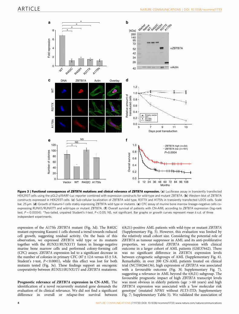
expression of the A175fs ZBTB7A mutant (Fig. 3d). The R402Cmutant expressing Kasumi-1 cells showed a trend towards reducedcell growth, suggesting residual activity. On the basis of thisobservation, we expressed ZBTB7A wild type or its mutantstogether with the RUNX1/RUNX1T1 fusion in lineage-negativemurine bone marrow cells and performed colony-forming cell(CFC) assays. ZBTB7A expression led to a significant decrease inthe number of colonies in primary CFC (87±12.6 versus 45±5.8,Student’s t-test, Po0.0001), while this effect was lost for bothmutants tested (Fig. 3e). These findings support an oncogeniccooperativity between RUNX1/RUNX1T1 and ZBTB7A mutations.
Prognostic relevance of ZBTB7A expression in CN-AML. Theidentification of a novel recurrently mutated gene demands theevaluation of its clinical relevance. We did not find a significantdifference in overall or relapse-free survival between
t(8;21)-positive AML patients with wild-type or mutant ZBTB7A(Supplementary Fig. 5). However, this evaluation was limited bythe relatively small cohort size. Considering the potential role ofZBTB7A as tumour suppressor in AML and its anti-proliferativeproperties, we correlated ZBTB7A expression with clinicaloutcome in a larger cohort of AML patients (GSE37642). Therewas no significant difference in ZBTB7A expression levelsbetween cytogenetic subgroups of AML (Supplementary Fig. 6).Remarkably, in over 200 CN-AML patients treated on clinicaltrial (NCT00266136), high expression of ZBTB7A was associatedwith a favourable outcome (Fig. 3f; Supplementary Fig. 7),suggesting a relevance in AML beyond the t(8;21) subgroup. Thefavourable prognostic impact of high ZBTB7A transcript levelswas most obvious in elderly patients (age 460 years) and highZBTB7A expression was associated with a ‘low molecular riskgenotype’ (mutated NPM1 without FLT3-ITD; SupplementaryFig. 7; Supplementary Table 5). We validated the association of
a b
dc
fe- ZBTB7A high (n=54)
ZBTB7A low (n=161)
P=0.0004
*NS
NS
**
**
αZBTB7A
260
Native
R402C
R402H
R377X
WT
A175f
s
Mar
ker
1409572
52
42
34
26
[kDa]
αActin
DNA ZBTB7A Actin Overlay
WT
R37
7XA
175f
s
42
1.2
8
6
4
2
0
WT
Fol
d re
pres
sion
R402C
R402H
R377X
A175f
s
1.0
1.0
0.8
0.8
0.6
0.6
0.4
0.4
0.2
0.2
0.0
0.00 12 24 36 48 60
Months
Ove
rall
surv
ival
72 84 96 108
120
100
Num
ber
of c
olon
ies
80
60
40
20
0Vector WT R402C A175fs
5 7 9 11 13Days post transduction
Rel
ativ
e am
ount
of
tran
sduc
ed c
ells
-
0 25μm
0 25μm
0 25μm
Empty vectorWTA175fsR402C
Figure 3 | Functional consequences of ZBTB7A mutations and clinical relevance of ZBTB7A expression. (a) Luciferase assay in transiently transfected
HEK293T cells using the pGL2-p19ARF-Luc reporter combined with expression constructs for wild-type and mutant ZBTB7A. (b) Western blot of ZBTB7A
constructs expressed in HEK293T cells. (c) Sub-cellular localization of ZBTB7A wild type, R377X and A175fs in transiently transfected U2OS cells. Scale
bar, 25mm. (d) Growth of Kasumi-1 cells stably expressing ZBTB7A wild type or mutants. (e) CFC assay of murine bone marrow lineage-negative cells co-
expressing RUNX1/RUNX1T1 and wild-type or mutant ZBTB7A. (f) Overall survival of patients with CN-AML according to ZBTB7A expression (log-rank
test, P¼0.0004). *Two-tailed, unpaired Student’s t-test, Po0.05; NS, not significant. Bar graphs or growth curves represent mean±s.d. of three
independent experiments.
ARTICLE NATURE COMMUNICATIONS | DOI: 10.1038/ncomms11733
4 NATURE COMMUNICATIONS | 7:11733 | DOI: 10.1038/ncomms11733 | www.nature.com/naturecommunications

high ZBTB7A expression with favourable outcome in an inde-pendent CN-AML patient cohort16,17 (Supplementary Fig. 8).
DiscussionIn summary, we have identified ZBTB7A as one of the mostfrequently mutated genes in t(8;21)-positive AML. Consistentwith our findings, ZBTB7A mutations in 3 of 20 (15%) AMLt(8;21) patients and 1 of 395 AML inv(16) patients werereported18 during the revision of the present manuscript. Ourfunctional analyses indicate that ZBTB7A mutations result in lossof function, due to alteration or loss of the zinc-finger motives.Beyond DNA binding, the zinc-finger domain of ZBTB7A is alsoknown to interact with TP53 and BCL6 (ref. 9). Thus, multiplepathways might be influenced by alteration or loss of the ZBTB7Azinc-finger domain. The N-terminal missense mutations in theBTB domain may result in failure of co-repressor recruitment.Considering that 4 of 13 of patients had more than one ZBTB7Amutation, our finding that overexpression of wild-type ZBTB7Aleads to reduced proliferation of Kasumi-1 cells and a decreasednumber of CFCs of murine bone marrow cells, we suggest thatZBTB7A acts as a tumour suppressor in t(8;21)-positive AML.Initial studies characterized ZBTB7A as proto-oncogene invarious tissues14,19. For example, Maeda et al. demonstratedthat transgenic mice with Zbtb7a overexpression in the immatureT- and B-lymphoid lineage develop precursor T-cell lymphoma/leukaemia14. In contrast, it was more recently shown thatZBTB7A can also act as a tumour suppressor. Overexpressionof Zbtb7a in murine prostate epithelium did not result inneoplastic transformation; unexpectedly, Zbtb7a inactivation leadto the acceleration of Pten-driven prostate tumorigenesis20.Recently, somatic zinc-finger mutations of ZBTB7A were foundat low frequencies (o5%) in a variety of solid cancers suggestinga common mechanism across tumour entities21. In fact, thede-repression of glycolytic genes upon deletion or mutation ofZBTB7A10,21 might underlie the loss of anti-proliferativeproperties that we observed for ZBTB7A mutants A175fsand R402C in the present study. Any inactivating alteration ofZBTB7A will likely increase glycolysis, and, thus, helps thetumour cells to produce more energy. Besides tumourmetabolism, it is known that ZBTB7A also plays an importantrole in haematopoietic lineage fate decisions9. Duringlymphopoiesis ZBTB7A regulates B-cell development22, whereasin the myeloid lineage it is essential for erythroiddifferentiation23. Thus, ZBTB7A mutations may contribute tothe block of differentiation in AML t(8;21).
The favourable prognostic relevance of high ZBTB7Aexpression in CN-AML, which accounts for half of all AMLpatients, may point towards a more general tumour suppressorrole of ZBTB7A in myeloid leukaemia. In particular, theanti-proliferative properties of ZBTB7A may slow down diseaseprogression. High ZBTB7A expression as a favourable prognosticmarker has been reported also in colorectal cancer10, consistentwith a clinicobiological role of ZBTB7A across malignancies ofmultiple tissue origins. Given that somatic mutations of ZBTB7Aseem to be absent or rare in CN-AML, other mechanisms,including epigenetic changes or alterations of upstreamregulators, may lead to inactivation or downregulation ofZBTB7A.
Our discovery of frequent ZBTB7A mutations in AML witht(8;21) translocation, one of the most common translocationsin AML and the first balanced translocation identified inleukaemia1, demonstrates that the mutational landscape of AMLis still not fully understood. Further studies will be required tounravel the mechanism underlying leukaemogenic cooperativitybetween mutated ZBTB7A and the RUNX1/RUNX1T1 fusion gene.
MethodsPatients. AML samples were collected within the German Cancer Consortium(DKTK) at the partner sites Munich and Dresden. Patients were treated accordingto the protocols of Acute Myeloid Leukemia Cooperative Group (AMLCG) orStudy Alliance Leukemia (SAL) multicentre clinical trials. Study protocols wereapproved by the Institutional Review Boards of the participating centres. Informedconsent was received in accordance with the Declaration of Helsinki.
Sequencing. Exome sequencing (mean coverage: 87x; range 80–90x) was per-formed on a HiSeq 2000 Instrument (Illumina), using the SureSelect Human AllExon V5 kit (Agilent). Pretreatment blood or bone marrow specimens from 56AML patients with t(8;21) translocation were sequenced using Haloplex customamplicons (Agilent) and a HiSeq 1500 instrument (Illumina). Target sequenceincluded the entire open-reading frame of ZBTB7A in addition to 45 leukaemia-related genes or mutational hotspots (Supplementary Table 3). Variant calling wasperformed as described previously24. Sanger sequencing of PCR-amplified genomicDNA was carried out using a 3500xL Genetic Analyzer (Applied Biosystems).Primer sequences are provided in Supplementary Table 2. Sequencing of messengerRNA was performed using the TruSeq RNA Sample Preparation protocol, followedby sequencing on a HiSeq 2000 Instrument (Illumina). RNA sequence reads werealigned to the human genome (hg19) using STAR25 (version 2.4.1b). Reads pergene were counted using HTseq26 (version 0.6.1) with intersection-strict mode andnormalized for the total number of reads per sample.
Structural modelling. Suitable templates for the modelling were searched withHHPRED27, using the zinc-finger domain of ZBTB7A as input sequence. Thehighest scoring homologue, for which a structure of a DNA complex is available,was the Wilms tumour suppressor protein28 (PDB accession code 2J9P, E-value4.8E–29, P-value 1.3E–30). The model for ZBTB7A was generated on the basis of2J9P using MODELLER29. Importantly, 2J9P also contains an arginine at theequivalent position of ZBTB7A’s R402, allowing us to model the function of R402as major groove binder with confidence.
Plasmids. The pcDNA3.1-His-ZBTB7A expression construct was a gift fromTakahiro Maeda (Boston). ZBTB7A A175fs, R377X, R402C and R402H mutantplasmids were generated using the QuikChange II XL Site-Directed MutagenesisKit (Agilent) and confirmed by Sanger sequencing. ZBTB7A wild type and mutantswere subcloned into pMSCV-IRES-YFP (pMIY), using the In-Fusion HD cloningkit (Clontech) and EcoRI restriction sites. The pMSCV-IRES-GFP(pMIG)-RUNX1/RUNX1T1 plasmid was provided by Christian Buske (Ulm).
DNA pull-down. HEK293T cells (DSMZ no.: ACC 635) were transfected withpcDNA3.1 His-Xpress-ZBTB7A (wild type or mutant). After 24 h, protein wasextracted using lysis buffer (50 mM Tris HCl, pH 8.5, 150 mM NaCl, 1% TritonX-100, cOmplete Protease Inhibitor Cocktail). For each reaction, 20 ml proteinlysate was incubated in binding buffer (PBS supplemented with 150 mM NaClresulting in a total salt concentration of nearly 300 mM, 0.1% NP40, 1 mM ETDA)with 10 pM biotinylated double-stranded oligonucleotides that contain either theZBTB7A consensus binding motif (POK WT; 50-GGTTAAAAGACCCCTCCCCGAATTCGGATC-30) or a mutant thereof (POK mut; 50-GGTTAAAATTTTTCTCCCCGAATTCGGATC-30). After 1 h of incubation at 4 �C, 10ml streptavidin agarosebeads (Sigma Aldrich) was added to each reaction and incubated for 30 min at 4 �C.Beads were washed three times with binding buffer and resupended in 10 mlLaemmli buffer for subsequent western blot analysis. ZBTB7A protein was detectedusing an antibody against the Xpress tag (1:5,000 dilution, clone R910-25; LifeTechnologies) and secondary goat anti-mouse IgG-HRP (1:10,000 dilution, clonesc-2060; Santa Cruz). The uncropped western blot scan underlying Fig. 2c is shownin Supplementary Fig. 9.
Reporter gene assay. HEK293T cells (DSMZ no.: ACC 635) were co-transfectedwith pcDNA3.1-His-ZBTB7A (wild type or mutant), pGL2-p19ARF-Luc (gift fromTakahiro Maeda, Boston) as well as pRL-CMV (Renilla luciferase; Promega) usingLipofectamine 2000 (ThermoFischer). After 24 h, cells were lysed; Firefly andRenilla luciferase activity was measured with the dual-luciferase reporter assaysystem (Promega) according to the manufacturer’s instructions. Three independentexperiments were each performed in triplicates.
Western blot. HEK293T cells (DSMZ no.: ACC 635) were transfected usingLipofectamine 2000 (ThermoFischer) with pcDNA3.1-His-ZBTB7A (wild typeor mutant). After 24 h, protein was either extracted by multiple freeze–thaw cyclesin lysis buffer (600 mM KCl, 20 mM Tris-Cl pH 7.8, 20% Glycerol, cOmpleteProtease Inhibitor Cocktail) or using the Qproteome Nuclear Protein Kit (Qiagen)for the analysis of nuclear and cytoplasmic protein fractions. From archived patientbone marrow samples, protein was isolated using the AllPrep DNA/RNA/ProteinMini Kit (Qiagen) according to the manufacturer’s instructions. FollowingSDS–polyacrylamide gel electrophoresis and protein transfer to polyvinylidenedifluoride membrane (Hybond PTM, Amersham Pharmacia biotech),
NATURE COMMUNICATIONS | DOI: 10.1038/ncomms11733 ARTICLE
NATURE COMMUNICATIONS | 7:11733 | DOI: 10.1038/ncomms11733 | www.nature.com/naturecommunications 5

immunoblots were blocked with 5% nonfat dried milk, probed with anti-humanPokemon (ZBTB7A) purified antibody (1:5,000 dilution, clone: 13E9; eBioscience)and secondary anti-Armenian hamster IgG-HRP (1:10,000 dilution, clone: sc-2443;Santa Cruz). As loading control immunoblots were incubated with rabbit anti-actin(1:5,000 dilution, clone: sc-1616- R; Santa Cruz) and secondary goat anti-rabbitIgG-HRP (1:10,000 dilution, clone: sc-2030; Santa Cruz). For analysis of thenuclear and cytoplasmic ZBTB7A protein fractions, we used mouse anti-Xpresstag (1:5,000 dilution, clone R910-25, Life Technologies) and secondary goatanti-mouse IgG-HRP (1:10,000 dilution, clone: sc-2060; Santa Cruz). Mouseanti-GAPDH (1:10,000 dilution, clone: sc-32233; Santa Cruz) served as loadingcontrol for the cytoplasmic protein fraction. Proteins were detected with enhancedchemiluminescence (ECL, Amersham, GE Healthcare).
Immunofluorescence staining. U2OS human osteosarcoma cells (ATTC no.:HTB-96) were grown on coverslips and transiently transfected with pcDNA3.1-His-ZBTB7A wild type and mutant constructs using PoliFect (Qiagen) according tothe manufacturer’s guidelines. Cells were fixed 48 h post transfection usingPBS 2% formaldehyde (37% stock solution; Merck Schuchardt) for 10 min,permeabilized with PBS 0.5% Triton X-100 (Carl Roth) for 10 min and blocked for1 h with PBS 2% bovine serum albumin (Albumin Fraction V, AppliChem).Cells were then incubated with polyclonal rabbit His-probe (H-15) antibody(1:50 dilution; Santa Cruz) for 1 h. After extensive washing with PBS 0.1% Tween20 (Carl Roth), secondary antibody incubation was performed for 1 h with goatanti-rabbit IgG (Hþ L), F(ab0)2 fragment Alexa Fluor 594 conjugate (1:500dilution; Cell Signaling Technology). Counterstaining was performed usingNucBlue Reagent and ActinGreen 488 ReadyProbes Reagent (Life Technologies;2 drops per ml) at room temperature for 20 min. Coverslips were mounted usingfluorescence mounting medium (DAKO). Specimens were analysed using aconfocal fluorescence laser scanning system (TCS SP5 II; Leica). For imageacquisition and processing, the LAS AF Lite Software (Leica) was used.
Retroviral transduction. Retroviral transduction of Kasumi-1 cells (DSMZ no.:ACC 220) was accomplished as outlined previously30. In brief, HEK293T cells wereco-transfected with pMSCV-IRES-YFP (pMIY) vectors containing either wild-typeor mutant (A175fs, R402C) ZBTB7A and packaging plasmids. After 48 h, the cellculture supernatant was collected, sterile filtered and used for viral loading ofRetroNectin (Takara Clontech)-coated plates. A total of 3� 105 Kasumi-1 cellswere transduced per well. The percentage of YFP-positive cells was assessed on aFACSCalibur flow cytometer (BD Biosciences). Three independent experimentswere each performed in duplicates.
Colony-forming cell assay. For in vitro CFC assays, bone marrow cells werecollected from the femur and pelvic girdle of wild-type mice (C57BL/6X129/J).Lineage-negative haematopoietic progenitors were isolated using magneticseparation (MACS, murine lineage depletion kit, Miltenyi biotech). Retrovirallytransduced cells were sorted for GFP/YFP and were plated in 1% myeloid-condi-tioned methylcellulose containing Iscove’s modified Dulbecco medium-basedMethocult (Methocult M3434; StemCell Technologies) at a concentration of500 cells per ml. Single-cell suspensions of colonies were serially replated at thesame concentration until the exhaustion of cell growth. Three independentexperiments were each performed in duplicates.
Analysis of clinical and gene expression data. Clinical relevance of ZBTB7Amutations or expression levels was evaluated using the Kaplan–Meier method andthe log-rank test. Fisher’s exact test was used to compare categorical variables,while Wilcoxon Mann–Whitney U-test was applied for continuous variables. Allpatients included in this analysis were treated intensively with curative intentaccording to the AMLCG protocols. Gene expression profiling was performed on215 adult patients with cytogenetically normal AML, using Affymetrix HumanGenome (HG) U133A/B (n¼ 155) and HG U133Plus2.0 microarrays (n¼ 60).The RMA method was used for data normalization, and probe set summarizationutilized custom chip definition files based on the GeneAnnot database (version2.2.0). Probe set GC19M004001_at was used to determine ZBTB7A expressionlevels. High ZBTB7A expression was defined as the highest (4th) quartile ofexpression values observed in CN-AML patients. Patients with ZBTB7A expressionlevels in the 1st to 3rd quartile were classified as having low expression. Thepatients analysed here represent a subset of the previously published data setGSE37642. Validation of the results was done using data sets from the HaematoOncology Foundation for Adults in the Netherlands (HOVON) study group(GSE14468 and GSE1159)16,17.
Data availability. Data referenced in this study are available in the GeneExpression Omnibus database with the accession codes GSE37642, GSE14468 andGSE1159. The next-generation sequencing data that support the findings of thisstudy are available on request from the corresponding author (P.A.G). The data arenot publicly available due to them containing information that could compromiseresearch participant privacy or consent. Explicit consent to deposit raw-sequencingdata was not obtained from the patients, many samples were collected 410 years
ago. Thus, the vast majority of patients cannot be asked to provide their consent fordeposit of their comprehensive genetic data.
References1. Rowley, J. D. Identificaton of a translocation with quinacrine fluorescence in a
patient with acute leukemia. Ann. Genet. 16, 109–112 (1973).2. Erickson, P. et al. Identification of breakpoints in t(8;21) acute myelogenous
leukemia and isolation of a fusion transcript, AML1/ETO, with similarity toDrosophila segmentation gene, runt. Blood 80, 1825–1831 (1992).
3. Vardiman, J. W. et al. The 2008 revision of the World Health Organization(WHO) classification of myeloid neoplasms and acute leukemia: rationale andimportant changes. Blood 114, 937–951 (2009).
4. Rhoades, K. L. et al. Analysis of the role of AML1-ETO in leukemogenesis,using an inducible transgenic mouse model. Blood 96, 2108–2115 (2000).
5. Schessl, C. et al. The AML1-ETO fusion gene and the FLT3 length mutationcollaborate in inducing acute leukemia in mice. J. Clin. Invest. 115, 2159–2168(2005).
6. Schwieger, M. et al. AML1-ETO inhibits maturation of multiplelymphohematopoietic lineages and induces myeloblast transformation insynergy with ICSBP deficiency. J. Exp. Med. 196, 1227–1240 (2002).
7. Yuan, Y. et al. AML1-ETO expression is directly involved in the developmentof acute myeloid leukemia in the presence of additional mutations. Proc. NatlAcad. Sci. USA 98, 10398–10403 (2001).
8. Higuchi, M. et al. Expression of a conditional AML1-ETO oncogene bypassesembryonic lethality and establishes a murine model of human t(8;21) acutemyeloid leukemia. Cancer Cell 1, 63–74 (2002).
9. Lunardi, A., Guarnerio, J., Wang, G., Maeda, T. & Pandolfi, P. P. Role ofLRF/Pokemon in lineage fate decisions. Blood 121, 2845–2853 (2013).
10. Liu, X. S. et al. ZBTB7A acts as a tumor suppressor through the transcriptionalrepression of glycolysis. Genes Dev. 28, 1917–1928 (2014).
11. Costoya, J. A. Functional analysis of the role of POK transcriptional repressors.Brief Funct. Genomic Proteomic 6, 8–18 (2007).
12. Morrison, D. J. et al. FBI-1, a factor that binds to the HIV-1 inducer of shorttranscripts (IST), is a POZ domain protein. Nucleic Acids Res. 27, 1251–1262(1999).
13. Micol, J. B. et al. Frequent ASXL2 mutations in acute myeloid leukemia patientswith t(8;21)/RUNX1-RUNX1T1 chromosomal translocations. Blood 124,1445–1449 (2014).
14. Maeda, T. et al. Role of the proto-oncogene Pokemon in cellular transformationand ARF repression. Nature 433, 278–285 (2005).
15. Pendergrast, P. S., Wang, C., Hernandez, N. & Huang, S. FBI-1 can stimulateHIV-1 Tat activity and is targeted to a novel subnuclear domain that includes theTat-P-TEFb-containing nuclear speckles. Mol. Biol. Cell 13, 915–929 (2002).
16. Valk, P. J. et al. Prognostically useful gene-expression profiles in acute myeloidleukemia. N. Engl. J. Med. 350, 1617–1628 (2004).
17. Wouters, B. J. et al. Double CEBPA mutations, but not single CEBPAmutations, define a subgroup of acute myeloid leukemia with a distinctive geneexpression profile that is uniquely associated with a favorable outcome. Blood113, 3088–3091 (2009).
18. Lavallee, V.-P. et al. RNA-sequencing analysis of core binding factor AMLidentifies recurrent ZBTB7A mutations and defines RUNX1-CBFA2T3 fusionsignature. Blood, pii: blood-2016-03-703868 (2016).
19. Jeon, B. N. et al. Proto-oncogene FBI-1 (Pokemon/ZBTB7A) represses transcriptionof the tumor suppressor Rb gene via binding competition with Sp1 and recruitmentof co-repressors. J. Biol. Chem. 283, 33199–33210 (2008).
20. Wang, G. et al. Zbtb7a suppresses prostate cancer through repression of aSox9-dependent pathway for cellular senescence bypass and tumor invasion.Nat. Genet. 45, 739–746 (2013).
21. Liu, X. S. et al. Somatic human ZBTB7A zinc finger mutations promote cancerprogression. Oncogene, doi:10.1038/onc.2015.371 (2015).
22. Maeda, T. et al. Regulation of B versus T lymphoid lineage fate decision by theproto-oncogene LRF. Science 316, 860–866 (2007).
23. Maeda, T. et al. LRF is an essential downstream target of GATA1 in erythroiddevelopment and regulates BIM-dependent apoptosis. Dev. Cell 17, 527–540 (2009).
24. Herold, T. et al. Isolated trisomy 13 defines a homogeneous AML subgroupwith high frequency of mutations in spliceosome genes and poor prognosis.Blood 124, 1304–1311 (2014).
25. Dobin, A. et al. STAR: ultrafast universal RNA-seq aligner. Bioinformatics 29,15–21 (2013).
26. Anders, S., Pyl, P. T. & Huber, W. HTSeq--a Python framework to work withhigh-throughput sequencing data. Bioinformatics 31, 166–169 (2015).
27. Hildebrand, A., Remmert, M., Biegert, A. & Soding, J. Fast and accurateautomatic structure prediction with HHpred. Proteins 77(Suppl 9): 128–132(2009).
28. Stoll, R. et al. Structure of the Wilms tumor suppressor protein zinc fingerdomain bound to DNA. J. Mol. Biol. 372, 1227–1245 (2007).
29. Webb, B. & Sali, A. Protein structure modeling with MODELLER. MethodsMol. Biol. 1137, 1–15 (2014).
ARTICLE NATURE COMMUNICATIONS | DOI: 10.1038/ncomms11733
6 NATURE COMMUNICATIONS | 7:11733 | DOI: 10.1038/ncomms11733 | www.nature.com/naturecommunications

30. Wichmann, C. et al. Activating c-KIT mutations confer oncogenic cooperativityand rescue RUNX1/ETO-induced DNA damage and apoptosis in humanprimary CD34þ hematopoietic progenitors. Leukaemia 29, 279–289 (2015).
31. Liu, W. et al. IBS: an illustrator for the presentation and visualization ofbiological sequences. Bioinformatics 31, 3359–3361 (2015).
AcknowledgementsWe thank all participants and recruiting centres of the AMLCG and SAL trials. Thiswork was supported by a grant to P.A.G. and H.B. from the Wilhelm-Sander-Stiftung(2014.162.1). K.H.M, K.S., P.A.G., W.H., K.-P.H. and H.B. acknowledge support from theGerman Research Council (DFG) within the Collaborative Research Centre (SFB) 1243‘Cancer Evolution’ (projects A06, A07, A08, A10 and Z02). We thank Christina Schreckand Robert A.J. Oostendorp for providing murine bone marrow.
Author contributionsL.H. and. P.A.G. conceived and designed the experiments. L.H., S.D., S.O., G.L., K.R.,K.B., C.P., L.C.-W. and S.K. performed the experiments. L.H., S.O., K.R., T.H., S.A.B.,K.H.M. and S.V. analysed the data. S.V. and A.G. provided the bioinformatics support.H.B. and S.W. managed the sequencing platforms. K.B., E.Z., N.P.K., S.S., J.B., S.K.B.,K.S., J.M.M., F.S. and C.T. characterized the patient samples. M.C.S., J.B., W.E.B., T.B.,B.J.W. and W.H. coordinated the AMLCG clinical trials. K.-P.H. performed the
structural modelling. P.A.G., C.W. and K.S. supervised the project. L.H. and P.A.G. wrotethe manuscript.
Additional informationSupplementary Information accompanies this paper at http://www.nature.com/naturecommunications
Competing financial interests: The authors declare no competing financial interests.
Reprints and permission information is available online at http://npg.nature.com/reprintsandpermissions/
How to cite this article: Hartmann, L. et al. ZBTB7A mutations in acute myeloidleukaemia with t(8;21) translocation. Nat. Commun. 7:11733 doi: 10.1038/ncomms11733(2016).
This work is licensed under a Creative Commons Attribution 4.0International License. The images or other third party material in this
article are included in the article’s Creative Commons license, unless indicated otherwisein the credit line; if the material is not included under the Creative Commons license,users will need to obtain permission from the license holder to reproduce the material.To view a copy of this license, visit http://creativecommons.org/licenses/by/4.0/
NATURE COMMUNICATIONS | DOI: 10.1038/ncomms11733 ARTICLE
NATURE COMMUNICATIONS | 7:11733 | DOI: 10.1038/ncomms11733 | www.nature.com/naturecommunications 7