zebrafish (danio rerio and japanese medaka oryzias latipes...
TRANSCRIPT
Zebrafish (Danio rerio)
and Japanese Medaka
(Oryzias latipes) as Model
Species for Evaluation of
Endocrine Disrupting Chemicals
Suresh Yamani
Master of Science Programme in Veterinary Medicine
for International Students
Faculty of Veterinary Medicine and Animal Science
Swedish University of Agricultural Sciences
Uppsala 2004
Report - Master of Science Programme in Veterinary Medicine for International Students Faculty of Veterinary Medicine and Animal Science Swedish University of Agricultural Sciences Report no. 47 ISSN 1403-2201
Zebrafish (Danio rerio) and Japanese Medaka (Oryzias latipes) as Model
Species for Evaluation of Endocrine Disrupting Chemicals
Suresh Yamani
Division of Pathology Department of Biomedical Sciences and Veterinary Public Health
Faculty of Veterinary Medicine and Animal Science
Swedish University of Agricultural Sciences Uppsala 2004

The present thesis is a partial fulfilment of the requirements for an
Master of Science Degree in Veterinary Medicine for International
Students at the Swedish University of Agricultural Sciences (SLU),
in the field of Pathology.
Suresh Yamani, Division of Pathology, Department of Biomedical Sciences and Veterinary Public Health Faculty of Veterinary Medicine and Animal Science Swedish University of Agricultural Sciences (SLU) P.O. Box 7028, SE- 750 07 Uppsala, Sweden Print: SLU Service/Repro, Uppsala 2004

To my Parents and God

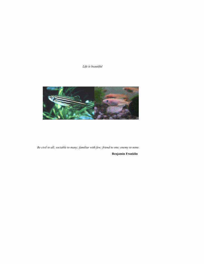
Life is beautiful
Be civil to all; sociable to many; familiar with few; friend to one; enemy to none.
Benjamin Franklin

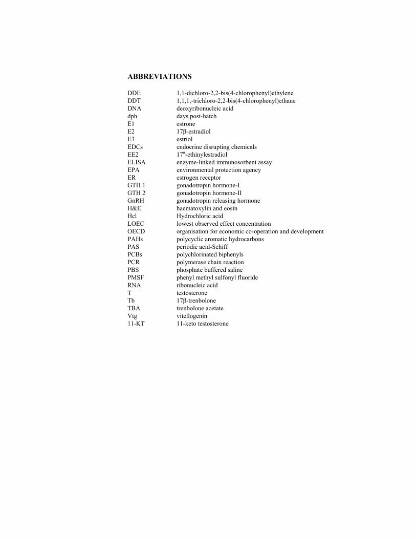
Contents
Abbreviations
Abstract
1 BACKGROUND 101.1 Endocrine disrupting chemicals (EDCs) 10 1.2 Deleterious effects of EDCs in vertebrates 10 1.3 Mechanism of endocrine disruption 11 1.4 Steroid 12
1.4.1 Estrogens 121.4.2 Androgens 14
1.5 Impact of EDCs on fish 151.6 Fish models for evaluation of EDCs 16
1.6.1 Zebrafish (Danio rerio) 161.6.2 Japanese Medaka (Oryzias latipes) 171.6.3 Fathead minnow (Pimephales promelas) 17
1.7 Fish life cycle assays 17 1.7.1 Fish screening assay 171.7.2 42-days Reproductive Fitness Test 181.7.3 Fish Sexual Develomental TestComplete Life
Cycle Test / fish-full-life-cycle-test 181.8 Biomarkers 18
1.8.1 Vitellogenin 181.8.2 Fish gonads & histology 20
1.8.2.1 Ovary 20 1.8.2.2 Testis 21
2 GENERAL AIM OF THE THESIS 223 MAJOR CONCLUSIONS 224 REFERENCES 23
5 RESEARCH PAPER I 3017 -ethinylestradiol (EE2) induced vitellogenin levels and normal gonad morphology during early stages of life cycle in Japanese medaka (Oryzias latipes)
6 RESEARCH PAPER II 41
Comparison of vitellogenin induction and gonad morphology between zebrafish and Japanese medaka after exposure to 17 -ethinylestradiol and 17 -trenbolone
7 ACKNOWLEDGEMENTS 56
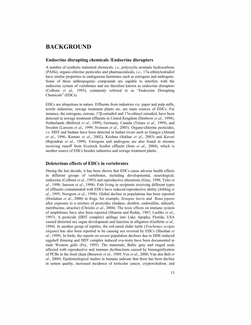
ABBREVIATIONS
DDE 1,1-dichloro-2,2-bis(4-chlorophenyl)ethylene DDT 1,1,1,-trichloro-2,2-bis(4-chlorophenyl)ethane DNA deoxyribonucleic acid dph days post-hatch E1 estrone E2 17 -estradiol E3 estriol EDCs endocrine disrupting chemicals EE2 17 -ethinylestradiol ELISA enzyme-linked immunosorbent assay EPA environmental protection agency ER estrogen receptor GTH 1 gonadotropin hormone-I GTH 2 gonadotropin hormone-II GnRH gonadotropin releasing hormone H&E haematoxylin and eosin Hcl Hydrochloric acid LOEC lowest observed effect concentration OECD organisation for economic co-operation and development PAHs polycyclic aromatic hydrocarbons PAS periodic acid-Schiff PCBs polychlorinated biphenyls PCR polymerase chain reaction PBS phosphate buffered saline PMSF phenyl methyl sulfonyl fluoride RNA ribonucleic acid T testosterone Tb 17 -trenbolone TBA trenbolone acetate Vtg vitellogenin 11-KT 11-keto testosterone

11
ABSTRACT
Endocrine disrupting chemicals (EDCs) are anthropogenic compounds that have similar properties as endogenous hormones in vertebrates and they are capable of interfering with the endocrine system. EDCs have been detected in sewage receiving effluents from industries and livestock feedlots. Fish living in the recipients contaminated with EDCs are reported to have disturbed physiological homeostasis and reproductive disabilities. The deleterious effects have also been reported in amphibians, reptiles, aves and mammals including humans. The natural estrogen, 17 -estradiol (E2) stimulates the fish liver to synthesize vitellogenin (Vtg), the protein that nourishes the maturation of eggs. 17 -ethinyl estradiol (EE2) is a potent, synthetic estrogen used in oral contraceptive preparations. It mimics E2 leading to enhanced circulatory Vtg concentrations and male-to-female sex reversal in fish. 17 -trenbolone (Tb), an androgenic steroid, is used as growth promoter in beef cattle. It has been reported to cause declined Vtg concentrations and masculinization in fish. In the present study (Paper I) on juvenile medaka (Oryzias latipes), after exposure to 100 ng EE2/l for 20 days period, there was time-dependent induction in Vtg was noticed. A significant increase in Vtg was observed in 48 hours after exposure. Normal gonadal development in early life stages of male and female medaka was illustrated in time-correlation with Vtg inductions of unexposed fish. The current study (Paper II) has proved the alteration in Vtg levels and sex reversal after experimental exposure to EE2 and Tb in two model fish species viz., zebrafish (Danio rerio) and Japanese medaka (Oryzias latipes). Complete masculinization of fish after 50 ng Tb/l and complete feminization of fish after 10 ng EE2/l exposure was observed in zebrafish. In conclusion, zebrafish was determined more sensitive than medaka to both of the test chemicals.
Keywords: 17 -ethinylestradiol (EE2); 17 -trenbolone (Tb); vitellogenin (Vtg); zebrafish (Danio rerio); medaka (Oryzias latipes).

12
13
BACKGROUND
Endocrine disrupting chemicals /Endocrine disrupters
A number of synthetic industrial chemicals, i.e., polycyclic aromatic hydrocarbons (PAHs), organo-chlorine pesticides and pharmaceuticals, i.e., 17 -ethinylestradiol have similar properties to endogenous hormones such as estrogens and androgens. Some of these anthropogenic compounds are capable to interfere with the endocrine system of vertebrates and are therefore known as endocrine disruptors (Colborn et al., 1993), commonly referred to as “Endocrine Disrupting Chemicals” (EDCs).
EDCs are ubiquitous in nature. Effluents from industries viz. paper and pulp mills, textile industries, sewage treatment plants etc. are main sources of EDCs. For instance, the estrogens, estrone, 17 -estradiol and 17 -ethinyl estradiol, have been detected in sewage treatment effluents in United Kingdom (Desbrow et al., 1998), Netherlands (Belfroid et al., 1999), Germany, Canada (Ternes et al., 1999), and Sweden (Larsson et al., 1999; Svenson et al., 2003). Organo-chlorine pesticides, i.e. DDT and lindane have been detected in Indian rivers such as Ganges (Ahmad et al., 1996; Kumari et al., 2002), Krishna (Sekhar et al., 2003) and Kaveri (Rajendran et al., 1999). Estrogens and androgens are also found in streams receiving runoff from livestock feedlot effluent (Soto et al., 2004), which is another source of EDCs besides industries and sewage treatment plants.
Deleterious effects of EDCs in vertebrates
During the last decade, it has been shown that EDCs cause adverse health effects in different groups of vertebrates, including developmental, neurological, endocrine (Colborn et al., 1993) and reproductive alterations (Gray, 1998; Tyler et
al., 1998; Janssen et al., 1998). Fish living in recipients receiving different types of effluents contaminated with EDCs have reduced reproductive ability (Jobling et
al., 1995; Norrgren et al., 1998). Global decline in populations has been reported (Houlahan et al., 2000) in frogs, for example, Xenopus laevis and Rana pipens
after exposure to a mixture of pesticides (lindane, dieldrin, endosulfan, aldicarb, metribuzine, atrazine) (Christin et al., 2004). The toxic effects on immune system of amphibians have also been reported (Sharma and Reddy, 1987; Luebke et al., 1997). A pesticide (DDT complex) spillage into Lake Apopka, Florida, USA caused distorted sex organ development and function in alligators (Guillette et al.,
1994). In another group of reptiles, the red-eared slider turtle (Trachemys scripta
elegans) has also been reported to be causing sex reversal by EDCs (Sheehan et
al., 1999). In birds, the reports on severe population declines due to DDE-induced eggshell thinning and DDT complex induced ova-testis have been documented in male Western gulls (Fry, 1995). The mammals, Baltic gray and ringed seals affected with reproductive and immune dysfunctions caused by biomagnification of PCBs in the food chain (Brouwer et al., 1989; Vos et al., 2000; Van den Belt et
al., 2004). Epidemiological studies in humans indicate that there has been decline in semen quality, increased incidence of testicular cancer, cryptorchidism, and
14
hypospadias over the past 50 years (Carlsen et al., 1992). These findings may be related to increased estrogenic exposure in utero (Sharpe and Skakkebaeck, 1993). An increased prevalence of breast cancer have been reported in females exposed to xenoestrogens (Colborn and Clement, 1992; Davis et al., 1993; Recchia et al., 2004).
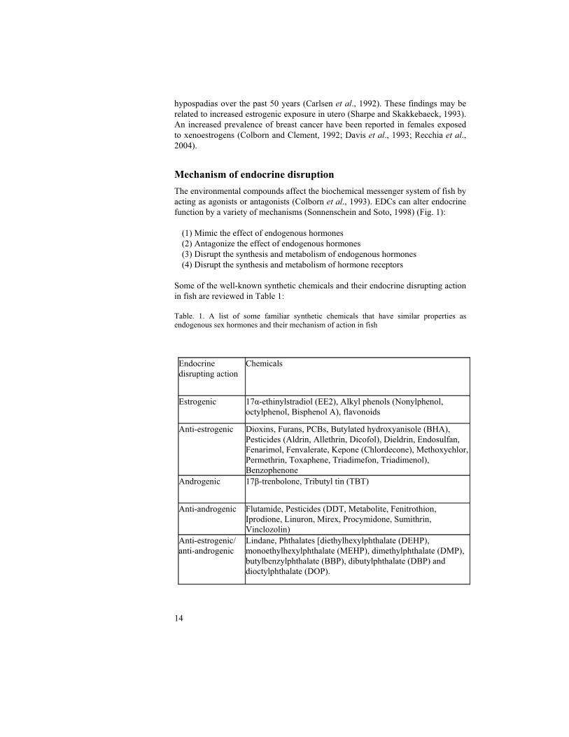
Mechanism of endocrine disruption
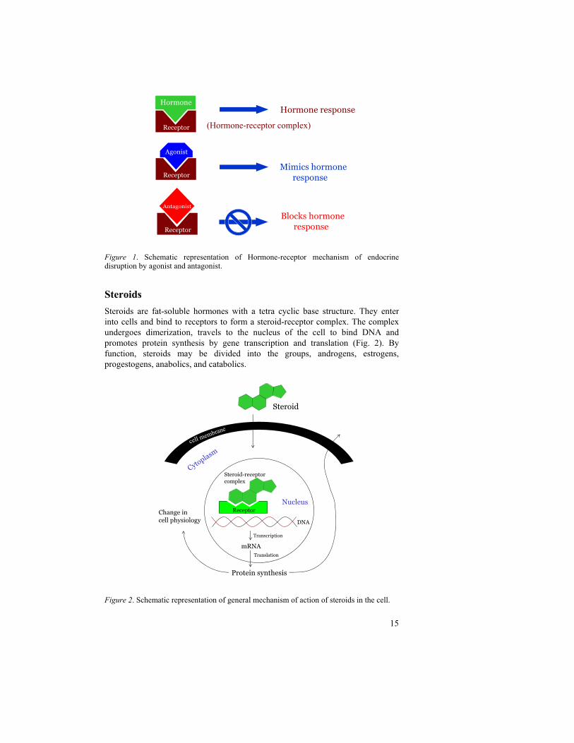
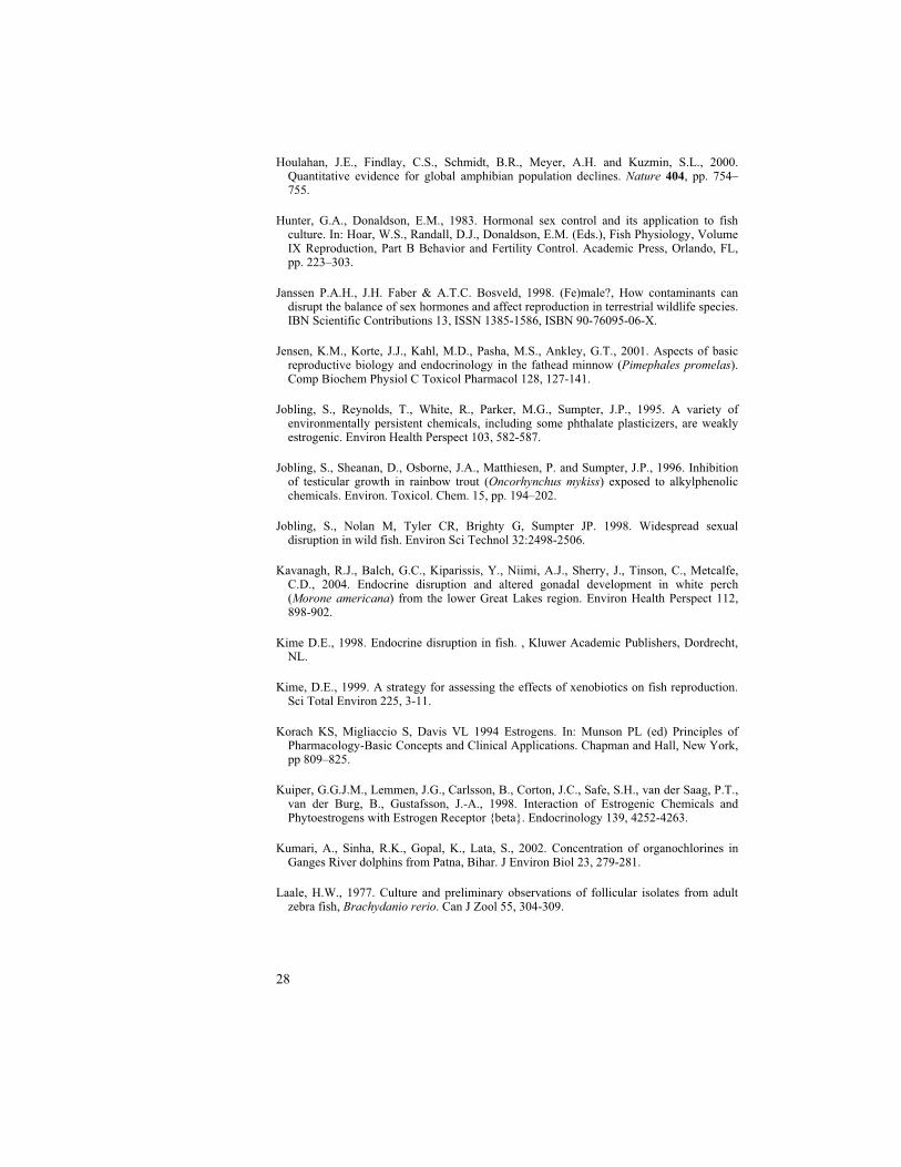
The environmental compounds affect the biochemical messenger system of fish by acting as agonists or antagonists (Colborn et al., 1993). EDCs can alter endocrine function by a variety of mechanisms (Sonnenschein and Soto, 1998) (Fig. 1):
(1) Mimic the effect of endogenous hormones (2) Antagonize the effect of endogenous hormones (3) Disrupt the synthesis and metabolism of endogenous hormones (4) Disrupt the synthesis and metabolism of hormone receptors
Some of the well-known synthetic chemicals and their endocrine disrupting action in fish are reviewed in Table 1:
Table. 1. A list of some familiar synthetic chemicals that have similar properties as endogenous sex hormones and their mechanism of action in fish
Endocrine disrupting action
Chemicals
Estrogenic 17 -ethinylstradiol (EE2), Alkyl phenols (Nonylphenol, octylphenol, Bisphenol A), flavonoids
Anti-estrogenic Dioxins, Furans, PCBs, Butylated hydroxyanisole (BHA), Pesticides (Aldrin, Allethrin, Dicofol), Dieldrin, Endosulfan, Fenarimol, Fenvalerate, Kepone (Chlordecone), Methoxychlor, Permethrin, Toxaphene, Triadimefon, Triadimenol), Benzophenone
Androgenic 17 -trenbolone, Tributyl tin (TBT)
Anti-androgenic Flutamide, Pesticides (DDT, Metabolite, Fenitrothion, Iprodione, Linuron, Mirex, Procymidone, Sumithrin, Vinclozolin)
Anti-estrogenic/ anti-androgenic
Lindane, Phthalates [diethylhexylphthalate (DEHP), monoethylhexylphthalate (MEHP), dimethylphthalate (DMP), butylbenzylphthalate (BBP), dibutylphthalate (DBP) and dioctylphthalate (DOP).
15
Figure 1. Schematic representation of Hormone-receptor mechanism of endocrine disruption by agonist and antagonist.
Steroids
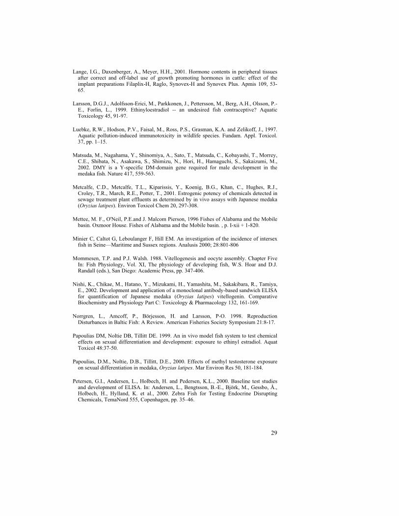
Steroids are fat-soluble hormones with a tetra cyclic base structure. They enter into cells and bind to receptors to form a steroid-receptor complex. The complex undergoes dimerization, travels to the nucleus of the cell to bind DNA and promotes protein synthesis by gene transcription and translation (Fig. 2). By function, steroids may be divided into the groups, androgens, estrogens, progestogens, anabolics, and catabolics.
Figure 2. Schematic representation of general mechanism of action of steroids in the cell.
Cytoplasm
cell membrane
Steroid
mRNA
DNA
Change incell physiology
Protein synthesis
Steroid-receptorcomplex
Transcription
Translation
NucleusReceptor
Hormone response
Mimics hormone response
Blocks hormone response
Receptor (Hormone-receptor complex)
Receptor
Agonist
Receptor
Antagonist
HormoneHormoneHormoneHormone
16
Estrogens
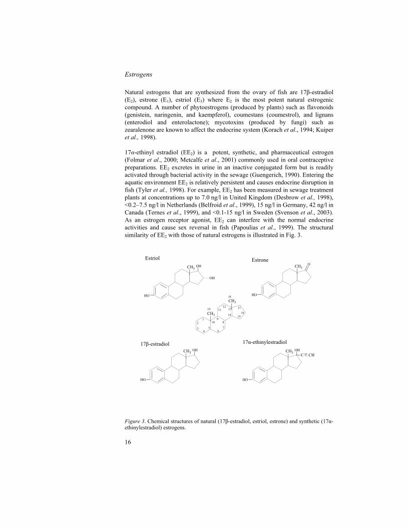
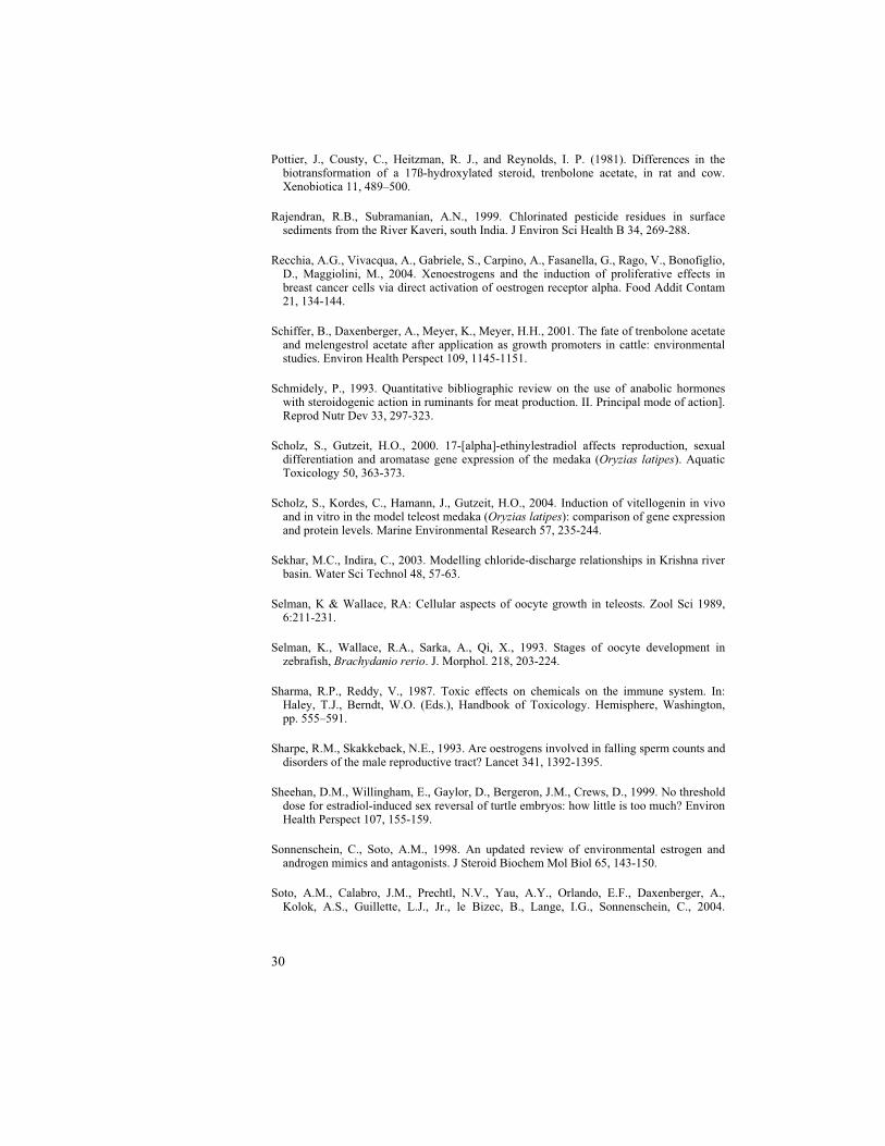
Natural estrogens that are synthesized from the ovary of fish are 17 -estradiol (E2), estrone (E1), estriol (E3) where E2 is the most potent natural estrogenic compound. A number of phytoestrogens (produced by plants) such as flavonoids (genistein, naringenin, and kaempferol), coumestans (coumestrol), and lignans (enterodiol and enterolactone); mycotoxins (produced by fungi) such as zearalenone are known to affect the endocrine system (Korach et al., 1994; Kuiper et al., 1998).
17 -ethinyl estradiol (EE2) is a potent, synthetic, and pharmaceutical estrogen (Folmar et al., 2000; Metcalfe et al., 2001) commonly used in oral contraceptive preparations. EE2 excretes in urine in an inactive conjugated form but is readily activated through bacterial activity in the sewage (Guengerich, 1990). Entering the aquatic environment EE2 is relatively persistent and causes endocrine disruption in fish (Tyler et al., 1998). For example, EE2 has been measured in sewage treatment plants at concentrations up to 7.0 ng/l in United Kingdom (Desbrow et al., 1998), <0.2–7.5 ng/l in Netherlands (Belfroid et al., 1999), 15 ng/l in Germany, 42 ng/l in Canada (Ternes et al., 1999), and <0.1-15 ng/l in Sweden (Svenson et al., 2003). As an estrogen receptor agonist, EE2 can interfere with the normal endocrine activities and cause sex reversal in fish (Papoulias et al., 1999). The structural similarity of EE2 with those of natural estrogens is illustrated in Fig. 3.
Figure 3. Chemical structures of natural (17 -estradiol, estriol, estrone) and synthetic (17 -ethinylestradiol) estrogens.
CH3OH
HO
CH3O
HO
CH3
CH3
12
34
56
7
89
10
1112
13
14 1516
17
18
19
CH3OH
HO
OH
CH3OH
HO
CHC
Estriol Estrone
17 -ethinylestradiol17 -estradiol

17
Androgens
The male reproductive hormones, androgens, are produced in the testes of fish to contribute secondary sexual characteristics. They include mainly, testosterone (T), androstenedione, 11-ketotestosterone (11-KT), and 11 -hydroxy testosterone. Female fish also produce testosterone, which aromatises to E2 by cytochrome P450 (CYP19) in the granulosa cells of ovarian follicles (Petersen et al., 2000).
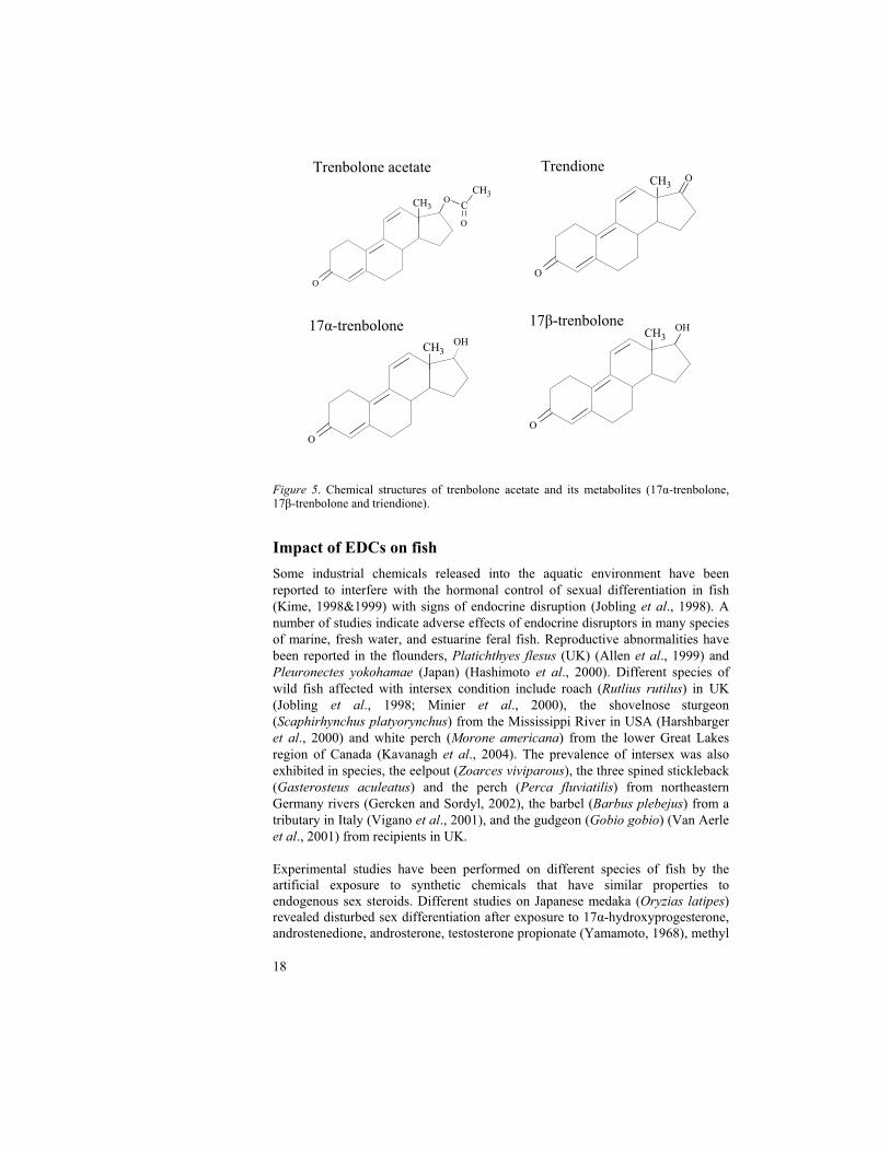
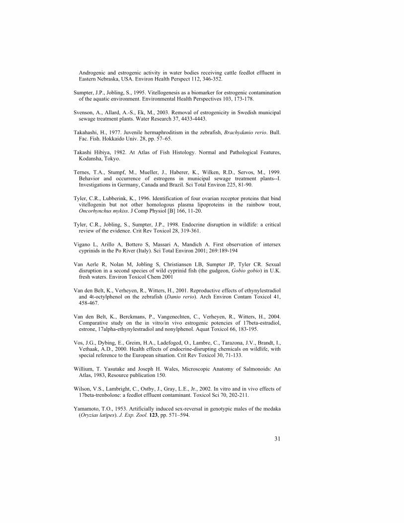
17 -trenbolone (17 -hydroxyestra-4, 9, 11-trien-3-one) is an anabolic, androgenic steroid, with the homology to testosterone structure (Fig. 4), can disturb the endocrine homeostasis by mimicking 11-KT and testosterone. Trenbolone acetate (TbA), an acetate form of trenbolone, degrades proteins through reduction in the activity of catabolic glucocorticoids (Schmidely, 1993). It is used as growth promoter in beef cattle in USA and Canada (Lange et al., 2001, Wilson et al., 2002). The anabolic androgenic steroids in urine are metabolised mostly as glucuronides and sulfates. TbA is excreted mainly as 17 -trenbolone, 17 -trenbolone and triendione in urine (Pottier et al., 1981, Wilson et al., 2002) (Fig. 5), and enter into environment through the excreta along with downstream from cattle farms. The metabolites of TbA can remain active for more than 270 days in the manure piles (Schiffer et al., 2001). TbA has been reported to masculinize channel catfish (Ictalurus punctatus) (Galvez et al., 1995) and blue tilapia (Oreochromis aureus) (Galvez et al., 1996). 17 -trenbolone is a potent androgen receptor agonist, caused masculinizing effect, and declined fecundity in fathead minnow (Pimephales promelas) (Ankley et al., 2003).
Figure 4. Chemical structure of natural androgen (testosterone) and anabolic steroidal androgen (17 -trenbolone).
CH3OH
O
CH3OH
CH3
O
Testosterone 17 -trenbolone
18
Figure 5. Chemical structures of trenbolone acetate and its metabolites (17 -trenbolone, 17 -trenbolone and triendione).
Impact of EDCs on fish
Some industrial chemicals released into the aquatic environment have been reported to interfere with the hormonal control of sexual differentiation in fish (Kime, 1998&1999) with signs of endocrine disruption (Jobling et al., 1998). A number of studies indicate adverse effects of endocrine disruptors in many species of marine, fresh water, and estuarine feral fish. Reproductive abnormalities have been reported in the flounders, Platichthyes flesus (UK) (Allen et al., 1999) and Pleuronectes yokohamae (Japan) (Hashimoto et al., 2000). Different species of wild fish affected with intersex condition include roach (Rutlius rutilus) in UK (Jobling et al., 1998; Minier et al., 2000), the shovelnose sturgeon (Scaphirhynchus platyorynchus) from the Mississippi River in USA (Harshbarger et al., 2000) and white perch (Morone americana) from the lower Great Lakes region of Canada (Kavanagh et al., 2004). The prevalence of intersex was also exhibited in species, the eelpout (Zoarces viviparous), the three spined stickleback (Gasterosteus aculeatus) and the perch (Perca fluviatilis) from northeastern Germany rivers (Gercken and Sordyl, 2002), the barbel (Barbus plebejus) from a tributary in Italy (Vigano et al., 2001), and the gudgeon (Gobio gobio) (Van Aerle et al., 2001) from recipients in UK.
Experimental studies have been performed on different species of fish by the artificial exposure to synthetic chemicals that have similar properties to endogenous sex steroids. Different studies on Japanese medaka (Oryzias latipes)revealed disturbed sex differentiation after exposure to 17 -hydroxyprogesterone, androstenedione, androsterone, testosterone propionate (Yamamoto, 1968), methyl
CH3O
O
CH3OH
O
17 -trenbolone17 -trenbolone
CH3OH
O
TrendioneTrenbolone acetate
CH3O
O
CH3
C
O
19
testosterone (Papoulias et al., 2000), and EE2 (Papoulias et al., 2000, Scholz & Gutzeit, 2000). In addition, reproductive disabilities such as testis-ova after exposure to p-nonylphenol (Gray & Metcalfe, 1997) and inhibited spermatogenesis after exposure to 4-tert-octylphenol (Gronen et al., 1999) in medaka were documented. Exposure of zebrafish (Danio rerio) to a mixture of PCBs (Örn et al., 1998), ethinylestradiol (EE2), and 4t-octylphenol (Van den belt et al., 2001) caused adverse effect on the reproductive success.
Fish models for evaluation of EDCs
A variety of fish species have been used as test organisms for the detection of EDCs. This means that interpretation of results and extrapolation between species may be difficult due to variation in sensitivity between species. The largest group of vertebrates, the teleost super-order, comprises more than 90% of the total number of described fish species. In order to harmonise test guidelines based on fish it is essential to develop mutual test protocols suitable for a small number of species. Today, OECD (Organisation for Economic Co-operation and Development) promotes three small laboratory freshwater species as model test species. They are zebrafish (Danio rerio), Japanese medaka (Oryzias latipes), and fathead minnow (Pimephales promelas). A short description of the test species is given below:
Zebrafish
Danio rerio (Hamiton-Buchanan, 1822/1823), a tropical Cypriniform (family Cyprinidae) is commonly called zebrafish because of its striped integument. The zebrafish is native of India (Ganges and Brahmaputra rivers) and extended to Pakistan and northern Burma. The zebrafish is a regular spawner normally ovulating approximately every fifth day. They are characterized by high fecundity providing between 100-500 eggs at each occasion. The eggs are non-adherent, transparent and have a developmental period from fertilization to hatching of 96h at 26°C (Laale, 1977). According to Takahashi (1977), male zebrafish pass through a phase of juvenile hermaphroditism. All fish start to develop ovaries at the age of 10–12 days and the period of sex differentiation begins at the age of approximately 23–25 days where in some of the fish the ovaries degenerate and transform into testes. The process of sex differentiation completes approximately at 40 days post-hatch and the final maturation of the gonads will generally be finished at an age of 60 days.
Japanese Medaka
Oryzias latipes, a freshwater killifish (family Cyprinodontidae), is native to the rice paddies of Japan, Taiwan, and Southeast Asia and is commonly called Japanese medaka or Medaka. They produce 10 to 30 eggs per spawning. In medaka, the undifferentiated gonad directly differentiates into either testis or ovary because of its’ stable genetic XX/XY sex determination system and a sex
20
determining gene (DMY) is isolated from Y chromosome (Matsuda et al., 2002). Medaka is considered as a powerful model species (Environmental Agency, Government of Japan, 2000) with a short reproduction cycle to enable the study of complete life stages in order to probe the effects of EDCs. EDCs have ability to alter the phenotypic sex of fish when exposed prior to or during sexual differentiation and cause sex reversal irrespective of genotype (Yamamoto, 1953; Hunter and Donaldson, 1983). If the medaka d-rR strain is used, genomic sex can be distinguished from functional sex by the hormone insensitive orange colour trait, which is specified by a Y-chromosomal gene (Yamamoto, 1975).
Fathead minnow
Fathead minnow, Pimephales promelas is a member of the family Cyprinidae and is widely distributed in North America. The name ‘fathead minnow’ is derived from the shape of the head (Pimephales –‘fat head’) and it's colour in breeding males (promelas – ‘forward’ and ‘black’) (Mettee et al., 1996). It produces 50-100 adhesive eggs every 3-5 days (Jensen et al., 2001) and the eggs hatch in 5 to 6 days. They have distinct secondary sexual characteristics in both sexes.
Fish life cycle assays
Today, OECD considers four new test guidelines for detection of EDCs. These are briefly described as:
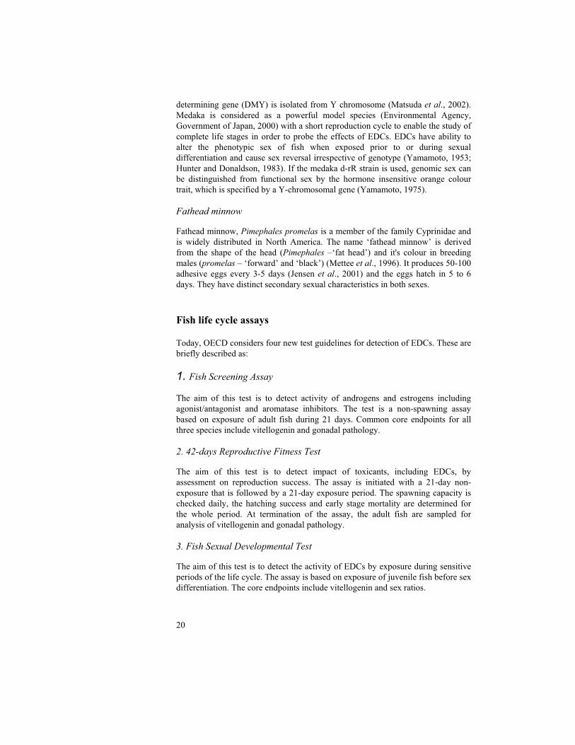
1. Fish Screening Assay
The aim of this test is to detect activity of androgens and estrogens including agonist/antagonist and aromatase inhibitors. The test is a non-spawning assay based on exposure of adult fish during 21 days. Common core endpoints for all three species include vitellogenin and gonadal pathology.
2. 42-days Reproductive Fitness Test
The aim of this test is to detect impact of toxicants, including EDCs, by assessment on reproduction success. The assay is initiated with a 21-day non-exposure that is followed by a 21-day exposure period. The spawning capacity is checked daily, the hatching success and early stage mortality are determined for the whole period. At termination of the assay, the adult fish are sampled for analysis of vitellogenin and gonadal pathology.
3. Fish Sexual Developmental Test
The aim of this test is to detect the activity of EDCs by exposure during sensitive periods of the life cycle. The assay is based on exposure of juvenile fish before sex differentiation. The core endpoints include vitellogenin and sex ratios.

21
4. Complete Life Cycle Test / fish-full-life-cycle-test (FFLT)
The aim of this test is to detect activity of EDCs in the parental fish and in their progeny. Core endpoints include spawning success, offspring survival, and sex ratios.
Biomarkers
The alteration of the normal physiology in response to EDCs and the potency of the xenoestrogens can be demonstrated by using in vivo markers.
Vitellogenin
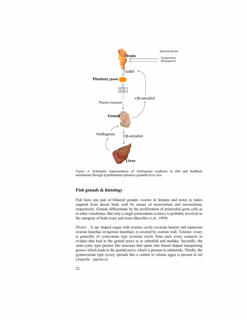
In oviparous vertebrates including fish, the eggs contain a large amount of yolk in the form of a precursor protein, vitellogenin (Vtg), which is produced by the liver under the control of the endocrine system. In fish brains, the hypothalamus is stimulated by external factors, such as water temperature and photoperiod, to secrete gonadotropin-releasing hormone (GnRH), which in turn stimulates the pituitary gland to secrete two gonado tropic hormones (GTH 1 and 2). GTH 1 and 2 trigger the gonads to release 17 -estradiol (E2) which binds to E2 receptors and activates the synthesis of Vtg protein in the liver. Vtg is circulated to ovaries (Bun and Idler, 1983) and is cleaved into lipovitellin and phosvitin to deposit in the oocyte by means of yolk during oocyte development. Vtg is the protein source that nourish in the maturation of the embryo and for developing larva (Mommsen & Walsh, 1988; Selman et al., 1989). The synthesis of Vtg is regulated by endocrine system under the control of feed back mechanisms through hypothalamus-pituitary-gonadal-liver axis, E2, and testosterone (Fig. 6). In general, Vtg is not found in male fish but it can be induced upon estrogenic exposure (Sumpter and Jobling, 1995; Folmar et al., 1996; Jobling et al., 1996; and Tyler et al., 1996). Hence, Vtg has become the most frequently used biomarker. Governmental organizations such as OECD and the US Environmental Protection Agency (EPA) consider Vtg as a core biomarker for the detection of endocrine disrupting effects in different species of fish. The estrogenic potency of EDCs can be determined by quantitative analysis of Vtg. Since Vtg levels in different tissues, i.e. liver, heart and blood plasma correlates (Nishi et al., 2002), there are different options to measure Vtg depending on fish species.
Different assays for the detection of Vtg include radio immunoassays (RIA), enzyme immunoassays (EIA), immunohistochemistry using monoclonal as well as polyclonal antibodies, RNA protection assay, and transcript analysis by Northern blotting or polymerase chain reaction (PCR).
22
Figure 6. Schematic representation of vitellogenin synthesis in fish and feedback mechanism through hypothalamus-pituitary-gonadal-liver axis.
Fish gonads & histology
Fish have one pair of bilateral gonads: ovaries in females and testes in males suspend from dorsal body wall by means of mesovarium and mesorchium, respectively. Gonads differentiate by the proliferation of primordial germ cells as in other vertebrates. But only a single primordium (cortex) is probably involved in the ontogeny of both ovary and testis (Baroiller et al., 1999).
Ovary: A sac shaped organ with ovarian cavity (ovarian lumen) and numerous ovarian lamellae (ovigerous lamellae), is covered by ovarian wall. Teleosts’ ovary is generally of cystovarian type (ovarian cavity from each ovary connects to oviduct that lead to the genital pore) as in zebrafish and medaka. Secondly, the semi-cystic type (pocket like structure that opens into funnel shaped transporting groove which leads to the genital pore), which is present in salmonids. Thirdly, the gymnovarian type (ovary spreads like a curtain to release eggs) is present in eel (Anguilla japonica).
Liver
Brain
GTH 1GTH 2
17 -estradiol
GnRH
Plasma transport
Pituitary gland
Gonad
17 -estradiolVitellogenin
External factors
TemperaturePhotoperiod

23
Cell types in ovary in the order of maturation according to the validation of the fish-screening assay for Endocrine active substances, OECD, 2004:
1. Oogonium: It is the smallest cell type with relatively large nucleus, small inapparent nucleolus, and minimal amounts of cytoplasm.
2. Chromatin nuclear oocyte: It is surrounded by prefollicular cells. It consists of relatively large nucleus with single large nucleolus and granular cytoplasm.
3. Perinucleolar oocyte: It is characterised by multiple nucleoli located at periphery of the nucleus (germinal vesicle).
4. Cortical alveolar oocyte: It is larger than perinucleolar oocyte and it can be recognised by the presence of cortical alveoli (yolk vesicles) and distinct chorion (vitelline envelope) between oocyte and perifollicular cells. A mature cortical alveolus consists of distinct central core surrounded by light flocculent material and a membrane. In this stage, irregular enlargement of germinal vesicle and proliferation of pleo-morphic nucleoli take place (Selman et al., 1993). Yolk vesicles appear earlier than yolk globules and they contain glycoprotein that stain slightly with eosin to give light red colour or deep red with PAS (Takashi Hibiya, 1982).
5. Early vitellogenic oocyte: It is larger than cortical alveolar oocyte and characterised by centralised appearance of spherical, eosinophilic, vitellogenic yolk granules or globules that resemble reddish nucleus. Presence of eosinophilic substances i.e. lipoprotein and carbohydrate in yolk globules gives positive staining with H&E but weekly to PAS. Glycerides and cholesterol present in oil droplet that appears as a circular vacuole in paraffin sections but stains black by osmic acid fixation.
6. Late vitellogenic oocyte: The increased volume of vitellogenic granules displaces the cortical alveolar material to periphery and nucleus starts migrating to periphery. Germinal vesicle is large and has smooth contour, spherical nucleoli and small nucleoli at centre. Lipid droplets are never seen within zebra fish oocytes (Selman et al., 1993).
7. Mature/Spawning oocyte: Larger cell type with nucleus migrated to periphery and it appears more hydrated due to vitellogenesis.
8. Atretic mature oocyte: This cell type is characterised by zona radiata breakdown and yolk resorption.
There are three layers surrounding the oocyte: inner zona radiata, middle follicle and outer vascularized theca (Willium and Joseph, 1983). Hyperplasia of follicle cells with inner and outer squamous theca cell layers is due to vitellogenesis i.e. accumulation of yolk substances (yolk vesicles, yolk globules, and oil droplets).
24
Testis: Testes are paired, sac-shaped organs and sperms are carried through sperm duct as in higher vertebrates. In zebrafish, a series of five parallel efferent ducts collects spermatozoa from seminiferous tubules and pass caudo-ventrally to unite at genital papilla (Ewing, 1972; Laale, 1977).
Spermatogenesis: Spermatogonium proliferates to give primary spermatocytes, which undergoes meiotic division leading to secondary spermatocyte, and it further divides to produce spermatids. Finally, spermatids mature to become spermatozoa.
Cell types of testis in the order of maturation according to the validation of the fish-screening assay for Endocrine active substances, OECD, 2004 are:
1. Spermatogonium: Largest cell type and consists of vesicular nuclei with distinct nuclear membrane and prominent nucleoli.
2. Spermatocyte: Primary spermatocyte is larger than secondary spermatocyte.
3. Spermatid: It is smallest cell type with dense nucleus and narrow rims of eosinophilic cytoplasm.
4. Spermatozoa: They are mature cells with dark round nuclei and tail.
Along with germinal cells, sertoli cells (cyst cells) which take part in nourishment during the spermatogenesis and interstitial cells (leydig cells) secreting sex steroids are present in testis.

25
GENERAL AIM OF THE THESIS
The aim of the present study was to determine the impact of endocrine disrupting chemicals on sex differentiation of fish by using two model test species, zebrafish and Japanese medaka, with the exposure of 17 -ethinylestradiol and 17 -trenbolone.
MAJOR CONCLUSIONS
The fish species, zebrafish (Danio rerio) and Japanese medaka (Oryzias latipes)are suitable model test species for evaluation of potential endocrine disruption. The two species showed a remarkable sensitivity difference after exposure to 17 -ethinylestradiol (EE2) and 17 -trenbolone (Tb).
In the time-related study on medaka, a significant Vtg induction was noticed after exposure to 100 ng EE2/l from 12 dph to 30 dph. The study was elicited that the Vtg induction was increased above the normal Vtg levels in 48 hours after exposure and reached a maximum stable Vtg induction in two weeks. The normal gonadal histology in medaka was illustrated with different stages of gonads and the observed maturation in gonads at 35 dph was correlated with the elevated Vtg levels in control fish between 30 to 38 dph.
The life cycle test with the vitellogenin (Vtg) analysis at 38 dph and sex ratio at 60 dph was resulted in high Vtg induction and complete feminization in zebrafish after 10 ng EE2/l exposure. Likewise, a significant decline in Vtg was correlated with complete masculinization after 50 ng Tb/l exposure. However, in medaka, the Tb concentration, 50 ng /l was resulted in unaffected sex ratio with no correlation to declined Vtg at 38 dph. Furthermore, regarding EE2 concentrations, medaka showed the Vtg induction and feminizing effect only at high concentration i.e., 100 ng/l. This delineates a lower sensitivity of medaka when compared to zebrafish towards the estrogen mimic as well as androgen mimic.

26
REFERENCES
Ahmad, S., Ajmal, M., Nomani, A.A., 1996. Organochlorines and polycyclic aromatic hydrocarbons in the sediments of Ganges River (India). Bull Environ Contam Toxicol 57, 794-802.
Allen, Y., Matthiessen, P., Scott, A.P., Haworth, S., Feist, S., Thain, J.E., 1999. The extent of oestrogenic contamination in the UK estuarine and marine environments -- further surveys of flounder. The Science of The Total Environment 233, 5-20.
Ankley, G.T., Jensen, K.M., Makynen, E.A., Kahl, M.D., Korte, J.J., Hornung, M.W., Henry, T.R., Denny, J.S., Leino, R.L., Wilson, V.S., Cardon, M.C., Hartig, P.C., Gray, L.E., 2003. Effects of the androgenic growth promoter 17-beta-trenbolone on fecundity and reproductive endocrinology of the fathead minnow. Environ Toxicol Chem 22, 1350-1360.
Baroiller, J. F., Guigen, Y. & Fostier, A. (1999). Endocrine and environmental aspects of sex differentiation in fish. Cellular and Molecular Life Sciences 55, 910–931.
Belfroid, A.C., Van der Horst, A., Vethaak, A.D., Schafer, A.J., Rijs, G.B.J., Wegener, J., Cofino, W.P., 1999. Analysis and occurrence of estrogenic hormones and their glucuronides in surface water and waste water in The Netherlands. The Science of The Total Environment 225, 101-108.
Brouwer, A., Reijnders, P.J.H. and Koeman, J.H., 1989. Polychlorinated biphenyl (PCB)-contaminated fish induces vitamin A and thyroid deficiency in the common seal (Phoca vitulina). Aquat. Toxicol. 15, pp. 99–106.
Bun Ng, T., Idler, D.R., 1983. Yolk formation and differentiation in teleost fishes. In: Hoar, W.S., Randall, D.J. (Eds.), Fish Physiology, vol. IXA. Academic Press, USA, pp. 373–397.
Carlsen, E., A. Giwercam, N. Keiding and N.E. Skakkebaeck, Evidence for decreasing quality of semen during the past 50 years. Br. Med. J. 305 (1992), pp. 609–613.
Christin, M.S., Menard, L., Gendron, A.D., Ruby, S., Cyr, D., Marcogliese, D.J., Rollins-Smith, L., Fournier, M., 2004. Effects of agricultural pesticides on the immune system of Xenopus laevis and Rana pipiens. Aquat Toxicol 67, 33-43.
Colborn, T., vom Saal, F.S., Soto, A.M., 1993. Developmental effects of endocrine-disrupting chemicals in wildlife and humans. Environ Health Perspect 101, 378-384.
Colborn, T., Clement, C. (Eds.). 1992. Chemically-induced alterations in sexual and functional development: The Wildlife/Human Connection. Princeton Scientific Pub. Co., Princeton, NJ, 402 pp.
Davis, D.L., Bradlow HL, Wolff M, Woodruff T, Hoel D.G., Anton-Culver H. 1993. Medical hypothesis: xenoestrogens as preventable causes of breast cancer. Environ Health Perspect 101:372-377.
Desbrow, C., Routledge, E.J., Brighty, G.C., Sumpter, J.P., Waldock, M., 1998. Identification of estrogenic chemicals in STW effluent. 1. Chemical fractionation and in vitro biological screening. Environmental Science and Technology 32, 1549-1558.
27
EDSTAC. 1998. Endocrine Disruptor Screening and Testing Advisory Committee (EDSTAC) Final Report. Washington, DC:U.S. Environmental Protection Agency.
Ewing, H. H., 1972. Spermatogenesis in zebrafish, Brachydanio rerio. Anat. Res. 172, 308.
Folmar, L.C., Denslow, N.D., Rao, V., Chow, M., Crain, D.A., Enblom, J., Marcino, J., Guillette L.J., J., 1996. Vitellogenin induction and reduced serum testosterone concentrations in feral male carp (Cyprinus carpio) captured near a major metropolitan sewage treatment plant. Environmental Health Perspectives 104, 1096-1101.
Folmar, L.C., Hemmer, M., Hemmer, R., Bowman, C., Kroll, K., Denslow, N.D., 2000. Comparative estrogenicity of estradiol, ethynyl estradiol and diethylstilbestrol in an in vivo, male sheepshead minnow (Cyprinodon variegatus), vitellogenin bioassay. Aquatic Toxicol 49, 77-88.
Fry, D.M., 1995. Reproductive effects in birds exposed to pesticides and industrial chemicals. Environ Health Perspect 103 Suppl 7, 165-171.
Galvez, J.I., Mazik, P.M., Phelps, R.P., Mulvaney, D.R., 1995. Masculinization of channel catfish (Ictalurus punctatus) by oral administration of trenbolone acetate. J. World Aquacult. Soc. 26, 378–383.
Galvez JI, Morrison JR. 1996. Efficacy of trenbolone acetate insex inversion of the blue tilapia (Oreochromis aureus) J. World Aquacult Soc 27:483–486.
Gercken, J., Sordyl, H., 2002. Intersex in feral marine and freshwater fish from northeastern Germany. Mar Environ Res 54, 651-655.
Gray, L.E., Jr., 1998. Xenoendocrine disrupters: laboratory studies on male reproductive effects. Toxicol Lett 102-103, 331-335.
Gray, M.A., Metcalfe, C.D., 1997. Induction of testis-ova in Japanese medaka (Oryzias latipes) exposed to p-nonylphenol. Environ. Toxicol. Chem. 16, 1082-1086.
Gronen, S., Denslow, N., Manning, S., Barnes, S., Barnes, D., Brouwer, M., 1999. Serum vitellogenin levels and reproductive impairment of male Japanese Medaka (Oryzias latipes) exposed to 4-tert-octylphenol. Environ Health Perspect 107, 385-390.
Guengerich, F.P., 1990. Metabolism of 17 alpha-ethynylestradiol in humans. Life Sci 47, 1981-1988.
Guillette, L.J., Jr., Gross, T.S., Masson, G.R., Matter, J.M., Percival, H.F., Woodward, A.R., 1994. Developmental abnormalities of the gonad and abnormal sex hormone concentrations in juvenile alligators from contaminated and control lakes in Florida. Environ Health Perspect 102, 680-688.
Harshbarger JC, Coffey MJ, Young MY. Intersexes in Mississippi River shovelnose sturgeon sampled below Saint Louis, Missouri, USA. Mar Environ Res 2000; 50:247-250
Hashimoto, S., Bessho, H., Hara, A., Nakamura, M., Iguchi, T., Fujita, K., 2000. Elevated serum vitellogenin levels and gonadal abnormalities in wild male flounder (Pleuronectes yokohamae) from Tokyo Bay, Japan. Mar Environ Res 49, 37-53.
28
Houlahan, J.E., Findlay, C.S., Schmidt, B.R., Meyer, A.H. and Kuzmin, S.L., 2000. Quantitative evidence for global amphibian population declines. Nature 404, pp. 754–755.
Hunter, G.A., Donaldson, E.M., 1983. Hormonal sex control and its application to fish culture. In: Hoar, W.S., Randall, D.J., Donaldson, E.M. (Eds.), Fish Physiology, Volume IX Reproduction, Part B Behavior and Fertility Control. Academic Press, Orlando, FL, pp. 223–303.
Janssen P.A.H., J.H. Faber & A.T.C. Bosveld, 1998. (Fe)male?, How contaminants can disrupt the balance of sex hormones and affect reproduction in terrestrial wildlife species. IBN Scientific Contributions 13, ISSN 1385-1586, ISBN 90-76095-06-X.
Jensen, K.M., Korte, J.J., Kahl, M.D., Pasha, M.S., Ankley, G.T., 2001. Aspects of basic reproductive biology and endocrinology in the fathead minnow (Pimephales promelas).Comp Biochem Physiol C Toxicol Pharmacol 128, 127-141.
Jobling, S., Reynolds, T., White, R., Parker, M.G., Sumpter, J.P., 1995. A variety of environmentally persistent chemicals, including some phthalate plasticizers, are weakly estrogenic. Environ Health Perspect 103, 582-587.
Jobling, S., Sheanan, D., Osborne, J.A., Matthiesen, P. and Sumpter, J.P., 1996. Inhibition of testicular growth in rainbow trout (Oncorhynchus mykiss) exposed to alkylphenolic chemicals. Environ. Toxicol. Chem. 15, pp. 194–202.
Jobling, S., Nolan M, Tyler CR, Brighty G, Sumpter JP. 1998. Widespread sexual disruption in wild fish. Environ Sci Technol 32:2498-2506.
Kavanagh, R.J., Balch, G.C., Kiparissis, Y., Niimi, A.J., Sherry, J., Tinson, C., Metcalfe, C.D., 2004. Endocrine disruption and altered gonadal development in white perch (Morone americana) from the lower Great Lakes region. Environ Health Perspect 112, 898-902.
Kime D.E., 1998. Endocrine disruption in fish. , Kluwer Academic Publishers, Dordrecht, NL.
Kime, D.E., 1999. A strategy for assessing the effects of xenobiotics on fish reproduction. Sci Total Environ 225, 3-11.
Korach KS, Migliaccio S, Davis VL 1994 Estrogens. In: Munson PL (ed) Principles of Pharmacology-Basic Concepts and Clinical Applications. Chapman and Hall, New York, pp 809–825.
Kuiper, G.G.J.M., Lemmen, J.G., Carlsson, B., Corton, J.C., Safe, S.H., van der Saag, P.T., van der Burg, B., Gustafsson, J.-A., 1998. Interaction of Estrogenic Chemicals and Phytoestrogens with Estrogen Receptor {beta}. Endocrinology 139, 4252-4263.
Kumari, A., Sinha, R.K., Gopal, K., Lata, S., 2002. Concentration of organochlorines in Ganges River dolphins from Patna, Bihar. J Environ Biol 23, 279-281.
Laale, H.W., 1977. Culture and preliminary observations of follicular isolates from adult zebra fish, Brachydanio rerio. Can J Zool 55, 304-309.
29
Lange, I.G., Daxenberger, A., Meyer, H.H., 2001. Hormone contents in peripheral tissues after correct and off-label use of growth promoting hormones in cattle: effect of the implant preparations Filaplix-H, Raglo, Synovex-H and Synovex Plus. Apmis 109, 53-65.
Larsson, D.G.J., Adolfsson-Erici, M., Parkkonen, J., Pettersson, M., Berg, A.H., Olsson, P.-E., Forlin, L., 1999. Ethinyloestradiol -- an undesired fish contraceptive? Aquatic Toxicology 45, 91-97.
Luebke, R.W., Hodson, P.V., Faisal, M., Ross, P.S., Grasman, K.A. and Zelikoff, J., 1997. Aquatic pollution-induced immunotoxicity in wildlife species. Fundam. Appl. Toxicol. 37, pp. 1–15.
Matsuda, M., Nagahama, Y., Shinomiya, A., Sato, T., Matsuda, C., Kobayashi, T., Morrey, C.E., Shibata, N., Asakawa, S., Shimizu, N., Hori, H., Hamaguchi, S., Sakaizumi, M., 2002. DMY is a Y-specific DM-domain gene required for male development in the medaka fish. Nature 417, 559-563.
Metcalfe, C.D., Metcalfe, T.L., Kiparissis, Y., Koenig, B.G., Khan, C., Hughes, R.J., Croley, T.R., March, R.E., Potter, T., 2001. Estrogenic potency of chemicals detected in sewage treatment plant effluents as determined by in vivo assays with Japanese medaka (Oryzias latipes). Environ Toxicol Chem 20, 297-308.
Mettee, M. F., O'Neil, P.E.and J. Malcom Pierson, 1996 Fishes of Alabama and the Mobile basin. Oxmoor House. Fishes of Alabama and the Mobile basin. , p. I-xii + 1-820.
Minier C, Caltot G, Leboulanger F, Hill EM. An investigation of the incidence of intersex fish in Seine—Maritime and Sussex regions. Analusis 2000; 28:801-806
Mommesen, T.P. and P.J. Walsh. 1988. Vitellogenesis and oocyte assembly. Chapter Five In: Fish Physiology, Vol. XI, The physiology of developing fish, W.S. Hoar and D.J. Randall (eds.), San Diego: Academic Press, pp. 347-406.
Nishi, K., Chikae, M., Hatano, Y., Mizukami, H., Yamashita, M., Sakakibara, R., Tamiya, E., 2002. Development and application of a monoclonal antibody-based sandwich ELISA for quantification of Japanese medaka (Oryzias latipes) vitellogenin. Comparative Biochemistry and Physiology Part C: Toxicology & Pharmacology 132, 161-169.
Norrgren, L., Amcoff, P., Börjesson, H. and Larsson, P-O. 1998. Reproduction Disturbances in Baltic Fish: A Review. American Fisheries Society Symposium 21:8-17.
Papoulias DM, Noltie DB, Tillitt DE. 1999. An in vivo model fish system to test chemical effects on sexual differentiation and development: exposure to ethinyl estradiol. Aquat Toxicol 48:37-50.
Papoulias, D.M., Noltie, D.B., Tillitt, D.E., 2000. Effects of methyl testosterone exposure on sexual differentiation in medaka, Oryzias latipes. Mar Environ Res 50, 181-184.
Petersen, G.I., Andersen, L., Holbech, H. and Pedersen, K.L., 2000. Baseline test studies and development of ELISA. In: Andersen, L., Bengtsson, B.-E., Björk, M., Gessbo, Å., Holbech, H., Hylland, K. et al., 2000. Zebra Fish for Testing Endocrine Disrupting Chemicals, TemaNord 555, Copenhagen, pp. 35–46.
30
Pottier, J., Cousty, C., Heitzman, R. J., and Reynolds, I. P. (1981). Differences in the biotransformation of a 17ß-hydroxylated steroid, trenbolone acetate, in rat and cow. Xenobiotica 11, 489–500.
Rajendran, R.B., Subramanian, A.N., 1999. Chlorinated pesticide residues in surface sediments from the River Kaveri, south India. J Environ Sci Health B 34, 269-288.
Recchia, A.G., Vivacqua, A., Gabriele, S., Carpino, A., Fasanella, G., Rago, V., Bonofiglio, D., Maggiolini, M., 2004. Xenoestrogens and the induction of proliferative effects in breast cancer cells via direct activation of oestrogen receptor alpha. Food Addit Contam 21, 134-144.
Schiffer, B., Daxenberger, A., Meyer, K., Meyer, H.H., 2001. The fate of trenbolone acetate and melengestrol acetate after application as growth promoters in cattle: environmental studies. Environ Health Perspect 109, 1145-1151.
Schmidely, P., 1993. Quantitative bibliographic review on the use of anabolic hormones with steroidogenic action in ruminants for meat production. II. Principal mode of action]. Reprod Nutr Dev 33, 297-323.
Scholz, S., Gutzeit, H.O., 2000. 17-[alpha]-ethinylestradiol affects reproduction, sexual differentiation and aromatase gene expression of the medaka (Oryzias latipes). Aquatic Toxicology 50, 363-373.
Scholz, S., Kordes, C., Hamann, J., Gutzeit, H.O., 2004. Induction of vitellogenin in vivo and in vitro in the model teleost medaka (Oryzias latipes): comparison of gene expression and protein levels. Marine Environmental Research 57, 235-244.
Sekhar, M.C., Indira, C., 2003. Modelling chloride-discharge relationships in Krishna river basin. Water Sci Technol 48, 57-63.
Selman, K & Wallace, RA: Cellular aspects of oocyte growth in teleosts. Zool Sci 1989, 6:211-231.
Selman, K., Wallace, R.A., Sarka, A., Qi, X., 1993. Stages of oocyte development in zebrafish, Brachydanio rerio. J. Morphol. 218, 203-224.
Sharma, R.P., Reddy, V., 1987. Toxic effects on chemicals on the immune system. In: Haley, T.J., Berndt, W.O. (Eds.), Handbook of Toxicology. Hemisphere, Washington, pp. 555–591.
Sharpe, R.M., Skakkebaek, N.E., 1993. Are oestrogens involved in falling sperm counts and disorders of the male reproductive tract? Lancet 341, 1392-1395.
Sheehan, D.M., Willingham, E., Gaylor, D., Bergeron, J.M., Crews, D., 1999. No threshold dose for estradiol-induced sex reversal of turtle embryos: how little is too much? Environ Health Perspect 107, 155-159.
Sonnenschein, C., Soto, A.M., 1998. An updated review of environmental estrogen and androgen mimics and antagonists. J Steroid Biochem Mol Biol 65, 143-150.
Soto, A.M., Calabro, J.M., Prechtl, N.V., Yau, A.Y., Orlando, E.F., Daxenberger, A., Kolok, A.S., Guillette, L.J., Jr., le Bizec, B., Lange, I.G., Sonnenschein, C., 2004.
31
Androgenic and estrogenic activity in water bodies receiving cattle feedlot effluent in Eastern Nebraska, USA. Environ Health Perspect 112, 346-352.
Sumpter, J.P., Jobling, S., 1995. Vitellogenesis as a biomarker for estrogenic contamination of the aquatic environment. Environmental Health Perspectives 103, 173-178.
Svenson, A., Allard, A.-S., Ek, M., 2003. Removal of estrogenicity in Swedish municipal sewage treatment plants. Water Research 37, 4433-4443.
Takahashi, H., 1977. Juvenile hermaphroditism in the zebrafish, Brachydanio rerio. Bull. Fac. Fish. Hokkaido Univ. 28, pp. 57–65.
Takashi Hibiya, 1982. At Atlas of Fish Histology. Normal and Pathological Features, Kodansha, Tokyo.
Ternes, T.A., Stumpf, M., Mueller, J., Haberer, K., Wilken, R.D., Servos, M., 1999. Behavior and occurrence of estrogens in municipal sewage treatment plants--I. Investigations in Germany, Canada and Brazil. Sci Total Environ 225, 81-90.
Tyler, C.R., Lubberink, K., 1996. Identification of four ovarian receptor proteins that bind vitellogenin but not other homologous plasma lipoproteins in the rainbow trout, Oncorhynchus mykiss. J Comp Physiol [B] 166, 11-20.
Tyler, C.R., Jobling, S., Sumpter, J.P., 1998. Endocrine disruption in wildlife: a critical review of the evidence. Crit Rev Toxicol 28, 319-361.
Vigano L, Arillo A, Bottero S, Massari A, Mandich A. First observation of intersex cyprinids in the Po River (Italy). Sci Total Environ 2001; 269:189-194
Van Aerle R, Nolan M, Jobling S, Christiansen LB, Sumpter JP, Tyler CR. Sexual disruption in a second species of wild cyprinid fish (the gudgeon, Gobio gobio) in U.K. fresh waters. Environ Toxicol Chem 2001
Van den Belt, K., Verheyen, R., Witters, H., 2001. Reproductive effects of ethynylestradiol and 4t-octylphenol on the zebrafish (Danio rerio). Arch Environ Contam Toxicol 41, 458-467.
Van den Belt, K., Berckmans, P., Vangenechten, C., Verheyen, R., Witters, H., 2004. Comparative study on the in vitro/in vivo estrogenic potencies of 17beta-estradiol, estrone, 17alpha-ethynylestradiol and nonylphenol. Aquat Toxicol 66, 183-195.
Vos, J.G., Dybing, E., Greim, H.A., Ladefoged, O., Lambre, C., Tarazona, J.V., Brandt, I., Vethaak, A.D., 2000. Health effects of endocrine-disrupting chemicals on wildlife, with special reference to the European situation. Crit Rev Toxicol 30, 71-133.
Willium, T. Yasutake and Joseph H. Wales, Microscopic Anatomy of Salmonoids: An Atlas, 1983, Resource publication 150.
Wilson, V.S., Lambright, C., Ostby, J., Gray, L.E., Jr., 2002. In vitro and in vivo effects of 17beta-trenbolone: a feedlot effluent contaminant. Toxicol Sci 70, 202-211.
Yamamoto, T.O., 1953. Artificially induced sex-reversal in genotypic males of the medaka (Oryzias latipes). J. Exp. Zool. 123, pp. 571–594.
32
Yamamoto, T., 1968. Effects of 17-alpha-hydroxyprogesterone and androstenedione upon sex differentiation in the medaka, Oryzias latipes. Gen Comp Endocrinol 10, 8-13.
Yamamoto, T.O., 1975. A YY male goldfish from mating estrone-induced XY female and normal male. J Hered 66, 2-4.
Örn, S., Andersson, P.L., Forlin, L., Tysklind, M., Norrgren, L., 1998. The impact on reproduction of an orally administered mixture of selected PCBs in zebrafish (Danio rerio). Arch Environ Contam Toxicol 35, 52-57.
33
Research paper I
17 -ethinylestradiol (EE2) induced vitellogenin levels and normal gonad morphology during early life cycle stages in Japanese medaka (Oryzias latipes)
Suresh Yamani a, *, Stefan Örn a, Leif Norrgren a
a Division of Pathology, Department of Biomedical Sciences and
Veterinary Public Health, Faculty of Veterinary Medicine, Swedish
University of Agricultural Sciences (SLU), SE-750 07 Uppsala;
*Corresponding author e-mail address: [email protected]
ABSTRACT
Japanese medaka (Oryzias latipes) is a cogent fish model to evaluate the potency of xenoestrogens that alter gonad morphology. It necessitates the proper description of normal gonadal development with various stages of cell types. 17 -ethinylestradiol (EE2), a synthetic potent medicative estrogen, is capable of modifying the sensitive parts of fish life cycle. It greatly influences the body physiology by endocrinal intervention together with reproductive alterations. Vitellogenin (Vtg) is the protein that responds with estrogenic exposure in fish. The study in medaka dealt with Vtg inductions at the regular occasions, 10, 12, 14, 16, 18, 22, 26, and 30 days post-hatch (dph) after EE2 exposure at 100ng/l for the 20 days period. About 270-fold increase in Vtg production was observed after 48 hrs of exposure. There was a significant (P < 0.001) increase in Vtg levels from 12 dph to 30 dph. The study indicates the high sensitivity of medaka to 100ng EE2/l throughout the exposure period. The developmental stages of gonadal cell types in medaka at 24, 29, 35, 40, 45, 60, 70, and 75 dph were described and they were correlated with the Vtg inductions in control fish.
Keywords: 17 -ethinylestradiol; EE2; vitellogenin; Japanese medaka; Oryzias latipes

34
INTRODUCTION
Most teleost species are gonochoristic with bipotential gonads, i.e., they cannot be distinguished as either testes or ovaries in advance to the sex differentiation (Nimrod and Benson, 1998; Parker et al., 1999). In Japanese medaka (Oryzias
latipes), the female (XX) and male (XY) sex chromosomal system operates the undifferentiated gonad to differentiate directly into either testis or ovary. However, xenoestrogens may induce sex-reversal in the genotypic males and transform them into functional females (Yamamoto, 1953) when exposed prior to or during sexual differentiation (Hunter and Donaldson, 1983).
17 -ethinylestradiol (EE2), the hormonal ingredient of many oral contraceptives, is a potent synthetic estrogen (Folmar et al., 2000; Metcalfe et al., 2001). Feminization (Papoulias et al., 1999; Scholz and Gutzeit, 2000; Örn et al, 2003) and appearance of intersex (ovatestes) gonads (Metcalfe et al., 2001; Länge et al., 2001; Seki et al., 2002) have been reported as the major consequences in fish after EE2 exposure. Other effects include suppression of testicular development, testicular fibrosis, and sterility in males (Zillioux et al., 2001; and Van Den Belt et al., 2002; Weber et al., 2003). In female fish, alterations in ovarian development, ovary size, and reduced egg production are reported as potential effects of EE2
(Piferrer and Donaldson, 1992; and Zillioux et al., 2001; Weber et al., 2003; Van Den Belt et al., 2003). Vitellogenin (Vtg), an egg protein, is synthesized in liver in response to the hormonal interference of EE2 when exposed in early/ sensitive stages of life cycle before/ during the sex differentiation. For example, an increase in Vtg induction was indicated in zebrafish (Danio rerio) exposed to EE2 during different developmental periods from hatching to 60 dph (Andersen et al., 2003). The plasma Vtg levels rises steadily in female fish during sexual maturation, but the levels rise remarkably in both sexes when exposed to estrogenic chemicals. Therefore, Vtg is one of the most frequently used biomarkers to explore estrogenic activity in oviparous vertebrates (Sumpter and Jobling 1995, Heppell et al., 1995, Tyler et al., 1996). Sandwich enzyme-linked immunosorbent assay (ELISA) is the favoured enzyme immunoassay (EIA) for precise quantification of whole body Vtg concentrations in fish. Medaka (Oryzias latipes) is considered as a powerful model species (Environmental Agency, Government of Japan, 2000) to evaluate estrogenic effects. The present study aimed to determine EE2 induced Vtg concentrations at regular occasions during early life cycle stages of medaka. Different developmental stages of gonads in unexposed medaka were depicted in order to correlate gonadal maturation with Vtg concentration.

35
MATERIALS AND METHODS
Test animals
The test animals, medaka fish were obtained from Japan. Adult fish were kept in a tank provided with recycling water system. The eggs of medaka were collected en masse with forceps directly from the vent region where a cluster of eggs remain firm after spawning. The eggs were transferred into glass beakers. After hatching, fifty free-swimming larvae were transferred to experimental 10-liters aquaria.
Test chemicals
The test chemical, 17 -ethinylestradiol (EE2), was purchased from Sigma Chemical Company®, Sweden and it was dissolved in methanol to make the stock solution at a concentration of 10 mg/l. A concentration of 100ng EE2/l was used to expose the juvenile medaka from 10 days post-hatch (dph) to 30 dph.
Test procedure
The fish were exposed to EE2 through an aqueous route starting 10 days after hatching until 30 dph, under semi-static water conditions. A control aquarium was used to compare the fish with the exposed fish. The water was renewed with 50% of the test volume every second day. Aerated dechlorinated tap water was used for the renewal of test volume. The water was at a temperature of 26±2oC, pH range 7-8 and a 12 h dark / 12 h light regime was maintained throughout the test procedure. Air was bubbled into the water through a syringe needle in each aquarium to maintain adequate dissolved oxygen concentration. Fish were fed ad libitum three times daily, once with live Artemia nauplii and twice with commercial food (Sera micron®, Nutrafin®, Tetra Min®).
Sampling for Vtg analysis
Five fish were sampled at each occasion viz. 10, 12, 14, 16, 18, 22, 26, and 30 days post-hatch where the first occasion i.e., 10 dph was sampled after 7 hrs of exposure. The control fish were sampled at 10, 20, 30, 34, 38, 46, 48, 50, and 52 days-post-hatch. They were anaesthetized before frozen in liquid nitrogen. The samples were kept at a temperature -80oC until Vtg analysis.
Homogenization and vitellogenin analysis
The frozen whole fish were weighed and homogenized individually in buffer (12ml of Tris Hcl + 2 mg of aprotinin + 120 µl of PMSF) using a manual homogenizer. The volume of the homogenization buffer was added in 10 times the weight of each fish. The homogenate was centrifuged at 16000×g (4ºC) for one hour and the supernatant below the fat layer was collected to determine whole-body homogenate Vtg concentration of each fish. Measurement of Vtg was performed by using a commercially available, pre-coated vitellogenin ELISA kit from Biosense laboratories® (Norway). The procedure was followed according to
36
the manufacturer's instructions in the booklet provided with the ELISA-kit. The absorbance of samples in the ELISA-plate-wells was measured using microtiter plate reader and the concentration of Vtg in each sample was calculated.
Sampling for histology
In order to perform the histological evaluation of gonads, unexposed fish were sampled at 24, 29, 35, 40, 45, 60, 70, and 75 days-post-hatch. They were fixed in neutral buffered formalin.
Histological preparation
The tail and head of each fish were cut to obtain the required central block of each fish. The blocks were placed in labelled plastic cassettes and then dehydrated using 70% to absolute ethanol, treated with xylene and finally embedded in paraffin. Each paraffin block contained 6-8 individuals. The paraffin blocks were sectioned sagittally on a microtome to get sections of size 3-4 microns. About six sections per fish were cut such that they could show different stages of gonadal development. The sections were transferred on to glass slides and placed on a heating plate for one hour to allow them settle by drying. Then the sections were deparaffinized with xylene and rehydrated using a graded series of ethanol and finally with tap water to make the sections ready for staining by hematoxylin and eosin. After the staining, sections were dehydrated again in ethanol and xylene and then mounted with cover slips to fix them for light microscopy.
Histopathological analysis
The histological sections from the unexposed fish were examined to determine the different cell types of testis and ovary at the different sampling occasions. The sections were pictured using Nikon® digital camera (DXM 1200) attached to the light microscope, Nikon eclipse® E600.
Statistical analysis
The data obtained from vitellogenin measurements were analysed by non-parametric Mann-Whitney U test for significant (P < 0.05) differences between the control and treatment groups.
37
RESULTS
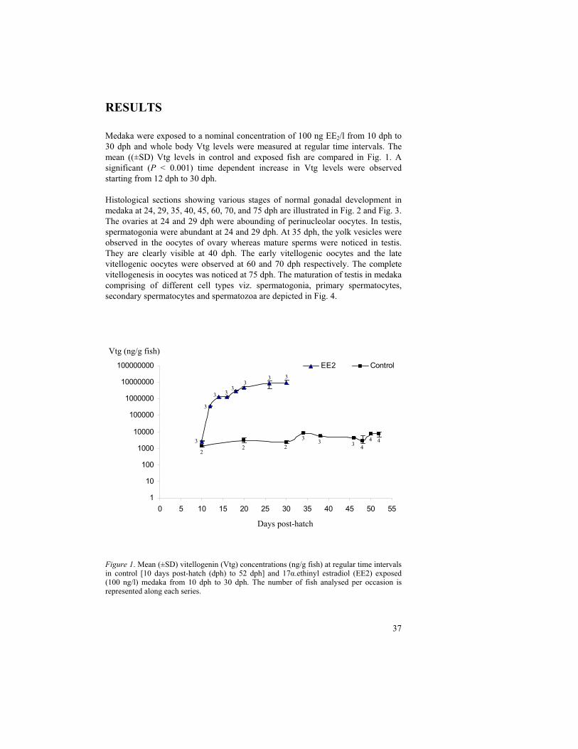
Medaka were exposed to a nominal concentration of 100 ng EE2/l from 10 dph to 30 dph and whole body Vtg levels were measured at regular time intervals. The mean ((±SD) Vtg levels in control and exposed fish are compared in Fig. 1. A significant (P < 0.001) time dependent increase in Vtg levels were observed starting from 12 dph to 30 dph.
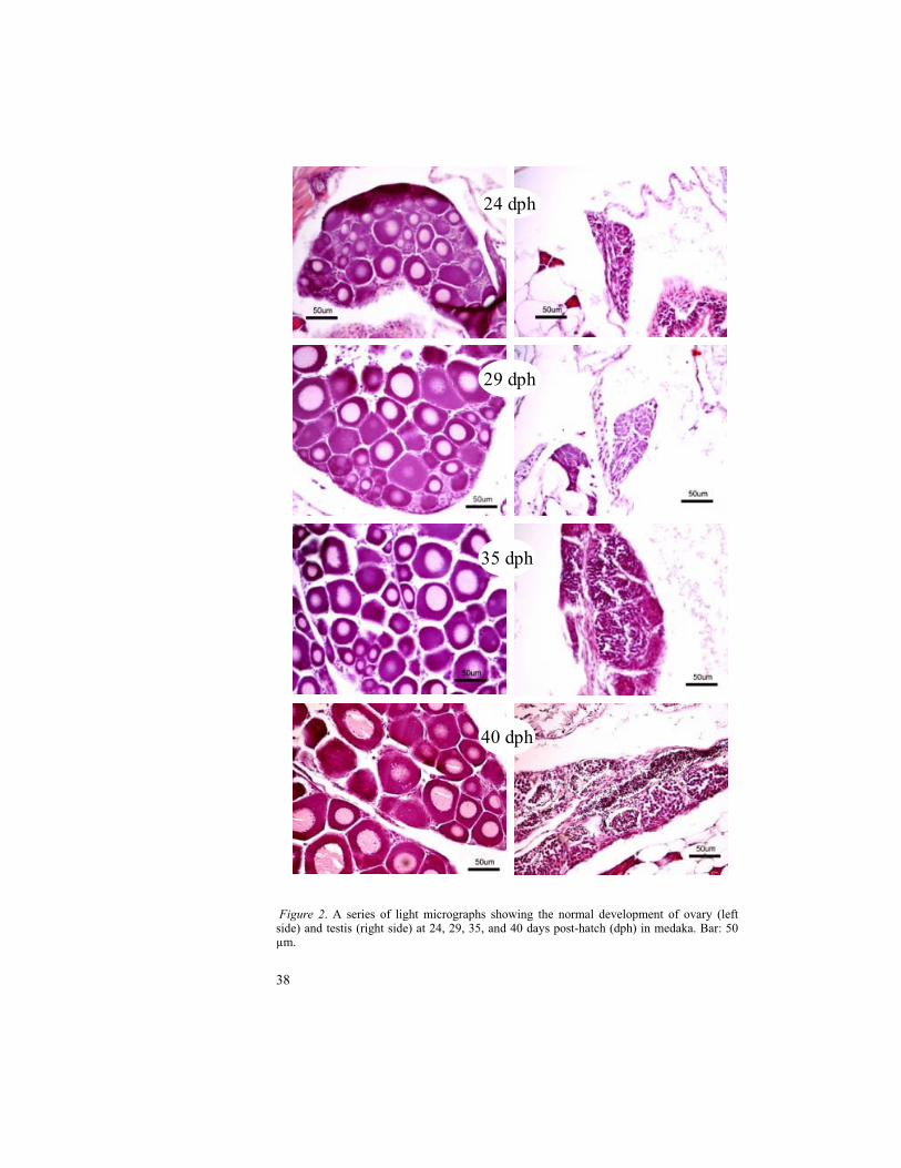
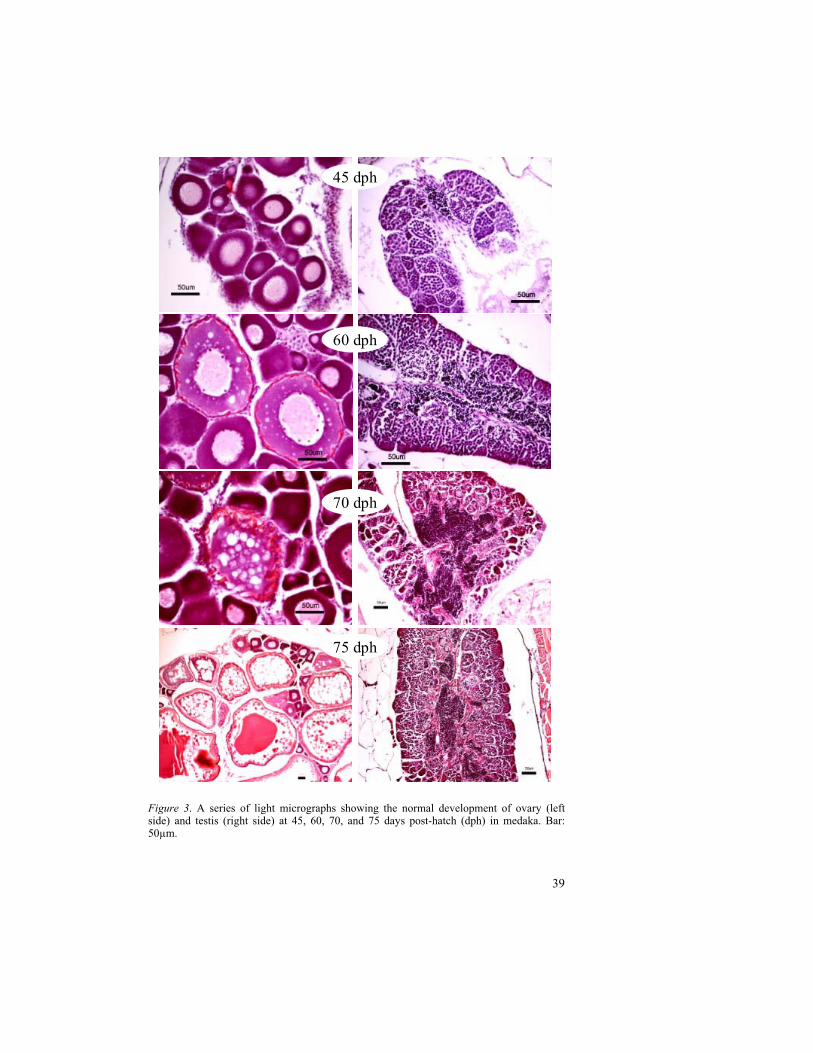
Histological sections showing various stages of normal gonadal development in medaka at 24, 29, 35, 40, 45, 60, 70, and 75 dph are illustrated in Fig. 2 and Fig. 3. The ovaries at 24 and 29 dph were abounding of perinucleolar oocytes. In testis, spermatogonia were abundant at 24 and 29 dph. At 35 dph, the yolk vesicles were observed in the oocytes of ovary whereas mature sperms were noticed in testis. They are clearly visible at 40 dph. The early vitellogenic oocytes and the late vitellogenic oocytes were observed at 60 and 70 dph respectively. The complete vitellogenesis in oocytes was noticed at 75 dph. The maturation of testis in medaka comprising of different cell types viz. spermatogonia, primary spermatocytes, secondary spermatocytes and spermatozoa are depicted in Fig. 4.
Figure 1. Mean (±SD) vitellogenin (Vtg) concentrations (ng/g fish) at regular time intervals in control [10 days post-hatch (dph) to 52 dph] and 17 .ethinyl estradiol (EE2) exposed (100 ng/l) medaka from 10 dph to 30 dph. The number of fish analysed per occasion is represented along each series.
EE2 Control
1
10
100
1000
10000
100000
1000000
10000000
100000000
0 5 10 15 20 25 30 35 40 45 50 55
Days post-hatch
Vtg (ng/g fish)
22 2
33 3
4
4 43
3
3 33
33 3
38
Figure 2. A series of light micrographs showing the normal development of ovary (left side) and testis (right side) at 24, 29, 35, and 40 days post-hatch (dph) in medaka. Bar: 50 µm.
29 dph
24 dph
35 dph
40 dph
39
Figure 3. A series of light micrographs showing the normal development of ovary (left side) and testis (right side) at 45, 60, 70, and 75 days post-hatch (dph) in medaka. Bar: 50µm.
45 dph
60 dph
70 dph
75 dph
40
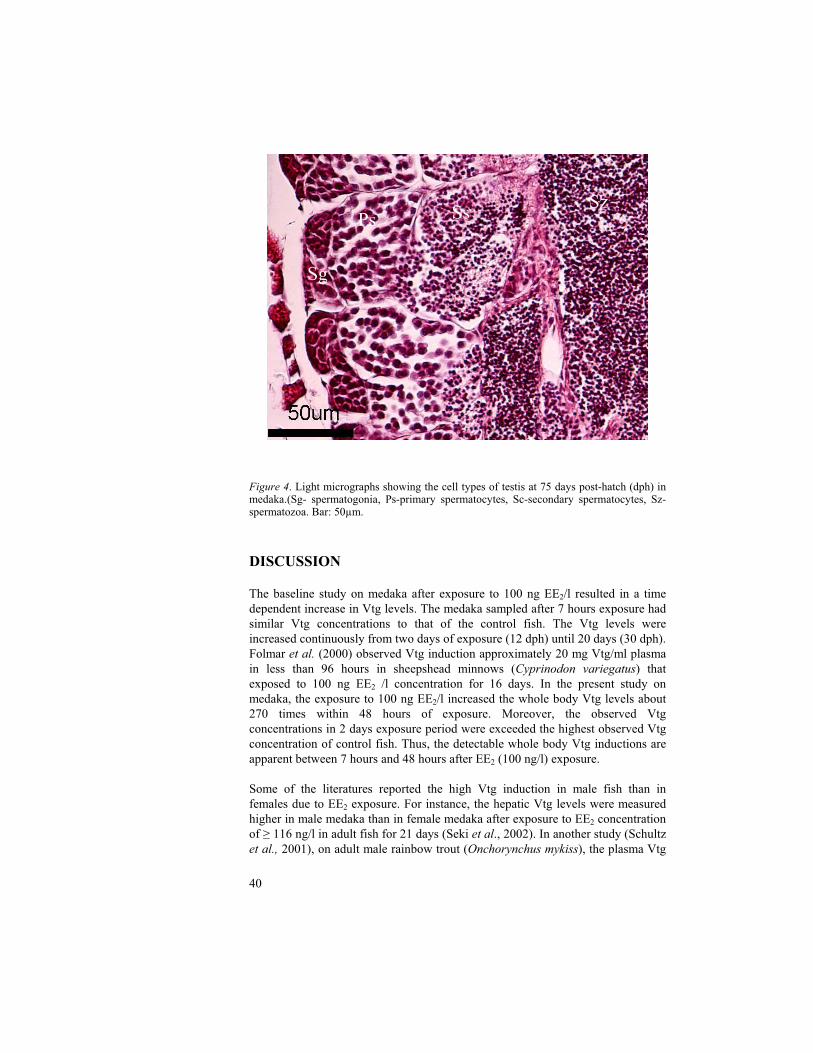
Figure 4. Light micrographs showing the cell types of testis at 75 days post-hatch (dph) in medaka.(Sg- spermatogonia, Ps-primary spermatocytes, Sc-secondary spermatocytes, Sz-spermatozoa. Bar: 50µm.
DISCUSSION
The baseline study on medaka after exposure to 100 ng EE2/l resulted in a time dependent increase in Vtg levels. The medaka sampled after 7 hours exposure had similar Vtg concentrations to that of the control fish. The Vtg levels were increased continuously from two days of exposure (12 dph) until 20 days (30 dph). Folmar et al. (2000) observed Vtg induction approximately 20 mg Vtg/ml plasma in less than 96 hours in sheepshead minnows (Cyprinodon variegatus) that exposed to 100 ng EE2 /l concentration for 16 days. In the present study on medaka, the exposure to 100 ng EE2/l increased the whole body Vtg levels about 270 times within 48 hours of exposure. Moreover, the observed Vtg concentrations in 2 days exposure period were exceeded the highest observed Vtg concentration of control fish. Thus, the detectable whole body Vtg inductions are apparent between 7 hours and 48 hours after EE2 (100 ng/l) exposure.
Some of the literatures reported the high Vtg induction in male fish than in females due to EE2 exposure. For instance, the hepatic Vtg levels were measured higher in male medaka than in female medaka after exposure to EE2 concentration of 116 ng/l in adult fish for 21 days (Seki et al., 2002). In another study (Schultz et al., 2001), on adult male rainbow trout (Onchorynchus mykiss), the plasma Vtg
Ps Ss
Sg
Sz
41
concentrations were reached the maximum above background levels within 7-9 days after exposure to intra-arterial doses ranging 0.001 to 0.1 mg EE2/kg. Nevertheless, EE2 was observed more potent estrogen in fish in responding Vtg with no regard to sex of the individual.
Gonadal development in medaka was observed from 24 dph to 75 dph in regular occasions. The stages, 24 and 29 dph showed the immature gonads with perinucleolar oocytes in ovary and spermatogonia in testis. The appearance of yolk vesicles in oocytes from 35 dph indicates the stage of cortical alveolar oocyte. In testis, the mature sperms were also started to appear at 35 dph. The beginning of gonadal maturation at 35 dph was correlated with the two-fold Vtg production between 30-38 dph of control fish in the time related study. The number of yolk vesicles in oocytes as well as number of mature sperms in testis was increased and clearly visible at 40 dph. The 60 dph stage indicates the early vitellogenic stage with yolk globules. The vitellogenesis of ovary with late vitellogenic oocytes was observed at 75 dph. Unlike the medaka, the late vitellogenic oocytes were detected at 60 dph in zebrafish (paper 2 of this thesis). It is probably due to the early hatching period (2 days) in zebrafish compared to medaka (8-10 days).
In summery, about 270-fold increase in Vtg production was observed after 48 hrs of exposure EE2 (100 ng/l) and the Vtg concentrations reached to a maximum level after two weeks exposure period. In conclusion, two-days exposure of EE2
(100 ng/l) is sufficient to cause Vtg induction in medaka whereas increased Vtg levels in control fish between 30-38 dph was correlated with the maturing gonads at 35 dph.
42
REFERENCES
Andersen, L., Holbech, H., Gessbo, A., Norrgren, L., Petersen, G.I., 2003. Effects of exposure to 17alpha-ethinylestradiol during early development on sexual differentiation and induction of vitellogenin in zebrafish (Danio rerio). Comp Biochem Physiol C Toxicol Pharmacol 134, 365-374.
Folmar, L.C., Hemmer, M., Hemmer, R., Bowman, C., Kroll, K., Denslow, N.D., 2000. Comparative estrogenicity of estradiol, ethynyl estradiol and diethylstilbestrol in an in vivo, male sheepshead minnow (Cyprinodon variegatus), vitellogenin bioassay. Aquatic Toxicol 49, 77-88.
Heppell, S.A., Denslow, N.D., Folmar, L.C., Sullivan, C.V., 1995. Universal assay of vitellogenin as a biomarker for environmental estrogens. Environ Health Perspect 103 Suppl 7, 9-15.
Hunter, G.A., Donaldson, E.M., 1983. Hormonal sex control and its application to fish culture. In: Hoar, W.S., Randall, D.J., Donaldson, E.M. (Eds.), Fish Physiology, Volume IX Reproduction, Part B Behavior and Fertility Control. Academic Press, Orlando, FL, pp. 223–303.
Länge, R., Hutchinson, T.H., Croudace, C.P., Siegmund, F., Schweinfurth, H., Hampe, P., Panter, G.H., Sumpter, J.P., 2001. Effects of the synthetic estrogen 17 alpha-ethinylestradiol on the life-cycle of the fathead minnow (Pimephales promelas). Environ Toxicol Chem 20, 1216-1227.
Metcalfe, C.D., Metcalfe, T.L., Kiparissis, Y., Koenig, B.G., Khan, C., Hughes, R.J., Croley, T.R., March, R.E., Potter, T., 2001. Estrogenic potency of chemicals detected in sewage treatment plant effluents as determined by in vivo assays with Japanese medaka (Oryzias latipes). Environ Toxicol Chem 20, 297-308.
Nimrod, A.C. and Benson, W.H., 1998. Reproduction and development of Japanese medaka following an early life stage exposure to xenoestrogens. Aquat. Toxicol. 44, pp. 141–156.
Nishi, K., Chikae, M., Hatano, Y., Mizukami, H., Yamashita, M., Sakakibara, R., Tamiya, E., 2002. Development and application of a monoclonal antibody-based sandwich ELISA for quantification of Japanese medaka (Oryzias latipes) vitellogenin. Comparative Biochemistry and Physiology Part C: Toxicology & Pharmacology 132, 161-169.
Norrgren, L., Amcoff, P., Börjesson, H. and Larsson, P-O. 1998. Reproduction Disturbances in Baltic Fish: A Review. American Fisheries Society Symposium 21:8-17.
Papoulias DM, Noltie DB, Tillitt DE. 1999. An in vivo model fish system to test chemical effects on sexual differentiation and development: exposure to ethinyl estradiol. Aquat Toxicol 48:37-50.
Parker, K.L., Schimmer, B.P., Schedl, A., 1999. Genes essential for early events in gonadal development. Cell Mol Life Sci 55, 831-838.
Piferrer, F. and Donaldson, E.M., 1992. The comparative effectiveness of the natural and a synthetic estrogen for the direct feminization of chinook salmon (Oncorhynchus tshawytscha). Aquaculture 106, pp. 183–193
43
Scholz, S., Kordes, C., Hamann, J., Gutzeit, H.O., 2004. Induction of vitellogenin in vivo and in vitro in the model teleost medaka (Oryzias latipes): comparison of gene expression and protein levels. Marine Environmental Research 57, 235-244.
Schultz, I.R., Orner, G., Merdink, J.L., Skillman, A., 2001. Dose-response relationships and pharmacokinetics of vitellogenin in rainbow trout after intravascular administration of 17alpha-ethynylestradiol. Aquat Toxicol 51, 305-318.
Seki, M., Yokota, H., Matsubara, H., Tsuruda, Y., Maeda, M., Tadokoro, H., Kobayashi, K., 2002. Effect of ethinylestradiol on the reproduction and induction of vitellogenin and testis-ova in medaka (Oryzias latipes). Environ Toxicol Chem 21, 1692-1698.
Sumpter, J.P., Jobling, S., 1995. Vitellogenesis as a biomarker for estrogenic contamination of the aquatic environment. Environmental Health Perspectives 103, 173-178.
Thompson S., F. Tilton, D. Schlenk and W.H. Benson , Comparative vitellogenic responses in three teleost species: extrapolation to in situ field studies. Mar. Environ. Res. 51(2000), pp. 185–189.
Tyler, C.R., Lubberink, K., 1996. Identification of four ovarian receptor proteins that bind vitellogenin but not other homologous plasma lipoproteins in the rainbow trout, Oncorhynchus mykiss. J Comp Physiol [B] 166, 11-20.
Van den Belt, K., Wester, P.W., van der Ven, L.T., Verheyen, R., Witters, H., 2002. Effects of ethynylestradiol on the reproductive physiology in zebrafish (Danio rerio): time dependency and reversibility. Environ Toxicol Chem 21, 767-775.
Van den Belt, K., Verheyen, R., Witters, H., 2003. Effects of 17alpha-ethynylestradiol in a partial life-cycle test with zebrafish (Danio rerio): effects on growth, gonads and female reproductive success. Sci Total Environ 309, 127-137.
Weber, L.P., Hill, J., Robert L., Janz, D.M., 2003. Developmental estrogenic exposure in zebrafish (Danio rerio): II. Histological evaluation of gametogenesis and organ toxicity. Aquatic Toxicology 63, 431-446.
Yamamoto, T.O., 1953. Artificially induced sex-reversal in genotypic males of the medaka (Oryzias latipes). J. Exp. Zool. 123, pp. 571–594.
Zillioux, E.J., Johnson, I.C., Kiparissis, Y., Metcalfe, C.D., Wheat, J.V., Ward, S.G., Liu, H., 2001. The sheepshead minnow as an in vivo model for endocrine disruption in marine teleosts: a partial life-cycle test with 17alpha-ethynylestradiol. Environ ToxicolChem20,1968-1978.
Örn, S., Holbech, H., Madsen, T.H., Norrgren, L., Petersen, G.I., 2003. Gonad development and vitellogenin production in zebrafish (Danio rerio) exposed to ethinylestradiol and methyltestosterone. Aquat Toxicol 65, 397-411.
44
Research paper II
Comparison of vitellogenin induction and gonad morphology between zebrafish and Japanese medaka after exposure to 17 -ethinylestradioland 17 -trenbolone
Suresh Yamani a, *, Stefan Örn a, Leif Norrgren a
a Division of Pathology, Department of Biomedicine and Veterinary
Public Health Sciences, Faculty of Veterinary Medicine, Swedish
University of Agricultural Sciences (SLU), SE-750 07 Uppsala;
*Corresponding author e-mail address: [email protected]
ABSTRACT
The pharmaceutical estrogen, 17 -ethinylestradiol (EE2) and the anabolic androgen, 17 -trenbolone (Tb) can interfere with the endocrine and reproductive systems of fish. Potency of these chemicals in zebrafish (Danio rerio) and Japanese medaka (Oryzias latipes) was assessed using the core endpoints, vitellogenin (Vtg) and sex ratio at 38 days post-hatch and 60 days post-hatch respectively. Sandwich enzyme-linked immunosordent assay (ELISA) was performed to measure the Vtg levels in fish whole body homogenate samples. High Vtg concentration and feminisation of fish after 10 ng EE2/l, as well as masculinisation after 50 ng Tb/l exposure were observed in zebrafish. Ova-testis (intersex) condition was observed in medaka exposed to EE2. A significant (P<0.05) decrease in Vtg production after Tb exposure (50 ng/l) was measured in both zebrafish and medaka. In contrast to the medaka, the high sensitivity of zebrafish to both EE2 and Tb was clearly indicated in the study.
Keywords: 17 -ethinylestradiol; 17 -trenbolone; vitellogenin; zebra fish; medaka
45
INTRODUCTIONA number of man-made substances with homology to natural hormones can interfere with the reproductive and endocrine systems causing adverse effects in wildlife and humans (Colborn et al., 1993; Gray, 1998; Tyler et al., 1998). Reduced reproductive ability was found in fish that are living in recipients receiving different types of effluents contaminated with the substances (Jobling et
al., 1995; Norrgren et al., 1998). Several of studies on different species of fish experimentally exposed to synthetic chemicals with properties similar to endogenous sex steroids has been shown to produce deleterious effects on reproduction. The hormonal active compounds are able to act as agonists or antagonists of the hormonal receptors, thereby causing endocrine disruption (Colborn et al. 1993; Sonnenschein and Soto, 1998). One effect of this leads to the disturbance in physiological homeostasis by increased/ decreased circulatory levels of vitellogenin (Vtg), an egg protein synthesized by liver in oviparous vertebrates. For instance, 17 -ethinylestradiol (EE2), a synthetic medicative estrogen, competes with 17 -estradiol (E2) to induce Vtg synthesis in fish leading to potentially harmful effects such as intersex (Metcalfe et al., 2001) and feminization (Papoulias et al., 1999; Örn et al., 2003). 17 -trenbolone (Tb) is an anabolic, androgenic steroid that is used as growth promoter in beef cattle in USA and Canada (Lange et al., 2001, Wilson et al., 2002). Tb acts as androgen mimic with its high affinity for androgen receptor (Pottier et al., 1981; Wilson et al., 2002) and eventually lowers the Vtg production. Tb has been repored to masculinize fathead minnow (Pimephales promelas) (Ankley et al., 2003), channel catfish (Ictalurus punctatus) (Galvez et al., 1995) and blue tilapia (Oreochromis aureus) (Galvez et al., 1996). Biomarkers and bioassays are necessary to detect the endocrine imbalance in fish caused by the anthropogenic chemicals. An in vivo screening test i.e., induction of Vtg (Sumpter and Jobling. 1995; Heppell et al., 1995; Tyler et al., 1996) in fish has become a valuable tool in laboratory experiments for the investigation of the potency of various chemicals. Some teleost fish species, such as zebrafish (Danio rerio), Japanese medaka (Oryzias latipes) and fathead minnow (Pimephales promelas), are considered as model test species for future risk assessment of chemicals.
The present study was aimed to compare the response in zebrafish (Danio rerio)and medaka (Oryzias latipes) by applying core end-points, i.e., vitellogenin and sex ratios, after exposure to model compounds viz. 17 -ethinylestradiol (EE2) and 17 -trenbolone during a sensitive part of the life-cycle.
MATERIALS AND METHODS
Test animals
Adult zebrafish aged 6–8 months were purchased from a commercial dealer. The fish were adapted to laboratory conditions for two months. A group of fish were placed in circular stainless steel spawning funnels provided with a net separating
46
the fish from the laid eggs. Newly fertilized eggs were collected within two hours after the onset of light. The eggs were transferred into glass beakers until the larvae become free swimming and then they were transferred to 10 litres stock aquaria.
The medaka fish were obtained from Japan and were kept in a tank provided with recycling water system. The eggs of medaka were collected en masse with forceps directly from the vent region where a cluster of eggs remain firm after spawning. The eggs were transferred into glass beakers. After hatching fifty free-swimming larvae were transferred to experimental 10-liters aquaria.
Test chemicals
The test chemicals, 17 -trenbolone (Tb) and 17 -ethinylestradiol (EE2) were purchased from Sigma Chemical Company®, Sweden and were dissolved in methanol to make the stock solutions at a concentration of 10 mg/l. The exposure of EE2 was performed in 10 ng/l and 100 ng/l for medaka, whereas only the low concentration i.e. 10 ng/l was used for zebrafish. Trenbolone was used in concentrations of 10 ng/l and 50 ng/l for both species.
Test procedure
The fish were exposed to the chemicals through an aqueous route starting one day after hatching until 60 days post-hatch, under semi-static water conditions. The water was renewed with 50% of the test volume every second day. Aerated dechlorinated tap water was used for the renewal of test volume. The water was at a temperature of 26±2oC, pH range 7-8 and a 12 h dark / 12 h light regime was maintained throughout the test procedure. Air was bubbled into the water through a syringe needle in each aquarium to maintain adequate dissolved oxygen concentration. Fish were fed ad libitum three times daily, once with live Artemia
nauplii and twice with commercial food (Sera micron®, Nutrafin®, Tetra Min®). All exposures, including controls, were performed in duplicates with 50 fish per aquarium.
Sampling
From each aquarium, five fish were sampled at 38 days post-hatch and were frozen in liquid nitrogen. The samples were kept at -80oC until Vtg analysis. At 60 days post-hatch, all the remaining fish were sampled, fixed in neutral buffered formalin and processed for light microscopy.
Homogenization and vitellogenin analysis
The frozen whole fish was weighed and homogenized individually using homogenization buffer (12ml of Tris Hcl + 2 mg of aprotinin + 120 µl of PMSF)
47
in 10 times the weight of each fish. The homogenate was centrifuged at 16000×g (4ºC) for one hour and the supernatant below the fat layer was collected individually to determine whole-body homogenate Vtg concentrations. Measurement of Vtg protein in the supernatant samples was performed by using a commercially available, pre-coated vitellogenin ELISA kit from Biosense laboratories® (Norway). The procedure was followed according to the manufacturer's instructions and the absorbance of samples in the ELISA-plate-wells was determined using microtiter plate reader and Vtg concentration whole body homogenate for each fish was calculated.
Histological preparation
The tail and head of each fish were cut to obtain the required central portion and placed in labeled plastic cassettes. They were dehydrated using 70% to absolute ethanol, treated with xylene and finally embedded in paraffin. Each paraffin block contained between 6-8 individuals. The paraffin blocks were sectioned sagittally on a microtome to get sections of size 3-4 microns. About six sections per fish were cut. The sections were transferred on to glass slides and placed on a heating plate for one hour to allow them settle by drying. Then the sections were deparaffinized with xylene and rehydrated using a graded series of ethanol and finally with tap water to make the sections ready for staining by hematoxylin and eosin. After the staining, sections were dehydrated again in ethanol and xylene and then mounted with cover slips to fix them for histological examination using light microscope.
Histopathological analysis
The processed histological sections of fish were examined to determine the sex and gonadal development of each fish. The number of oocytes at different stages was counted and the percentage of each cell type was calculated. In testes, the stages of spermatogenesis were examined. The sections were pictured using Nikon® digital camera (DXM 1200) attached to the light microscope, Nikon eclipse® E600.
Statistical analysis
The data obtained from vitellogenin measurements were analysed by non-parametric Mann-Whitney U test for significant (P < 0.05) differences between the controls and the the exposed groups. Fisher’s Exact Test was used to recognize differences in sex ratios between control and each treatment group.
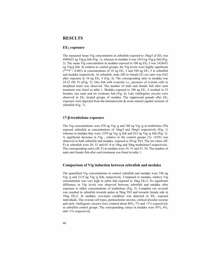
48
RESULTS
EE2 exposure
The measured mean Vtg concentration in zebrafish exposed to 10ng/l of EE2 was 4986622 ng Vtg/g fish (Fig. 1), whereas in medaka it was 2414 ng Vtg/g fish (Fig. 2). The mean Vtg concentration in medaka exposed to 100 ng EE2 /l was 1424432 ng Vtg/g fish. In relation to control groups, the Vtg levels were highly significant (***P < 0.001) at concentrations of 10 ng EE2 /l and 100 ng EE2 /l in zebrafish and medaka respectively. In zebrafish, male (M) to female (F) sex ratio was 0:62 after exposure to 10 ng EE2 /l (Fig. 4). The corresponding ratio in medaka was 24:22 (M: F) ((Fig. 5). One fish with ovatestis i.e., presence of ovarian cells in atrophied testis was observed. The number of male and female fish after each treatment was listed in table 1. Medaka exposed to 100 ng EE2 /l resulted in 53 females, one male and six ovatestes fish (Fig. 6). Late vitellogenic oocytes were observed in EE2 treated groups of medaka. The suppressed gonads after EE2
exposure were depicted from the haematoxylin & eosin stained sagittal sections of zebrafish (Fig. 7).
17- -trenbolone exposure
The Vtg concentrations were 478 ng Vtg /g and 386 ng Vtg /g in trenbolone (Tb) exposed zebrafish at concentrations of 10ng/l and 50ng/l respectively (Fig. 1) whereas in medaka they were 1539 ng Vtg /g fish and 813 ng Vtg /g fish (Fig. 2). A significant decrease in Vtg , relative to the control groups (*p <0.05) was observed in both zebrafish and medaka exposed to 50 ng Tb/l. The sex ratios (M: F) in zebrafish were 20: 51 and 65: 0 at 10ng and 50ng trenbolone/l respectively. The corresponding ratios (M: F) in medaka were 54: 51 and 51: 59. The number of male and female fish after each treatment was listed in table 1.
Comparison of Vtg induction between zebrafish and medaka
The quantified Vtg concentrations in control zebrafish and medaka were 546 ng Vtg /g and 2119 ng Vtg /g fish, respectively. Compared to medaka, relative Vtg concentration was very high in zebra fish exposed to 10ng EE2/l. No significant difference in Vtg levels was observed between zebrafish and medaka after exposure to either concentrations of trenbolone (Fig. 3). Complete sex reversal was resulted in zebrafish towards males at 50ng Tb/l and towards female side at 10ng EE2/l. In medaka, ova-testis condition was detected in EE2 exposed individuals. The ovarian cell types, perinucleolar oocytes, cortical alveolar oocytes and early vitellogenic oocytes were counted about 86%, 7% and <1% respectively in zebrafish control groups. The corresponding values in medaka were 93%, 4%, and <1% respectively.
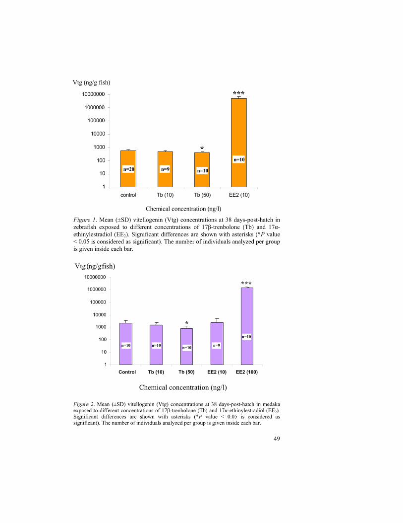
49
Figure 1. Mean (±SD) vitellogenin (Vtg) concentrations at 38 days-post-hatch in zebrafish exposed to different concentrations of 17 -trenbolone (Tb) and 17 -ethinylestradiol (EE2). Significant differences are shown with asterisks (*P value < 0.05 is considered as significant). The number of individuals analyzed per group is given inside each bar.
Figure 2. Mean (±SD) vitellogenin (Vtg) concentrations at 38 days-post-hatch in medaka exposed to different concentrations of 17 -trenbolone (Tb) and 17 -ethinylestradiol (EE2). Significant differences are shown with asterisks (*P value < 0.05 is considered as significant). The number of individuals analyzed per group is given inside each bar.
1
10
100
1000
10000
100000
1000000
10000000
control Tb (10) Tb (50) EE2 (10)
Chemical concentration (ng/l)
n=9 n=10
n=10
n=20
Vtg (ng/g fish)
***
*
1
10
100
1000
10000
100000
1000000
10000000
control Tb (10) Tb (50) EE2 (10) EE2 (100)
Vtg ( ng/g fish)
***
*
Chemical concentration (ng/l)
1
10
100
1000
10000
100000
1000000
10000000
Control Tb (10) Tb (50) EE2 (10) EE2 (100)
***
*
n=10 n=10 n=10 n=9
n=10
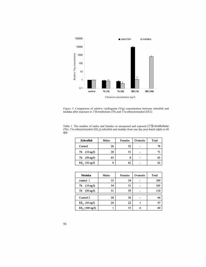
50
Figure 3. Comparison of relative vitellogenin (Vtg) concentration between zebrafish and medaka after exposure to 17 -trenbolone (Tb) and 17 -ethinylestradiol (EE2)
Table 1. The number of males and females in unexposed and exposed [17 -trenbolone (Tb), 17 -ethinylestradiol (EE2)] zebrafish and medaka from one day post-hatch (dph) to 60 dph
0.1
1
10
100
1000
10000
100000
control Tb (10) Tb (50) EE2 (10) EE2 (100)
zebra fish medaka
Chemical concentration (ng/l)
Rel
ativ
e V
tgco
ncen
trat
ion
110-5951Tb (50 ng/l)
105-5154Tb (10 ng/l)
109-5455control 1
TotalOvatestisFemalesMales Medaka
110-5951Tb (50 ng/l)
105-5154Tb (10 ng/l)
109-5455control 1
TotalOvatestisFemalesMales Medaka
62-620EE2 (10 ng/l)
65-065Tb (50 ng/l)
71-5120Tb (10 ng/l)
78-5226Control
TotalOvatestisFemalesMales Zebrafish
62-620EE2 (10 ng/l)
65-065Tb (50 ng/l)
71-5120Tb (10 ng/l)
78-5226Control
TotalOvatestisFemalesMales Zebrafish
606531EE2 (100 ng/l)
4712224EE2 (10 ng/l)
64-3628Control 2
606531EE2 (100 ng/l)
4712224EE2 (10 ng/l)
64-3628Control 2
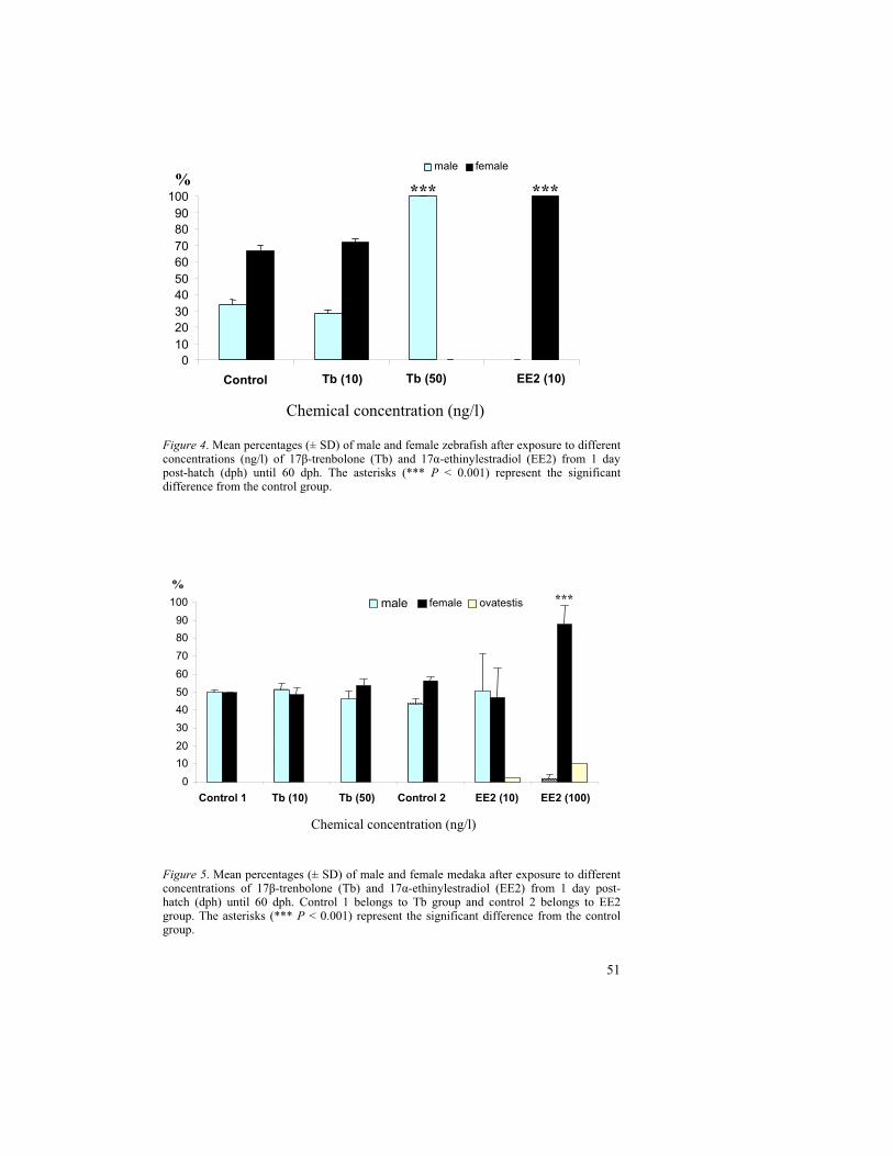
51
Figure 4. Mean percentages (± SD) of male and female zebrafish after exposure to different concentrations (ng/l) of 17 -trenbolone (Tb) and 17 -ethinylestradiol (EE2) from 1 day post-hatch (dph) until 60 dph. The asterisks (*** P < 0.001) represent the significant difference from the control group.
Figure 5. Mean percentages (± SD) of male and female medaka after exposure to different concentrations of 17 -trenbolone (Tb) and 17 -ethinylestradiol (EE2) from 1 day post-hatch (dph) until 60 dph. Control 1 belongs to Tb group and control 2 belongs to EE2 group. The asterisks (*** P < 0.001) represent the significant difference from the control group.
0
10
20
30
40
50
60
70
80
90
100
Control Tb (10) Tb (50) EE2 (10)
male female
%*** ***
Chemical concentration (ng/l)
0
10
20
30
40
50
60
70
80
90
100
Control 1 Tb (10) Tb (50) Control 2 EE2 (10) EE2 (100)
male female ovatestis
%***
Chemical concentration (ng/l)
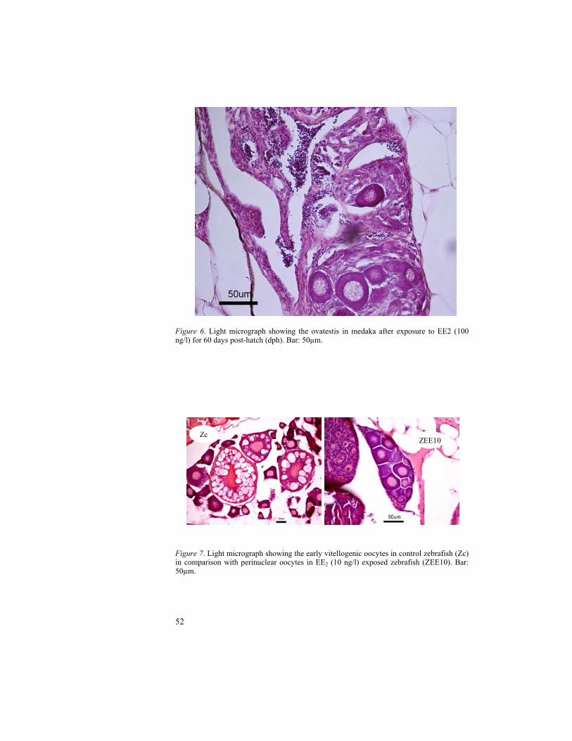
52
Figure 6. Light micrograph showing the ovatestis in medaka after exposure to EE2 (100 ng/l) for 60 days post-hatch (dph). Bar: 50µm.
Figure 7. Light micrograph showing the early vitellogenic oocytes in control zebrafish (Zc) in comparison with perinuclear oocytes in EE2 (10 ng/l) exposed zebrafish (ZEE10). Bar: 50µm.
ZEE10Zc

53
DISCUSSION
Recent studies investigating fish in response to EDCs during the early developmental period indicate that there are considerable differences in sensitivity among fish species. Genetic variation and disparate patterns of sexual differentiation are probable causes for the interspecific differences in sensitivity. Nevertheless, an alteration in sensitivity also depends on the route of exposure (i.e., aqueous, dietary, direct injection through intra-muscular or intra-peritoneal route) and exposure system viz. flow-through, semi-static/ static renewal system.
The present study clearly shows the difference in sensitivity between the zebrafish and the medaka to EE2 exposure and supports the statement that zebrafish is a highly estrogen sensitive member of the cyprinid family (Rose et al., 2002). As an estrogen agonist, EE2 binds to the estrogen receptor (ER) by imitating 17 -estradiol. This leads to enhanced levels of circulatory vitellogenin (Vtg). The lowest observed effect concentration (LOEC) of EE2 for Vtg induction in zebrafish was 1.67ng/l after 21 days exposure (Fenske et al., 2001) and 2.97ng/l after 8 days exposure (Rose et al., 2002) periods. Vtg induction is highly variable and influenced by several factors such as duration of exposure, stage of life cycle and species. Örn et al. (2003) indicated the dose dependent Vtg production in EE2
exposed zebrafish from 20 dph to 38 dph at concentrations 2-25ng/l. A significant (P<0.05) increase in Vtg production was indicated during different exposure periods between hatching to 60 dph of zebrafish exposed to 15.4±1.4ng EE2/l (Andersen et al., 2003). However, diversified results are associated with indifferent experimental conditions of the test protocol with regard to water temperature, pH, photoperiod etc (Baroiller et al., 1999).
Van den belt et al. (2003) reported that the measured Vtg concentrations in EE2
exposed (hatch to 3 months) zebrafish whole body homogenates were significant at 10 (**P<0.01) and 25 ng/l (***P<0.001). However, in the present study (hatch to 38 dph), the Vtg induction was highly significant (***P < 0.001) even at the concentration of 10ng EE2 /l when compared to unexposed group. Örn et al.
(2003) reported that the mean Vtg concentration was nearly one million ng/g fish in EE2 (10ng/l) exposed zebrafish form 20 dph to 38 dph, whereas the Vtg concentration in the current study (hatch to 38 dph) was about five million ng/g fish. This clearly indicates the enhanced Vtg induction when EE2 exposure was initiated right from hatching. In contrast, the adult zebrafish are also pliable to respond to EE2 exposure irrespective of their sex. For instance, about 200-fold increase in Vtg levels was found when adult male zebrafish were exposed to 10ng EE2/l for 7 days (Holbech et al., 2001). Petersen et al. (2000) reported the induced Vtg concentration in adult zebrafish as approximately 2270000 ng /g fish after exposure for 30 days at 20 ng EE2/l.
With regard to the medaka, the LOEC of EE2 for hepatic Vtg induction in exposed adult males was 63.9 ng/l (Seki et al., 2002). In the present medaka study, there were two exposure concentrations i.e., 10 and 100ng/l to determine the whole
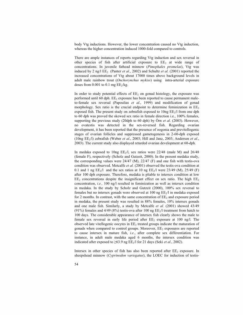
54
body Vtg inductions. However, the lower concentration caused no Vtg induction, whereas the higher concentration induced 1000-fold compared to controls.
There are ample instances of reports regarding Vtg induction and sex reversal in other species of fish after artificial exposure to EE2 at wide range of concentrations. In juvenile fathead minnow (Pimephales promelas), Vtg was induced by 2 ng/l EE2 (Panter et al., 2002) and Schultz et al. (2001) reported the increased concentrations of Vtg about 17000 times above background levels in adult male rainbow trout (Onchorynchus mykiss) using intra-arterial exposure doses from 0.001 to 0.1 mg EE2/kg.
In order to study potential effects of EE2 on gonad histology, the exposure was performed until 60 dph. EE2 exposure has been reported to cause permanent male-to-female sex reversal (Papoulias et al., 1999) and modification of gonad morphology. Sex ratio is the crucial endpoint to determine feminization in EE2
exposed fish. The present study on zebrafish exposed to 10ng EE2/l from one dph to 60 dph was proved the skewed sex ratio in female direction i.e., 100% females, supporting the previous study (20dph to 60 dph) by Örn et al. (2003). However, no ovatestis was detected in the sex-reversed fish. Regarding ovarian development, it has been reported that the presence of oogonia and previtellogenic stages of ovarian follicles and suppressed gametogenesis in 2-60-dph exposed (10ng EE2/l) zebrafish (Weber et al., 2003; Hill and Janz, 2003; Andersen et al., 2003). The current study also displayed retarded ovarian development at 60-dph.
In medaka exposed to 10ng EE2/l, sex ratios were 22/48 (male M) and 26/48 (female F), respectively (Scholz and Gutzeit, 2000). In the present medaka study, the corresponding values were 24/47 (M), 22/47 (F) and one fish with testis-ova condition was observed. Metcalfe et al. (2001) observed the testis-ova condition at 0.1 and 1 ng EE2/l and the sex ratios at 10 ng EE2/l were 23/49 (M), 25/49 (F) after 100 dph exposure. Therefore, medaka is pliable to intersex condition at low EE2 concentrations despite the insignificant effect on sex ratio. The high EE2
concentration, i.e., 100 ng/l resulted in feminization as well as intersex condition in medaka. In the study by Scholz and Gutzeit (2000), 100% sex reversal to females but no intersex gonads were observed at 100 ng EE2/l in medaka exposed for 2 months. In contrast, with the same concentration of EE2 and exposure period in medaka, the present study was resulted in 88% females, 10% intersex gonads and one male fish. Similarly, a study by Metcalfe et al. (2001) showed 43/49 (91%) females and 4/49 (8%) testis-ova after 100 ng EE2/l treatment from hatch to 100 days. The considerable appearance of intersex fish clearly shows the male to female sex reversal in early life period after EE2 exposure at 100 ng/l. The observed late vitellogenic oocytes in EE2 treated groups indicate the maturation of gonads when compared to control groups. Moreover, EE2 exposures are reported to cause intersex in mature fish, i.e., after complete sex differentiation. For instance, in adult male medaka aged 6 months, the intersex condition was indicated after exposed to 63.9 ng EE2/l for 21 days (Seki et al., 2002).
Intersex in other species of fish has also been reported after EE2 exposure. In sheepshead minnow (Cyprinodon variegatus), the LOEC for induction of testis-
55
ova was 20 ng EE2/l (Zillioux et al., 2001). A male:female ratio of 5:84 with intersex in 11 % of fish was observed in fathead minnow (Pimephales promelas)at 56 days post hatch after exposure to 4 ng EE2 /l (Länge et al., 2001).
The growth promoter in cattle, trenbolone has recently been reported to produce masculinizing effects in fish. Ankley et al. (2003) noticed that a minimum concentration of 27ng Tb/l can cause a significant decrease in Vtg concentrations, masculinizing effect and decline in fecundity in female fathead minnow (Pimephales promelas) after exposure for 21 days. In the present study, zebrafish and medaka were exposed to 17 -trenbolone in order to assess the crucial endpoints, Vtg and sex ratio. The higher concentration of trenbolone, i.e., 50 ng/l resulted in significant decrease in Vtg levels in both zebrafish and medaka at 38 days post hatch. Contradictory, the sex ratios varied between the two species. In zebrafish, the higher concentration caused skewed sex reversal in male direction, whereas no effect on sex ratio was observed in medaka. It elucidates the suppressive effect of trenbolone at physiological level despite the unapparent phenotypic expression in medaka. The lower concentration (10 ng/l) of trenbolone caused no significant effect on either Vtg or sex ratios in both the species. The higher concentration (50 ng/l) indicates the possible trenbolone associated androgen mimicking endocrine disruption in zebrafish with regard to Vtg as well as sex ratios.
The present study was purposed to compare the potential effects of Tb and EE2
with similar concentrations for both the test species. However, a study by Hill and Janz (2003) reported that EE2 induced high mortality rate (>90%) in zebrafish exposed to 100 ng/l during 2–60 dph. In fact, unlike the medaka, the EE2
concentration, 100 ng/l is highly toxic in zebrafish and it is impractical to assign the sex ratio with few survivals after exposure. Thus, the high EE2 concentration was not used in the present zebrafish exposure study to compare with medaka. The background mean Vtg levels in medaka were about 4 times higher to that of zebrafish at 38 days post hatch. It is probably due to the difference in hatching time i.e., 8 days in medaka and 3 days in zebrafish.
In summary, the relative Vtg concentration in zebrafish was observed approximately 10000 times higher than medaka after exposure to 10 ng EE2 /l. Moreover, there was 100% feminization in zebrafish at the same concentration. Similarly, exposure to 50 ng/l of trenbolone resulted in 100% masculinization. Therefore, the difference in sensitivity between zebrafish and medaka with regard to the core endpoints, Vtg and sex ratio was apparent. In conclusion, the present study disclosed that the zebrafish is highly sensitive to estrogen mimics as well as androgen mimics when compared to the medaka.
56
REFERENCES
Andersen, L., Holbech, H., Gessbo, A., Norrgren, L., Petersen, G.I., 2003. Effects of exposure to 17alpha-ethinylestradiol during early development on sexual differentiation and induction of vitellogenin in zebrafish (Danio rerio). Comp Biochem Physiol C Toxicol Pharmacol 134, 365-374.
Ankley, G.T., Jensen, K.M., Makynen, E.A., Kahl, M.D., Korte, J.J., Hornung, M.W., Henry, T.R., Denny, J.S., Leino, R.L., Wilson, V.S., Cardon, M.C., Hartig, P.C., Gray, L.E., 2003. Effects of the androgenic growth promoter 17-beta-trenbolone on fecundity and reproductive endocrinology of the fathead minnow. Environ Toxicol Chem 22, 1350-1360.
Baroiller, J. F., Guigen, Y. & Fostier, A., 1999. Endocrine and environmental aspects of sex differentiation in fish. Cellular and Molecular Life Sciences 55, 910–931.
Colborn, T., vom Saal, F.S., Soto, A.M., 1993. Developmental effects of endocrine-disrupting chemicals in wildlife and humans. Environ Health Perspect 101, 378-384.
Fenske, M., R. van Aerle, S. Brack, C.R. Tyler, and H. Segner, Development and validation of a homologous zebrafish (Danio rerio Hamilton-Buchanan) vitellogenin enzyme-linked immunosorbent assay (ELISA) and its application for studies on estrogenic chemicals. Comp Biochem Physiol C Toxicol Pharmacol, 2001. 129(3): p. 217-32.
Galvez, J.I., Mazik, P.M., Phelps, R.P., Mulvaney, D.R., 1995. Masculinization of channel catfish (Ictalurus punctatus) by oral administration of trenbolone acetate. J. World Aquacult. Soc. 26, 378–383.
Galvez JI, Morrison JR. 1996. Efficacy of trenbolone acetate insex inversion of the blue tilapia (Oreochromis aureus) J. World Aquacult Soc 27:483–486.
Gray, L.E., Jr., 1998. Xenoendocrine disrupters: laboratory studies on male reproductive effects. Toxicol Lett 102-103, 331-335.
Heppell, S.A., Denslow, N.D., Folmar, L.C., Sullivan, C.V., 1995. Universal assay of vitellogenin as a biomarker for environmental estrogens. Environ Health Perspect 103 Suppl 7, 9-15.
Hill, R.L., Jr., Janz, D.M., 2003. Developmental estrogenic exposure in zebrafish (Danio rerio): I. Effects on sex ratio and breeding success. Aquat Toxicol 63, 417-429.
Holbech, H., Andersen, L., Petersen, G.I., Korsgaard, B., Pedersen, K.L., Bjerregaard, P., 2001. Development of an ELISA for vitellogenin in whole body homogenate of zebrafish (Danio rerio). Comp Biochem Physiol C Toxicol Pharmacol 130, 119-131.
Jobling, S., Reynolds, T., White, R., Parker, M.G., Sumpter, J.P., 1995. A variety of environmentally persistent chemicals, including some phthalate plasticizers, are weakly estrogenic. Environ Health Perspect 103, 582-587.
Lange, I.G., Daxenberger, A., Meyer, H.H., 2001. Hormone contents in peripheral tissues after correct and off-label use of growth promoting hormones in cattle: effect of the implant preparations Filaplix-H, Raglo, Synovex-H and Synovex Plus. Apmis 109, 53-65.

57
Länge, R., Hutchinson, T.H., Croudace, C.P., Siegmund, F., Schweinfurth, H., Hampe, P., Panter, G.H., Sumpter, J.P., 2001. Effects of the synthetic estrogen 17 alpha-ethinylestradiol on the life-cycle of the fathead minnow (Pimephales promelas). Environ Toxicol Chem 20, 1216-1227.
Metcalfe, C.D., Metcalfe, T.L., Kiparissis, Y., Koenig, B.G., Khan, C., Hughes, R.J., Croley, T.R., March, R.E., Potter, T., 2001. Estrogenic potency of chemicals detected in sewage treatment plant effluents as determined by in vivo assays with Japanese medaka (Oryzias latipes). Environ Toxicol Chem 20, 297-308.
Norrgren, L., Amcoff, P., Börjesson, H. and Larsson, P-O. 1998. Reproduction Disturbances in Baltic Fish: A Review. American Fisheries Society Symposium 21:8-17.
Papoulias DM, Noltie DB, Tillitt DE. 1999. An in vivo model fish system to test chemical effects on sexual differentiation and development: exposure to ethinyl estradiol. Aquat Toxicol 48:37-50.
Papoulias, D.M., Noltie, D.B., Tillitt, D.E., 2000. Effects of methyl testosterone exposure on sexual differentiation in medaka, Oryzias latipes. Mar Environ Res 50, 181-184.
Petersen, G.I., Andersen, L., Holbech, H. and Pedersen, K.L., 2000. Baseline test studies and development of ELISA. In: Andersen, L., Bengtsson, B.-E., Björk, M., Gessbo, Å., Holbech, H., Hylland, K. et al., 2000. Zebra Fish for Testing Endocrine Disrupting Chemicals, TemaNord 555, Copenhagen, pp. 35–46.
Pottier, J., Cousty, C., Heitzman, R. J., and Reynolds, I. P. (1981). Differences in the biotransformation of a 17ß-hydroxylated steroid, trenbolone acetate, in rat and cow. Xenobiotica 11, 489–500.
Panter, G.H., Hutchinson, T.H., Lange, R., Lye, C.M., Sumpter, J.P., Zerulla, M., Tyler, C.R., 2002. Utility of a juvenile fathead minnow screening assay for detecting (anti-)estrogenic substances. Environ Toxicol Chem 21, 319-326.
Rose, J., Holbech, H., Lindholst, C., Norum, U., Povlsen, A., Korsgaard, B., Bjerregaard, P., 2002. Vitellogenin induction by 17beta-estradiol and 17alpha-ethinylestradiol in male zebrafish (Danio rerio). Comp Biochem Physiol C Toxicol Pharmacol 131, 531-539.
Scholz, S., Gutzeit, H.O., 2000. 17-[alpha]-ethinylestradiol affects reproduction, sexual differentiation and aromatase gene expression of the medaka (Oryzias latipes). Aquatic Toxicology 50, 363-373.
Schultz, I.R., Orner, G., Merdink, J.L., Skillman, A., 2001. Dose-response relationships and pharmacokinetics of vitellogenin in rainbow trout after intravascular administration of 17alpha-ethynylestradiol. Aquat Toxicol 51, 305-318.
Sonnenschein, C., Soto, A.M., 1998. An updated review of environmental estrogen and androgen mimics and antagonists. J Steroid Biochem Mol Biol 65, 143-150.
Soto, A.M., Calabro, J.M., Prechtl, N.V., Yau, A.Y., Orlando, E.F., Daxenberger, A., Kolok, A.S., Guillette, L.J., Jr., le Bizec, B., Lange, I.G., Sonnenschein, C., 2004. Androgenic and estrogenic activity in water bodies receiving cattle feedlot effluent in Eastern Nebraska, USA. Environ Health Perspect 112, 346-352.
Sumpter, J.P., Jobling, S., 1995. Vitellogenesis as a biomarker for estrogenic contamination of the aquatic environment. Environmental Health Perspectives 103, 173-178.
58
Seki, M., Yokota, H., Matsubara, H., Tsuruda, Y., Maeda, M., Tadokoro, H., Kobayashi, K., 2002. Effect of ethinylestradiol on the reproduction and induction of vitellogenin and testis-ova in medaka (Oryzias latipes). Environ Toxicol Chem 21, 1692-1698.
Tyler, C.R., Lubberink, K., 1996. Identification of four ovarian receptor proteins that bind vitellogenin but not other homologous plasma lipoproteins in the rainbow trout, Oncorhynchus mykiss. J Comp Physiol [B] 166, 11-20.
Tyler, C.R., Jobling, S., Sumpter, J.P., 1998. Endocrine disruption in wildlife: a critical review of the evidence. Crit Rev Toxicol 28, 319-361.
Van den Belt, K., Verheyen, R., Witters, H., 2003. Effects of 17alpha-ethynylestradiol in a partial life-cycle test with zebrafish (Danio rerio): effects on growth, gonads and female reproductive success. Sci Total Environ 309, 127-137.
Weber, L.P., Hill, J., Robert L., Janz, D.M., 2003. Developmental estrogenic exposure in zebrafish (Danio rerio): II. Histological evaluation of gametogenesis and organ toxicity. Aquatic Toxicology 63, 431-446.
Wilson, V.S., Lambright, C., Ostby, J., Gray, L.E., Jr., 2002. In vitro and in vivo effects of 17beta-trenbolone: a feedlot effluent contaminant. Toxicol Sci 70, 202-211.
Zillioux, E.J., Johnson, I.C., Kiparissis, Y., Metcalfe, C.D., Wheat, J.V., Ward, S.G., Liu, H., 2001. The sheepshead minnow as an in vivo model for endocrine disruption in marine teleosts: a partial life-cycle test with 17alpha-ethynylestradiol. Environ ToxicolChem20,1968-1978.
Örn, S., Holbech, H., Madsen, T.H., Norrgren, L., Petersen, G.I., 2003. Gonad development and vitellogenin production in zebrafish (Danio rerio) exposed to ethinylestradiol and methyltestosterone. Aquat Toxicol 65, 397-411.
59
ACKNOWLEDGEMENTS
Firstly, I would greatly thankful to the Swedish Foundation for International Co-operation in Research and Higher Education (STINT) for providing me the scholarship to pursue my Master’s degree.
I would like to express my gratitude to:
Karin Östensson, Marie Sundberg and all the staff (SIPAR) for my acceptation to the special Master of Science Programme in Veterinary Medicine for International Students;
Professor Leif Norrgren, my supervisor, Head of the department, for the given opportunity of proposed Master’s project, valuable suggestions, guidance to the thesis and for the financial assistance to continue my research with another project: Tack så mycket;
Stefan Örn, my sub-supervisor, friend for his technical assistance in my experimentation, for his patience in answering to my questions ( ), discussions all the time and for statistical, linguistic help to the thesis: Tack så jätte mycket;
Åsa Gessbo, for the preparation of nice histological sections of fish and making them available in time;
Anne-Sofie Lundquist, for the cordial reception at the airport on my arrival to Sweden, for assistance in all the practical matters, and for lending me the bike;
My examination committee members, Patric Amcoff, Eva Janssen and Hans Tjälve for their patience to review this thesis and the given suggestions;
Dr. Maria Hurst, Assoc. Prof. Ronny Lindberg, Prof. Wilhelm Engström and Prof. Stina Ekman, Prof. Wilhelm Tham and Prof. Marie-Louise with your kind approachability and perceptiveness emancipated my mind;
My aquatic family members (Norrgren et al.), Anna Norman, Gunnar Carlsson, Stephanie and Jenny Rydh, for their good companionship and fun with working together;
Britta Ojava, Tapio Nikkilä for sharing their knowledge in general (long) discussions about.... bla..... bla….. bla…..making me to get relief from work;
Sten, the computer professional, for his quick response to fulfill the requirements of my computer;
Susan Pajuluoma and Mats Forsberg, your valuable lessons on scientific english helped me a lot in writing this thesis;
60
Prof. Agneta Oskarsson, Jonas Talkvist, Pia Larsson, Elisabet Ekman, Ulla Hammarström, Fredrik Södersten, Susanne, Helena Öhrvik, Eva Tyden, Cecilia ley, Lena Olsen, Agneta Boström with your nice greetings making me refresh everyday;
My friends, international co-Master’s students, Amitha Baddamwar, Vishal Singh Parihar, Tuempong Wongtawan, Duong Chi Mai, Carlos Eduardo Hernandez, and Pushkar Kulkarni with their mutual co-operation, get-togethers, tours etc making my travel in a real friend-SHIP;
All my corridor friends for their Swedish language lessons, assistance in practical problems, parties, going out for downhill on snow etc. making me joy;
My brother, Naresh for inspiration to come to Sweden with enough information to start my life in this cold country and your regular contact to solve my practical problems and advice for my future;
My sister, Niveditha for your encouragement, directions that making my study well and taking me to the right path;
My mother, Rushendra Mani and my father, Rama Murthy with your love and support keeping my mental health in good condition to drive my life smoothly;
Finally, beseeching the God to forgive me for sacrificing the fish that are greatly contributed to this thesis.

