aggressive malignant hematopoietic disorders accumulation of abnormal blasts ( immature precursors...
TRANSCRIPT
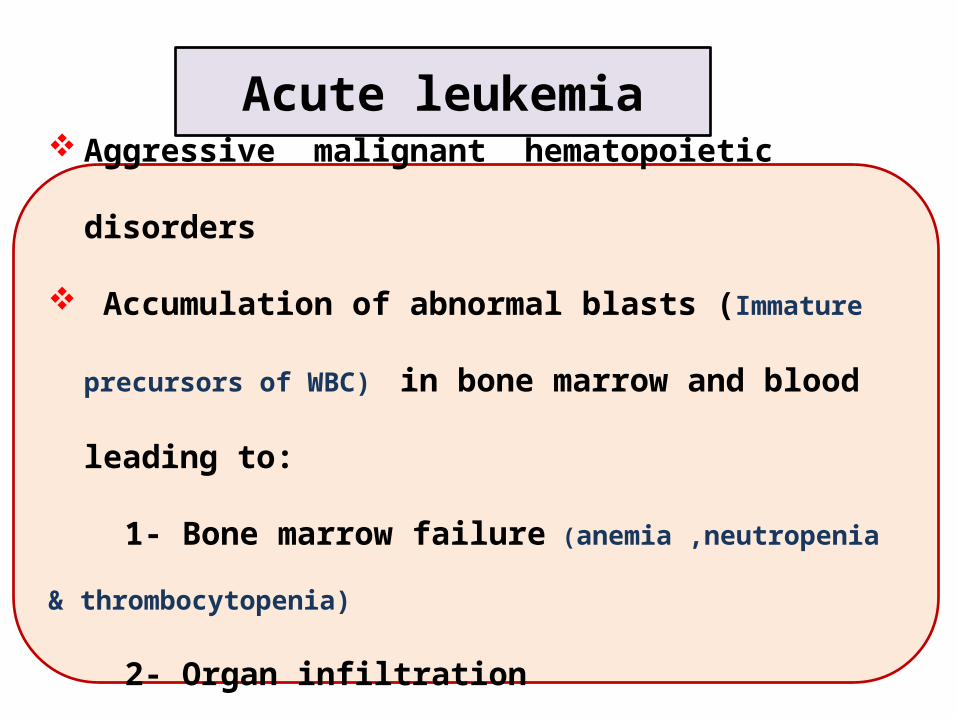
Aggressive malignant hematopoietic disorders
Accumulation of abnormal blasts (Immature precursors of WBC)
in bone marrow and blood leading to:
1- Bone marrow failure (anemia ,neutropenia & thrombocytopenia)
2- Organ infiltration ( hepatosplenomegy ,lymphadenopathy )
Acute leukemia

• Means “white blood” in Greek.
• Named by pathologist Virchow in 1845.
• Classified by FAB classification systems in 1976.
• Reclassified by World Health Organization in 2001 & 2008.
HISTORY
PATHOGENESIS

Genetic alteration in the immature precursors
Block of differentiation ,Enhanced proliferation & Decreased apoptosis
Genetic predisposition
Environmental factors Infection Previous
therapy
Other hematological
disorders
Unknown Mechanism
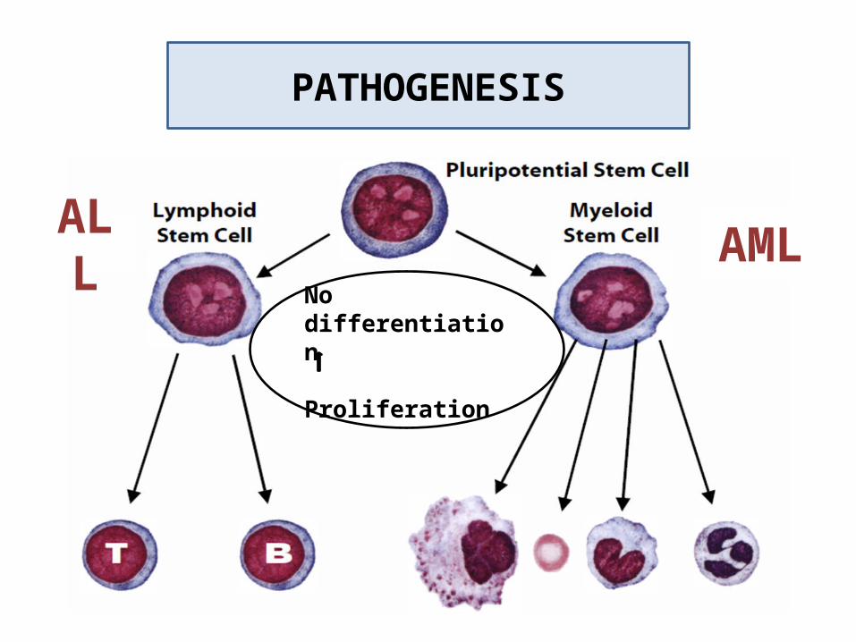
No differentiation Proliferation
AMLALL
PATHOGENESIS

• AL represent about 8% of neoplastic disease & cause about 4% of malignancy related deaths !
• AML has an incidence of 2 – 3 per 100 000 per year in children, rising to 15 per 100 000 in adults.
•ALL has an incidence of 30 per million & represent about 76% of childhood leukemia .
Epidemiology
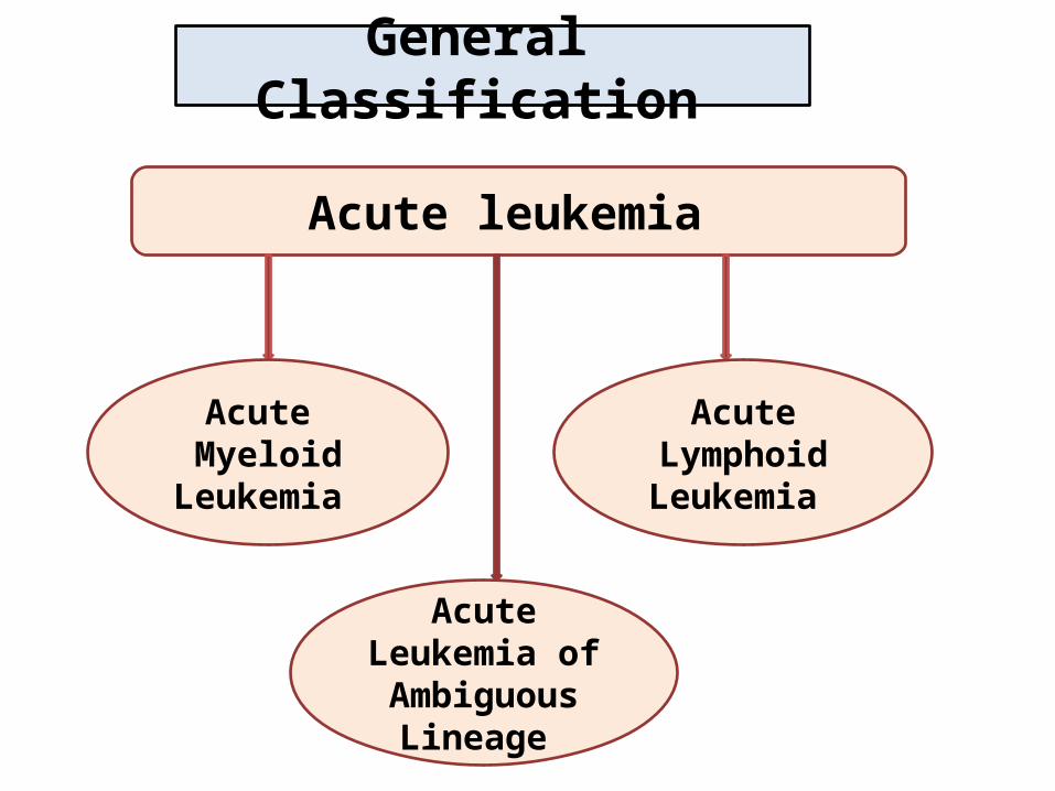
Acute leukemia
Acute Lymphoid Leukemia
Acute Myeloid Leukemia
Acute Leukemia of Ambiguous
Lineage
General Classification

1. Clinical history (Previous therapy)
2. Morphology 3. Flow cytometry 4. Chromosomal Karyotyping 5. Molecular study
Basis of Classification
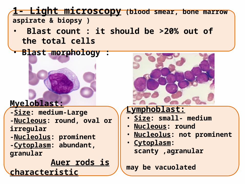
Myeloblast:-Size: medium-Large -Nucleous: round, oval or irregular-Nucleolus: prominent-Cytoplasm: abundant, granular Auer rods is characteristic
Lymphoblast:• Size: small- medium • Nucleous: round• Nucleolus: not prominent• Cytoplasm: scanty ,agranular may be vacuolated
1- Light microscopy (blood smear, bone marrow aspirate & biopsy )
• Blast count : it should be >20% out of the total cells • Blast morphology :
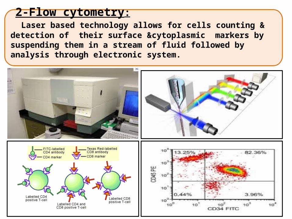
2-Flow cytometry: Laser based technology allows for cells counting & detection of their surface &cytoplasmic markers by suspending them in a stream of fluid followed by analysis through electronic system.

Basis of Classification
Stem Cell Markers: (CD34& TDT)
MPOCD13CD33CD14CD64CD41
CD235a
CD10CD19CD22
CD79a
CD3CD4CD5CD7CD8
Myeloid B-Lymphoid T-Lymphoid

3-Chromosomal KaryotypeSet of the chromosomes from one cell during metaphase to study the numerical(deletion &trisomy) and structural ( translation &inversion ) abnormality
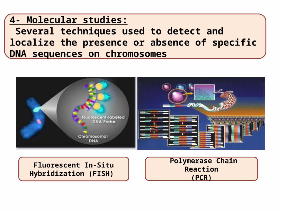
4- Molecular studies: Several techniques used to detect and localize the presence or absence of specific DNA sequences on chromosomes
Fluorescent In-Situ Hybridization (FISH)
Polymerase Chain Reaction(PCR)
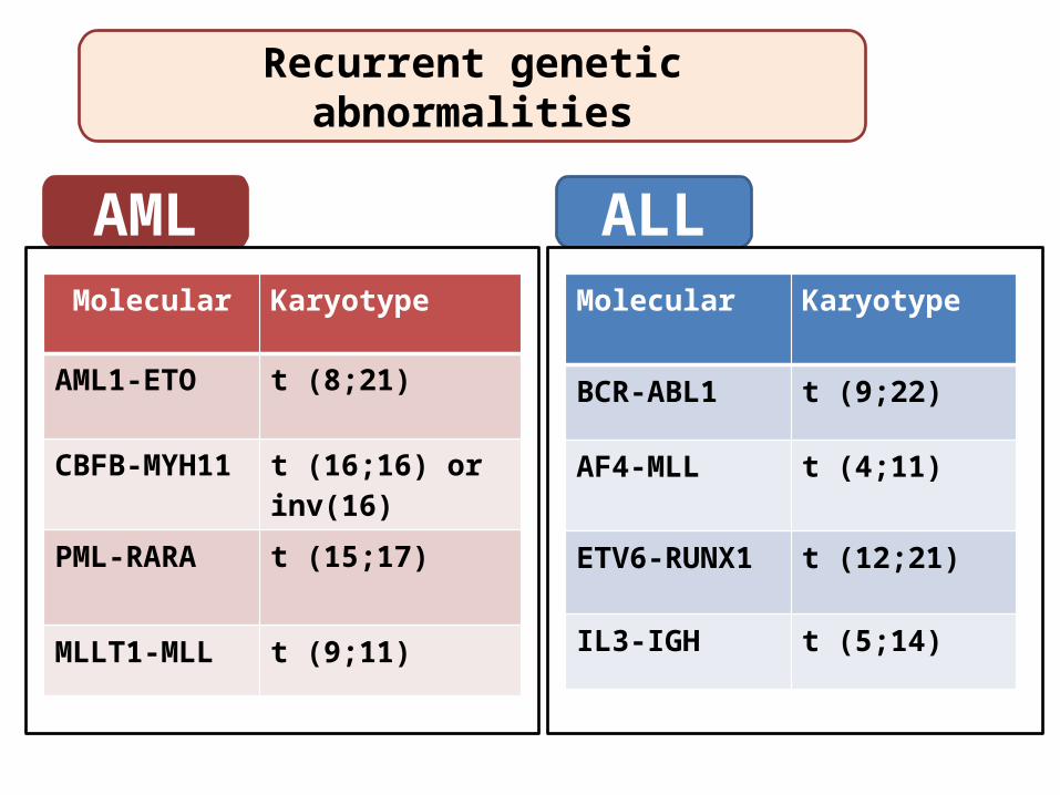
Recurrent genetic abnormalities
AML ALLMolecular Karyotype
BCR-ABL1 t (9;22)
AF4-MLL t (4;11)
ETV6-RUNX1 t (12;21)
IL3-IGH t (5;14)
Molecular Karyotype
AML1-ETO t (8;21)
CBFB-MYH11 t (16;16) or inv(16)
PML-RARA t (15;17)
MLLT1-MLL t (9;11)

Acute Myeloid Leukemia
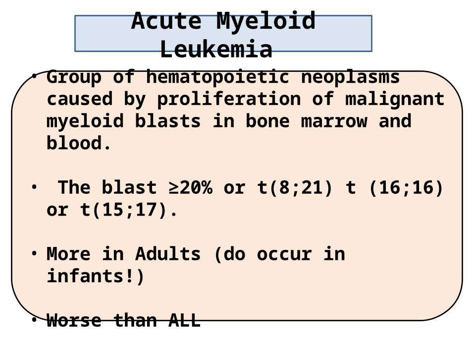
• Group of hematopoietic neoplasms caused by proliferation of malignant myeloid blasts in bone marrow and blood.
• The blast ≥20% or t(8;21) t (16;16) or t(15;17).
• More in Adults (do occur in infants!)
• Worse than ALL
Acute Myeloid Leukemia

ErythroblastMegakaryoblast
HSC
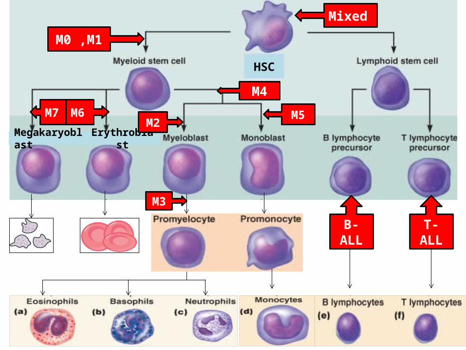
ErythroblastMegakaryoblast
HSC
Mixed
M0 ,M1
M7 M6
M4
M2M5
M3
B-ALL T-ALL

FAB Classification
• Based on morphology& flow cytometry
CD235a
CD41

AML Classification (WHO)
AML with recurrent genetic abnormalities
Myelodysplasia related AML
Therapy related AML
AML, not otherwise specified
(FAB)
1- t(8;21)2- t(16;16)3- t(15;17)Prognosis:Good
•Blasts≥ 20%•Significant dysplasia Prognosis:poor
•Blasts≥ 20%•Previous chemotherapy Prognosis:poor
•Blasts≥ 20%•Genetic: N•No dysplasia Prognosis:Standard

M0 M1
M2
M3
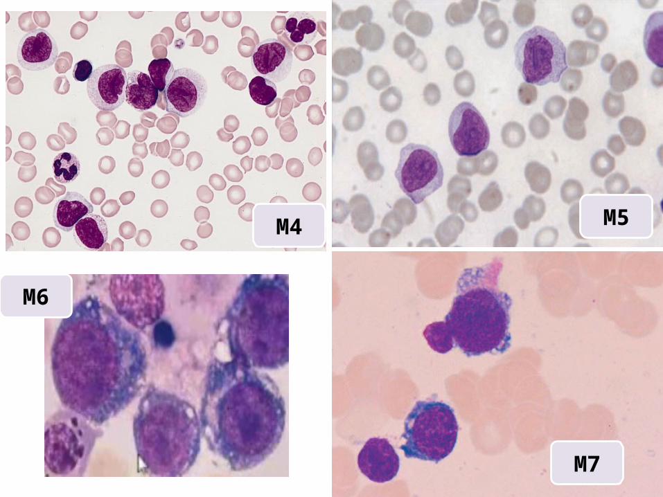
M4
M6
M7
M5

1-Pancytopenia:WBC infection (fever ,septic shock)Hb anemia (fatigue , headache , pallor ,SOB….) platelets bleeding (bruises , epistaxis ,menorrhagia…)
2-Organ infiltration:• Hepatosplenomegally. • Lymphadenopathy (rare)• Myeloid sarcoma• Gum hypertrophy• CNS disease
More with Acute Monoblastic Leukemia
Clinical Features of AML
Acute onset

4-Disseminated Intravascular Coagulation (DIC):Widespread activation of coagulation system leading to intravascular fibrin deposition &consumption of platelet and coagulation factors which can be manifested as bleeding (85%) or thrombosis (15%)
More with Acute Promyelocytic leukemia (M3)
3-Leucostasis (increased blood viscosity)
Clinical Features of AML
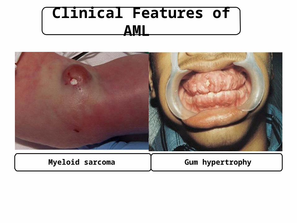
Myeloid sarcoma Gum hypertrophy
Clinical Features of AML
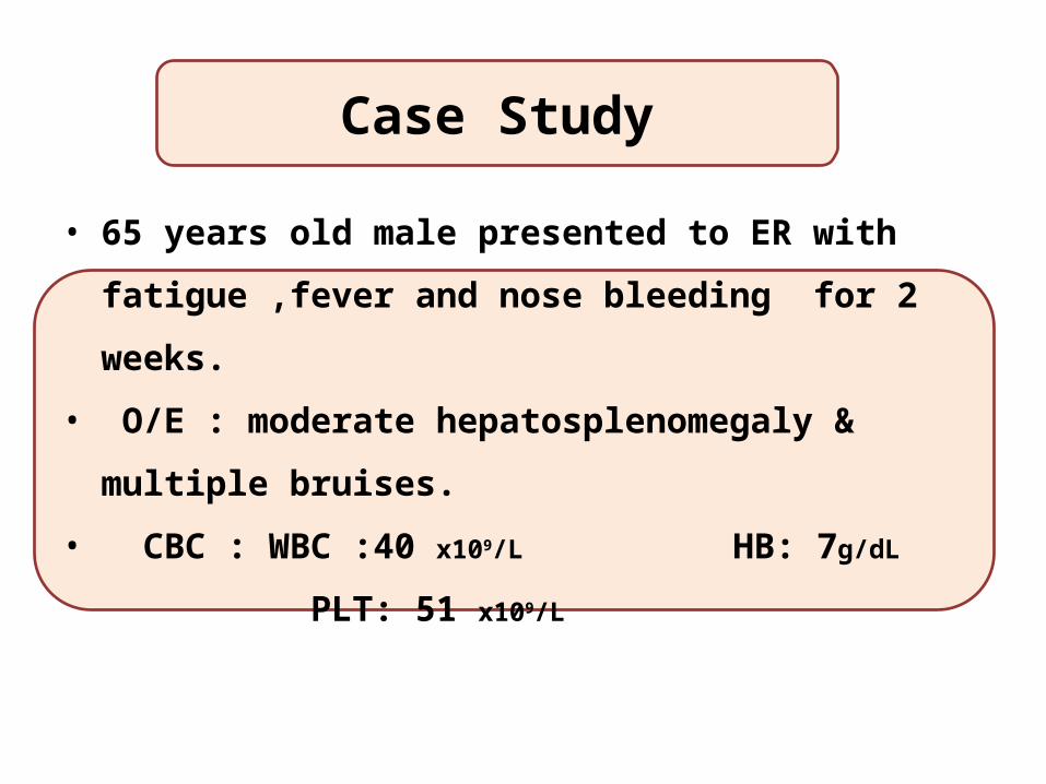
Case Study
• 65 years old male presented to ER with fatigue ,fever and
nose bleeding for 2 weeks.
• O/E : moderate hepatosplenomegaly & multiple bruises.
• CBC : WBC :40 x109/L HB: 7g/dL PLT: 51 x109/L

Flow cytometry :The blast are positive for CD34 ,CD13,CD33,CD117 and MPOThey are negative for CD3,CD10,CD19&CD79a
Blood smear & bone marrow:
?

AML with maturation (M2) (FAB)

Karyotype :
The final diagnosis: AML with t(8;21) (WHO)

Better prognosis:• Genetics: t(8;21), inv(16;16) or t(15;17)• Age: < 60 years• Primary better than secondary
Treatment• Chemotherapy:
AML: M0-M8 but not M3 ( same protocol) AML: M3 (ATRA or arsenic)
• Stem cell transplantation
Prognosis and treatment

ACUTE LYMPHOBLASTIC LEUKEMIA (ALL)

Acute leukemia characterized by proliferation of
malignant lymphoid blasts in bone marrow and blood.
B and T cells
More common in Children
Better than AML
Acute Lymphoblastic Leukemia (ALL)
1-Pancytopenia:WBC infection (fever ,septic shock)Hb anemia (fatigue , headache , pallor ,SOB….) platelets bleeding (bruises , epistaxis ,menorrhagia…)
2-Organ infiltration:• Lymphadenopathy (very common)• Hepatosplenomegally. • testicles involvement • CNS disease• Mediastinal mass
Clinical Features of ALL
Acute onset
Characteristic for T-ALL
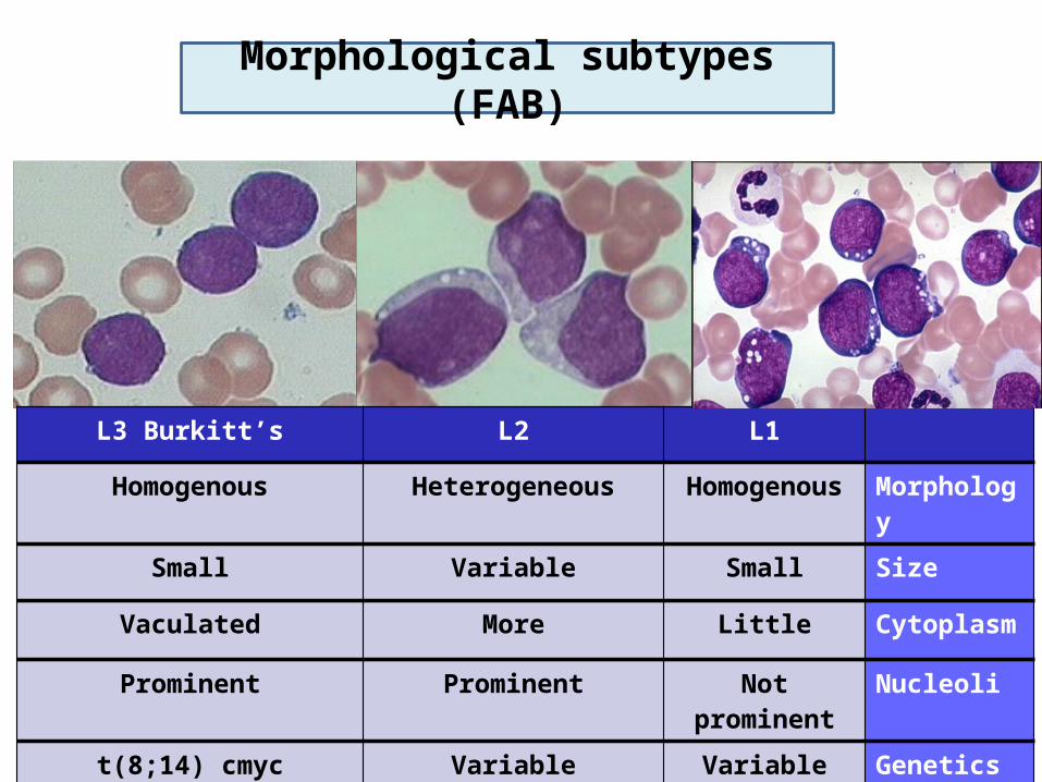
Morphological subtypes (FAB)
L3 Burkitt’s L2 L1
Homogenous Heterogeneous Homogenous Morphology
Small Variable Small Size
Vaculated More Little Cytoplasm
Prominent Prominent Not prominent Nucleoli
t(8;14) cmyc Variable Variable Genetics
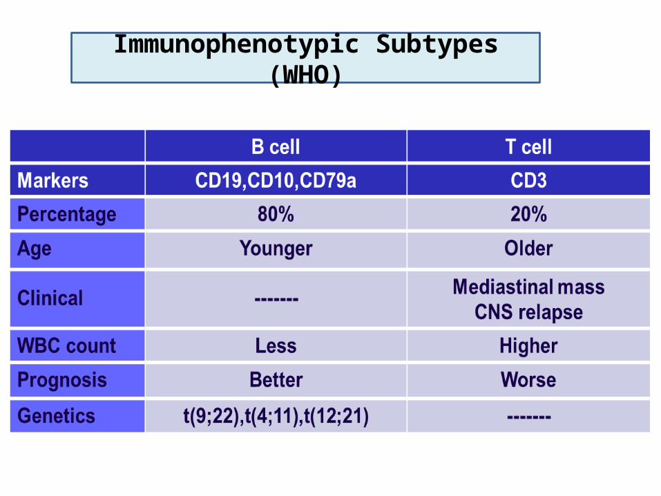
Immunophenotypic Subtypes (WHO)

L3 (Burkitt’s) represents mature lymphoid neoplasmso it is a type of lymphoma
not Acute lymphoblastic leukaemia

Precursor B cell Mature B cell
CD34& TDT
Surface Immunoglobulin
CD10
CD19,CD20 &CD79a
B- ALL Burkitt’s
Common B-ALL
B-ALL
3838
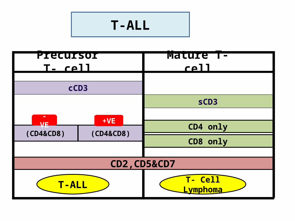
sCD3
cCD3
(CD4&CD8) (CD4&CD8)
- VE +VECD4 only
CD8 only
CD2,CD5&CD7
T-ALL
Precursor T- cell Mature T- cell
T-ALL T- Cell Lymphoma

Treatment: Chemotherapy (high cure rate) Stem cell transplantation
Prognosis &treatment
Worse Better
<2 - >10 yrs 2 - 10 yrs Age
M F Gender
High Low WBC count
T cell B cell Cell type
Others Common B-ALL phenotype
Hypodiploidyt(9;22)
Hyperdiploidyt(12;21)
B-ALL genetics
Yes No CNS involvement

Acute leukaemia is a fatal neoplastic condition
20% or more blasts = Acute leukaemia
Diagnosis requires special investigations
Auer rods = AML
AML M3 = DIC &target therapy
Gum hypertrophy = mostly M4 or M5,
Mediastinal = T-ALL
Remember !
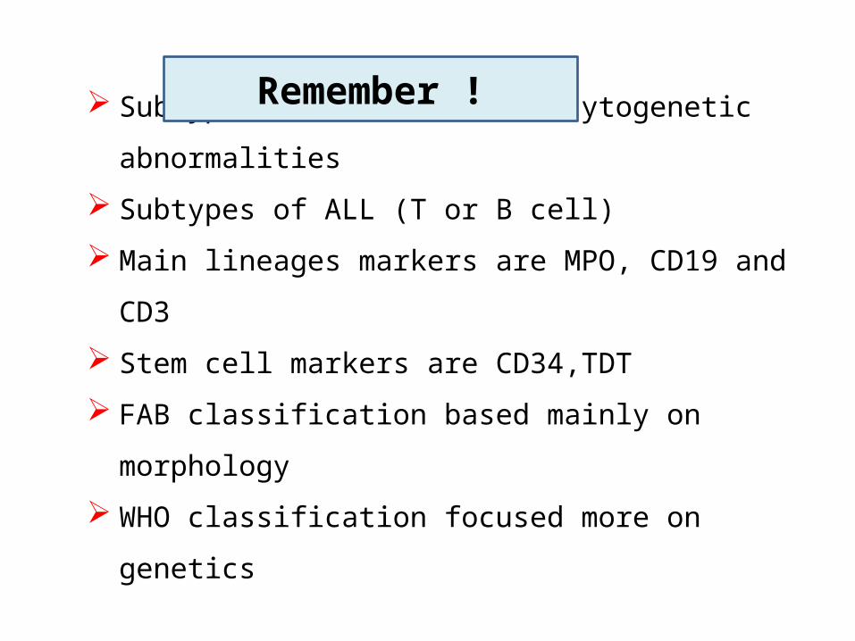
Subtypes of AML (M0-M8) + cytogenetic abnormalities
Subtypes of ALL (T or B cell)
Main lineages markers are MPO, CD19 and CD3
Stem cell markers are CD34,TDT
FAB classification based mainly on morphology
WHO classification focused more on genetics
Remember !

THANK YOU