thegunnybag.files.wordpress.com · web viewchediak-higashi syndrome (chs) 4.swhachman syndrome...
TRANSCRIPT
SEMINAR ON PRIMARY IMMUNODEFICIENCY
BY DR MOHANMODERATOR – DR PUSHPALATA
The immune system, which protects the body from disease, works through a complicated web of cells and chemicals. A defect in any one of these parts can damage the body's ability to fight off disease. Such a defect is called an immunodeficiency disease.
IMMUNE SYSTEMInnate immunity – phagocytic cells, natural killer (NK) cells, complement system, and other plasma factors Adaptive immunity – T and B lymphocytes and their secreted products
TYPES OF IMMUNODEFICIENCY• PRIMARY• SECONDARY
The immune system is not fully mature at birth and may not be well developed in some aspects until a child reaches school age. Even with a well-functioning immune system, young children can have up to six upper respiratory tract infections per year for the first 3 to 5 years of life . Typically, children with an intact immune system and no other predisposing factors handle these infections well, with rapid resolution of bacterial infections using appropriate antibiotics
Several factors contribute to the risk for infectionsduring childhood -
• Increased infectious agent exposure, school-aged siblings, peer group
• passive smoking• Atopy , hyper reactive air –way disease• Anatomic factors, structural or ciliary defects• Foreign body• Gastroesophageal reflux
Primary immunodeficiencies are generally the result of genetic defects in the immune system cells. These disorders are rare, with the exception of IgA deficiency, which occurs with a frequency of approximately 1 : 500-700 among the white population. The estimated range of prevalence for other primary immunodeficiencies is 1 : 10,000 to 1 : 200,000 depending on the specific diagnosis.
CHARESTERISTICS OF INFECTION
• Increasing susceptibility to infections• Increasing severity of infection• Increasing duration of infections• Unusual infection• Infection with opportunistic agents• Continuous illness • Dependence to antibiotics
10 WARNING SIGNS OF PRIMARY IMMUNODEFICIENCY
1. Eight or more new ear infections within 1 year.2. Two or more serious sinus infections within 1 year.3. Two or more months on antibiotics with little effect.4. Two or more pneumonias within 1 year.5. Failure of an infant to gain weight or grow normally.6. Recurrent, deep skin or organ abscesses7. Persistent thrush in mouth or elsewhere on skin, after age 1.8. Need for intravenous antibiotics to clear infections9. Two or more deep-seated infections10. A family history of Primary Immunodeficiency.
PRIMARY IMMUNODEFICIENCY1) B-cell defects 2) T-cell defects 3) complement system defects 4) phagocytic system defects .
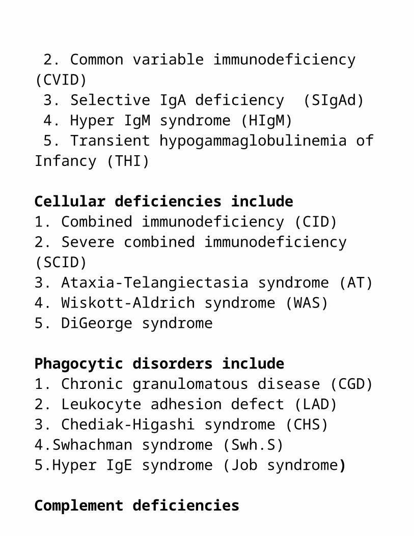
Antibody deficiencies include: 1. X-linked agammaglobulinemia (XLA) 2. Common variable immunodeficiency (CVID) 3. Selective IgA deficiency (SIgAd) 4. Hyper IgM syndrome (HIgM) 5. Transient hypogammaglobulinemia of Infancy (THI) Cellular deficiencies include1. Combined immunodeficiency (CID)2. Severe combined immunodeficiency (SCID)3. Ataxia-Telangiectasia syndrome (AT)4. Wiskott-Aldrich syndrome (WAS)5. DiGeorge syndrome Phagocytic disorders include1. Chronic granulomatous disease (CGD)2. Leukocyte adhesion defect (LAD)3. Chediak-Higashi syndrome (CHS)4.Swhachman syndrome (Swh.S)5.Hyper IgE syndrome (Job syndrome)
Complement deficiencies

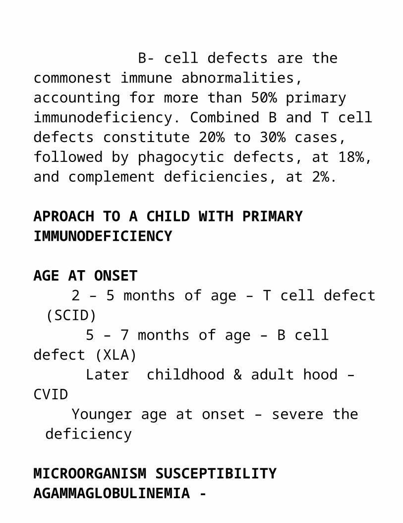
B- cell defects are the commonest immune abnormalities, accounting for more than 50% primary immunodeficiency. Combined B and T cell defects constitute 20% to 30% cases, followed by phagocytic defects, at 18%, and complement deficiencies, at 2%.
APROACH TO A CHILD WITH PRIMARY IMMUNODEFICIENCY
AGE AT ONSET 2 – 5 months of age – T cell defect (SCID)
5 – 7 months of age – B cell defect (XLA) Later childhood & adult hood –CVID
Younger age at onset – severe the deficiency
MICROORGANISM SUSCEPTIBILITYAGAMMAGLOBULINEMIA -
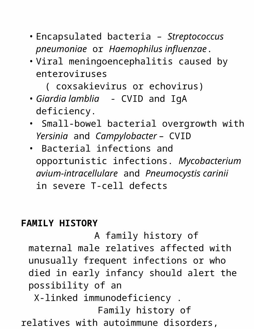
• Encapsulated bacteria – Streptococcus pneumoniae or Haemophilus influenzae.
• Viral meningoencephalitis caused by enteroviruses ( coxsakievirus or echovirus)• Giardia lamblia - CVID and IgA deficiency.• Small-bowel bacterial overgrowth with Yersinia and
Campylobacter – CVID• Bacterial infections and opportunistic infections.
Mycobacterium avium-intracellulare and Pneumocystis carinii in severe T-cell defects
FAMILY HISTORY A family history of maternal male relatives affected with unusually frequent infections or who died in early infancy should alert the possibility of an X-linked immunodeficiency .
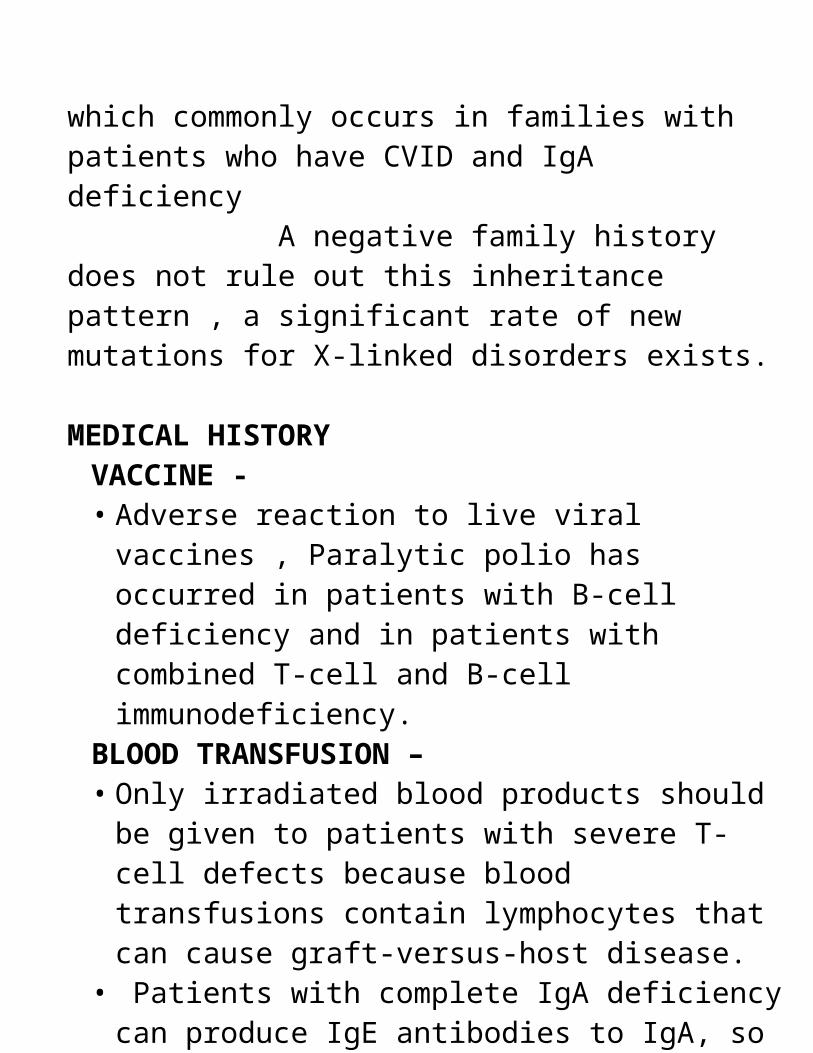
Family history of relatives with autoimmune disorders, which commonly occurs in families with patients who have CVID and IgA deficiency A negative family history does not rule out this inheritance pattern , a significant rate of new mutations for X-linked disorders exists.
MEDICAL HISTORYVACCINE -• Adverse reaction to live viral vaccines , Paralytic polio
has occurred in patients with B-cell deficiency and in patients with combined T-cell and B-cell immunodeficiency.
BLOOD TRANSFUSION –• Only irradiated blood products should be given to
patients with severe T-cell defects because blood transfusions contain lymphocytes that can cause graft-versus-host disease.
• Patients with complete IgA deficiency can produce IgE antibodies to IgA, so they are at risk for an anaphylactic reaction to plasma or blood transfusions
PHISICAL EXAMINATION• A normal physical examination does not rule out an
underlying immunodeficiency .EX - In children with X-linked lymphoproliferative disease, symptoms or signs of disease typically do not develop before Epstein-Barr virus infection develops
• Patients with antibody-deficiency syndromes can demonstrate normal growth and development despite frequent and severe RTIs. Antibody-deficiency syndromes can be characterized by asymptomatic periods
• Some children with underlying immunodeficiency appear chronically ill and underweight. If initial onset of the disease occurs early in life, growth and development may be delayed, leading to failure to thrive.
SKIN• Eczema and petechiae Wiskott-Aldrich syndrome• Telangiectasia Ataxia-telangiectasia syndrome• Dermatomyositis-like rash B-cell dysfunction • Generalized molluscum contagiosumT-cell deficiency• Extensive warts T-cell deficiency• Candidiasis T-cell deficiency
DYSMORPHIC FEATURES In patients with DiGeorge anomaly, abnormalities in the embryologic development of the third and fourth pharyngeal pouches produce dysmorphic features, including hypoplastic mandible, small mouth, hypertelorism and antimongoloid slant, and low-set and posteriorly rotated ears. DiGeorge anomaly also is associated with hypoparathyroidism; an aplastic or hypoplastic thymus; and conotruncal abnormalities of the heart, such as tetralogy of Fallot, ventricular septal defect/atrial septal defect (VSD/ASD), and pulmonic artery atresia or stenosis.
ENT EXAMINATION Extensive mucous membrane candidiasis suggests a T-cell defect. Examination of the pharynx and nasal cavities for signs of sinusitis like postnasal drainage, or purulent nasal discharge. Tympanic membranes can appear scarred and disfigured as a sign of previous recurrent and chronic infection of the middle ear.
SYSTEMIC EXAMINATION
RESPIRATORY SYSTEM –
• Rales on auscultation of the chest may suggest bronchiectasis occurring as a complication of recurrent lung infections. Digital clubbing points to significant lung disease.
CARDIOVASCULAR SYSTEM –
• Pulmonary hypertension can occur in patients with chronic lung disease
LYMPHIOD SYSTEM
• Absence of tonsils and lymph nodes suggests a severe immunodeficiency, as seen in patients with XLA or SCID.
• Cervical adenopathy and enlarged liver or spleen can be seen in patients with a B-cell deficiency, such as CVID or IgA deficiency,
• Lymphoreticular malignancies occur more commonly in certain primary immunodeficiencies, including Wiskott-Aldrich syndrome, ataxia-telangiectasia, and CVID
NEUROLOGICAL EXAMINATION
• Progressive ataxia in a young child could be the first sign of ataxia-telangiectasia even before immunodeficiency becomes clinically apparent.
• Signs of posterior and lateral column involvement of the spinal cord with loss of vibratory sense in the lower extremities, positive Babinski's response, or poor finger coordination can be signs of pernicious anemia complicating the course of CVID or IgA deficiency.
LAB DIAGNOSIS• CBC, ESR•
B CELL DEFECTS
SCREENING TESTS
1. IgA, IgG, IgM measurement 2. Isohemagglutinins 3.Antibody titres to tetanus, diphtheria,S. pneumoniae, H. influenzae
ADVANCED TESTS
1. B cell enumeration(CD19 or CD20)2. IgG subclass estimation3. IgD and IgE measuremen4. In vitro stimulation of B cells to produce immunoglobulins5. Coculture of T and B cells to assess help and suppression
T CELL DEFECTS
SCREENING TESTS
1. Delayed skin tests: e.g.,Trichophyton. Candida2. Lymphocyte count and 3. Morphology Chest x-ray for thymic size
ADVANCED TESTS 1. T-cell enumeration and phenotyping by flow cytometry 2. In vitro proliferative responses to mitogens, specific antigens, or allogeneic cells 3.Intracellular cytokine production by flow cytometry 4.T-cell cytotoxicity assays
Anemia of chronic disease can develop in patients with chronic infections, whereas pure erythrocyte aplasia can be seen in patients with thymoma and CVID. Persistent lymphopenia can be a sign of cellular immunodeficiency. Lymphopenia is defined as less than 3000 cells/mm3 in infants, whereas in older children or adults, a total lymphocyte count of less than 1500 cells/mm3 is abnormal. Thrombocytopenia and small platelet size are characteristic of patients with Wiskott-Aldrich syndrome. Autoantibodies causing autoimmune hemolytic anemia, thrombocytopenia, or neutropenia can occur in some of the B-cell immunodeficiencies Quantitation of serum immunoglobulins (IgG, IgM, IgA) is the first step in evaluating humoral or B-cell immunity Low IgA level - IgA deficiency or other immunoglobulin deficiency diseases. High IgM level - hyper-IgM syndrome
The IgE level commonly is elevated in Atopy Wiskott-Aldrich syndrome. Specific antibody titers against glycoprotein antigens, such as tetanus and diphtheria, or polysaccharide antigens, such as pneumococcal polysaccharide, can be assessed
ROLE OF PEDIATRICIAN
• Prompt recognition of infection and aggressive treatment are essential to avoid life-threatening complications and improve prognosis and quality of life. Initiation of early empiric coverage for suspected pathogens till appropriate cultures obtained.
• Prophylactic antibiotics are recommended for children with significant T-cell defects because of the risk for Pneumocystis carinii pneumonia with Co-trimaxazole .
• Children with B-cell defects who continue to experience recurrent infections despite adequate intravenous immunoglobulin therapy , should be considered for antimicrobial therapy to avoid complications, such as bronchiectasis .
• Live-attenuated vaccines, such as oral polio, varicella, and BCG should not be given to children with suspected or diagnosed antibody or T-cell defects, because vaccine-induced infection is a risk in these patients. Inactivated polio vaccine should be given to household members to prevent transmission of the virus that can occur by shedding of the attenuated virus in the stool.
• Measles-mumps-rubella, varicella, and BCG vaccines can be given to family members

• Only irradiated, leukocyte-poor, and virus-free (i.e., cytomegalovirus) products should be used in patients with T-cell defects to avoid graft-versus-host disease and cytomegalovirus infection.
SPECIFIC TREATMENT OPTIONS
• Currently available treatment techniques include bone marrow transplantation, immunoglobulin replacement, and enzyme-replacement. Gene therapy for some diseases has been initiated in clinical trials.
• Genetic counseling is important not only for a child's parents also for siblings.
• Prenatal diagnosis can be established by performing analyses on fetal blood samples, amniotic fluid cells, or chorionic villus biopsy specimens.
B - CELL DEFECTS
AGAMMAGLOBULINEMIA
• Agammaglobulinemia is the severe of the antibody-deficiency syndromes
• Significant decreases in all major classes of immunoglobulins.
• An absence of circulating B cells .• Small tonsils and no palpable lymphnodes. • T cells are present normally, with preservation of
delayed hypersensitivity and other cell-mediated immune functions.
• neutropeniaEARLY B- CELL MATURATION FAILS IN AGAMMAGLOBULINEMIA
ETIOLOGY• X-linked recessive - commonest form• XLA is caused by mutations in the Btk gene, located on
chromosome Xq 21.3-22• Autosomal recessive - caused by abnormalities in the
mu-chain gene that codes for the heavy chain of IgM or the B-cell linker protein
CLINICAL FEATURES• Extracellular pyogenic bacterial infections, particularly
otitis, sinusitis, and pneumonia, may begin as early as age 4 to 6 months, when the maternal IgG level decreases. Approximately 20% of patients present with overwhelming sepsis.
• Fatal meningoencephalitis with enteroviruses can occur due to lack of Ig A
DIAGNOSIS• The diagnosis is supported by the presence of affected
maternal male cousins, uncles, or nephews. • The serum IgG level is usually less than 200 mg/dL .• IgM and IgA levels typically are less than 20 mg/DL• Test for natural antibodies to A & B RBC antigens will
be abnormal . • Tests for antibodies to routine vaccines will be abnormal• Flow cytometry showing less than 2% CD19+ B cells in
circulation .• Normal number of Pre B - cells in bone marrow
COMMON VARIABLE IMMUNODEFICIENCY
CVID is also called • hypogammaglobulinemia • adult-onset agammaglobulinemia • late-onset hypogammaglobulinemia • acquired agammaglobulinemia
• CVID is a late-onset, highly variable hypogammaglobulinemic primary immune deficiency that can occur after age 18 months, with two peaks at approximately ages 1 to 5 years and 16 to 20 years.
• It affects approximately 1 in 10,000 to 100,000 general population
It is characterised by –• Variable deficiency of immunoglobulins .• Normal number of B – cells . • Normal sized or enlarged tonsils ,lymph nodes &
spleenomegaly.• Autoimmune diseases .• Malignancies .
ETIOLOGY• The exact cause is unknown.• Most cases of CVID occur sporadically; however,
familial inheritance ( Chromosome 6 ) may be found in as many as 25% of cases. In 10% of patients, CVID or a
related immunodeficiency disease (e.g., IgA deficiency) is found in more than one family member
• CVID is commonly associated with HLA – B8 & HLA – DR3.
• T- cell signaling to B- cells is defective in CVID• B – cells do not function properly and fail to receive
proper signals from T – cells .• T- cell defects have not been well defined.• Frequent bacterial infections of the ears, sinuses,
bronchi, and lungs • painful swollen joints in the knee, ankle, elbow, or wrist • an enlarged spleen and swollen glands or lymph nodes
CLINICAL FEATURES An increased susceptibility to Respiratory tract infections and gastrointestinal infection is the commonest clinical presentation of CVID. RTI may be caused by H. influenzae and S. pneumoniae, whereas G. lamblia and Campylobacter jejuni are responsible for most gastrointestinal infection
Autoimmune disorders, such as idiopathic thrombocytopenia (ITP), autoimmune hemolytic anemia, pernicious anemia, rheumatoid arthritis, systemic lupus erythematosus, autoimmune thyroiditis, vitiligo, and primary biliary cirrhosis also may develop in patients with CVID. Sarcoid-like granulomata of the lungs, liver, spleen, and conjunctivae also may affect patients with CVID.
Malignancies are increased in patients with CVID. A
100-fold increased risk for malignant lymphoma and a 50-fold increased risk for gastric cancer with CVID.
DIAGNOSIS• Serum concentrations of IgM ,IgG , IgA are reduced . • A normal number of B cells .• A variable degree of T-cell dysfunction .• Isohemagglutinins are absent. • Responses to protein and polysaccharide vaccines are
poor.
SELECTIVE IgA DEFICIENCY
It is the most prevalent primary immunodeficiency disease, occurring in approximately 1/500 to 1/1000 general population. • Serum IgA levels less than 7 mg/dl with normal levels of
other immunoglobulin classes• Normal serum antibody responses. • Normal cell mediated immunity • The exact cause is unknown• Since the associated factors like malignancy, family
history, autoimmunity are common to both CVID & IgA deficiency , same genetic cause may be present .
• Terminal differention of B – cells fails to result in IgA deficiency.
TYPES• ISOLATED IgA DEFICIENCY• ASSOCIATED WITH
IgE DEFICIENCY IgG2 OR IgG4 DEFICIENCY
Many people with IgA-deficiency are healthy, with no more than the usual number of infections. Those who do have symptoms typically have recurring ear, sinus, or lung infections that may not respond to regular treatment with antibiotics. People with IgA-deficiency are likely to have other problems, including allergies, asthma, chronic diarrhea, and autoimmune diseases.
CLINICAL FEATURES• Susceptibility to recurrent RTI ,GIT infections.• RTI may be caused by H. influenzae and S.
pneumoniae, whereas G. lamblia and Campylobacter jejuni are responsible for most gastrointestinal infection.
• Associated autoimmune disorders may be present.DIAGNOSIS
• Serum IgA level of less than 7 mg/Dl• Normal serum IgG and IgM levels and a normal IgG
antibody response to vaccination.• Normal number of B – cells.• Diagnosed reliably only after age 4 years.• Differentiate between
(1) patients in whom no IgA is detected (2) patients who have low but detectable IgA concentrations
HYPERIgM SYNDROME
Hyper-IgM is a rare immunodeficiency disease in which the immune system fails to produce IgA and IgG antibodies The faulty T cells do not give B cells a signal they need to switch from making IgM to making IgA and IgG. Most cases of hyper-IgM syndrome are linked to the X chromosome. Most male patients with the hyper-IgM syndrome have a mutation in the CD40L gene on the X chromosome. The interaction between CD40 on the B cell and the CD40L on the activated T cell is essential for the switch from IgM to IgG production, which explains why a deficiency in CD40L leads to hyper-IgM production with deficient IgG. These patients have normal or elevated numbers of B cells, normal numbers of T cells and normal T-cell proliferation
In addition to the recurrent RTIs, patients with CD40L deficiency also have an increased susceptibility to infections with some intracellular pathogens, such as P.
carinii pneumonia, CNS histoplasmosis, and toxoplasmosis. The increased susceptibility to intracellular pathogens is caused by the role of the CD40L in host defenses against some intracellular pathogens
The hyper-IgM syndrome also should be suspected in the presence of cryptosporidium-related diarrhea, sclerosing cholangitis, or parvovirus-induced aplastic anemia. Hyper-IgM syndrome also is associated with neutropenia Another aspect of Hyper-IgM Syndrome is autoimmune disease. Autoimmune attacks on red blood cells lead to anemia, while autoimmune destruction of infection-fighting neutrophils further increases the risk of infection Infants usually develop recurring upper and lower respiratory infections within the first year of life. Other signs of the disease include enlarged tonsils, liver, and spleen, chronic diarrhea, and an increased risk of unusual or opportunistic infections and non-Hodgkin's lymphoma DIAGNOSIS-• Normal numbers of T and B cells.• high levels of IgM and very low IgG and IgA. • Patients also may have neutropenia
HYPOGAMMAGLOBULINEMIA OF INFANCY
Transient hypogammaglobulinemia of infancy is an ill-defined entity in which a child's postnatal decrease in serum IgG level is accentuated A delay in the onset of endogenous immunoglobulin synthesis occurs, possibly because of deficiency in T helper cells Most patients have normal IgM and IgA concentrations and a normal circulating B-cell level. The initial serum IgG levels are typically higher than those of patients with agammaglobulinemia.• IgG concentrations usually normalize by the time these
patients reach age 2 or 3 years.
TREATMENT OF B CELL DEFECTS
General management of patients with immunodeficiency requires an extraordinary amount of care to maintain optimal health and nutrition, manage infections, prevent emotional problems related to their illness, and cope with costs. Antibiotics are lifesaving for treating infections; selection and dosage are identical to those used normally.
Fever or other manifestations of infection are assumed to be secondary to bacterial infection, and antibiotic treatment is begun immediately. Throat, blood, or other cultures are obtained before most therapy; these are especially useful subsequently when the infection does not respond to the initial antibiotic and when the infectious organism is unusual. Continuous prophylactic antibiotics often are beneficial, particularly in recurrent infection in agammaglobulinemia despite IG therapy. Immune globulin (IG) is effective replacement therapy in most forms of antibody deficiency. It is a 16.5% solution of IgG with trace quantities of IgM and IgA for IM or subcutaneous injection, or a 3 to 12% solution for IV infusion (IVIG). The loading dose is 200 mg/kg given in 2 or 3 doses over 2 to 5 days followed at monthly intervals by 100 mg/kg .
High doses of IVIG (400 to 800 mg/kg/mo) can be given and are beneficial to some antibody-deficient patients not responding well to conventional doses, particularly those with chronic lung disease. The aim with high-dose IVIG is to keep IgG trough levels in the normal range (ie, > 500 mg/dL) • Plasma has been used as an alternative to IG, but
because of the risk of disease transmission, it is rarely indicated
T CELL DEFECTS
Primary T-cell immunodeficiencies are rare inherited disorders that affect T-cell development and function. These disorders usually present in infancy or early childhood; however, the age of symptom onset may vary depending on the underlying gene defect. Although T-cell immune responses may be selectively affected, abnormal B-cell function often is associated, in part because of concomitant intrinsic B-cell defects but also because production of most antibodies is dependent on T-cell help
SELECTIVE T-CELL DEFECTS
DiGeorge Syndrome
DGS classically includes conotruncal cardiac malformations
persistent hypocalcemia and cellular immunodeficiency secondary to a defect in the development of third and fourth pharyngeal pouches that affects the parathyroid glands and thymus The specific cardiac anomalies most frequently associated with DGS are interrupted aortic arch, tetralogy of Fallot, and truncus arteriosus DiGeorge (DGS), velocardiofacial, and conotruncal anomaly face syndromes, have been demonstrated to share a microdeletion of one copy of chromosome 22q.• CATCH 22 syndrome(cardiac, abnormal facies, thymic
hypoplasia, cleft palate, hypocalcemia) includes broad spectrum of conditions with 22q11 deletions.
The dysmorphic features include, Hypertelorism, antimongloid slant of eyes,Low set notched ears, short philtrum of the upper lip, Mandibular hypoplasia.
The immune defects in DGS include,
• decreased (<1500 cells/mm3 ) CD3+ T lymphocytes, a CD4+ T-cell count of less than 1000 cells/mm3 , and mildly impaired cellular immunity
• 50% or less of normal T-cell numbers, • 20% have in vitro T-cell proliferative responses of less
than 50% of normal • the immune defect improves with age, and many
children produce normal numbers of functional T lymphocytes by the end of the second year of life
TREATMENT
Bone marrow transplantation
SEVERE COMBINED IMMUNODEFICIENCY SYNDROMES
Severe combined immunodeficiency syndrome (SCID) is a heritable disorder in children characterized by profoundly defective or absent T-cell and B-cell function • fatal within the first year of life unless curative
hematopoietic stem cell transplantation or, in the case of adenosine deaminase (ADA) deficiency, enzyme replacement is accomplished
• X- LINKED RECESSIVE.• It accounts for 45% of the cases.• Occurs due to mutation at Xq 13 which codes for gamma
chain of the cytokine receptors,( IL-2, IL-4, IL-7, IL-9, IL15, IL-21) which mediates intracellular signalling.
• Characterised by T-, B+, NK-.• AUTOSOMAL RECESSIVE FORM• There are 6 subtypes.1. Adenosine deaminase deficiency –• Accounts for 15% of cases due to mutation at 20q 13.• Accumulation of adenosine 2’ deoxy adenosine, 2’ 0
methyladenosine leads to T cell apoptosis.• Characterised by T-, B-, NK-.2. Jak 3- accounts for 6% of cases• Occurs due to mutation at 19p13.• Jak 3 is required for the transduction of gamma chain
cytokine receptors.• Characterised by T-, B+, NK-.
3.IL -7 R alpha deficiency.• Accounts for 10% cases, due to mutation at 5p13.• Characterised by T-, B+, NK+.4.RAG1 or RAG2 (RECOMBINASE ACTIVATING GENES) DEFICIENCY.• Accounts for 10% caeses.• Due to mutation at 11p13.• The genes are essential for generation of T cell and B
cell antigen receptors.
• Characterised by T-, B-, NK+.5. CD45 DEFICIENCY.• Recently identified entity, accounting for very few cases.• Gene not yet mapped. • Required for T and B cell antigen receptor transduction.
6. ARTIMIS DEFICIENCY.• Occurs due to mutation at 10p13.• Defect in repair of DNA following cuts produced by
products of RAG1 or RAG2.• Characterised by T-, B-, NK+.
CLINICAL FEATURES• Despite underlying genetic heterogeneity, patients with
SCID present similarly within the first 6 months of life with recurrent diarrhoea, pneumonia, otitis media, sepsis, cutaneous infections.
• In general, the following infections may develop in affected infants:
Bacteria Gram-negative sepsis,Disseminated BCG after immunizationFungi and protozoa Candidiasis, AspergillusPneumocystis carinii pneumoniaViruses Cytomegalovirus, Parainfluenza viruses, Adenovirus, Respiratory syncytial virus, Disseminated varicella, Vaccine-acquired paralytic poliomyelitis,Molluscum contagiosum.

Failure to thrive secondary to diarrhea and malabsorption also may be present. The appearance of an early-onset erythematous maculopapular rash unresponsive to medical management may suggest chronic graft-versus-host disease (GVHD) from engrafted maternal T cells. Most SCID patients have thymic hypoplasia and absent or small, poorly developed lymph nodes and tonsils; hepatosplenomegaly may be detected in affected infants with maternal GVHD.A diagnosis of SCID is suggested when an affected infant has
• lymphopenia (<1500 cells/mm3 ; normal range, 4000-13,500 cells/mm3 ),
• less than 20% CD3+ T lymphocytes, • and severe hypogammaglobulinemia (IgG, <150
mg/dL). The presence of circulating, transplacentally acquired maternal T cells detected by histocompatibility (i.e., human leukocyte antigen, HLA) testing is definitive evidence for a diagnosis of SCID until proven otherwise. TREATMENT
• Bone marrow transplantation
OMENN SYNDROME
OS is a rare AR disorder described in 1965 by Omenn as SCID characterized by profound susceptibility to infection with T cell infiltration into skin, intestine, liver and spleen leading toErythrodermaLymphadenopathyHepatosplenomegalyFailure to thrive secondary to diarrheaFever
Mutations in RAG1 or RAG2 that result in partial recombinase activity and the development of rare activated, but anergic, oligoclonal T cells were identified in patients with OS.

Laboratory findings
HypoalbuminemiaEosinophilia (>1000 cells/mm3 )Variable lymphocyte countsDecreased to elevated CD3+ T-cell countlow or Absent B cellsNormal NK-cell countMarkedly defective T-cell and B-cell functionHypogammaglobulinemiaSeverely decreased IgG, IgM, and IgA levelsHyper-IgE (>1000 IU/mL)
Wiskott-Aldrich Syndrome Wiskott-Aldrich syndrome (WAS) is an X-linked inherited immunodeficiency characterized by eczema, congenital thrombocytopenia with small platelets, and recurrent infections.
• Due to mutation at Xp11 coding for WASP which is required for microvesicles formation in blood cells.
CLINICAL FEATURES Presents in the newborn period or early infancy with petechiae,bloody diarrhea, intracranial hemorrhage, and excessive bleeding from an umbilical stump or after circumcision .Eczema develops in more than 80% of patients and often is seen before 6 months of age.Recurrent sinopulmonary infections with encapsulated organisms develop within the first 2 years of life. Opportunistic infections, such as Pneumocystis carinii pneumonia and recurrent herpesvirus infections.
• The prevalence of leukemia, lymphomas of the abdomen and CNS, and Epstein-Barr virus (EBV)-associated tumors is markedly increased
• Autoimmune disease, including hemolytic anemia, arthritis, vasculitis, inflammatory bowel disease, and glomerulonephritis, is seen in approximately 40% of patients with WAS.
LAB DIAGNOSIS• Lymphopenia (<1000 cells/mm3 ) with declining
numbers of CD3+ and CD8+ T cells • B-cell and NK-cell numbers remain normal. • Normal serum IgG but decreased IgM, in association
with defective production of pneumococcal antibodies and absent isohemagglutinins.
Ataxia Telangiectasia
Ataxia-telangiectasia (AT) is a complex AR disorder characterized by cerebellar ataxia, oculocutaneous telangiectasia, radiosensitivity, predisposition to malignancy, hepatic & endocrine involvement and combined immunodeficiency
• Due to mutation at 11q22.• There is incraesed sensitivity to ionising radiation and
defective DNA repair.
CLINICAL FEATURES
The presenting symptom in AT is ataxic gait seen in more than 85% of patients 4 years of age. The ataxia progressively involves the trunk, extremities, and palatal muscles, resulting in dysarthric speech, drooling, ocular apraxia, and inability to ambulate independently by 10 years of age. Telangiectasia of the sclerae and skin subsequently develops between the ages of 4 and 8 years .Recurrent upper and lower respiratory tract infections and chronic lung disease.
Increased predisposition to T-cell and B-cell malignancies, particularly leukemia and Hodgkin and non-Hodgkin lymphomas . Growth retardation, hypogonadism and pubertal delay, and insulin-resistant nonketotic diabetes mellitus. Progressive lymphopenia involving both T and B cells, including selective loss of CD4+ T cells, with inversion of the normal CD4-CD8 ratio. IgA and IgE deficiency in most AT patients and decreased isohemagglutinins and serum IgG2 levels
REFERRENCES• Nelson’s textbook of pediatrics• PCNA • Ganong’s textbook of physiology• Harrison’s textbook of internal medicine