distribution pattern in chediak-higashi · jc/in pathol 1982;35:496-501 distribution pattern...
TRANSCRIPT
J C/in Pathol 1982;35:496-501
Distribution pattern of lysosomal granules infibroblasts of the Chediak-Higashi syndromeK ABE, S ARASHIMA, M HONMA
From the Department of Anatomy and Pediatrics, Hokkaido University School of Medicine, Sapporo,Japan
SUMMARY Cultured fibroblasts from a patient with the Chediak-Higashi syndrome, the mother ofthe patient, and a normal control were studied by light and electron microscopy. The distributionpattern of PAS-positive and acid phosphatase-containing granules in the cytoplasm differed signifi-cantly in the fibroblasts from the patient when compared with those from the mother and control.The granules in the fibroblasts from the patient were clustered in the perinuclear area, whereas thegranules in the fibroblasts from the mother and control were dispersed throughout the cytoplasm.After incubation with ascorbic acid, the clustered granules in the fibroblasts of the Chediak-Higashisyndrome showed a tendency to spread throughout the cytoplasm. The distribution pattern of thegranules was studied by quantitative morphology.
The Chediak-Higashi syndrome is a rare geneticdisorder characterised by the presence of abnormallylarge inclusions in polymorphonuclear leucocytes.1-3Certain mutants of Aleutian minks and beige miceare known to be homologues of the Chediak-Higashisyndrome in humans.45 Although the pathogenesisof this syndrome remains uncertain, recently it hasbeen reported that the Chediak-Higashi syndromeshows abnormal function of microtubules,0 and thefunction is restored by ascorbic acid.7 The patho-genesis of some genetic disorders has been explainedwith cell culture.8-10 Abnormally large inclusionshave been observed in cultured fibroblasts from apatient with the Chediak-Higashi syndrome'1and beige mice.6 Thus, a culture of the fibroblastsderived from the patient with the Chediak-Higashisyndrome may be useful for understanding itspathogenesis.We recently had a patient with the Chediak-
Higashi syndrome.12 The case showed abnormalinclusions in the polymorphonuclear leucocytesand lymphocytes, and died 110 days after birth.From skin biopsy of the patient, we established thecell lines of fibroblasts, and the cultured fibroblastsshowed characteristic distribution pattern of lyso-somal granules. In this study, the cultured skinfibroblasts of the Chediak-Higashi syndrome, andthe effects of ascorbic acid on the fibroblasts wereexamined cytologically.
Accepted for publication 7 September 1981
Material and methods
Skin fibroblasts from a 95-day-old girl with theChediak-Higashi syndrome, a 26-year-old mother ofthe patient, and a 37-year-old healthy male as acontrol were cultured in monolayer on cover slips.The fibroblasts were grown in Eagle's medium with15% calf serum. Eight different subcultures out of 26subcultures in each cell line were examined at thesecond day of the culture by light and electron mi-croscopy.
Fibroblasts on the cover glasses were fixed with 2%glutaraldehyde in 0 1 M cacodylate buffer (pH 7 4)at 4°C for between 30 min and two hours. For lightmicroscopy, they were stained with periodic acid-Schiff reagent (PAS) and haematoxylin, or reactedby Gomori's method for acid phosphatase. Some ofthem were stained with Giemsa, toluidine blue, orSudan black B. For electron microscopy, afterfixation with glutaraldehyde, they were fixed with 2%OS04 in 0-1 M cacodylate buffer (pH 7-4) at 4°Cfor one hour and embedded in Epon. Ultrathinsections were stained with uranyl acetate and leadcitrate, and examined by electron microscopy.Effects of ascorbic acidFibroblasts from the patient, mother of the patient,and the control were incubated in the mediumcontaining 0 1, 1, 10, or 100 mM ascorbic acid for15 min before fixation and examined by lightmicroscopy. Two cultures for each source wereexamined.
496
on 15 Novem
ber 2018 by guest. Protected by copyright.
http://jcp.bmj.com
/J C
lin Pathol: first published as 10.1136/jcp.35.5.496 on 1 M
ay 1982. Dow
nloaded from
Fibroblasts of Chediak-Higashi syndrome
Results
The cultured fibroblasts showed no significantdifferences in cellular shape and number for thecultures derived from the patient with the Chediak-Higashi syndrome, the mother of the patient, andthe healthy control (Figs. 1-3). They were spindle-
,*
,,,KsN-A *
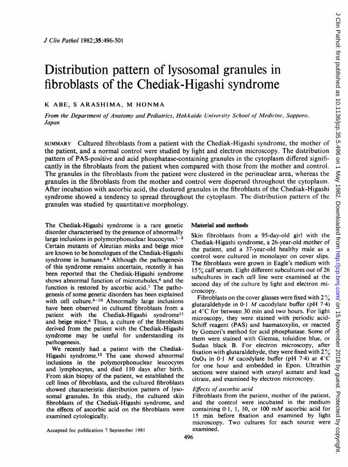
Fig. 1 Cultured fibroblast from the control. Lysosomalgranules are dispersed throughout the cytoplasm. Acidphosphatase demonstrated by Gomori's method.Haematoxylin x 700.
A...4i 2. .
A ~ ~ j~~
Fig. 2 Cultured fibroblast from the patient withChediak-Higashi syndrome. Lysosomal granules areclustered adjacent to the nucleus. Gomori's method.Haematoxylin x 700.
shaped or stellate with a few cytoplasmic processes,and the nuclei were oval and had several nucleoli.The cytoplasm contained a large number of smallgranules which were PAS-positive and showed acidphosphatase activity (Figs. 1-3). These granules werenot stained with Giemsa, toluidine blue, or Sudanblack B. The distribution of these cytoplasmicgranules in the fibroblasts, however, exhibitedsignificant differences between the fibroblasts de-rived from the patient and those from healthyindividuals.
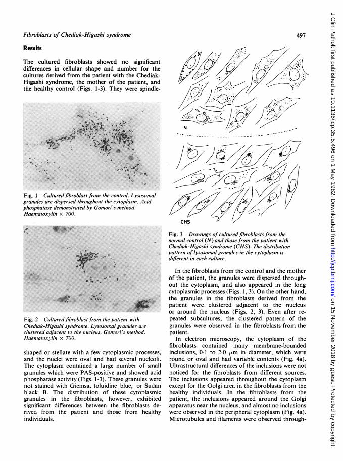
Fig. 3 Drawings of cultured fibroblasts from thenormal control (N) and those from the patient withChediak-Higashi syndrome (CHS). The distributionpattern of lysosomal granules in the cytoplasm isdifferent in each culture.
In the fibroblasts from the control and the motherof the patient, the granules were dispersed through-out the cytoplasm, and also appeared in the longcytoplasmic processes (Figs. 1, 3). On the other hand,the granules in the fibroblasts derived from thepatient were clustered adjacent to the nucleusor around the nucleus (Figs. 2, 3). Even after re-peated subcultures, the clustered pattern of thegranules were observed in the fibroblasts from thepatient.
In electron microscopy, the cytoplasm of thefibroblasts contained many membrane-boundedinclusions, 01 to 2-0 ,um in diameter, which wereround or oval and had variable contents (Fig. 4a).Ultrastructural differences of the inclusions were notnoticed for the fibroblasts from different sources.The inclusions appeared throughout the cytoplasmexcept for the Golgi area in the fibroblasts from thehealthy individuals. In the fibroblasts from thepatient, the inclusions appeared around the Golgiapparatus near the nucleus, and almost no inclusionswere observed in the peripheral cytoplasm (Fig. 4a).Microtubules and filaments were observed through-
497
on 15 Novem
ber 2018 by guest. Protected by copyright.
http://jcp.bmj.com
/J C
lin Pathol: first published as 10.1136/jcp.35.5.496 on 1 M
ay 1982. Dow
nloaded from
Abe, Arashima, Honma
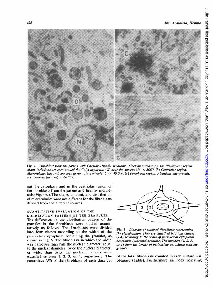
Fig. 4 Fibroblast from the patient with Chediak-Higashi syndrome. Electron microscopy. (a) Perinuclear region.Many inclusions are seen around the Golgi apparatus (G) near the nucleus (N) x 8000. (b) Centriolar region.Microtubules (arrows) are seen around the centriole (C) x 40 000. (c) Peripheral region. Abundant microtubulesare observed (arrows) x 60 000.
out the cytoplasm and in the centriolar region ofthe fibroblasts from the patient and healthy individ-uals (Fig. 4bc). The shape, amount, and distributionof microtubules were not different for the fibroblastsderived from the different sources.
QUANTITATIVE EVALUATION OF THE
DISTRIBUTION PATTERN OF THE GRANULESThe differences in the distribution pattern of thegranules in the fibroblasts were studied quanti-tatively as follows. The fibroblasts were dividedinto four classes according to the width of theperinuclear cytoplasm containing the granules, asshown in Fig. 5. The fibroblasts in which the widthwas narrower than half the nuclear diameter, equalto the nuclear diameter, twice the nuclear diameter,or wider than twice the nuclear diameter wereclassified as class 1, 2, 3, or 4, respectively. Thepercentage (Pi) of the fibroblasts of each class cut
Fig. 5 Diagram of cultured fibroblasts representingthe classification. They are classified into four classes(1-4) according to the width ofperinuclear cytoplasmcontaining lysosomal granules. The numbers (1, 2, 3,or 4) show the border ofperinuclear cytoplasm with thegranules.
of the total fibroblasts counted in each culture wasobtained (Table). Furthermore, an index indicating
498
* VcC
4,OD
.:4N
#?'
on 15 Novem
ber 2018 by guest. Protected by copyright.
http://jcp.bmj.com
/J C
lin Pathol: first published as 10.1136/jcp.35.5.496 on 1 M
ay 1982. Dow
nloaded from
Fibroblasts of Chediak-Higashi syndrome
Proportion offibroblasts in each class representing thedistribution pattern of lysosomal granules meanranges are in parentheses
Class N CHSM CHS
1 1-0 (0-2) 0-6 (0-1) 76-0 (64-86)2 13 5 (5-21) 14-0(8-21) 20-0 (12-28)3 48-0 (38-61) 48-7 (44-53) 4-3 (2-8)4 37-5 (24-56) 36-7 (29-42) 0-3 (0-1)
CHS = patient with Chediak-Higashi syndrome.CHSM = patient's mother.N = normal control.
the distribution pattern of the granules in thefibroblasts for each culture was represented by4U(i- 1) Pi/3. The index becomes 0 if all fibroblasts
belong to class 1, or 100 if all fibroblasts belong toclass 4.As seen in the Table and Fig. 6, in the culture from
80--0-- CHS
--* N + A(mM).-. CHS A(mM)
60-0.1 Q .\10
% 40 a
1 2 3 4Class
Fig. 6 Proportion offibroblasts in each class. N =
normal control; CHS = Chediak-Higashi syndrome.Broken lines represent the changes in the distributionpattern after incubation with 0-1, 1, 10, or 100 mMascorbic acid (A) in the medium.
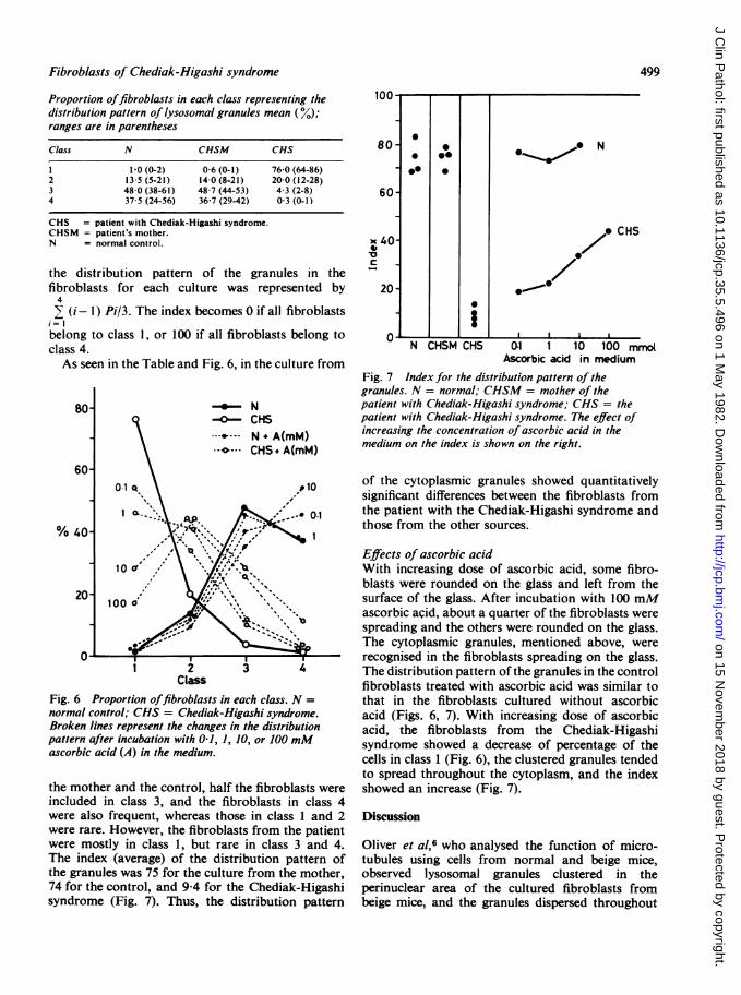
the mother and the control, half the fibroblasts wereincluded in class 3, and the fibroblasts in class 4were also frequent, whereas those in class 1 and 2were rare. However, the fibroblasts from the patientwere mostly in class 1, but rare in class 3 and 4.The index (average) of the distribution pattern ofthe granules was 75 for the culture from the mother,74 for the control, and 9 4 for the Chediak-Higashisyndrome (Fig. 7). Thus, the distribution pattern
80-
60-
x 40-to'Uc -
20-
0
00
0go
0
i" N CHSM CHS
* N
, CHS
00~~
01 1 10 100 mmotAscorbic acid in medium
Fig. 7 Index for the distribution pattern of thegranules. N = normal; CHSM = mother of thepatient with Chediak-Higashi syndrome; CHS = thepatient with Chediak-Higashi syndrome. The effect ofincreasing the concentration of ascorbic acid in themedium on the index is shown on the right.
of the cytoplasmic granules showed quantitativelysignificant differences between the fibroblasts fromthe patient with the Chediak-Higashi syndrome andthose from the other sources.
Effects of ascorbic acidWith increasing dose of ascorbic acid, some fibro-blasts were rounded on the glass and left from thesurface of the glass. After incubation with 100 mMascorbic acid, about a quarter of the fibroblasts werespreading and the others were rounded on the glass.The cytoplasmic granules, mentioned above, wererecognised in the fibroblasts spreading on the glass.The distribution pattern of the granules in the controlfibroblasts treated with ascorbic acid was similar tothat in the fibroblasts cultured without ascorbicacid (Figs. 6, 7). With increasing dose of ascorbicacid, the fibroblasts from the Chediak-Higashisyndrome showed a decrease of percentage of thecells in class 1 (Fig. 6), the clustered granules tendedto spread throughout the cytoplasm, and the indexshowed an increase (Fig. 7).
Discussion
Oliver et al,6 who analysed the function of micro-tubules using cells from normal and beige mice,observed lysosomal granules clustered in theperinuclear area of the cultured fibroblasts frombeige mice, and the granules dispersed throughout
inn{
Ul . I I
499
. vv
on 15 Novem
ber 2018 by guest. Protected by copyright.
http://jcp.bmj.com
/J C
lin Pathol: first published as 10.1136/jcp.35.5.496 on 1 M
ay 1982. Dow
nloaded from
500
the cytoplasm in the normal fibroblasts. We noticedsimilar findings in the cultured fibroblasts from apatient with the Chediak-Higashi syndrome andnormal individuals. The presence of acid phos-phatase in the PAS-positive granules in our culturedfibroblasts indicates that the granules are lysosomal.'3These lysosomal granules in the Chediak-Higashifibroblasts were clustered adjacent to the nucleus,whereas those in the normal fibroblasts were distri-buted throughout the cytoplasm. Thus, the Chediak-Higashi fibroblast cultures were readily distinguishedfrom the others.
It is said that lysosomes arise from the Golgiapparatus and are scattered in the cytoplasm likesecretory granules.14 It has been known that cellularmovement and intracellular migration of secretorygranules, organelles, and inclusions depend on thefunction of microtubules and microfilaments.15However, recent studies concerning the Chediak-Higashi syndrome have suggested that impairedbacteriocidal activities and chemotaxis of poly-morphonuclear leucocytes, and abnormal migrationof concanavalin A receptor complexes on the poly-morphonuclear leucocytes are due to abnormalmicrotubular assembly in the syndrome.16-19 There-fore, genetic disorder of the Chediak-Higashisyndrome seems to be included in the intracellularmechanism regulating microtubular assembly. Mor-phologically it has been reported that no micro-tubules were detectable in the centriolar region ofthe polymorphonuclear leucocytes in the Chediak-Higashi syndrome,6 but microtubules in our Chediak-Higashi fibroblasts seemed normal in number andstructure. Thus, the cluster of the granules in thefibroblasts from the patient indicate dysfunctionof microtubules in the Chediak-Higashi syndrome.
In this study, we used the index to express thedistribution pattern of the granules in the fibroblasts,and the index clearly differentiated the Chediak-Higashi fibroblasts from the mother's or normalfibroblasts. The index of the Chediak-Higashifibroblasts rose towards the normal level aftertreatment with ascorbic acid, which is reflected inimproved microtubular function.7 20 Therefore, theindex for the distribution pattern of lysosomalgranules in cultured fibroblasts is considered to beuseful for the study of the impaired function ofmicrotubular assembly in the Chediak-Higashisyndrome; it may also suggest the degree of thedisease.The index in the fibroblasts from the mother,
carrier of the abnormal gene, showed a normal level.On the other hand, Danes and Bearn1 observed largeinclusions in the cultured fibroblasts from theparents of the patient, and recommended cellculture in detecting the carrier. Such inclusions were
Abe, Arashima, Honma
not shown in our Chediak-Higashi fibroblasts.Abnormally large inclusions shown by them" seemslightly different in stainability from the granulesin our fibroblasts. The differences between ourfibroblasts and those observed by Danes and Bearn"may be related to differences in the onset of thedisease. Their fibroblasts were obtained from a14-year-old patient, while our patient died 110 daysafter birth. However, large granules in the culturedfibroblasts from beige mice were prominent inconfluent cultures in 10 to 14 days,6 and it wassuggested that such abnormal granules arose byfusion of developing granules.21 We observed thefibroblasts in two days of subculture. The granulesin our Chediak-Higashi fibroblasts may becomelarger in confluent culture.
It is hoped that the fetal genetic diseases can bediagnosed during early pregnancy,10 22 but thevalue of amniocentesis in Chediak-Higashi syndromehas not been established. The perinuclear congrega-tion of the lysosomal granules in the fibroblasts ofthe Chediak-Higashi syndrome persists duringrepeated subculture. Cultured cells usually containsuch granules. Therefore, the amniotic cell culture,in which fibroblast-like cells and epithelial cellsgrow,23 is also considered to be useful in prenataldiagnosis of the syndrome.
References
Chediak MM. Nouvelle anomalie leucocytaire de caractereconstitutionnel et familial. Rev Hematol 1952;7:362-7.
2 Higashi 0. Congenital gigantism of peroxidase granule-first case ever reported of qualitative abnormality ofperoxidase. Tohoku J Exp Med 1954;59:315-32.
3 Douglas SD, Blume RS, Wolff SM. Fine structural studiesof leukocytes from patients and heterozygotes withChediak-Higashi syndrome. Blood 1969;33:527-40.
4 Lutzner MA, Tierney JH, Benditt EP. Giant granules andwidespread cytoplasmic inclusions in a genetic syndromeof Aleutian mink: an electron microscopic study.Lab Invest 1966;14:2063-79.
5Bennett JM, Blume RS, Wolff SM. Characterisation andsignificance of abnormal leukocyte granules in thebeige mouse: a possible homologue for Chediak-HigashiAleutian trait. J Lab Clin Med 1969 ;73 :235-43.
6 Oliver JM, Krawiec JA, Berlin RD. Carbamylcholineprevents giant granule formation in cultured fibroblastsfrom beige (Chediak-Higashi) mice. J Cell Biol 1976;69:205-10.
7 Boxer LA, Watanabe AM, Rister M, Besch HR Jr,Allen J, Baehner RL. Correction of leukocyte functionin Chediak-Higashi syndrome by ascorbate. N Engl JMed 1976;295:1041-5.
Leory JG, De Mars RI. Mutant enzymatic and cytologicalphenotypes in cultured human fibroblasts. Science1967 ;157 :804-6.
9Wiesman UN, Herschkowitz NN. Studies on the pathogenicmechanism of I-cell disease in cultured fibroblasts.Pediatr Res 1974;8:865-70.
10 Abe K, Matsuda 1, Arashima S, Mitsuyama T, Oka Y,Ishikawa M. Ultrastructural studies in fetal I-celldisease. Pediatr Res 1976;10:669-76.
on 15 Novem
ber 2018 by guest. Protected by copyright.
http://jcp.bmj.com
/J C
lin Pathol: first published as 10.1136/jcp.35.5.496 on 1 M
ay 1982. Dow
nloaded from
Fibroblasts of Chediak-Higashi syndrome
Danes BS, Bearn AG. Cell culture and the Chediak-Higashi syndrome. Lancet 1967 ;ii :65-7.
12 Tanakawa N. Anakura M, lizuka S, et al. A case of theChediak-Higashi syndrome (in Japanese). Shonika-rinsho 1980;33:1531-6.
13 Dingle JT. ed. Lysosomes in biology and pathology, 3.North-Holland, American Elsevier, 1973.
"4Novikoff AB, Essner E, Quintana N. Golgi apparatusand lysosomes. Fed Proc 1964;23:1010-22.
15 Stossel TP. Phagocytosis. N Engl J Med 1974;290:717-23.16 Clark R, Kimball H. Defective granulocyte chemotaxis
in the Chediak-Higashi syndrome. J Clin Invest 1971;50 :2645-52.
' Stossel TP, Root RK, Vaughan M. Phagocytosis in chronicgranulomatous disease and the Chediak-H igashisydrome. N Engl J Med 1972;286:120-3.
Wolff SM, Dale DC, Clark RA, Root RK, Kimball HR.The Chediak-Higashi syndrome: studies of host defenses.Ann Intern Med 1972;76:293-306.
19 Oliver JM. Impaired microtubule assembly in Chediak-Higashi syndrome neutrophils correctable by cyclicGMP and cholinergic agents. Am J Pathol 1976;85:395-418.
20 Sandler JA, Gallin JI, Vaughan M. Effects of serotonin,carbamylcholine and ascorbic acid on leukocyte cyclicGMP and chemotaxis. J Cell Biol 1975;67:480-4.
21 Davis WC, Spicer SS, Greene WB, Padgett GA. Ultra-structure of bone marrow granulocytes in normal minkand mink with the homolog of the Chediak-Higashitrait of human. 1. Origin of the abnormal granulespresent in the neutrophils of mink with C-HD trait.Lab Invest 1971 ;24:303-17.
22 Simpson NE, Daillaire L, Miller JR, Siminovich L,Hamaerton JL, Miller J. McKeen C. Prenatal diagnosisof genetic disease in Canada: report of a collaborativestudy. Can Med Assoc J 1976;23:739-48.
23 Gerbie AB, Melancon SB, Ryan C, Nadler HL. Cultivatedepithelial-like cells and fibroblasts from amniotic fluid:their relationship to enzymatic and cytologic analysis.Am J Obstet Gynecol 1972;114:314-20.
Requests for reprints to: Dr K Abe, Department ofAnatomy, Hokkaido University School of Medicine,Sapporo, 060 Japan.
501
on 15 Novem
ber 2018 by guest. Protected by copyright.
http://jcp.bmj.com
/J C
lin Pathol: first published as 10.1136/jcp.35.5.496 on 1 M
ay 1982. Dow
nloaded from