membrane fluidity in human mouse chediak-higashi...
TRANSCRIPT
Membrane Fluidity in Human and Mouse
Chediak-Higashi Leukocytes
RICHARDA. HAAK, LEAH M. INGRAHAM, ROBERTL. BAEHNER,andLAURENCEA. BOXER, Department of Microbiology and Immunology andDepartment of Pediatrics, Indiana University School of Medicine, Indianapolis,Indiana 46223
A B S T RA C T Polymorphonuclear leukocytes fromhumans and mice with the Chediak-Higashi syndromewere characterized by spin label electron spin reso-nance spectrometry. Our results suggest that cells fromafflicted mice and humans have membranes more fluidthan controls. Order parameters for a spin label thatprobes near the membrane surface were 0.652 fornormals and 0.645 for two Chediak-Higashi patients.Cells from Chediak-Higashi mice showed similar dif-ferences, as did isolated plasma memiibrane fractions.An increased membrane fluidity was also detected witha spin label that probes deeper in the bilayer. In vitrotreatment of Chediak-Higashi mouse cells with 0.01 Mascorbate increased the order parameter to normallevels. In vitro incubation of mouse Chediak-Higashicells with glucose oxidase increased the order para-meter, similar to the effect of ascorbate. This increasewas abolished when catalase was added to the incuba-tion medium. In vitro incubation with dibutyryl cyclicguanosine monophosphate (1 ,uM to 0.1 mM) did notnormalize order parameters. These results indicate thatfluidity of Chediak-Higashi cell membranes was af-fected by treatments expected to alter the oxidation:reduction potential of the environment but was not af-fected by treatments expected to alter the ratio of intra-cellular cyclic nucleotides. The latter treatment wouldaffect microtubule assembly. Therefore, it appears thatthe membrane fluidity abnormalities as demonstratedby electron spin resonance and the earlier demon-strated microtubule dysfunction s characteristic ofChediak-Higashi cells are coexistinig defects and areprobably not directly related.
This study was done during the tenure of Dr. Boxer as anEstablished Investigator of the American Heart Association.
Received for publication 20 November 1978 and in revisedform 15 February 1979.
138
INTRODUCTION
The Chediak-Higashi syndrome (CHS)' of man is anautosomal recessive disorder in which polymorpho-nuclear leukocytes (PMN) exhibit a variety of ab-normalities (1). A similar disorder has been describedin mice (2). Phenotypic characteristics of the syndromeinclude: (a) decreased pigmentation brought about byaggregation of melanin granules; (b) enlarged lyso-somnes; and (c) recurrent problems with bacterial in-fections secondary to disorders of PMNchemotaxis,degranulation, adherence, and bacterial killing. Treat-ment with ascorbate corrects these functional defects ofCHSPMN(3-5).
Membranie abnormiialities may contribute to thesevarious defects. Concanavalin A (Con A) treatment ofCHSPMNreveals spontaneous formation of polar capsof surf:ace glycoproteins (6, 7), suggesting that thesecells have abnormal mobility of memnbranie constitu-ents. Normal cells do not undergo capping unlesstreated with colchicine, which inhibits microtubule(MT) assemnbly. Thus, membrane alterations in CHSmay coexist along with MT dysfutnction or they maybe directly related to it.
In this study we have uised spin label electron spinlresonance (ESR) spectrometry to characterize abnor-malities in CHSPMNmemnbranes. Ouir restults indicatethat these cells from CHSmice have an altered mem-brane environmenit compared to those from normal ani-mals. Further, study of PMNfiomn patients with CHS
I Abbreviations used inl this paper: cAMP, cyclic AMP;eGMP, cyclic GMP; CHS, Chediak-Higashi synd(Irome; ConA, concanavalin A; ESR, electroni spinl resoniancee; G, gauss;HBG, modified Hanks' solutioni descril)ed in reference 8;MT, microtuh)ules; PMN, polymorphoiiuclear leukocytes;5DS, 2-(3-carloxypropyl)-4,4-dimethyl-2-tridecyl-3-oxa-zolidinyloxyl; 12DS, 2-(10-carloxy deevl)-2-hexyl-4,4-dimethyl-3-oxazolidinyloxyl.
J. Clin. Invest. (© The American Society for Clinical Investigatiown, Inic. * 0021-9738/79/07/0138/07 $1.00Volutmze 64 JulJ 1979 138-144
suggests that this same membrane alteration occurs inhuman cells.
NI ETHODS
Chemicals. Dibutyryl cyclic GMP (cGMP), glucoseoxidase, and catalase were obtained from Sigma ChemicalCo. (St. Louis, Mo.). The spinl labels (Fig. 1) 2-(3-carboxv-propyl)-4,4-dimethyl-2-tridecyl-3-oxazolidinyloxyl (5DS) and2-(10-carboxydecyl)-2-hexyl-4,4-dimethyl-3-oxazolidinyloxyl(12DS) were purchased from Syva (Palo Alto, Calif.). Stocksolutions of spin labels at 5 mMNwere made up in ethanol.
Animals. Normal (C576J +/+) and CHS (C576J bg/bg)mice were obtained from The Jackson Laboratory (Bar Harbor,Maine).
Humansubjects. Two white female patients were studied.The diagnosis of CHShad been established by clinical historyand examination and in the presence of giant lysosomes inPXIN. Reports of the success of ascorbic acid therapy withthese patients have been made (3, 5). Before the presentESR studies (2 m11o for patient 1; 5 wk for patient 2) ascorbicacid treatment had been discontinued as a check on thecontinued efficacy of this therapeutic approach. Degranula-tion, bacterial killing, and chemotaxis have been demon-strated to return to abnormal levels 4 wk after cessation ofascorbic acid therapy (3). Cells from normal white adultfemales were used as controls for our present investigation.Protocols involving human subjects were approved by theIndiana University/Purdue University-Indianapolis Coim-mittee on Protection of Human Subjects. Informed consentwas obtained and the research was conducted according tothe Declaration of Helsinki.
Preparation and incubation of PMN. Peritoneal exudateleukocytes were elicited from mice by intraperitoneal in-jection of 2-3 ml 12% soclium caseinate (Difco Laboratories,Detroit, NMich.). 18 h after injection, mice were sacrificed andperitoneal contents collected. Cells were suspended in ice-cold 0.85% NaCl and centrifuged (200 g, 10 min, 4°C), re-
suspended and washed twice in saline. Final suspension(3.5 to 4.5 x 10(7 leukocvtes/ml) was in modified Hanks' solu-tion (8) (HBG) with addled glucose (0.1%) and bovine serumalbumin (0.01%). Typically suspensions were 80-90% PMN.Human PMNwere prepared from venous blood bv dextransedimentation and separation by Ficoll-Hypaque gradient(Pharmacia Fine Chemicals, Piscataway, N. J.) (9). Final sus-pension was in HBGat 3.0 x 1(7 leukocvtes/ml vith 98% ofcells being PM1N.
1 ml PMNsuspenision was placed in a snap-cap tube. Forsomile experiments additions of ascorbate, cGMP, glucoseoxidase, or catalase were made. Controls received HBG inplace of these agents. Incubation was carried out at 37°C for15 min with tumbling (12 rpm). In experiments with ascorbate,cells were washed with 9 ml HBGafter incubation and resus-pended in 1 ml HBGafter centrifugation (200 g, 5 min, 20)C).
PMNmembrane fractions. Mouse peritoneal PMNcol-lected as described above were suspended in 10 mMTris HCI(2 vol buffer to 1 vol cell pellet), pH 7.4, containing 0.25 NMsucrose (8.3% sucrose). The cells were homogenizedl in aDounce homogenizer (Kontes Co., Vineland, N. J.) until90% of the cells were disrupted as monitored by phasemicroscopy and then centrifuged at 600 g, at 4°C for 10 min toremove unbroken cells, debris, and intact nuclei. The super-nate was layered on a sucrose gradient composed of succes-sive layers of 30, 40, and 50% sucrose. The tubes werecentrifuged at 30,000 rpm (111,OOOg) in a Beckman S. W.41 rotor (Beckman Instruments, Inc., Spinco Div., Palo Alto,Calif.) for 5 h at 4°C. The bands at the interfaces between8.3 and 30%, 30 and 40%, and 40 and 50% were collected andwashed in 25 ml ice-cold distilled water. Assays were per-formed for 5'-nucleotidase as plasma membrane marker (10),beta gluc1Uronidase (3) and lysozyme (Worthington kit No.27978; Worthington Biochemical Corp., Freehold, N. J.) asgranule markers, and glucose-6-phosphatase as microsomalmarker (11). The 8.3/30% interface band showed a 10-fold en-richment for 5'-nucleotidase as compared to the originalsupernate, an(d >50% of total activity of this enzyme was
SDS 12DS
nJ',
U
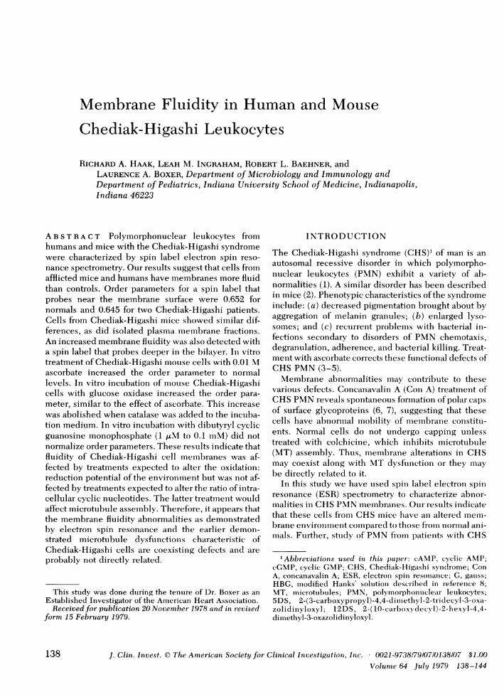
FIGURE 1 Diagrammatic representation of a generalized membrane. Proteins (P) and phospho-lipids (R ) are indicated along with the structures and probable locations of the spin labels used inthis study. The stearic acid analogs, 5DS and 12DS, probe different levels of the membraneinterior.
Membrane Fluidity of Chediak-Higashi Polymorphonuclear Leukocytes 139
located in this fraction. Little enrichment (_4% for thegranule or microsomal markers were located in the 8.3/30% band.
Spin labeling. Fig. 1 shows the spin labels, 5DSand 12DS,and their orientation in the membrane. The former has thenitroxide "reporter" group near the membrane surface,whereas the latter reports from deeper within the bilayer.The amphophilic nature of these molecules determines theirvertical position in the bilayer. The hyperfine coupling con-stants for labeled cells were consistent with the locationshown in Fig. 1.
For labeling intact cells "label tubes" were prepared by ad-dition of 8 ,l 5DS or 15 gl 12DS stock solution; the solventwas evaporated under a stream of N2 gas to provide a thinfilm of label in the bottom of each tube. The PMNsuspen-sion was transferred to a label tube. After 5 min at 20°C, thelabeled PMNwere centrifuged (40 s, 400g, 20°C) into thesealed tip of a Pasteur pipette and the ESRspectrum of the pel-let was recorded. Effects of spin labeling on viability ofPMNwere determined by trypan blue exclusion. Cell suspen-sions labeled with either 5DS or 12DS were 95% viable.
Membrane fractions were suspended in Krebs-Ringersphosphate at a concentration of _40 mg protein/ml. A 30-Al sample of membrane was labeled by placing it in a glasstube in which 3 ,ul of stock 5DS had previously been evap-orated. The membrane was taken up in a 100-,ul capillary,and the distal tip of the capillary was heat sealed and cooled.After centrifugation to displace the membranes into the sealedtip, the tube was placed in the ESR spectrometer and thespectrum recorded.
ESRdata acquisition and analysis. ESRspectra were ob-tained on a standard balanced-bridge spectrometer with diodedetection operating at 9.1 GHz. Phase-sensitive detection witha 50-kHz magnetic field modulation frequency was used.Sample heating and broadening of spectral lines were avoidedby recording all spectra at low microwave power (12 mW)in-cident on the Varian V4535 large access cavity (Varian Asso-ciates, Palo Alto, Calif.). A peak-to-peak modulation amplitudeof 1.5 gauss (G) was used for 5DS-labeled PMNand 0.5 G for12DS-labeled cells. Other appropriate values were some-times used after determination that comparisons betweensamples were independent of amplitude at the values used. Allinstrument settings were identical for a particular set ofsamples being compared. First derivative absorption spectrawere recorded with a 100-G field sweep, a scan time of 5 min,and a time constant of 0.2 s. The magnetic field sweep wascalibrated with a NMRgaussmeter (Varian Associates, PaloAlto, Calif.). Sample temperature was monitored with athermocouple, and was maintained to within +0.5°C of thedesired temperature (usually 25°C) through use of a chilled orheated N2 gas-flow system.
Two spectra from each sample were recorded and theresultant parameters averaged. All samples were coded andread blind. For experiments in which identical sampleswere run on the same day, the standard procedures for deter-mining mean, S error, and SDwere used. For experiments inwhich several samples were measured and data from severaldays were available, a two-way analysis of variance wascarried out. All P values given herein are for the probabilitythat a null hypothesis is true.
The order parameter (S), a measure of spin label order andmotion, was calculated (12, 13) for 5DS-labeled PMN.
TI- T'-CII= - l-b .1.66, (1)TI, + 2T1 + 2C
where C = 1.4-0.053 (T1l - T'). The measurement of thehyperfine spitting parameters TI, (T parallel) and TL (T' per-
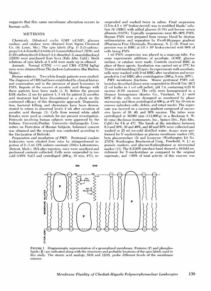
pendicular) from a typical ESR spectrum of 5DS-labeledPMNfrom Chediak-Higashi mice is shown in Fig. 2A. Asflexibility of the hydrocarbon chain of the spin label de-creases, the order parameter increases. This implies that as theenvironment surrounding the nitroxide group of the labelbecomes more ordered (i.e., less fluid), the order parameterincreases. The theoretical limits of S are S = 0 for a com-pletely fluid, isotropic system and S = 1 for a completelyrigid or ordered environment. Typical values for spin-labeledmembranes are from 0.5 to 0.7. Changes in S as small as0.5% can be shown to be statistically significant at a 95%con-fidence level (14).
The class of spectra resulting from isotropic rapid motion of aspin label is most amenable to quantitative assessment by thecalculation of the rotational correlation time, Tr (15, 16):
T, = (6 x 10⁻¹⁰ s/G (W+,)[(h+1/h-1)1/2 - 1]. (2)
The line-width and line-heights necessary for this calcula-tion are obtained from the ESRspectrum as shown in Fig. 2B.
B
AABSORPTION
AH
MAGNETICFIELD(H) I"
W+i
FIGURE 2 First derivative ESR spectrum at 25°C of spin-labeled PMNfrom Chediak-Higashi mice. (A) 5DS-labeledcells. The hyperfine splitting parameters 2T11 and 2T1 are theseparation between outer and inner extrema respectivelyand are used in the calculation of order parameter (Eq. 1). (B)12DS-labeled cells. The line-width W+1 and line-heights,h+, and h.1, are used in calculation of a empirical motionalparameter, Ri (Eq. 3).
140 R. A. Haak, L. M. Ingraham, R. L. Baehner, and L. A. Boxer
However, the assumptions made in deriving Eq. 2 are com-pletely satisfied only for rapid, isotropic motion (rT < 10-9 s).Because 12DS in PMNmembranes has a TC near this limitand also probably has some anisotropic motion, for com-parative purposes the 12DS results are reported with anoperationally defined, empirical parameter, R,:
Ri = (W+1)[(h+1/h- )l/2 - 1].s
(3)Ri can be related to the degree of spin label motion in themembrane by noting that near 25°C Ri changes by 0.2 G/°C. Asthe temperature increases, R1 decreases; this suggests that thespin label motion is more rapid and that the membrane en-vironment is more fluid.
RESULTS
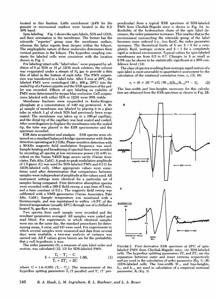
PMNprepared from venous blood of human subjectswere spin labeled with 5DS. In sets of paired samples,CHScells consistently had a lower S than did normalcells (Fig. 3) (P < 0.065 for patient 1; P < 0.004 for pa-tient 2). The relatively large number of cells requiredfor our determinations precluded intensive experi-mentation with PMNof these patients, but the similar-ity between their cells and those of the CHSmice en-couraged us to use cells from these animals in studiesdesigned to help elucidate the molecular and(or) sub-cellular basis for the fluidity difference.
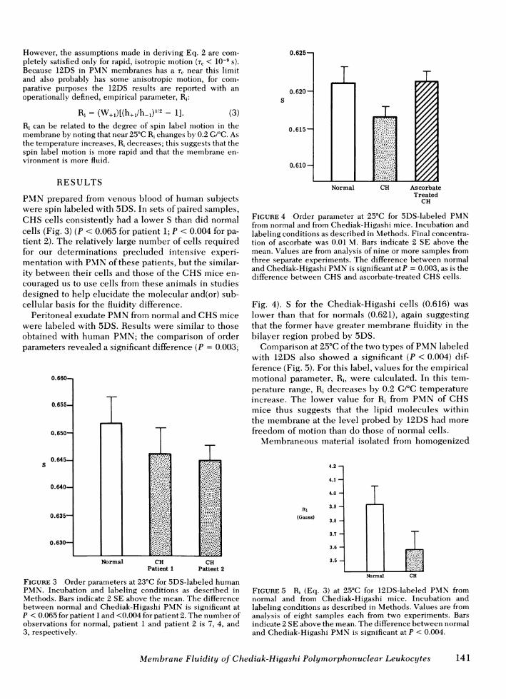
Peritoneal exudate PMNfrom normal and CHSmicewere labeled with 5DS. Results were similar to thoseobtained with human PMN; the comparison of orderparameters revealed a significant difference (P = 0.003;
Normal CH CHPatient 1 Patient 2
FIGURE 3 Order parameters at 23°C for 5DS-labeled humanPMN. Incubation and labeling conditions as described inMethods. Bars indicate 2 SE above the mean. The differencebetween normal and Chediak-Higashi PMNis significant atP < 0.065 for patient 1 and <0.004 for patient 2. The number ofobservations for normal, patient 1 and patient 2 is 7, 4, and3, respectively.
Normal CH As corbateTreated
CH
FIGURE 4 Order parameter at 25°C for 5DS-labeled PMNfrom normal and from Chediak-Higashi mice. Incubation andlabeling conditions as described in Methods. Final concentra-tion of ascorbate was 0.01 M. Bars indicate 2 SE above themean. Values are from analysis of nine or more samples fromthree separate experiments. The difference between normaland Chediak-Higashi PMNis significant at P = 0.003, as is thedifference between CHSand ascorbate-treated CHScells.
Fig. 4). S for the Chediak-Higashi cells (0.616) waslower than that for normals (0.621), again suggestingthat the former have greater membrane fluidity in thebilayer region probed by 5DS.
Comparison at 25°C of the two types of PMNlabeledwith 12DS also showed a significant (P < 0.004) dif-ference (Fig. 5). For this label, values for the empiricalmotional parameter, R1, were calculated. In this tem-perature range, R1 decreases by 0.2 G/CC temperatureincrease. The lower value for R1 from PMNof CHSmice thus suggests that the lipid molecules withinthe membrane at the level probed by 12DS had morefreedom of motion than do those of normal cells.
Membraneous material isolated from homogenized
4.2-
4.1-
4.0-
Ri 33.9-(Gauss) 3.8
3.7-
3.6-
3.5
Normal CH
FIGURE 5 R, (Eq. 3) at 25°C for 12DS-labeled PMN fromnormal and from Chediak-Higashi mice. Incubation andlabeling conditions as described in Methods. Values are fromanalysis of eight samples each from two experiments. Barsindicate 2 SE above the mean. The difference between normaland Chediak-Higashi PMNis significant at P < 0.004.
Membrane Fluidity of Chediak-Higashi Polymorphonuclear Leukocytes 141
mouse peritoneal PMNwas labeled with 5DS. The Svalue for the CHSmembrane fraction that banded at the8.3/30% sucrose interface was 0.648 (average of twoseparate determinations), whereas that for the samefraction from normal animals was 0.659. This fractionwas significantly enriched for the plasma membranemarker 5'-nucleotidase and showed little enrichmentfor granule or microsomal markers (Methods). Wetherefore conclude that the lower order parameter ob-served for intact CHS PMNas compared to intactnormal PMNis due, at least in part, to differencesin the plasma membrane fluidity of the two cell types.
Ascorbate treatment of some CHSpatients (4, 5) hasled to normalized in vitro function of PMN, which hascoincided with a marked diminuation of clinicallyapparent bacterial infections. We were interested,therefore, in examining the effect ascorbic acid mighthave on the abnormal membrane fluidity of CHSPMN.Incubation of 5DS-labeled CHS mouse PMNwith0.01 M ascorbate increased their order parameter(P < 0.003; Fig. 4) compared to untreated CHSPMN.The values were not significantly different (P = 0.92)from those for cells from normal mice. Ascorbate treat-ment of normal cells did not alter their S.
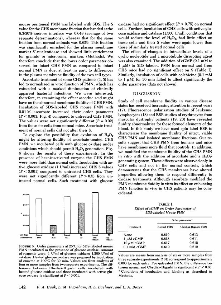
To explore the possibility that evolution of H202might be altering fluidity of ascorbate-treated CHSPMN, we incubated cells with glucose oxidase underconditions which should permit H202 generation. Fig.6 shows the results of these experiments. In thepresence of heat-inactivated enzyme the CHS PMNwere more fluid than normal cells. Incubation with ac-tive glucose oxidase (1 U/ml) increased CHSS values(P < 0.001) compared to untreated CHS cells. Theywere not significantly different (P > 0.5) from un-treated normal cells. Such treatment with glucose
Cell Type Normal CH Normal CHIncubated with: AGO AGO GO GO GO+ GO+
catalase catalase
FIGURE 6 Order parameters at 25°C for 5DS-labeled mousePMNincubated in the presence of glucose oxidase. Amountof reagents were: 1 U/ml of glucose oxidase; 1,500 U/ml ofcatalase. Heated glucose oxidase was prepared by incubationof enzyme at 100°C for 30 min. Values are from analysis offour or more samples from two separate experiments. The dif-ference between Chediak-Higashi cells incubated withheated glucose oxidase and those incubated with active glu-cose oxidase is significant at P < 0.001.
oxidase had no significant effect (P > 0.75) on normalcells. Further, incubation of CHScells with active glu-cose oxidase and catalase (1,500 U/ml), conditions thatwould reduce the level of H202, had little effect onthese cells and their S value were again lower thanthose of similarly treated normal cells.
The effect of changes in intracellular levels of acyclic nucleotide and a microtubule disrupting agentwas also examined. The addition of cGMP(0.1 mMto1 ,M) to 5DS-labeled PMNfrom normal and fromCHS mice had no significant effect on S (Table I).Similarly, incubation of cells with colchicine (0.1 mMto 1 ,uM) for 30 min failed to affect significantly theorder parameter (data not shown).
DISCUSSION
Study of cell membrane fluidity in various diseasestates has received increasing attention in recent years(17). Fluorescence polarization studies of leukemiclymphocytes (18) and ESRstudies of erythrocytes frommuscular dystrophy patients (19, 20) have revealedfluidity abnormalities for these formed elements of theblood. In this study we have used spin label ESR tocharacterize the membrane fluidity of intact, viableCHS PMNand isolated membrane fractions. Our re-sults suggest that CHSPMNfrom humans and micehave membranes more fluid that controls. In addition,we modified the membrane fluidity of the CHSPMNin vitro with the addition of ascorbate and a H202-generating system. These effects were observed only inCHS cells and not in the normal controls, wvhichdemonstrates that the CHS membranes have alteredproperties allowing them to respond differently tooxidase treatments. Although ascorbate modified thePMNmembrane fluidity in vitro its effect on enhancingPMNfunction in vivo in CHSpatients may be coin-cidental.
TABLE IEffect of cGMPon Order Parameter of
5DS-labeled Mouse PMN
Order parameter*
Treatment Normal PMN Chediak-Higashi PMN
None 0.619 0.6131 ,uM cGMP 0.618 0.61110 ,uM cGMP 0.617 0.6120.1 mMcGMP 0.619 0.612
Values are means from analysis of six or more samples fromthree separate experiments. 2 SE correspond to approximately0.003 for each entry. For untreated PMN, the difference be-tween normal and Chediak-Higashi is significant at P < 0.08.* Conditions of incubation and labeling as described inMethods.
142 R. A. Haak, L. M. Ingraham, R. L. Baehner, and L. A. Boxer

The increase in membrane fluidity observed in CHSPMNmembrane could be caused by many factors.Those of particular pertinence to the CHS includedecreased cholesterol content, a decreased sphingo-myelin:lecithin ratio, and an increased degree of un-saturation of membrane fatty acids. A decreased totalcholesterol level in the fasting serum of one CHSpa-tient has been reported (21). Through exchangeprocesses this serum decrease may be reflected in asimilar decrease of membrane cholesterol levels. De-creased cholesterol content would be expected to giverise to increased membrane fluidity (17) such as wereport here. An accelerated turnover of sphingolipidsin CHSleukocytes has been demonstrated (22), and isconsistent with previous reports of a decreased sphingo-lipid content in CHS PMNmembrane (23) and inserum (21). This decrease, together with the reportedconcurrent increase in lecithin content (21), would beexpected to give rise to increased membrane fluidity.
Molecular movements within the membrane seemcritical to many cellular functions. The lower S valuesfor CHSPMNmay reflect the same membrane altera-tions that lead to the abnormal fusion of azurophilicand specific granules (24, 25) found in these cells. Dis-ordered fluid portions of the membrane may favor fu-sion. Lucy (26) has suggested that factors which bringabout localized regions of micellular lipid arrange-ments in the bilayer will also favor membrane fusion.
In vitro ascorbate treatment might have resulted in adecrease in membrane fluidity in CHSPMNthrough avariety of mechanisms. Studies with cells in culturehave previously suggested that ascorbate-triggered re-actions led to production of H202, which in turn wasresponsible for membrane alteration (27). Membranesof PMNhigh in unsaturated fatty acid content, specifi-cally of arachidonic acid, have greater susceptibility tolipid peroxidation (28). In phospholipid liposomesascorbate-induced lipid peroxidation has been showni toincrease memlbrane rigidity (29). If, as we presentlybelieve, the ascorbate effects on CHS fluidity aremediated via H202, a likely biochemical mechanismii islipid peroxidation and the likely targets include un-saturated fatty acids. Whatever the mechanism, the ab-sence of a marked response to ascorbate treatment bynormal PMN, in contrast to CHS PMN, suggests thatinitially more fluid membranes of CHS cells have analtered in vitro structure or organization which may bethe basis, at least in part, of altered function in vivo.
An alternate mechanism for ascorbate effect onfluidity might be related to changes in intracellularlevels of cyclic nucleotides. Earlier studies had demon-strated that ascorbate treatmenit resulted in partial cor-rection of the elevated cAMP levels in human CHScells (3, 5). Treatment of CHSPMNof either human(30) or mouse (7) with cGMP also normalized func-tion, and it therefore appeared that cAMP:cGCIP ratios
might be important in defect repairs. Incubation ofPMNin concentrations of cGMPup to 0.1 mM, how-ever, had no significant effect on order parameters ofeither normal or CHS PMN. Thus, it appeared thatparticipation of ascorbate in oxidation:reduction reac-tions was more important in altering the fluidity ofCHS cells than was any effect related to potentialalteration of intracellular cyclic nucleotides.
A role for MTin defects of CHSPMNhas been sug-gested (7). PMN from CHS subjects undergo spon-taneous capping of membrane glycolipids as revealedby treatment with Con A (6, 7). Normal cells show nosuch tendency unless incubated with agents that pre-vent MTassembly. Cyclic GMPtreatment, which nor-malized capping in CHScells (6, 7,30), also influencedfunctions in normal PMNthat are colchicine-sensitive,such as lysosomal enzyme release (31). Electron micros-copy revealed an absence of MTassembly in Con A-treated PMNfrom a CHS patient but normal MT as-sembly in CHS PMN incubated with cGMP andcholinergic agonists before Con A treatment (32).Thus, in these studies it appeared that CHS abnor-malities were related to colchicine-binding structures,presumably MT.
On the other hand, giant lysosomes, pathonomic forCHS syndrome, persisted in human patients under-going ascorbate therapy (24), and we have not de-tected any consistent effects of cGMPor colchicine onthe fluidity of PMNfrom normal or CHSmice. In addi-tion, a recent study of capping in various mammaliancells suggests no clear role for MT (33). It appearsthat the membrane fluidity abnormalities as demon-strated by electron spin resonance and the earlierdemonstrated microtubule dysfunction s characteristicof Chediak-Higashi cells are coexisting defects and areprobably not directly related. However, the possiblerelations aimong abnormial NIT, altered memiibraneproperties, and defective PMNfunction in CHS re-main to be elucidated.
ACKNOWLEDGMENTS
The authors gratefully acknowledge J. M. Allen andl M. Stinefor expert technical assistance and thank F. W. Kleinhanisand W. D. Sawyer for helpful disccussioni.
This work was supported in part by National Instittutes ofHealth grant PHS ROI 13586-03, a granit from Indiaina Elksand a Biomedical Research Grant from Indiana UniversitySchool of Medicine. ESR data were obtained at the Depart-ment of Physics, Indiana University/Purdue University-Indianapolis.
REFERENCES
1. Blumlle, R. S., aind S. M. Wolff. 1972. The Chediak-Higashi syndrome: studies in four patients and a reviewof the literature. Medicine (Baltimore). 51: 247-280.
2. Gallin, J. I., J. S. Bujak, E. Patten, acndl S. M. Wolflf 1974.
Membrane Fluidity of Chediak-Higashi Polymorphonuclear Leukocytes 143
Granulocyte function in Chediak-Higashi syndrome ofmice. Blood. 43: 201-206.
3. Boxer, L. A., A. M. Watanable, M. Rister, H. R. Besch,J. Allen, and R. L. Baehner. 1976. Correction of leuko-cyte function in Chediak-Higashi syndrome by ascorbate.N. Engl. J. Med. 295: 1041-1045.
4. Boxer, L. A., J. M. Allen, A. M. Watanabe, H. R. Besch, andR. L. Baehner. 1978. Role of microtubules in granulo-cyte adherence. Blood. 51: 1045-1050.
5. Boxer, L. A., D. F. Albertini, R. L. Baehner, and J. M.Oliver. 1979. Impaired microtubule assembly and poly-morphonuclear leukocyte function in the Chediak-Higashi Syndrome correctable by ascorbic acid. Br. J.Hematol In press.
6. Oliver, J. M., R. B. Zurier, and R. D. Berlin. 1975. Con-conavalin A cap formation on polymorphonuclear leuko-cytes of normal and beige (Chediak-Higashi) mice. Na-ture (Lond.). 253: 471-473.
7. Oliver, J. M., and R. B. Zurier. 1976. Correction of char-acteristic abnormalities of microtubule function andgranule morphology in Chediak-Higashi syndrome withcholinergic agonists. Studies in vitro in man and in vivoin the beige mouse.J. Clin. Invest. 57: 1239-1247.
8. Martin, S. P., and R. Green. 1958. Methods for the studyof surviving leukocytes. A. Preparation of cell siuspen-sions. Methods Med. Res. 7: 136-138.
9. Boyum, A. 1968. Separation of leukocytes from blood andbone marrow. Scand. J. Clin. Lab Invest. Suppl. 21:77-89.
10. Avruck, J., and D. F. H. Wallach. 1971. Preparation andproperties of plasma membrane and endoplasmicreticulum fragments from isolated rat fat cells. Biochim.Biophys. Acta. 233: 344-347.
11. Bagenski, E. S., P. P. Foa, and B. Zak. 1974. Glucose-6-phosphatase. In Methods of Enzymatic Analysis. H. U.Bergmeyer, editor. Academic Press, Inc., NewYork. 876.
12. Gaffney, B. J., and C. M. McNamee. 1974. Spin-labelmeasurements in membranes. Methods Enzymol. 323:161-198.
13. Berliner, L. J. 1976. Spin labeling: Theory and Applica-tions. Academic Press, Inc., New York. 592 pp.
14. Kury, P. G., and H. M. McConnell. 1975. Regulation ofmembrane flexibility in human erythrocytes. Bio-chemistry. 14: 2798-2502.
15. Kivelson, D. 1960. Theory of ESR linewidths of freeradicals.J. Chem. Phys. 33: 1094-1106.
16. Stone, T. J., T. Buckman, P. L. Nordio, and H. M. Mc-Connell. 1965. Spin-labeled biomolecules. Proc. Natl.Acad. Sci. U. S. A. 54: 1010-1017.
17. Cooper, R. A. 1977. Abnormalities of cell-membranefluidity in the pathogenesis of disease. N. Engl. J. Med.297: 371-377.
18. Inbar, M., R. Goldman, L. Inbar, I. Bursuker, B. Goldman,E. Akstein, P. Segal, E. Ipp, and I. BenBassat. 1977.Fluidity differences of membrane lipids in human and
leukemic lymphocytes as controllecl by serum comn-ponents. Cancer Res. 37: 3037-3041.
19. Butterfield, D. A. 1977. Electron spin resonance studiesof erythrocyte membranes in mutscular dystrophy. Acc.Chem. Res. 10: 111-116.
20. Sato, B., K. Nishikida, L. T. Samnuels, and F. H. Tyler.1978. Electron spin resonance studies of erythrocytesfrom patients with Duchenne muisctular dystrophy. J.Clin. Invest. 61: 251-259.
21. Kritzler, R. A., J. Y. Terner, J. Lindenbaum, J. Magidson,R. Williams, R. Preisig, and G. B. Phillips. 1964. Chediak-Higashi syndrome. Cytologic and serum lipicl observa-tions in a case and family. Am. J. Med. 36: 583-594.
22. Kanfer, J. N., R. S. Blutne, R. A. Yankee, andl S. M. Wolff.1968. Alteration of sphingolipid metabolism in leuikocytesfrom patients with the Chediak-Higashi synd(rome. N.Engl. J. Med. 279: 410-413.
23. Kanfer, J. N., R. Richards, J. P. Kampine, S. Han(ldnaker,and R. A. Yankee. 1967. Alteration of the sphingolipiclcontent in leukocytes from patients with Chediak-Higashi syndrome. Life Sci. 6: 2661-2664.
24. Rausch, P. G., K. B. Pryzwansky, and J. K. Spitznagel.1978. Immunocytochemical identification of azurophilicand specific granule markers in the giant granules ofChediak-Higashi neutrophils. N. Engl. J. Med. 298:693-698.
25. Rozenszajn, L. A., E. B. David, and S. B. Sela. 1977. Largegranules and lysosomal fuision in humani Chediak-Higashiwhite blood cells. Acta Haematol. (Basel). 57: 279-289.
26. Lucy, J. A. 1970. The fusion of biological membranes.Nature (Lond.). 227: 815-817.
27. Peterkofsky, B., and W. Prather. 1977. Cytotoxicity ofascorbate and other reclucing agents towards culturedfibroblasts as a result of hydlrogen peroxide formation.
J. Cell Physiol. 90: 61-70.28. Stossel, T. P., R. J. Mason, and A. L. Smith. 1974. Lipid
peroxidation by human blood phagocytes.J. Clin. Invest.54: 638-645.
29. Dobretsov, G. E., T. A. Borschevskaya, V. A. Petrov, andY. A. Vladimirov. 1977. The increase of phospholipid bi-layer rigidity after lipid peroxidation. FEBS (Fed. Eur.Biochem. Soc.) Lett. 84: 125-128.
30. Boxer, L. A., M. Rister, J. M. Allen, and R. L. Baehner.1977. Improvement of Chediak-Higashi leukocyte func-tion by cyclic guanosine monophosphate. Blood. 49:9-17.
31. Weissman, G., I. Goldstein, S. Hoffstein, and P. K. Tsung.1975. Reciprocal effects of cAMP and cGMPon micro-tubule-dependent release of lysosomal enzymes. Ann.N. Y. Acad. Sci. 253: 750-762.
32. Oliver, J. M. 1976. Impaired microtubule function cor-rectable by cyclic GMPand cholinergic agonists in theChediak-Higashi syndrome. Am. J. Pathol. 85: 395-418.
33. Bourguignon, L. Y. W., and S. J. Singer. 1977. Transmem-brane interactions and the mechanism of capping surfacereceptors by their specific ligands. Proc. Natl. Acad. Sci.U. S. A. 74: 5031-5035.
144 R. A. Haak, L. M. Ingraham, R. L. Baehner, and L. A. Boxer