(hnk-1l) cells in chediak-higashi are normal numbersbut are
TRANSCRIPT

RAPID
PUBLICATIONS
Natural Killer (HNK-1l) Cells in Chediak-Higashi
Patients Are Present in Normal Numbers but Are
Abnormal in Function and Morphology
TORu ABO, JOHN C. RODER, WATARUABO, MAXD. COOPER,andCHARLESM. BALCH, Cellular Immunology Unit of the Tumor Institute,Departments of Surgery, Pediatrics and Microbiology, University of Alabamain Birmingham, Birmingham, Alabama 35294; Department of Microbiologyand Immunology, Queen's University, Kingston, Ontario, Canada K7L 3N6;Department of Pediatrics, Sapporo Medical College, Sapporo 060, Japan
A B S T R A C T Children with the Chediak-Higashi(CH) syndrome are known to have abnormalities ofnatural killer (NK) cell function. Weused the HNK-1 monoclonal antibody that reacts specifically withhuman NK and K cells to distinguish whether this ab-normality was due either to a numerical deficiency ofNK cells or a defect in their ability to function. Ineight CH patients, a significant proportion of theirblood mononuclear cells (10-19%) expressed the HNK-1 differentiation antigen. The level of NK cells in thefive children with CH syndrome was higher than forage-matched normal controls (15.8% vs. 5.8%, P <0.001). When HNK-1+ cells were isolated with a flu-orescence-activated cell sorter, the NK cells from CHpatients were a homogeneous population of lympho-cytes with a single large granule rather than the mul-tiple small granules seen in NK cells from normal in-dividuals. The purified HNK-1+ cells from the CHpatients had minimal NK or K cell function. The CHsyndrome thus includes a functionally defective pop-
Received for publication 17 February 1982 and in revisedform 31 March 1982.
ulation of NK cells that retain the capability of ex-pressing the HNK-1 differentiation antigen.
INTRODUCTION
Children with the Chediak-Higashi (CH)' syndrome,an autosomal recessive disorder, have an increased fre-quency of infections and decreased pigmentation (1).The characteristic giant granules in the cytoplasm ofmelanocytes and granulocytes is associated with thefunctional derangement of these specialized cells. Aselective deficiency of natural killer (NK) cell functionhas been described in CHpatients that may be relatedto their susceptibility to infections and their increasedrisk of malignancy (2-5). It has been suggested thatNK cells may be present but functionally defective inaffected individuals (2-4). Alternatively, it is possiblethat CH patients lack normal numbers of NK cells.These two possibilities were examined with a mono-
' Abbreviations used in this paper: ADCC, antibody-de-pendent cell-mediated cytotoxicity; CH, Chediak-Higashisyndrome; FACS, fluorescence-activated cell sorter; NK, nat-ural killer.
J. Clin. Invest. © The American Society for Clinical Investigation, Inc. * 0021-9738/82/07/0193/05 $1.00Volume 70 July 1982 193-197
193

a.
b.
CH HNK-l+
Normal HNIK-I+
FIGURE 1 Characteristic large granule in NK cells from a CH patient. Mononuclear bloodcells, stained by the HNK-1 antibody and fluoresceinated F(ab)'2 anti-mouse immunoglobulinwere separated into HNK-1+ and HNK-1- cells by FACS. (a) HNK-1+ cells from a 4-yr-oldCHpatient, N.S. (b) HNK-1+ cells from a normal individual. (c) HNK-1- cells from the patient.A single large cytoplasmic granule was seen in virtually all HNK-1+ cells from the patient,while the HNK-1+ cells from the normal control contained multiple small granules. The HNK-1- cells from both patient and normal control were agranular small lymphocytes.
clonal antibody (HNK-1) that selectively reacts witha differentiation antigen on granular lymphocytes withNK and K cell function (6, 7).
METHODSPatients. The CHpatients and their clinical features have
been described elsewhere (8, 9). The characterisitic giant
T. Abo, J. C. Roder, W. Abo, M. D. Cooper, and C. M. Balch194
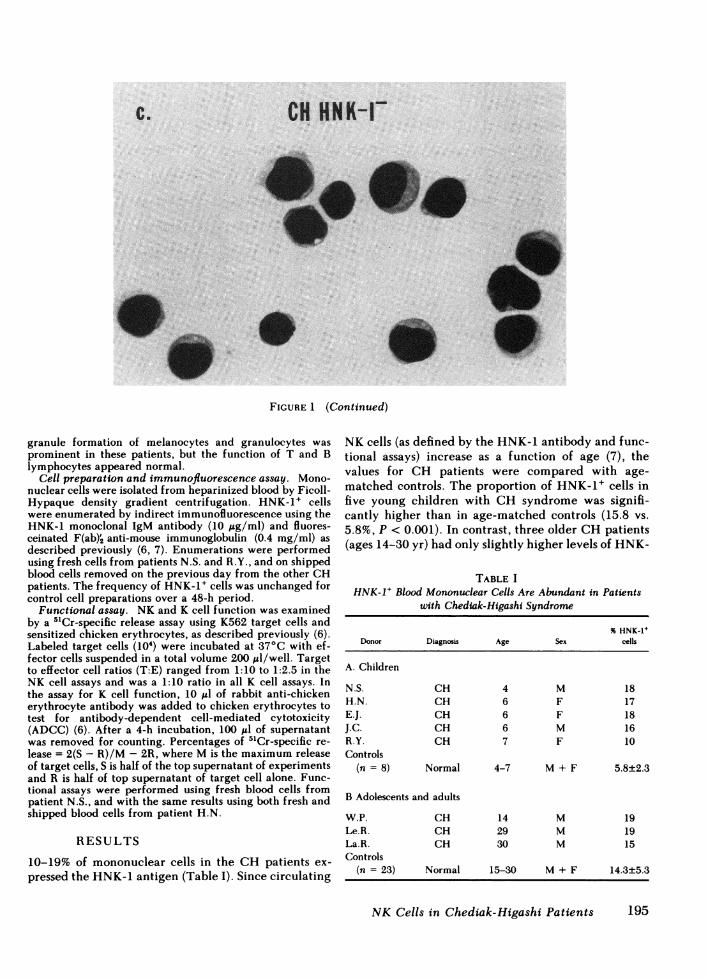
CH HNK-I-
FIGURE 1 (Continued)
granule formation of melanocytes and granulocytes wasprominent in these patients, but the function of T and Blymphocytes appeared normal.
Cell preparation and immunofluorescence assay. Mono-nuclear cells were isolated from heparinized blood by Ficoll-Hypaque density gradient centrifugation. HNK-1+ cellswere enumerated by indirect immunofluorescence using theHNK-1 monoclonal IgM antibody (10 ug/ml) and fluores-ceinated F(ab)2 anti-mouse immunoglobulin (0.4 mg/ml) asdescribed previously (6, 7). Enumerations were performedusing fresh cells from patients N.S. and R.Y., and on shippedblood cells removed on the previous day from the other CHpatients. The frequency of HNK-1+ cells was unchanged forcontrol cell preparations over a 48-h period.
Functional assay. NKand K cell function was examinedby a 51Cr-specific release assay using K562 target cells andsensitized chicken erythrocytes, as described previously (6).Labeled target cells (10') were incubated at 37°C with ef-fector cells suspended in a total volume 200 Il/well. Targetto effector cell ratios (T:E) ranged from 1:10 to 1:2.5 in theNK cell assays and was a 1:10 ratio in all K cell assays. Inthe assay for K cell function, 10 ;J of rabbit anti-chickenerythrocyte antibody was added to chicken erythrocytes totest for antibody-dependent cell-mediated cytotoxicity(ADCC) (6). After a 4-h incubation, 100 Ml of supernatantwas removed for counting. Percentages of 5'Cr-specific re-lease = 2(S - R)/M - 2R, where M is the maximum releaseof target cells, S is half of the top supernatant of experimentsand R is half of top supernatant of target cell alone. Func-tional assays were performed using fresh blood cells frompatient N.S., and with the same results using both fresh andshipped blood cells from patient H.N.
RESULTS
10-19% of mononuclear cells in the CH patients ex-pressed the HNK-1 antigen (Table I). Since circulating
NK cells (as defined by the HNK-1 antibody and func-tional assays) increase as a function of age (7), thevalues for CH patients were compared with age-matched controls. The proportion of HNK-1+ cells infive young children with CH syndrome was signifi-cantly higher than in age-matched controls (15.8 vs.5.8%, P < 0.001). In contrast, three older CHpatients(ages 14-30 yr) had only slightly higher levels of HNK-
TABLE IHNK-I+ Blood Mononuclear Cells Are Abundant in Patients
with Chediak-Higashi Syndrome
% HNK-1+Donor Diagnosis Age Sex cells
A. Children
N.S. CH 4 M 18H.N. CH 6 F 17E.J. CH 6 F 18J.C. CH 6 M 16R.Y. CH 7 F 10Controls
(n = 8) Normal 4-7 M + F 5.8±2.3
B Adolescents and adults
W.P. CH 14 M 19Le.R. CH 29 M 19La.R. CH 30 M 15Controls
(n = 23) Normal 15-30 M + F 14.3±5.3
NK Cells in Chediak-Higashi Patients 195
Ca

100r
50F
HNK-I +
Normal
CH
1 2.5 1:5T E Ratios
HNK- +
Normal
CH
no 10-2 10-3onti-CRBC (mg/mI)
A. NK100-
Cell Assoy (K-562)..... - , ._0loorHNK- I-
50F
0
50
01725 :5 110T:E Ratios
B. K Cell Assay (CRBC)IOO[ HNK- 1-
5OF
no 10-2 10-3onti -CRBC (mg/ml)
Whole MNC
1:2.5 1 5TE Ratios
1 10
no 10-2 10-3onti-CRBC (mg/rrml)
FIGURE 2 Impairment of NK and K cell function by HNK-1+ cells from a CH patient. HNK-1+ cells from CHpatient N.S. (- 0 -) were markedly defective in both NK and K cell functionscompared with HNK-1+ cells from a healthy donor (- * -). The sorted HNK-1+ and HNK-1-cell fractions contained <1% monocytes; the whole mononuclear cell fraction in the patientcontained -15% monocytes.
1+ cells than did age-matched controls (17.7 vs. 14.3%,P > 0.2).
The morphological and functional characteristics ofHNK-1+ cells were examined in greater detail usingmononuclear blood cells from the 4-yr-old patient N.S.HNK-1+ and HNK-1- cells were purified with a flu-orescence-activated cell sorter (FACS) and then stainedwith May-Grunwald-Giemsa (Fig. 1). The sorted HNK-1+ cells were a homogenous population of lymphocytescontaining a single giant granule (Fig. la). In contrast,HNK-1+ cells from normal individuals contained mul-tiple small granules in their cytoplasm (Fig. lb). HNK-1- cells from the patient and from a normal individualwere primarily small lymphocytes containing no gran-
ules (Fig. lc). The morphological homogeneity of eachsorted fraction exceeded 96%.
Studies of HNK-1+ cells from normal individualshave revealed the expression of HNK-1 antigen on
both the cell surface and in the cytoplasm (7). Thecytoplasmic HNK-1 staining appeared to increase as
a function of NK cell maturation. We therefore ex-
amined the cytoplasmic distribution of HNK-1 deter-
minants in surface HNK-1+ cells from CH patients.A normal diffuse pattern of HNK-1 antigen expressionwas noted in the majority (60%) of cells from affectedindividuals. There was no concentration of HNK-1antigen within the giant cytoplasmic granule.
Sorted HNK-1+ and HNK-1- blood mononuclearcells were then examined for their NKand K cell func-tional activities using K562 target cells and sensitizedchicken erythrocytes. The cytotoxic capability of ef-fector cells was minimal in the 4-yr-old patient N.S.(who had 16% HNK-1+ cells) compared with a normal19-yr-old male with 10% HNK-1+ cells (Fig. 2). TheNK functional activity was depressed in the CH pa-
tient using both a total mononuclear cell preparationand the purified fraction of HNK-1+ cells. Interest-ingly, significant ADCCactivity was observed in thewhole mononuclear cell fraction (Fig. 2). This was
probably due to a 15% contamination by monocytes,since minimal K cell activity was noted both in HNK-1+ and HNK-1- cell fractions, which were depletedof large unstained cells during the cell sorter separa-
tions. This data is consistent with previous studies dem-
196 T. Abo, J. C. Roder, W. Abo, M. D. Cooper, and C. M. Balch
4)0
4'
C,)
in
.0
100
50
C

onstrating that ADCCactivity by monocytes was intactin the CH patients, whereas K cell function in thelymphocyte fraction was abnormally low (4).
The dissociation between the HNK-1+ cell level andabsent NK cell function was also demonstrated in an-other CH patient H.N., using a discontinuous Percolldensity gradient technique as previously described(10). HNK-1+ cells from this 6-yr-old CHpatient wereenriched into a low density fraction similar to thoseof the normal donor. However, this fraction had adepressed NK cell functional capability comparedwith the control (data not shown).
DISCUSSIONThe present results provide direct evidence in supportof previous proposals (2-4) that the abnormality in CHpatients is due to functionally defective NK cells. Wehave found that lymphocytes expressing the HNK-1differentiation antigen and characteristic granularmorphology, are present in normal or elevated levelsin CH patients. The expanded population of circulat-ing HNK-1+ cells that we observed in young CH pa-tients could reflect accelerated development of HNK-1+ cells or excessive accumulation of functionally ab-normal NK cells.
Highly enriched (96%), HNK-1+ CH cells isolatedon the FACSexhibited defective NKand ADCCfunc-tion and were characterized by the presence of a singlelarge granule in their cytoplasm. On the other handneither macrophages nor T cells from CH patientsexhibited giant granules (Fig. lc and unpublished ob-servations), whereas both effector cell types exhibitednormal frequencies and cytolytic functions (Fig. 2 [4]).It seems likely, therefore, that giant granule in NKcells may be closely linked to the functional defect.The underlying mechanism of giant granule formationis not known but may involve an abnormal inter-action between the lysosome membrane and micro-tubules (11).
The function of the HNK-1 antigen is not yet knownbut it is probably not involved directly in the NK-target cell interaction because NK activity was not in-hibited by the HNK-1 antibody and inhibition of NKcytolysis by mild pronase treatment did not affect theexpression of HNK-1 (6). It is interesting to note thatthe distribution of intracellular HNK-1 antigen in CH-NK cells was normal.
This study thus demonstrates that the recessive ge-netic defect underlying the CHsyndrome affects lym-phocytes expressing the HNK-1 differentiation anti-gen. Previous studies have revealed abnormalities ofother granule-containing cells, e.g., granulocytes andmelanocytes (1). The CHdefect also affects functionsof these other cell types rather than their populationsize. By contrast, T lymphocytes and macrophages in
CHpatients do not contain the giant granule, lack theHNK-1 antigen and exhibit normal function. Our stud-ies identify the mononuclear blood cells displaying thecharacteristic giant granules, all of which expressHNK-1 antigen, as functionally defective NK cells.These observations are consistent with the idea thatNK cells belong to a separate pathway of lymphoiddifferentiation.
ACKNOWLEDGMENTS
Wewish to thank Drs. W. Crist in Birmingham, T. Orii inJapan, L. Boxer in Indianapolis, A. Fauci in Bethesda, D.Matheson in Calgary, and J. White in Minneapolis for supplyof blood from CH patients, Dr. G. Larry Gartland for op-eration of FACS, and Ms. Sharon Garrison and Ms. FrancesSmith for editorial assistance.
This work was supported by grants from the National In-stitutes of Health (CA-13148, CA-27197, CA-16673, and RR-32) and Medical Research Council and NCI of Canada.
REFERENCES
1. Windhorst, D. B., and G. J. Padgett. 1973. The Chediak-Higashi syndrome and the homologous trait in animals.Invest. Dermatol. 60: 529-537.
2. Roder, J. C., T. Haliotis, M. Klein, S. Korec, J. R. Jett,J. Ortaldo, R. B. Herberman, P. Katz, and A. S. Fauci.1980. A new immunodeficiency disorder in humans in-volving NK cells. Nature (Lond.). 284: 553-555.
3. Haliotis, T., J. Roder, M. Klein, J. Ortaldo, A. S. Fauci,and R. B. Herberman. 1980. Chediak-Higashi gene inhumans I. Impairment of natural-killer function. J. Exp.Med. 151: 1039-1048.
4. Klein, M., J. Roder, T. Haliotis, S. Korec, J. Jett, R. B.Herberman, P. Katz, and A. S. Fauci. 1980. Chediak-Higashi gene in humans II. The selectivity of the defectin natural-killer and antibody-dependent cell-mediatedcytotoxicity function. J. Exp. Med. 151: 1049-1058.
5. Dent, P. B., L. A. Fish, J. G. White, and R. A. Good.1966. Chediak-Higashi syndrome. Observation on thenature of the associated malignancy. Lab. Invest. 15:1634-1642.
6. Abo, T., and C. M. Balch. 1981. A differentiation antigenof human NK and K cells identified by a monoclonalantibody (HNK-1). J. Immunol. 127: 1024-1029.
7. Abo, T., M. D. Cooper, and C. M. Balch. 1982. Postnatalexpansion of the natural killer and killer cell populationin humans identified by the monoclonal HNK-1 anti-body. J. Exp. Med. 155: 321-326.
8. Prchal, J. T., W. M. Crist, A. Malluh, A. Vitek, W. N.Tauxe, and A. J. Carroll. 1980. A new glucose-6-phos-phate dehydrogenase deficient variant in a patient withChediak-Higashi syndrome. Blood. 56: 476-480.
9. Roder, J. C., L. Laing, T. Haliotis, and D. Kozbor. 1981.Genetic control of human NK function. In NK cells:Fundamental Aspects and Role in Cancer. R. B. Her-berman, editor. Elsevier/North-Holland Publishers,Amsterdam. In press.
10. Timonen, T., and E. Saksela. 1980. Isolation of humanNK cells by density gradient centrifugation. J. Immu-nol. Methods. 36: 285-291.
11. White, J. G., and C. C. Clawson. 1979. The Chediak-Higashi syndrome: Microtubules in monocytes and lym-phocytes. Am. J. Hematol. 1: 349-356.
NK Cells in Chediak-Higashi Patients 197