1 the breast sara sukumar pathobiology, september 6, 2013
TRANSCRIPT
1
The BREAST
Sara SukumarPathobiology,
September 6, 2013
Breast Cancer
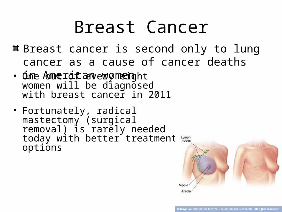
• One out of every eight women will be diagnosed with breast cancer in 2011
• Fortunately, radical mastectomy (surgical removal) is rarely needed today with better treatment options
2
Breast cancer is second only to lung cancer as a cause of cancer deaths in American women

Trends since 1950 in age-standardized
death rates comparing breast and selected other types
of cancer, among women in the USA
EBCTCG, Lancet, 2010
BREAST CANCER IN THE WORLD
1.15 million new cases
Incidence increasing in most countries
470 000 deaths
Half of the global burden in low- and medium-resourced countries

Outline- Part 1
• Development of the Breast
• Female Breast Anatomy
• Breast Cancer
• Risk Factors- Sporadic and Hereditary Breast Cancer
• Biology of Breast Cancer
5
Outline- Part 2
• How breast cancer is:
• Detected
• Diagnosed
• Treated
6
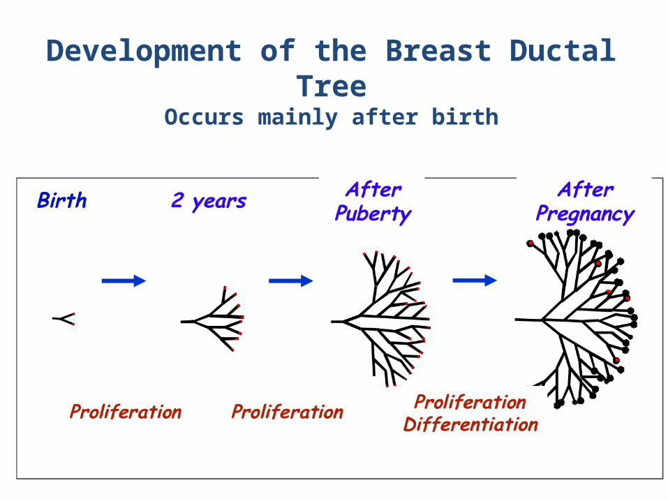
Development of the Breast Ductal Tree
Occurs mainly after birth
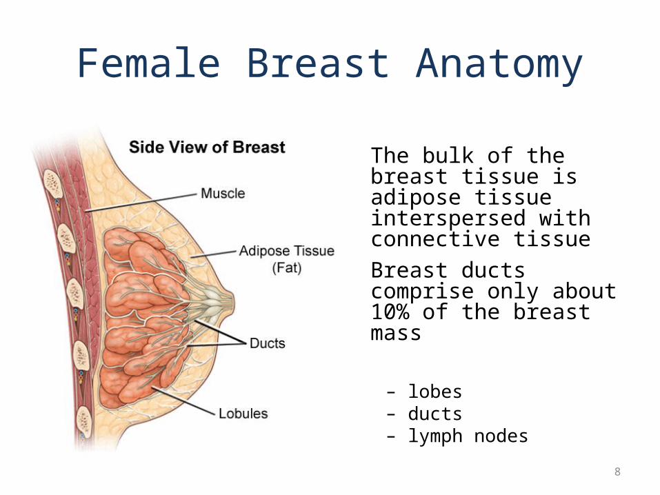
Female Breast Anatomy
• The bulk of the breast tissue is adipose tissue interspersed with connective tissue
• Breast ducts comprise only about 10% of the breast mass
– lobes– ducts– lymph nodes
8
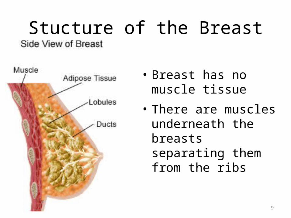
Stucture of the Breast
• Breast has no muscle tissue
• There are muscles underneath the breasts separating them from the ribs
9
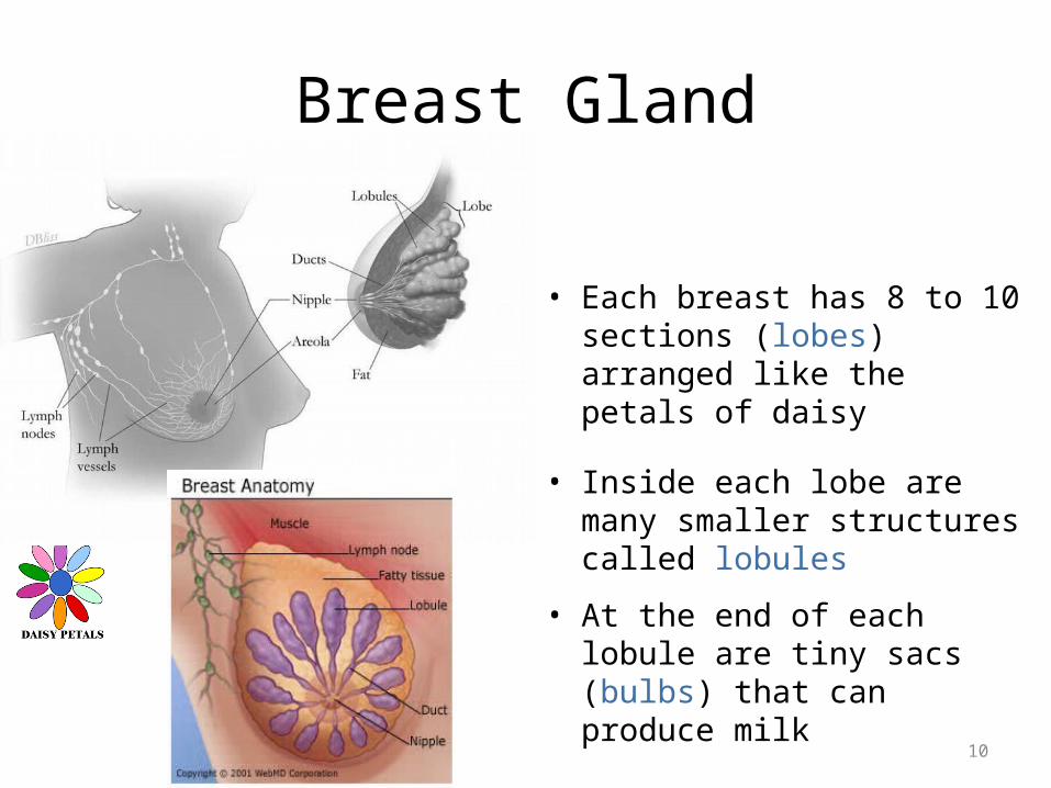
Breast Gland
• Each breast has 8 to 10 sections (lobes) arranged like the petals of daisy
• Inside each lobe are many smaller structures called lobules
• At the end of each lobule are tiny sacs (bulbs) that can produce milk
10

Ducts
• Lobes, Lobules and bulbs, are linked by a network of thin tubes (ducts)
• Ducts carry milk from bulbs toward dark area of skin in the center of the breast (areola)
11
Ducts join together into larger ducts ending at the nipple, where milk is delivered
Duct
Areola
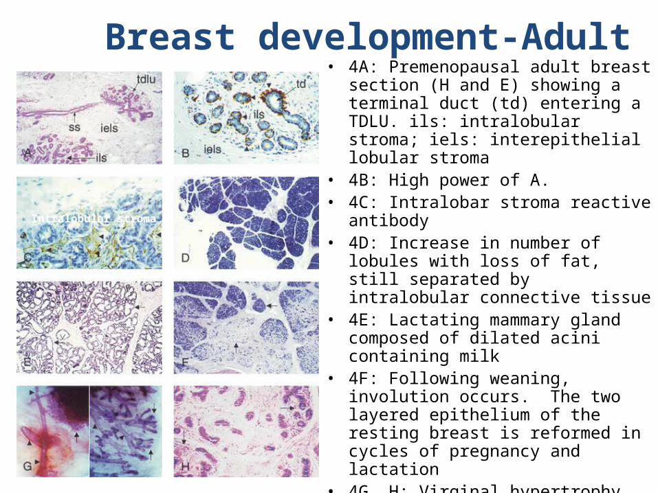
Breast development-Adult• 4A: Premenopausal adult breast
section (H and E) showing a terminal duct (td) entering a TDLU. ils: intralobular stroma; iels: interepithelial lobular stroma
• 4B: High power of A.• 4C: Intralobar stroma reactive
antibody • 4D: Increase in number of
lobules with loss of fat, still separated by intralobular connective tissue
• 4E: Lactating mammary gland composed of dilated acini containing milk
• 4F: Following weaning, involution occurs. The two layered epithelium of the resting breast is reformed in cycles of pregnancy and lactation
• 4G, H: Virginal hypertrophy
Intralobular stroma
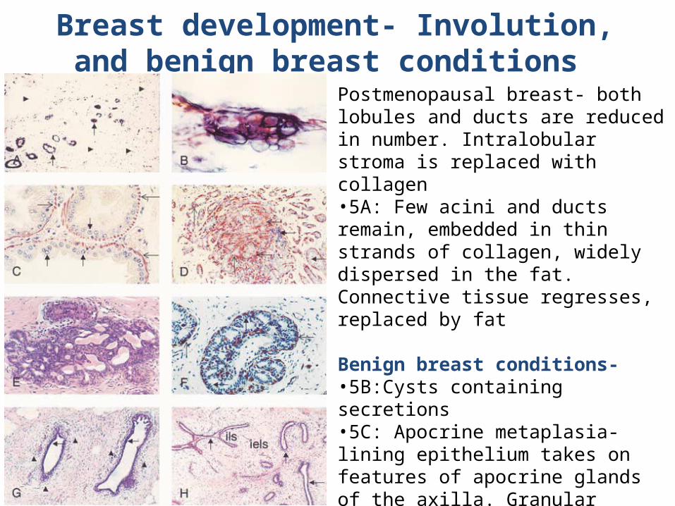
Breast development- Involution, and benign breast conditions
Postmenopausal breast- both lobules and ducts are reduced in number. Intralobular stroma is replaced with collagen•5A: Few acini and ducts remain, embedded in thin strands of collagen, widely dispersed in the fat. Connective tissue regresses, replaced by fat
Benign breast conditions-•5B:Cysts containing secretions•5C: Apocrine metaplasia-lining epithelium takes on features of apocrine glands of the axilla. Granular cytoplasm, large nuclei, nucleoli.•5D:Sclerosing adenosis-lobular proliferation with acini are infiltrative at the margins•5F: Epithelial hyperplasia-expansion of lobules
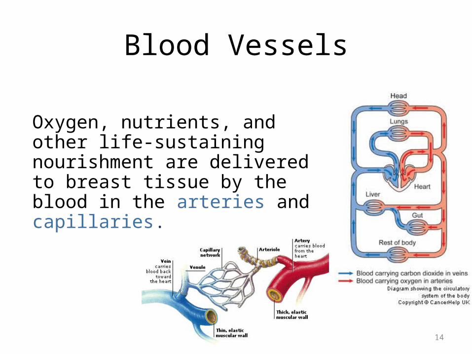
Blood Vessels
Oxygen, nutrients, and other life-sustaining nourishment are delivered to breast tissue by the blood in the arteries and capillaries.
14

Lymphatic System• Lymph ducts: Drain
fluid that carries white blood cells (that fight disease) from the breast tissues into lymph nodes under the armpit and behind the breastbone
• Lymph nodes: Filter harmful bacteria and play a key role in fighting off infection
15
A network of vessels
Lymph ductLymph node
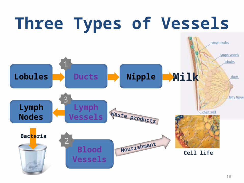
Three Types of Vessels
16
Bacteria
NourishmentBlood
VesselsCell life
2
Waste products
LymphNodes
LymphVessels
3
MilkLobules Ducts Nipple
1

Signs and Symptoms
17
Most common: lump or thickening in breast. Often painless
Change in color or appearance of areola
Redness or pitting of skin over the breast, like the skin of an orange
Discharge or bleeding
Change in size or contours of breast
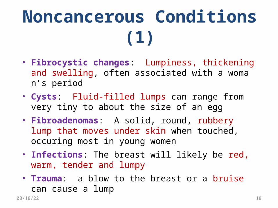
Noncancerous Conditions (1)
• Fibrocystic changes: Lumpiness, thickening and swelling, often associated with a woman’s period
• Cysts: Fluid-filled lumps can range from very tiny to about the size of an egg
• Fibroadenomas: A solid, round, rubbery lump that moves under skin when touched, occuring most in young women
• Infections: The breast will likely be red, warm, tender and lumpy
• Trauma: a blow to the breast or a bruise can cause a lump04/18/23 18
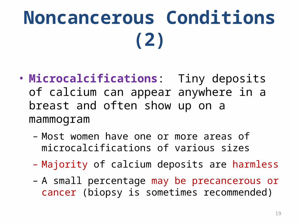
Noncancerous Conditions (2)
• Microcalcifications: Tiny deposits of calcium can appear anywhere in a breast and often show up on a mammogram– Most women have one or more areas of
microcalcifications of various sizes
– Majority of calcium deposits are harmless
– A small percentage may be precancerous or cancer (biopsy is sometimes recommended)
19
Causes
• Some of the cells begin growing abnormally
• These cells divide more rapidly than healthy cells do and may spread through the breast, to the lymph or to other parts of the body (metastasize)
• The most common type of breast cancer begins in the milk-production ducts, but cancer may also occur in the lobules or in other breast tissue
04/18/23 20
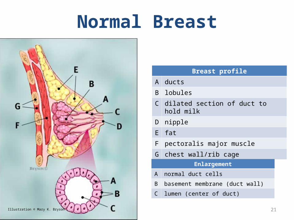
Normal Breast
Breast profile
A ducts
B lobules
C dilated section of duct to hold milk
D nipple
E fat
F pectoralis major muscle
G chest wall/rib cage
21
Enlargement
A normal duct cells
B basement membrane (duct wall)
C lumen (center of duct)
Illustration © Mary K. Bryson

Ductal Carcinoma in situ (DCIS)
22Illustration © Mary K. Bryson
Ductal cancer cells
Normal ductal cell
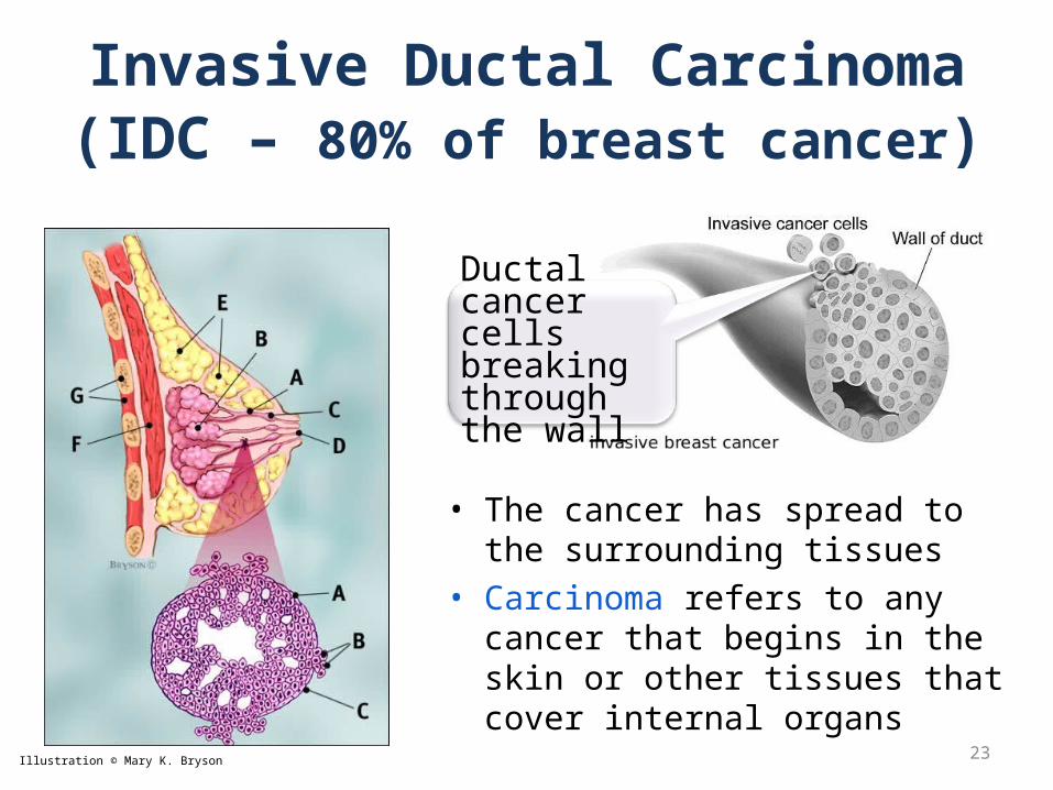
Invasive Ductal Carcinoma (IDC – 80% of breast cancer)
• The cancer has spread to the surrounding tissues
• Carcinoma refers to any cancer that begins in the skin or other tissues that cover internal organs
23Illustration © Mary K. Bryson
Ductal cancer cells breaking through the wall
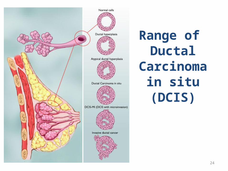
Range of Ductal
Carcinoma in situ (DCIS)
24
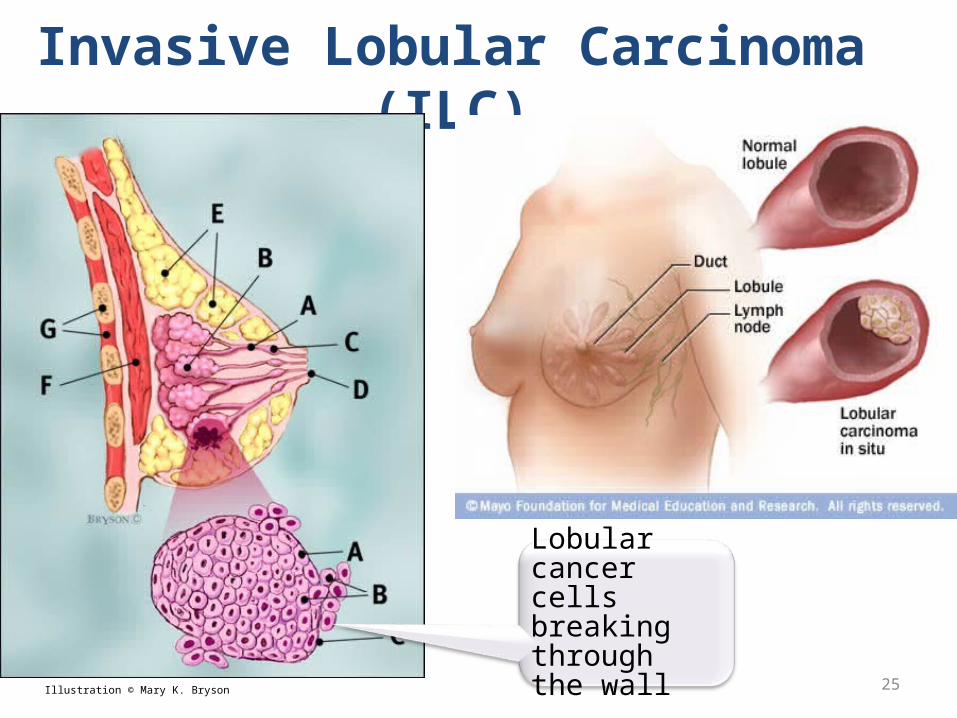
Invasive Lobular Carcinoma (ILC)
25Illustration © Mary K. Bryson
Lobular cancer cells breaking through the wall
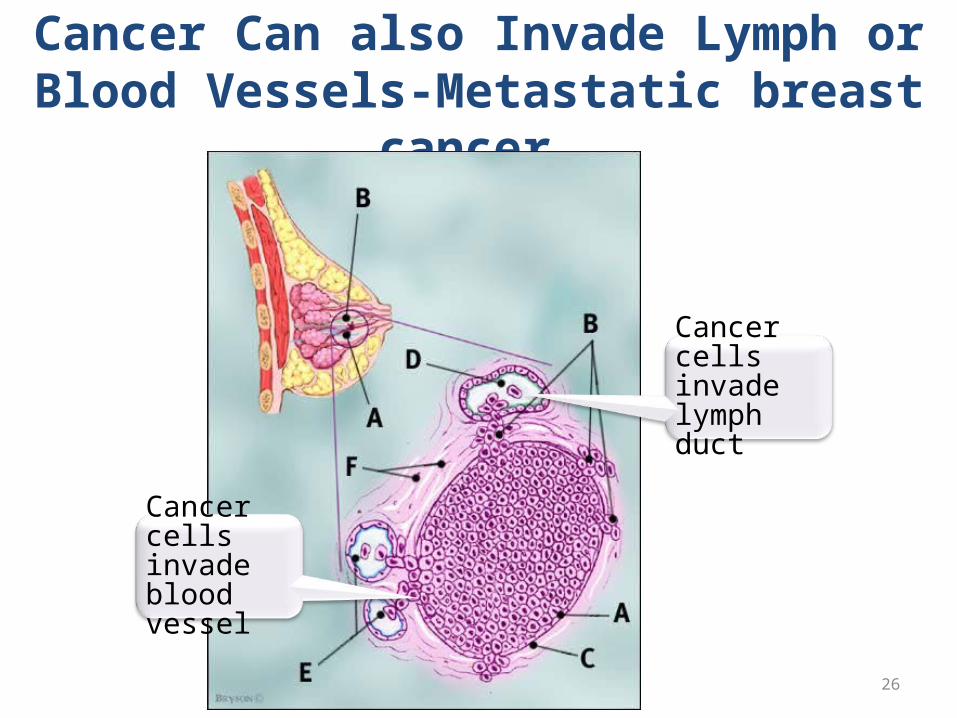
Cancer Can also Invade Lymph or Blood Vessels-Metastatic breast
cancer
26Illustration © Mary K. Bryson
Cancer cells invade lymph duct
Cancer cells invade blood vessel
Factors determining risk of developing
Breast Cancer
27

GENDER - All women are
at risk
Age
Family/PersonalHistory
ReproductiveHistory
MenstrualHistoryRace
Genetic Factors
Breast Cancer Risk Factorsunalterable factors
Radiation
Treatment withDES
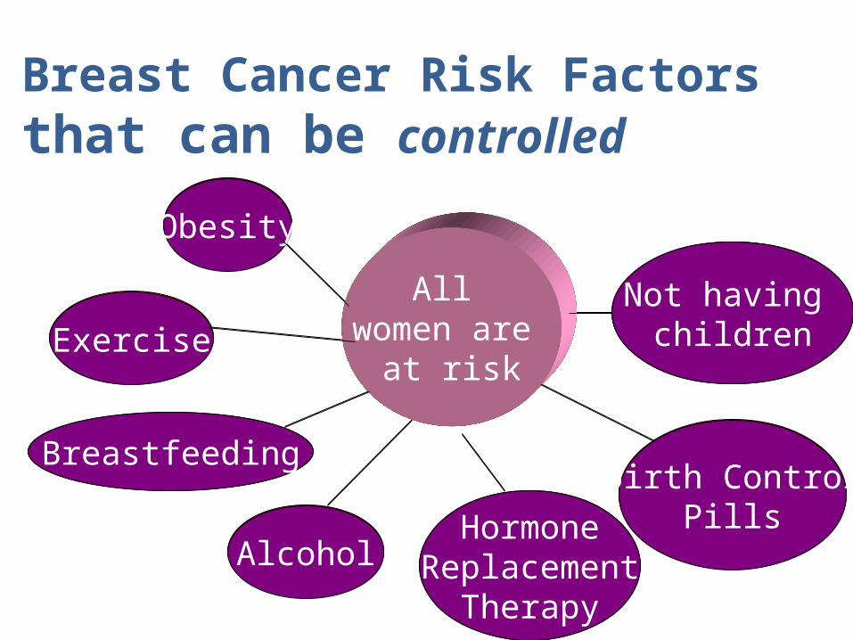
All women are
at risk
Obesity
Breastfeeding
Not having children
Birth ControlPills
AlcoholHormone
ReplacementTherapy
Exercise
All women are
at risk
Obesity
Breastfeeding
Not having children
Birth ControlPills
AlcoholHormone
ReplacementTherapy
Breast Cancer Risk Factorsthat can be controlled
Exercise

Potential Applications forBreast Cancer Biology
• Predict risk of cancer development
• Estimate prognosis for established cancer
• Predict response to therapy• Identify therapeutic targets• Identify early detection markers
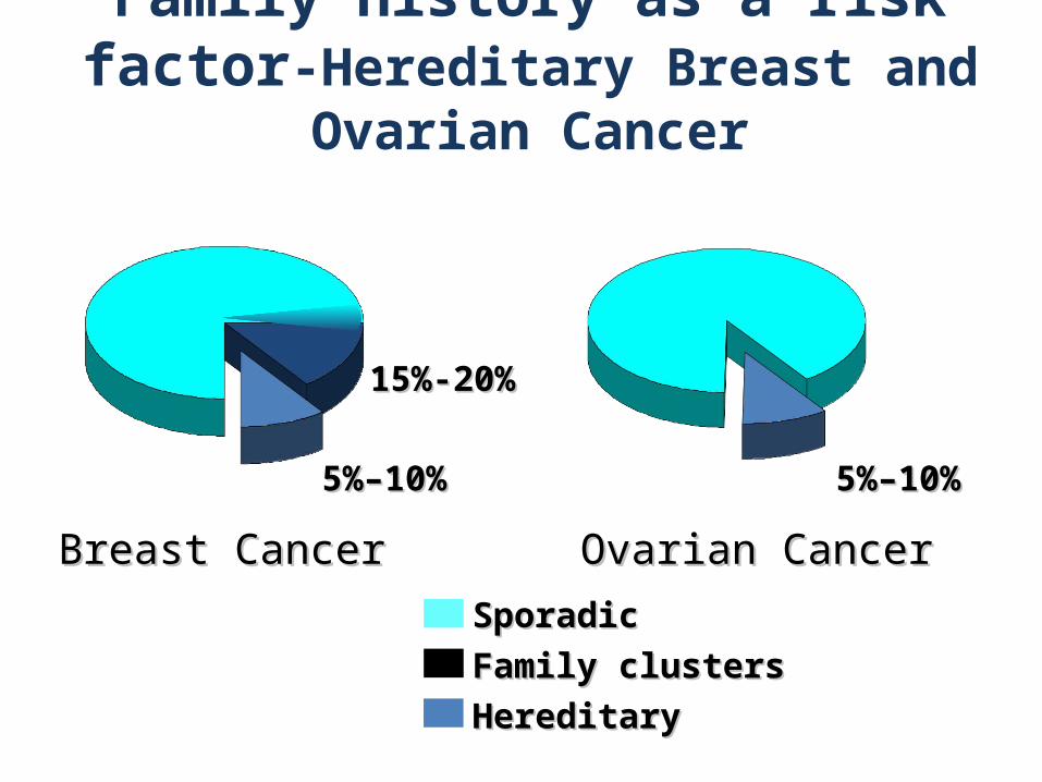
Family history as a risk factor-Hereditary Breast and Ovarian
Cancer
SporadicSporadic
Family clustersFamily clusters
HereditaryHereditary
Ovarian CancerOvarian CancerBreast CancerBreast Cancer
5%–10%5%–10% 5%–10%5%–10%
15%-20%15%-20%
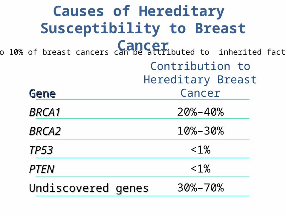
Causes of Hereditary Susceptibility to Breast
Cancer
GeneGene
BRCA1BRCA1
BRCA2BRCA2
TP53TP53
PTENPTEN
Undiscovered genesUndiscovered genes
Contribution to Hereditary Breast
Cancer
20%–40%
10%–30%
<1%
<1%
30%–70%
5 to 10% of breast cancers can be attributed to inherited factors
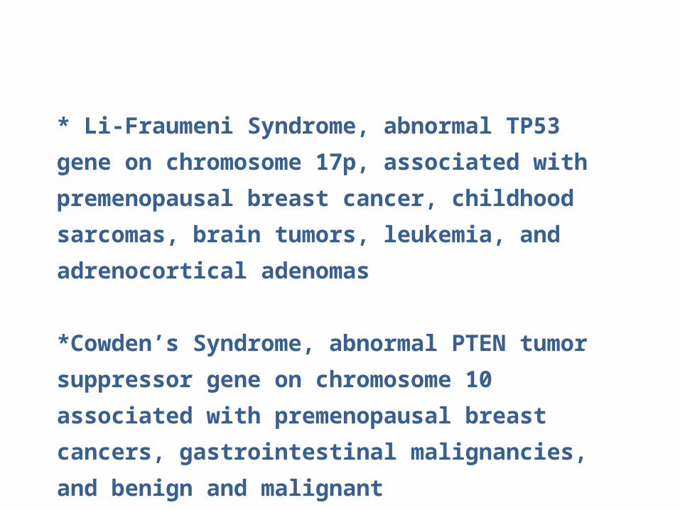
* Li-Fraumeni Syndrome, abnormal TP53
gene on chromosome 17p, associated with
premenopausal breast cancer, childhood
sarcomas, brain tumors, leukemia, and
adrenocortical adenomas
*Cowden’s Syndrome, abnormal PTEN
tumor suppressor gene on chromosome 10
associated with premenopausal breast
cancers, gastrointestinal malignancies, and
benign and malignant
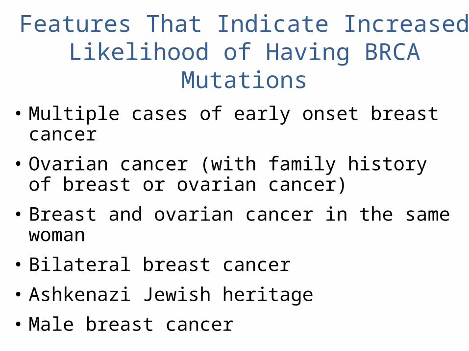
Features That Indicate Increased Likelihood of Having BRCA Mutations
• Multiple cases of early onset breast cancer
• Ovarian cancer (with family history of breast or ovarian cancer)
• Breast and ovarian cancer in the same woman
• Bilateral breast cancer
• Ashkenazi Jewish heritage
• Male breast cancer
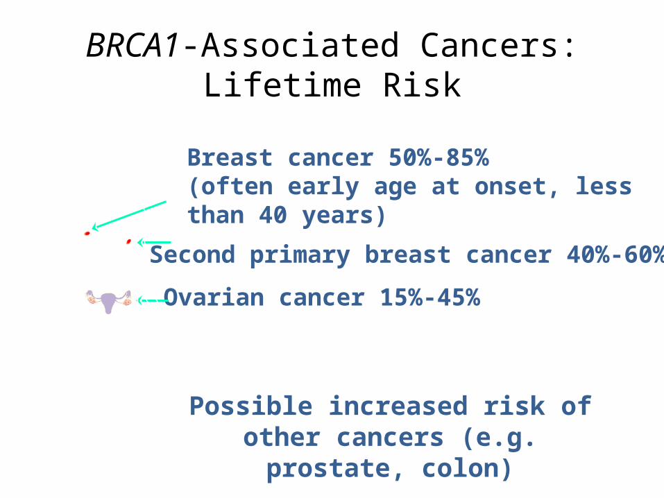
BRCA1-Associated Cancers:Lifetime Risk
Possible increased risk of other cancers (e.g. prostate, colon)
Breast cancer 50%-85%(often early age at onset, less than 40 years)
Second primary breast cancer 40%-60%
Ovarian cancer 15%-45%
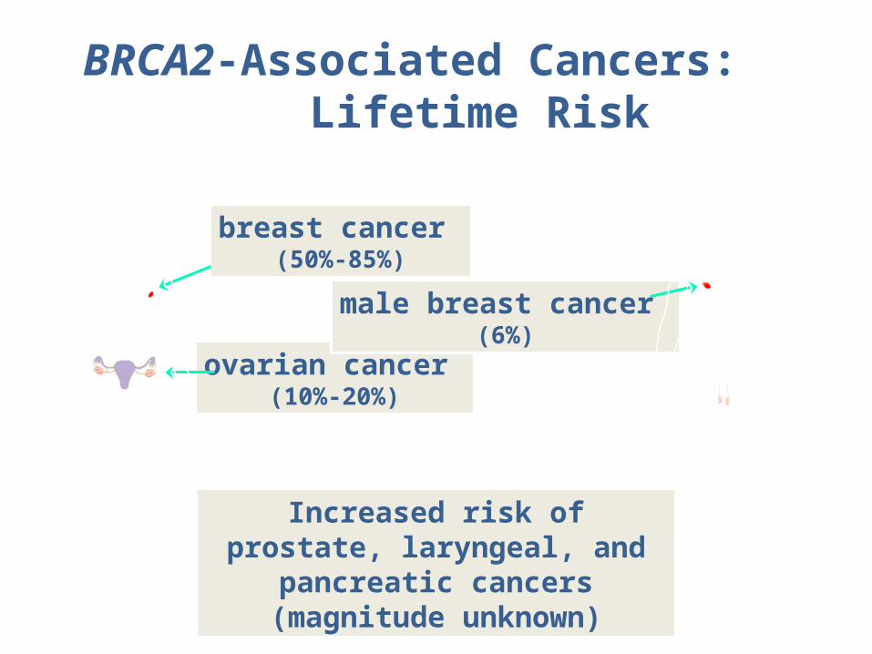
BRCA2-Associated Cancers: Lifetime Risk
Increased risk of prostate, laryngeal, and pancreatic
cancers (magnitude unknown)
breast cancer (50%-85%)
ovarian cancer (10%-20%)
male breast cancer (6%)
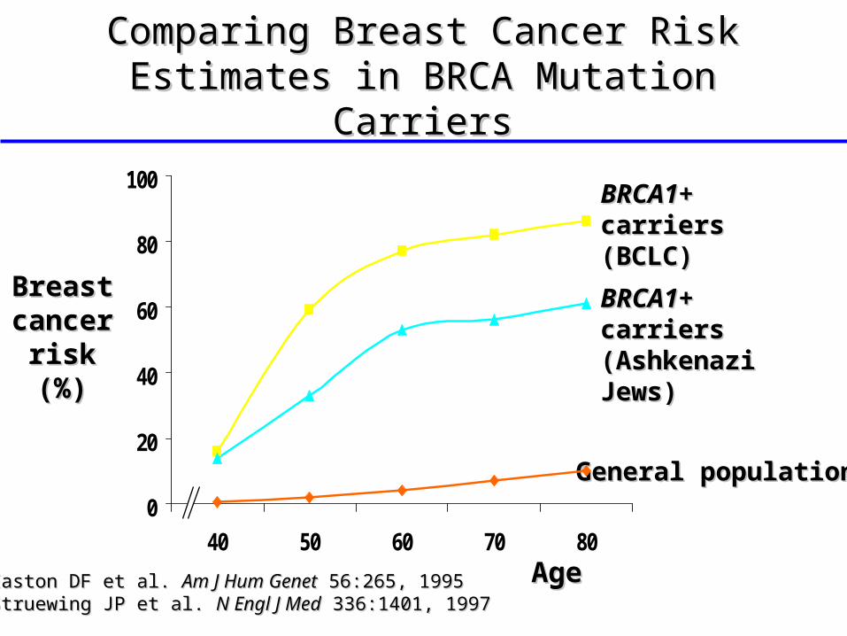
Comparing Breast Cancer Risk Comparing Breast Cancer Risk Estimates in BRCA Mutation CarriersEstimates in BRCA Mutation Carriers
Breast Breast cancer cancer
risk risk (%)(%)
General populationGeneral population
BRCA1BRCA1+ + carrierscarriers(BCLC)(BCLC)
BRCA1BRCA1+ + carrierscarriers(Ashkenazi (Ashkenazi Jews)Jews)
AgeAgeEaston DF et al. Easton DF et al. Am J Hum GenetAm J Hum Genet 56:265, 1995 56:265, 1995Struewing JP et al. Struewing JP et al. N Engl J MedN Engl J Med 336:1401, 1997 336:1401, 1997
0
20
40
60
80
100
40 50 60 70 80
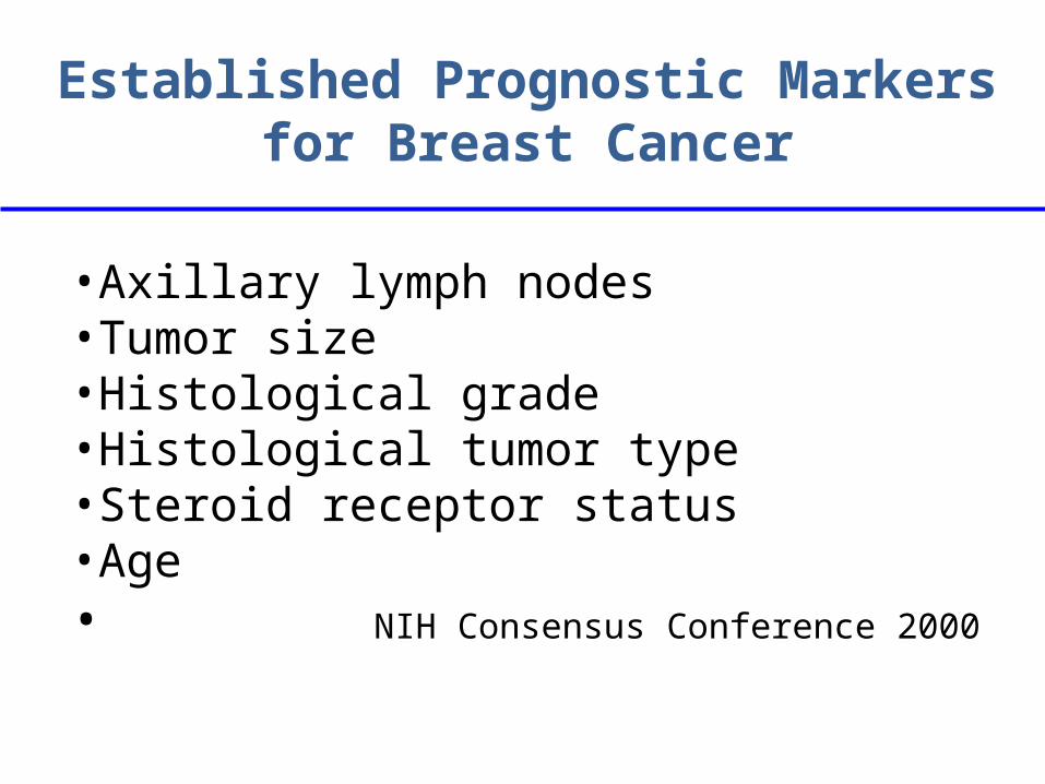
Established Prognostic Markers for Breast Cancer
•Axillary lymph nodes•Tumor size•Histological grade•Histological tumor type•Steroid receptor status•Age• NIH Consensus Conference 2000

Potential Applications forBreast Cancer Biology
• Predict risk of cancer development
• Estimate prognosis for established cancer
• Predict response to therapy• Identify therapeutic targets• Identify early detection markers

Molecular Portrait of Breast CancersHER-
2Basal-
likeLumina
l A
Luminal B
“Normal”
Sorlie T et al, PNAS 2001
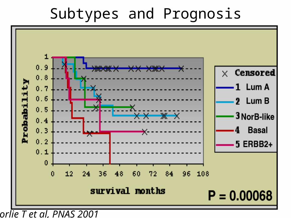
Subtypes and Prognosis
Sorlie T et al, PNAS 2001

Potential Applications forBreast Cancer Biology
• Predict risk of cancer development
• Estimate prognosis for established cancer
• Predict response to therapy• Identify therapeutic targets• Identify early detection markers
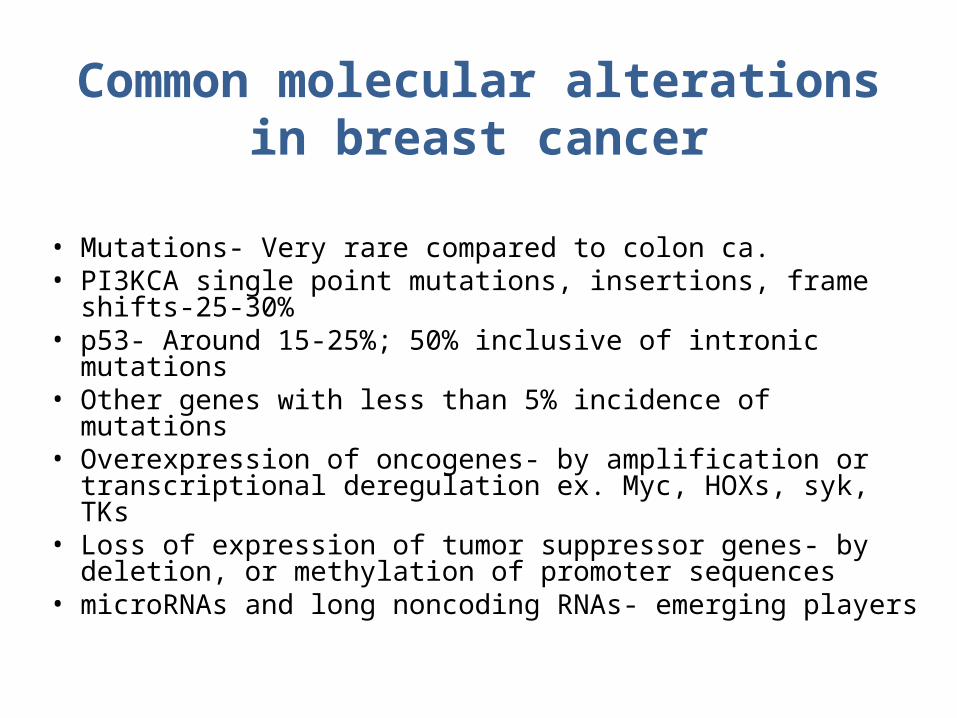
Common molecular alterations in breast cancer
• Mutations- Very rare compared to colon ca.• PI3KCA single point mutations, insertions, frame
shifts-25-30%• p53- Around 15-25%; 50% inclusive of intronic
mutations• Other genes with less than 5% incidence of mutations• Overexpression of oncogenes- by amplification or
transcriptional deregulation ex. Myc, HOXs, syk, TKs• Loss of expression of tumor suppressor genes- by
deletion, or methylation of promoter sequences• microRNAs and long noncoding RNAs- emerging
players

The Estrogen Receptors
Tx activation domain
2 cys-rich zinc fingersRecognize EREs, and stabilize
Hinge region
Variable

Activation of Estrogen Receptor
JM Hall et al, JBC 2001.
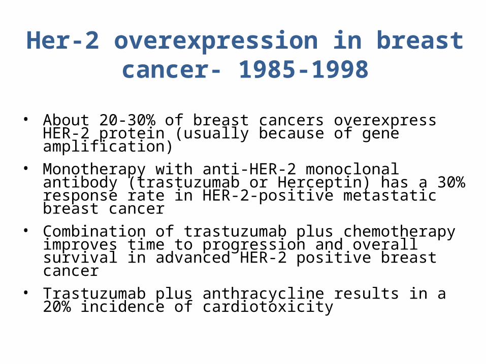
Her-2 overexpression in breast cancer- 1985-1998
• About 20-30% of breast cancers overexpress HER-2 protein (usually because of gene amplification)
• Monotherapy with anti-HER-2 monoclonal antibody (trastuzumab or Herceptin) has a 30% response rate in HER-2-positive metastatic breast cancer
• Combination of trastuzumab plus chemotherapy improves time to progression and overall survival in advanced HER-2 positive breast cancer
• Trastuzumab plus anthracycline results in a 20% incidence of cardiotoxicity
Potential Applications forBreast Cancer Biology
• Predict risk of cancer development• Estimate prognosis for established cancer• Predict response to therapy• Identify therapeutic targets• Identify early detection markers

EGFTGF
Amphiregulin-cellulinHB-EGF
Epiregulin Heregulins
NRG2NRG3
Heregulins-cellulin
Cysteine-richdomains
Tyrosine kinasedomain
ErbB-1Her1
EGFR
ErbB-2Her2neu
ErbB-3Her3
ErbB-4Her4
C-terminus
100
100
100
44
82
33
36
59
24
48
79
28
The EGFR (ErbB) family and ligands
www.astrazeneca.com
Copyright ©2004 AlphaMed Press
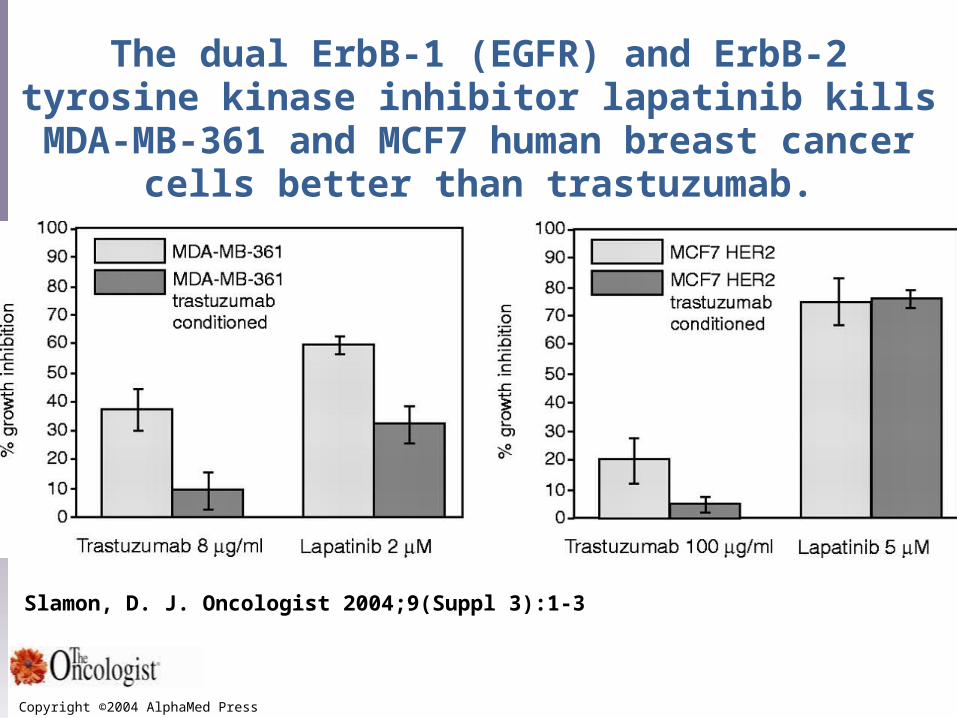
Slamon, D. J. Oncologist 2004;9(Suppl 3):1-3
The dual ErbB-1 (EGFR) and ErbB-2 tyrosine kinase inhibitor lapatinib kills MDA-MB-361 and MCF7 human breast cancer cells better than trastuzumab.
Applications of Expression Microarrays in Predicting Response
to Therapy
• Different profile of sporadic vs hereditary breast cancer (Heldenfalk, NEJM 2001)
• Identify subset of young women with poor prognosis early breast cancer (van’t Veer, Nature 2002)
• Subset outcomes for women with node-negativeER-positive breast cancer treated with tamoxifen (Paik, NEJM 2004, SABCS 2004)
Now available--$3400Should we use it?
For whom?How?
So What Good is All this Molecular Analysis??
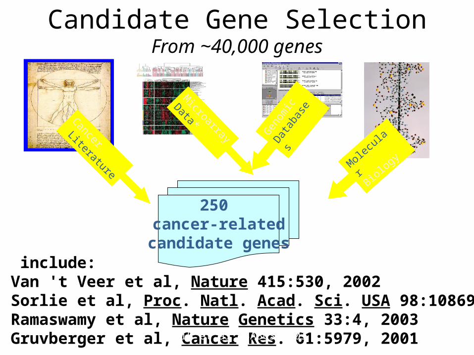
Candidate Gene SelectionFrom ~40,000 genes
Cancer
Literature
Microarray
Data*G
enom
ic
Dat
abas
es
250 cancer-related
candidate genes
Molecu
lar
Biology
*Sources include:1) Van 't Veer et al, Nature 415:530, 20022) Sorlie et al, Proc. Natl. Acad. Sci. USA 98:10869, 20013) Ramaswamy et al, Nature Genetics 33:4, 2003 4) Gruvberger et al, Cancer Res. 61:5979, 2001Paik et al, SABCS 2003

Three Breast Cancer Studies Used to Select 16 Cancer and 5 Reference Genes
PROLIFERATIONKi-67STK15
SurvivinCyclin B1MYBL2
ESTROGENER
PGRBcl2
SCUBE2
INVASIONStromelysin 3Cathepsin L2
HER2GRB7HER2
GSTM1REFERENCEBeta-actin
GAPDHRPLPOGUSTFRC
Best RT-PCR performance and most robust predictors
CD68
BAG1
Paik et al NEJM 2004

Three Breast Cancer Studies Used to Develop Recurrence Score (RS) Algorithm
Paik et al, SABCS 2003
RS = + 0.47 x HER2 Group Score - 0.34 x ER Group Score + 1.04 x Proliferation Group Score + 0.10 x Invasion Group Score + 0.05 x CD68 - 0.08 x GSTM1 - 0.07 x BAG1
Paik et al, SABCS 2003
Recurrence Recurrence CategoryCategory RS (0 – 100)RS (0 – 100)
Low riskLow risk < 18< 18
Intermediate riskIntermediate risk 18 – 3018 – 30
High riskHigh risk ≥ ≥ 3131
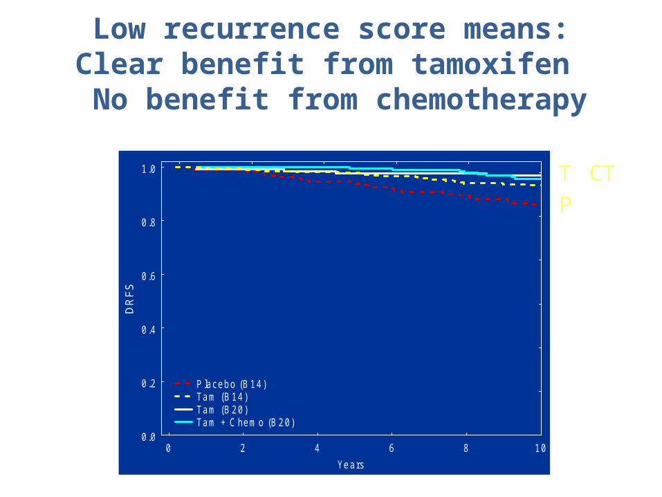
Low recurrence score means:Clear benefit from tamoxifen
No benefit from chemotherapy
0 2 4 6 8 10
Years
0.0
0.2
0.4
0.6
0.8
1.0
DR
FS
P lacebo (B 14) Tam (B 14) Tam (B 20) Tam + C hemo (B 20)
N355668227424
P
T CT
Paik, SABCS, 2004
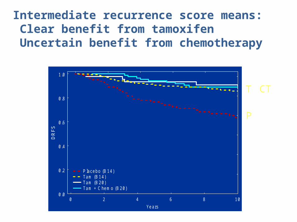
Intermediate recurrence score means: Clear benefit from tamoxifen Uncertain benefit from chemotherapy
0 2 4 6 8 10
Years
0.0
0.2
0.4
0.6
0.8
1.0
DR
FS
P lacebo (B 14) Tam (B 14) Tam (B 20) Tam + C hemo (B 20)
N355668227424
P
T CT
Paik, SABCS, 2004

High recurrence score means: No benefit from tamoxifen Clear benefit from chemotherapy
0 2 4 6 8 10
Years
0.0
0.2
0.4
0.6
0.8
1.0
DR
FS
P lacebo (B 14) Tam (B 14) Tam (B 20) Tam + C hemo (B 20)
N355668227424
P T
CT
Paik, SABCS, 2004

Potential Applications forBreast Cancer Biology
• Predict risk of cancer development
• Estimate prognosis for established cancer
• Predict response to therapy• Identify therapeutic targets
Outline- Part 2
• How is breast cancer:
• Detected
• Diagnosed
• Treated
59
Mammography
• Use a low-dose x-ray system to examine breasts
• Digital mammography replaces x-ray film by solid-state detectors that convert x-rays into electrical signals. These signals are used to produce images that can be displayed on a computer screen (similar to digital cameras)
• Mammography can show changes in the breast up to two years before a physician can feel them
60

Mammography Equipment
61
Computer-Aided Diagnosis
• Mammography allows for efficient diagnosis of breast cancers at an earlier stage
• Radiologists misdiagnose 10-30% of the malignant cases
• Of the cases sent for surgical biopsy, only 10-20% are actually malignant
• CAD systems can assist radiologists to reduce the above problems
62
National Cancer Institute
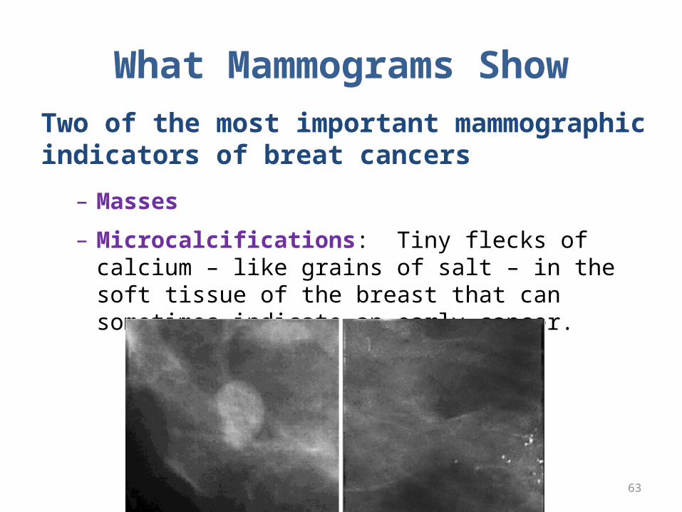
What Mammograms ShowTwo of the most important mammographic indicators of breat cancers
– Masses
– Microcalcifications: Tiny flecks of calcium – like grains of salt – in the soft tissue of the breast that can sometimes indicate an early cancer.
63
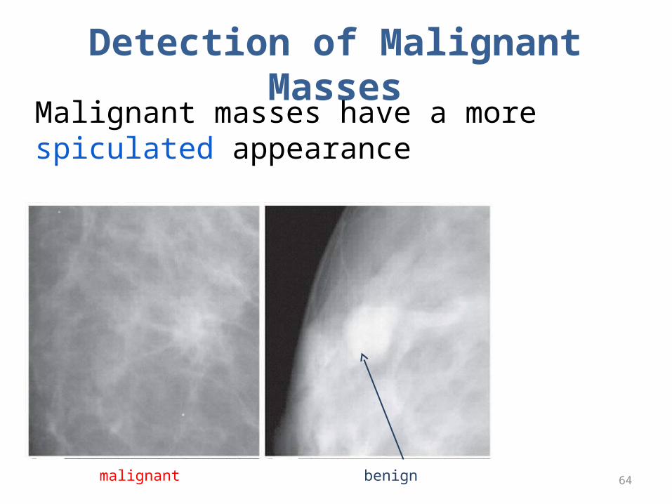
Detection of Malignant Masses
Malignant masses have a more spiculated appearance
64malignant benign
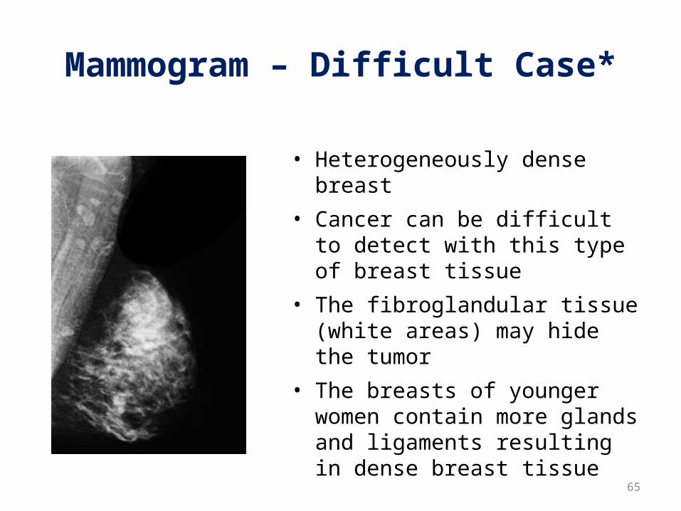
Mammogram – Difficult Case*
• Heterogeneously dense breast
• Cancer can be difficult to detect with this type of breast tissue
• The fibroglandular tissue (white areas) may hide the tumor
• The breasts of younger women contain more glands and ligaments resulting in dense breast tissue 65

Mammogram – Easier Case*
• With age, breast tissue becomes fattier and has fewer glands
• Cancer is relatively easy to detect in this type of breast tissue
66

Different Views
67
Top-to-Bottom
Side-to-Side
MRI - Cancer can have a uniqueappearance – many small irregularwhite areas that turned out to becancer (used for diagnosis)
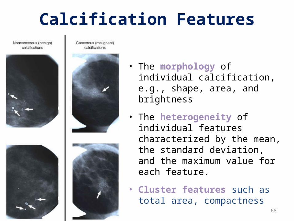
Calcification Features
• The morphology of individual calcification, e.g., shape, area, and brightness
• The heterogeneity of individual features characterized by the mean, the standard deviation, and the maximum value for each feature.
• Cluster features such as total area, compactness
68
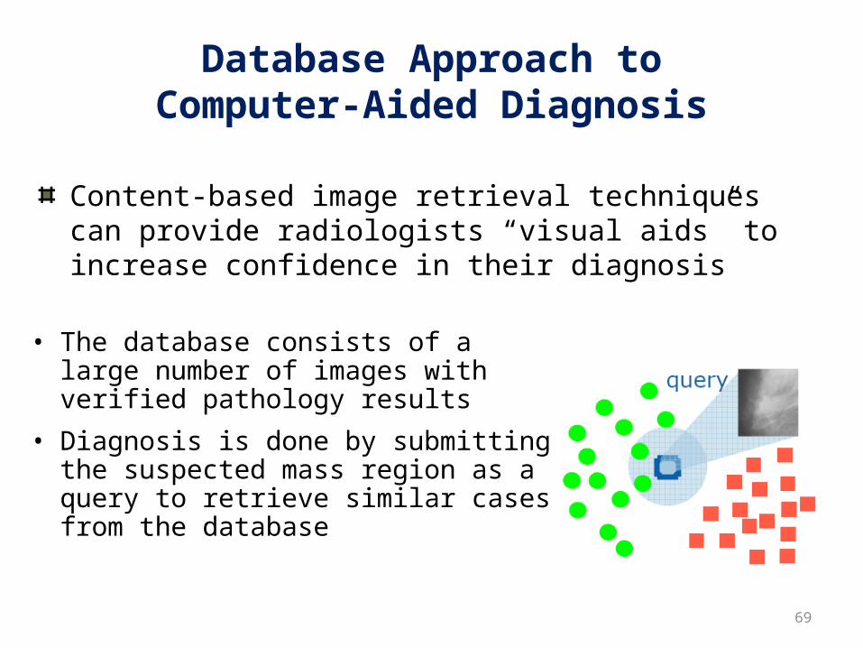
Database Approach toComputer-Aided Diagnosis
• The database consists of a large number of images with verified pathology results
• Diagnosis is done by submitting the suspected mass region as a query to retrieve similar cases from the database
69
Content-based image retrieval techniques can provide radiologists “visual aids” to increase confidence in their diagnosis

Outline- Part 2
• How is breast cancer:
• Detected
• Diagnosed
• Treated
70

Diagnosis and Treatment
• . Patient feels a breast mass or has an abnormal radiologic screening exam
• . Surgical biopsy or aspiration • . Observation (LCIS), lumpectomy or
mastectomy • . Staging • . Delivery of adjuvant therapies—
radiation and/or chemotherapy,hormonal therapies
04/18/23 71

Tumor characteristics
• Invasive vs. Non-invasive . • Histologic Type-Ductal (85%) vs.
Lobular . • Grade (estimate of the aggressiveness
under microscope) . • Size . • Margins .• Lymph Nodes . • Estrogen/ Progesterone Receptor (2/3
positive) . • Her-2/ neu 04/18/23 72

04/18/23 CBMS2006 73
Stages ofBreastCancer
Stage 0 --carcinoma in situ
Stage I – tumor < 2 cm, no nodes Stage II – tumor 2 to 5 cm, +/-nodes
Stage III – locally advanced disease, fixed or matted lymph nodes and variable tumor size
Stage IV – distant metastases (bone, liver, lung, brain)

What now?
74
Stage 0-III
Risk of recurrence is individual
What can we do to reduce the risk of recurrence in the breast, and systemically ?
Meet with Radiation Oncologist and Medical Oncologist
How is breast cancer treated?
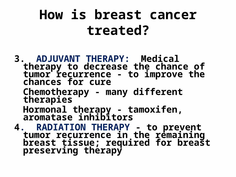
3. ADJUVANT THERAPY: Medical therapy to decrease the chance of tumor recurrence - to improve the chances for cureChemotherapy - many different therapiesHormonal therapy - tamoxifen, aromatase inhibitors
4. RADIATION THERAPY - to prevent tumor recurrence in the remaining breast tissue; required for breast preserving therapy
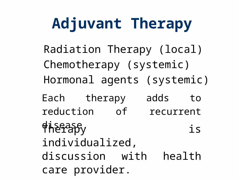
Adjuvant Therapy
Radiation Therapy (local)
Chemotherapy (systemic)
Hormonal agents (systemic)
Each therapy adds to reduction of
recurrent disease.
Therapy is individualized, discussion with health care provider.
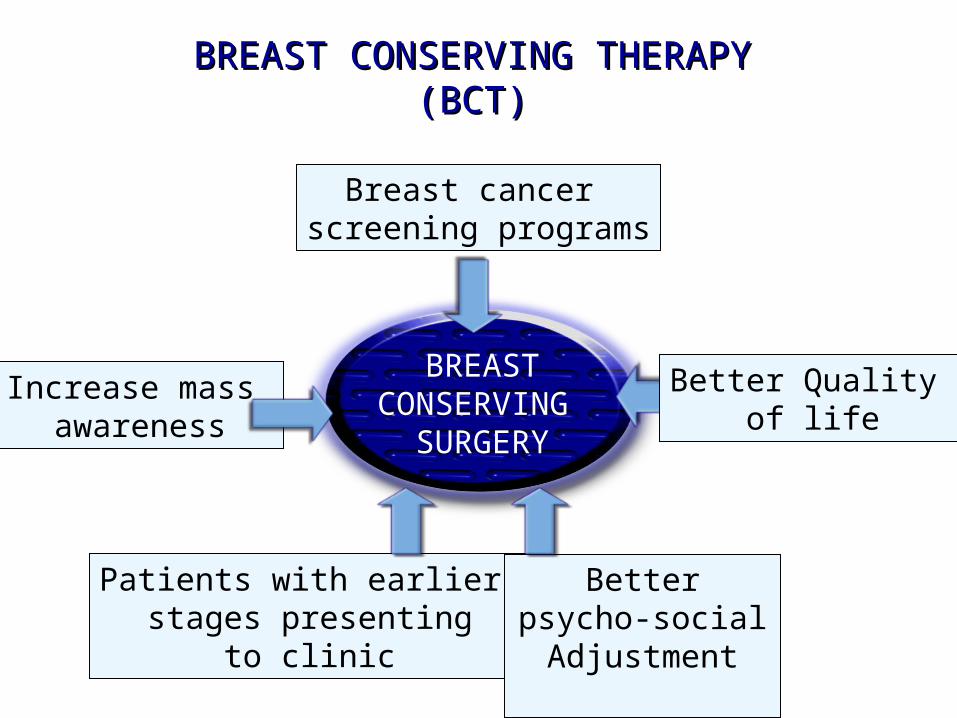
BREASTCONSERVING
SURGERY
Breast cancer screening programs
Increase mass awareness
Patients with earlier stages presenting
to clinic
Better psycho-social Adjustment
BREAST CONSERVING THERAPYBREAST CONSERVING THERAPY(BCT)(BCT)
Better Quality of life
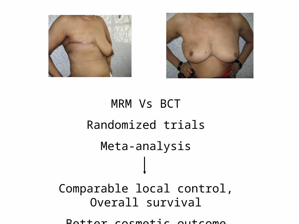
MRM Vs BCT
Randomized trials
Meta-analysis
Comparable local control, Overall survival
Better cosmetic outcome
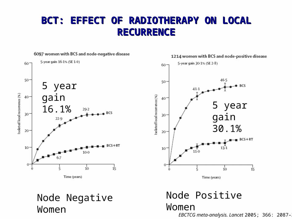
BCT: EFFECT OF RADIOTHERAPY ON LOCAL BCT: EFFECT OF RADIOTHERAPY ON LOCAL RECURRENCERECURRENCE
EBCTCG meta-analysis. Lancet 2005; 366: 2087–2106
5 year gain 16.1%
5 year gain 30.1%
Node Negative Women Node Positive Women
80
Chemotherapy Drugs
Adriamycin, Epirubicin
Cytoxan
Methotrexate, 5-fluorouracil
Taxol, Taxotere
Navelbine
Intravenous
Nausea, hair loss, low blood counts, cardiac toxicity,
bladder toxicity, nerve damage
Given for adjuvant or recurrent disease.

Tamoxifen*
81
Can be given to pre or post menopausal women
Works by blocking estrogen receptors in breast cells, inhibiting their growth
Side effects include hot flashes, depression, increased risk of uterine cancer and blood clots
Taken daily by mouth for 5 years

Aromatase is the enzyme that converts androgens to estrogen
82
AIs are only given to postmenopausal women
“May” be more effective than Tamoxifen
Examples: Anastrozole/Arimidex, Letrozole/Femara, Exemestane/Aromasin
Side effects include hot flashes, depression, osteoporosis, joint pains
Taken daily by mouth for variable periods of time
Aromatase Inhibitors*
Trastuzumab/Herceptin
83
Given to patients whose cancer cells overexpress Her-2-neu as measured by IHC or FISH (25 to 30% of patients)
Bisphosphonates
•Bone strengtheners
•Given for therapy-induced osteoporosis or for
cancer that has spread to bone
•Zometa (Zoledronic acid)
•Aredia (Pamidronate)
•Each lowers calcium and has been shown to
reduce the risk of fracture in pts with cancers
metastatic to bone.
Summary• The breast is a dynamic organ- undergoes cyclical
proliferative changes throughout life under the influence of hormones and growth factors- so may be likely to be more altered by environmental carcinogens
• Key function for ER and PR in breast cells. The same hormones that are important for breast growth during pregnancy are also important for breast cancer.
• ER function in signaling through other growth factor receptor pathways becomes very important in cancer. Production of estrogen through alternate sources keeps E supply ongoing in postmenopausal women.