2 review of literature -...
TRANSCRIPT

2
REVIEW
OF
LITERATURE

9
2.1Biodiversity
The idea of diversity was defined by Ramon Margalef several years ago
(Margalef, 1994). He proposed to use two words: biodiversity and diversity.
Biodiversity would be the total non-redundant genetic information in an ecosystem,
while diversity would be the collection of components which are active and abundant
at one particular time and place. The objectives of such techniques are to compare
communities, to identify the members of a community, and to quantify the abundance
of some or all of the members in the community.
2.1.1 Microbial Diversity
Biodiversity scrutiny is one of the current research topics in environmental
biology, trying to cover under-evaluated eco-systems and aimed basically to
understanding their individuals and their application in various aspects (Hertel, 2011).
Studies in biodiversity involves ecology, abundance of individuals, identification,
relation to each other, importance of individual in the ecosystem, and characterization
of individuals with number of species and individual’s in each species. Assessment of
the microbial diversity in the environment has long challenged microbiologists and
microbial ecologists. The initial focus of attention was directed to unravelling the
biodiversity of microorganisms inhabiting them. This may lead to the identification of
new interesting model organisms, enabling in-depth molecular studies (Aguilar,
1996).
Similarly, exploration of the ecology of the Rann of Kutch and studying the
diversity would entail about the individuals in microbial communities, abundance,
growth and metabolic activity, interaction between groups and adaptation with
physiochemical properties. While examining the samples, we find halophiles all over
the phylogenetic tree, in each of the three domains and in different branches of each
domain. The present study mainly focused on domain Archaea, in which many
species are still awaiting isolation and characterization.
2.1.2 Microbial Diversity of Different Ecosystems
So far, a large number of environments have been analysed for the evaluation
of microbial community which provide intensive output about microbial population
thriving there and their interaction. Many novel phylotypes from three domains have

10
been isolated and characterized from different microbial habitats, especially in recent
years many haloarchaeal isolates were obtained from ecoregions including open ocean
waters (Fuhrman et al., 1992), salt crystallizers (Maturrano et al., 2006; Mutlu et al.,
2008; Ochsenreiter et al., 2002), coastal waters (DeLong, 1992; Massana et al., 1997),
polar seas (Reysenbach et al., 2000; Vetriani et al., 1998, 1999), salt marshes
(Munson et al., 1997), freshwater lakes (Hershberger et al., 1996; Jurgens et al.,
2000), agricultural fields, forest soils, rhizosphere (Borneman and Triplett, 1997;
Buckley et al., 1998), paddy field soil (Chin et al., 1999; Gro kopf et al., 1998), hot
springs (Barns et al., 1994, 1996), deep-sea hydrothermal vents (Takai and Horikoshi,
1999a), mine water (Takai et al., 2001) and deep subsurface geothermal pools (Takai
and Horikoshi, 1999b).
Life is represented by all the three domains in hyper-saline environments
(Benlloch et al., 2002). The microbial flora of hyper-saline waters, with salinities
close to saturation, have been studied for many years, but only relatively recently has
it been possible to identify and to culture many of the dominant groups (Antón et al.,
2002; Burns et al., 2004a, b). The majority are extreme halophiles belonging to
domain Archaea, specially the family Halobacteriaceae (Oren, 2002), although
halophiles belongs to Eubacteria, and Eukaryota also are present, pointing to a diverse
group of microorganisms that can thrive in hyper-saline environments. In the Dead
Sea ecosystem, there are reports of characterized members belong to archaea, bacteria
and eukaryota (Oren, 1995; 2001; Blum et al., 2001). Many novel genus and novel
species of the halophilic archaea were isolated and characterized in recent years as
result of taxonomical analyses performed on such ecosystems.
Several new genera and new species have been discovered recently and
included into Halobacteriaceae family. Many unaffiliated groups isolated from
different geographical location areas are under characterization (Montalvo-Rodríguez
et al., 2000; Asker and Ohta, 2002; Ihara et al., 1997). New representatives from the
domain eubacteria can also be found from these environments. The genus
Thermohalobacter was isolated from a saltern (Cayol et al., 2000). Many members of
halophilic bacteria, like Salinibacter, were isolated and characterized (Antón et al.,
2002) from a hyper-saline environment. Today, huge lists of new species of
prokaryotes have been isolated from hyper-saline waters between few decades which
are considered as “lifeless”. Eukaryotic microorganisms can also be present in solar

11
salterns. Members belonging to domain eukaryota, fungi, algae and protozoa were
also isolated recently from different hyper-saline environments (Abdel-Hafez et al,.
1978; Buchalo et al,. 1998; Butinar et al., 2005; Gunde-Cimerman et al., 2000).
Diversity of species tends to decrease as salinity increases and therefore, only
a few microscopic organisms survive in hyper-saline region as brine acquires
saturation of salts. Some macroscopic organism also thrives in lower salinities
between 10% and 30% like brine shrimp (Artemia salina), larvae of brine flies (family
Ephydridae) (Grant 2004) and the alga, Dunaliella sp. (Cho, 2005) and they can reach
high populations densities. Dunaliella sp. can be observed at high salt concentrations
(up to 1.5 M NaCl) (Grant, 2004), though they are halotolerant rather than halophilic
(Oren, 2002a). Dunaliella salina is a phototrophic alga known for its β-carotene
pigments, which contributes to the red coloration of some salt lakes and saline
systems.
2.1.3 Cultureable Biodiversity and Uncultureable Diversity
Diversity of macro and micro organisms are differing significantly as one
group is visible to our naked eye and can be easily estimated but other is
complementary. During the last few decades, rapid and significant advances in
positive reception of the diversity of prokaryotic organisms have resulted from the
ecological surveys worldwide. Our current view of prokaryotic diversity of particular
ecosystem is mainly of two types i.e., culturabe diversity and diversity directly
retrieved by analysis of rRNA genes amplified from the environment (community).
Both point towards same direction, diversity, but there is a wide gap in result obtained
from both (Brambilla et al., 2001). Both diversity analysis, describing an ecosystem
or environment, have positive impact on microbial diversity over the last decade,
discovering the hidden treasure of microbial diversity. Only a few organisms in
environmental samples are, but it is unknown whether the cultured isolates from an
environment yield communities that resemble the communities from the original
habitat (Hallam et al., 2006). Obviously diversity is always defined and always less
than the actual representatives of an ecosystem. This issue in diversity is clearly
visible on comparing gene libraries from both cultured bacteria and uncultured
environmental samples.
12
Culture-based approaches give basic understanding of diversity, physiology,
metabolism and interaction, in a specific environment (Zengler et al., 2002; Webster
et al., 2006,). This study should be baseline for developing culturing methodologies,
media formulation, isolation strategies and basic standards. Diversity analysis should
start from “culturable” species and with this basic information proceed for
unculturable community. The long term goal of a diversity study is to understand each
and every member of community and understanding its cell mechanics and
interactions, which can only fulfilled after isolation and characterization of individual
species.
Only 0.1% to 10% of the microbial community in a soil sample is retrievable
(Head et al., 1998). This drawback greatly reflects in all diversity anlaysis while
characterising the microbial community in a niche. Despite this, development in
cultivation methods are still required to isolate and characterise unculturable
microorganisms, which provide an insight into evolution and microbial ecology
(community structure and function) as well as physiology, metabolic pathways and
genetics of particular organisms. For example, the recently cultivated Haloquadratum
walsbyi was an important step in understanding the microbial ecology in hyper-saline
environments. The discovery of Salinibacter ruber provided a better understanding of
bacterial ecology in hyper-saline environments and refuted previous assumptions that
bacteria were outcompeted at high salinities (Antón et al., 2002). The completed
genome of this bacterium provided insights into the co-evolution of this bacterium
with the haloarchaea (Mongodin et al., 2005). In addition, the cultivation of the
dominant species in the environment may allow the isolation and characterisation of
novel haloviruses also present in the environment.
Culture independent methods adopted for diversity study by different aspects
include metagenomic sequencing, probe based microarrays, PLFA, DGGE, TGGE,
TRFLP, and DDRT-PCR with community DNA (Zheng 1996; Takai et al., 2000;
Raskin et al., 1994; Lozupone et al., 2007; Lueders et al., 2003). The haloarchaeal
diversity in various hyper-saline regions has been analyzed and compared with the
community culture independent biodiversity. Thus, the rRNA based approach for
studying in situ prokaryotic diversity has led to finding the methodology for the novel
unculturable individuals (Podar et al., 2007). It is a real fact that there are still plenty
of unknown “unculturable” species, hidden in different environments. With the advent
13
of these molecular techniques and the ease of sequencing reactions, microbial
communities of many environments have been characterised, including soil (McCaig
et al., 1999), water (Homer-Devine et al., 2003), the human mouth (Kroes et al.,
1999) and the human gut (Suau et al., 1999).
The traditional culture based techniques fail to recover those largely
predominant microorganisms basically due to lack of understanding of the basic
environmental factors, physiology and metabolism of individuals. Techniques so far
used for isolating haloarchaea are quite simple and do not mimic the natural growth
conditions (Kaeberlein et al., 2002). In some cases lower diversity is recovered by
molecular techniques than the actual diversity, while in other instances the cultivated
isolates are not related to the sequences retrieved directly from the environmental
samples (Benlloch et al., 2001; Ochsenreiter et al.,2002; Burns et al., 2004;
Maturrano et al., 2006). It has allowed detection of uncultivated organisms such as the
Korarchaeota and has given insights into community structure and function.
Culture-dependent and culture-independent methods were used to assess the
diversity of haloarchaea in hyper-saline regions. Haloarchaeal diversity of different
ecosystem provides a large number of individual to family Halobacteriaceae in last
few decades. This archaeal community are explored by means of cultivation
dependent and cultivation independent methods. The two methods yielded slightly
different results, an observation commonly shared by many similar studies (Benlloch
et al., 2001; Ochsenreiter et al., 2002; Burns et al., 2004 b; Maturrano et al., 2006).
Archaeal rRNAs had been recovered from either nonviable or viable but unculturanle
populations, i.e., the extremophilic archaea might be microbial relics. Halophilic
archaeal rRNA likely reflects the microbial activity and species distribution during the
period of deposition over the past two million years. Members of two main phyla,
Euryarchaeota and Crenarchaeota are deeply investigated species amoung archaeal
groups. Almost all previous studies describing the microbial diversity of different
ecosystems have relied on the identification and characterization of cells which may
only represent 0.1–1% of the total microbial community (Rapp´e and Giovannoni,
2003).
Culture-independent methods are increasingly used in environmental
microbiology to bypass the shortcomings of studying microorganisms, which is
14
laborious and high microbial skills are needed basically in culture handling and
characterization, but community based method is comparatively more easy by
extracting and analysing total nucleic acids, theoretically representing the entire
microbial population from environmental samples (Spiegelman et al., 2005). Many
studies have reported that the assessment of bacterial diversity by cultivation-
dependent methods generate erroneous information with regard to bacterial diversity,
owing to the existence of a host of non- bacterial species. The analysis of microbial
diversity has, therefore, shifted in the last two decades, from cultivation-dependent
approaches to 16S rRNA-based cultivation-independent approaches. This shift in
method has resulted in the discovery of many novel microbial taxa, theoretically, but
none in hand.
Nevertheless, this approach also has important limitations, and is often
confined to naming 16S rRNA gene clones or DGGE (denaturing gradient gel
electrophoresis) bands based on sequence similarity, and speculation as to the
ecophysiology of species, on the grounds of such similarities. Therefore, as PCR-
based approaches have biases which can distort community composition (Martin-
Laurent et al., 2001; Hur and Chun, 2004) cultivation remains the preferred method
for the acquisition of an accurate picture of the physiology and complex ecological
interactions in which microorganisms engage.
The impact of DNA-based methods has remarkably improved our knowledge
by providing both a new alternative classification system and critically, new
experimental strategies to identify non- species (Amann et al., 1995). The resulting
data have not only highlighted the true breadth of prokaryotic diversity but have also
changed some of our previous views of biological evolution. Phylogenetic analysis of
gene sequences retrieved from both cultured and uncultured bacteria has shown that
all cellular life can be ordered into three taxa (termed Domains) - bacteria, archaea,
and eukaryota. Intriguingly, this has resulted in a major taxonomic promotion for the
Archaea, which were previously thought to be a series of unusual bacterial species. In
addition, the use of DNA-based methods (Yokouchi et al., 2006; Uchiyama et al.,
2006; Rondon et al., 2000) to identify and catalogue non- species has radically
improved our knowledge of the diversity found within living prokaryotes.
15
The results obtained by culture-dependent techniques covered only those few
organisms that could be cultivated. Due to this well documented disparity between
cultivable and in situ diversity, it is often difficult to assess the significance of
cultured members in microbial communities. Several studies have employed culture-
independent techniques to show that cultivated microorganisms from diverse
environments often may represent very minor components of the microbial
community as a whole (Kimura et al., 2006; Abulencia et al., 2006). It is generally
accepted that cultivation methods recover less than 1% of the total microorganisms
present in environmental samples (Amann et al., 1995; Ward et al., 1990), therefore,
microbial investigations based only on cultivation strategies cannot be regarded as
reliable in terms of reflecting the microbial diversity present in art samples (Von
Wintzingerode et al., 1997; Singleton et al., 2001).
Differences in operon copy number causes bias towards microbes with higher
copy number, thereby overestimating diversity by counting multiple signals from
single organism. There is now a database providing information on operon copy
number in different organisms (Klappenbach et al., 2001). Heterogeneity of 16S
rRNA sequences can also overestimate diversity (Crosby and Criddle, 2003). For
example, Haloarcula marismortui has an inter operon difference up to 5% between
two 16S rRNA gene sequences (Mylvaganam and Dennis, 1992). Hence, slightly
different genes could originate from one strain, but may be interpreted as genes from
two closely related organisms. Although culture dependent method is a laborious and
time consuming method, it provides almost full information about organisms, full
biochemical, physiological, metabolic, full length 16S rRNA genes, other
phylogenitic markers available for analysis, revealing a complete picture regarding
structure, genetic diversity of the community and taxonomic identification of
populations.
2.2 Halophiles
Halophiles are one class of extreme organisms, which live in saline
environments. Microbial life can be found over the whole range of salt concentrations
from freshwater and marine biotopes to hyper-saline environments with NaCl
concentrations up to saturation (Brown, 1976, 1978; Tokuoka, 1993; Buchalo et al.,
1998, Ventosa et al., 1998; Almeida, 1999; Oren, 1999, 2002; Antón et al., 2000;

16
Gunde-Cimerman et al., 2000; Sleator and Hill, 2001; Lahav et al., 2002; Roberts,
2004). These organisms can be found in a wide range of environments from low-
saline marine environments to hyper-saline lakes such as the Dead Sea and Great Salt
Lake. Generally Halophiles fall into three categories depending on the salinity optimal
for growth: halotolerant (1-6%), moderate halophile (6-15%), and extreme halophile
(15-30%) (Garabito et al., 1998). Halophilic and halotolerant nature of
microorganisms vary significantly in three domains. (Buchalo et al., 1998; Antón et
al., 2000; Oren, 1999, 2002).
The study of microbial community and organisms from saline environments is
a field of research that has found increased interest in the past few decades. As with
all extreme environments, microbial diversity decreases as the salinity increases,
water stress of an aqueous environment increases, water activity (Aw) decrease and
only a few species grow under conditions of extreme water stress i.e. saturation of
salt. However, colonization of salt-loving or salt-tolerant microorganisms in hyper-
saline environments such as salt lakes (Buchalo et al., 1998; Gunde- Cimermann, et
al., 2000) , salted food products (Onishi, 1957; Takashina et al., 1994) is often highly
successful and may reach high population densities in such ecosystems (Oren, 1999).
New organisms are being identified and characterized from marine and salt lake
environments throughout the world, which has helped shed light on the ecology of
these microbes (Oren 1994; Ventosa et al., 1998; Dyall-Smith, 2004). Within the
domain Archaea, except few including methanogens, almost all halophiles belong to
the family Halobacteriaceae.
Table 2.1 Classification of microorganisms based on salt tolerance according to
Kushner (Kushner, 1978).
Category Range (M) Optimum salt concentration (M)
Non halophile
Halotolerant
Haloversatile
Slight halophile
Moderate halophile
Borderline extreme halophile
Extreme halophile
0-0.1
0->1.0
0->3.0
0.2-2.0
0.4-3.5
1.4-4.0
2.0-5.2
<0.2
<0.2
0.2-0.5
0.2-0.5
0.5-2.0
2.0-3.0
>3.0
17
In extreme salinities, halophilic archaea have been shown to be the dominant
population and most bacterial species have been shown to prefer lower levels of
salinity for optimal growth (Oren, 2002b). The bacteria are generally more diverse,
with halophilic groups found in several phyla. Although knowledge about these
organisms and their metabolic processes has increased over the years, it is limited by
the ability to culture halophiles. Many of these organisms have unique growth
requirements that can be hard to grow in the laboratory, and also with slow growth
rates, can hamper the isolation of certain halophilic strains and is reflected in the
outcome of non-culturing molecular methods and culturing methods (Amann, 1995;
Benlloch et al., 2001; Burns et al., 2004). By learning more about what they require
and finding better ways to imitate their environment, culturing techniques have
improved over the past few years and more “uncultivables” are being isolated
(Kaeberlein et al., 2002)
Among halophilic members of Archaea, the family Halobacteriaceae been
extensively studied. The extreme halophilic archaeal group are diverse, required salt
ranging from 8-32% and pH ranging from acidic to basic. Majority of Halobacteriales
are nonmotile, stained gram negative, reproduced by binary fission, do not form
resting stages, obligate aerobes, chemoorganotrophs and have very high GC content
in their genome. Some members of halobacteriales have multiple copies of genome,
and have remarkable DNA repairing mechanisms. Several members of this order have
unique light mediated ATP synthesis mechanisms and some thrive in salt crystals for
thousands of years (Vreeland et al., 2000; Leuko et al., 2011).
2.2.1 Extremophilic Halophiles in Three Domains
During the last hundreds of years, several discoveries were made in the field
of biodiversity in different ecosystems. From the starting ages of science, living
organisms are grouped based on their similarities and dissimilarities. The unique
properties of microbial extremophiles have also encouraged interest in several other
fields beside ecology and taxonomy. Evolutionists have been interested in extreme
organisms as the primordial earth was a very hostile environment, and some believe
that extremophiles are the best possible candidates for representing the first organisms
to grow and survive. Another emerging area of extremophile research is astrobiology
or looking for alternative life forms on other planets. Comparisons have been drawn

18
between Earth’s hostile environments and those found on Mars, Europa (Galilean
satellite), and comets (Litchfield, 1998; Landis, 2001; Stan-Lotter et al., 1999). By
researching more about the organisms found in Earth’s extreme areas, scientists hope
to be able to understand more about what other life could be discovered elsewhere.
Archaea have been recognized as the third domain of life in this planet since the late
1970s. Archaebacteria are phylogenetically distinct from eubacteria and eukaryotes.
Origin of Archaebacteria is estimated to be 3.5 billion years.
First taxonomist Linnaeus divided the world in to Plants, animals and minerals
(Linnaeus, 1735). In 1969 Whittaker proposed a five kingdom model consisting of
Monera, Protista, Plantae, Fungi and Animalia (Whittaker, 1969) Three Domain
classification is based on different classes of ribosomal RNA (rRNA) and is believed
to be evolved from the Last Universal Common Ancestor which is the root base of
“Universal Tree of life”. The revision distinguished three domains called Eukaryota,
Bacteria and Archaebacteria (Woese et al., 1977) and in 1990, later was subsequently
renamed Archaea (Woese et al., 1990).
The knowledge about the archaea was little when the other two domains were
studied in detail and also it was assumed that the occurrence of archaea was taught to
be restricted to extreme environments, either with low or high pH, extreme
temperature, anaerobic conditions and salinity. Several studies in last decade, using
new cultivation independent molecular ecological studies revealed the existence of
archaea in all environments, cold environments, ocean water columns and sediments,
fresh water environments, etc.
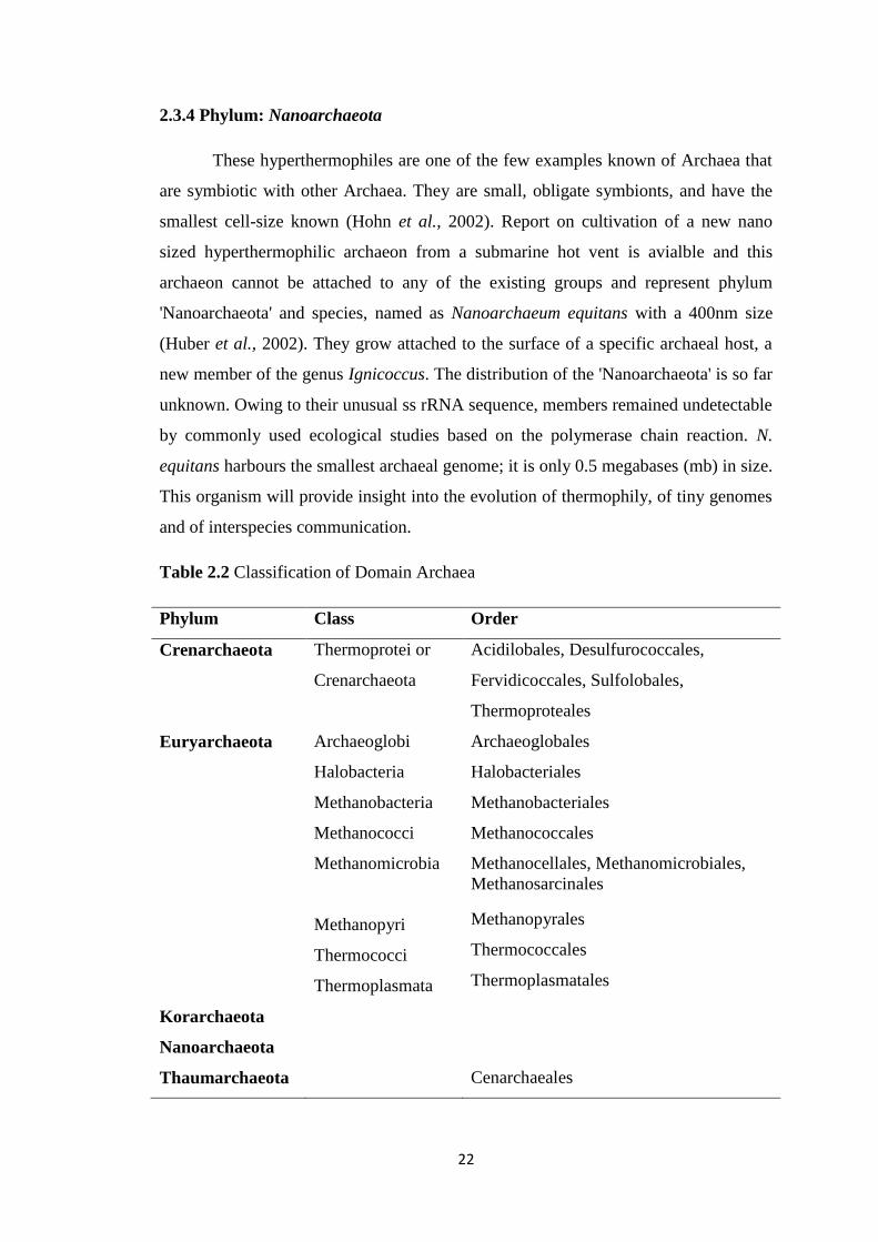
Archaea domain is divided into five phyla, Euryarchaeota, Crenarchaeota,
Korarchaeota, Nanoarchaeota and Thaumarchaeota, (uncultivable Group), the
presence of which has been determined only by environmental DNA sequences
(Woese, 2007; Barns et al., 1996; Bano et al., 2004; Huber et al., 2002; Cavalier-
Smith 2002). Well-known and cultivated archaea generally fall into several major
phenotypic groupings like halophiles, methanogens, thermophiles, thermoacidophilic
and nano-archaea (DeLong, 2003). Halophilic archaea are classified within the order
Halobacteriales, family Halobacteriaceae which consists of a number of aerobic
extreme halophiles that live normally in hyper saline environments, such as solar
salterns, soda lakes, sea beds, etc. Phylum Euryarchaeota is best known and
19
extensively studied phylum in archaea, includes halophiles, methonogens and some
hyperthermophiles. The archaeal members of hypothermophiles on marine ENV
lineage are least known in archaeal domain. Most known members of halobacteriales
are extreme halophiles flourish from 8% to saturation point of salt. Metabolic
diversity of halophiles includes oxygenic and anoxygenic phototrophs, aerobic
heterotrophs, fermenters, denitrifiers, sulphate reducers methanogens, etc.
The characterization of archaea is based on many internal and external
characters including chromatin organizing histone proteins, long thought to be absent
from Crenarchaea, is present in Euryarchaea. This has been interpreted as a support
for the euryarchaea as an origin of the eukaryotic nucleus (Martin and Müller, 1998).
Recent analysis of the Crenarchaeum symbiosum genome (Hallam et al., 2006) has
identified histones, ftsZ and the previously euryarchaea-specific DNA polymerase
polD, in conflict with the former distribution of these genes. Analysis of
environmental samples also indicates that histones were developed in ancestral
archaea (Cˇubonˇova´ et al., 2005). A group of DNA sequences from environmental
samples have been described as a third branch, the Korarchaeota. Korarchaea have
been sampled from geothermal environments (Barns et al., 1996), as well as from
hydrothermal regions (Marteinsson et al., 2001; Auchtung et al., 2006). A fourth
branch, Nanoarchaeota, has also been suggested (Huber et al., 2002). This group is
composed of extremely small organisms, only 400 nm in diameter, which grows in a
symbiotic/parasitic way on the surface of other archaeal species. Only one species of
this branch, Nanoarchaeum equitans, has been cultivated to date (Huber et al., 2002),
but sequences of others have been identified (Hohn et al., 2002).
Recent studies have suggested that the Korarchaea and Nanoarchaea may
represent fast evolving species within the Crenarchaeota and Euryarchaeota phylum,
respectively (Brochier et al., 2005; Robertson et al., 2005). Among three major
evolutionary domains of life on earth, members of the archaeal domain are the least
understood in terms of their diversity, physiology, genetics, and ecology.
Molecular phylogenetic studies have revealed that environmental archaeal
populations are diverse, complex and widespread, and that they frequently consist of
uncultivated and unidentified members (DeLong and Pace, 2001). As it is currently
impossible to construct culture-based phenotypic characterizations of many
20
environmental Archaea, the physiological significance of Archaea in nature has
remained unknown for a long time. When the phylogenetic features intrinsic to
archaeal communities are related to the environment, they may provide important
insights into the physiological functions and ecological roles of communities (Takai et
al., 2001). Several recent molecular studies (Bintrim et al., 1997; Jurgens et al., 1997;
Buckley et al., 1998) have demonstrated the ubiquity of Archaea in soil, particularly
those organisms belonging to the non-thermophilic Crenarchaeota lineage which
forms a deeply branching group with no close affiliation with any cultivated member
of Archaea. These organisms may constitute approximately 1% of the total soil
population (Whitman et al., 1998).
2.3 Domain Archaea
Archaea have long been considered as bacteria due to their prokaryotic
morphology, circular genomes, and gene organization in operons, but in 1977 Woese
could clearly distinguish archaea as a third domain of life by applying rRNA
phylogeny (Woese et al., 1990). Their status as a separate domain is further supported
by their unique features such as distinctive cell membrane phospholipids (Boris et al.,
2006). Genome -sequencing projects gave further insights into the nature of Archaea.
Haloarchaeal taxonomical studies started only few decades back with few isolates, in
last two decades, to a number of haloarchaea isolated and get characterized. Recently
a number of isolates were obtained from different ecosystems and is continuing by
adding new individuals to the Halobacteriaceae family. Current views of prokaryote
phylogeny are mainly based on 16S rRNA sequence data, which is only a small part
of the genome, which supposed to answer all our questions on archaeal evolution. In
prokaryote classification, polyphasic taxonomies are important in maintaining
flexibility and achieving a balanced picture.
2.3.1. Phylum: Euryarchaeota
In domain archaea, Halobacteriaceae familiy cover majority of extreme
halophiles, although several halophilic methanogenes are also known for its halophilic
nature. Only members of the Halobacteriaceae are usually considered as halophilic
archaea or haloarchaea. Up to now 33 genera of Halobacteriaceae have been defined
by rRNA sequencing and other criteria. The family is often collectively referred to as

21
“halobacteria” since the genus Halobacterium was the first halophilic archaeon to be
described and remains the best-studied representative of this group Archaea.
2.3.2. Phylum: Crenarchaeota
Several of this group are hypothermophiles (psychrophiles), but not much is
known about their taxonomy (Fuhrman et al., 1992). The Crenarchaeota has been
classified as a phylum of the Archaea domain, initially, the Crenarchaeota were
thought to be extremophiles but recent studies have identified them as the most
abundant archaea in the marine environment. Most of those are found in the 'Marine
ENV' branch on the Archaea tree. The hyperthermophiles have been studied more
thoroughly among cultured Archaea, in Crenarchaeota. Most are chemolithotrophs,
and in many extremely hot environments, they are the most important primary
producers (Könneke et al., 2005). The hyperthermophilic Crenarchaeota cluster
together on short branches on the tree, suggesting slow evolution, but not much is
known about their taxonomy. Most of these members are found in the 'Marine ENV'
branch on the Archaeal tree. They were separated from the other archaea based on
rRNA sequences; since then physiological features, such as lack of histones have
supported this division. However, some crenarchaea were found to have histones.
2.3.3 Phylum: Korarchaeota
The Korarchaeota have only been found in high temperature hydrothermal
environments. They appear to have diversified at different phylogenetic levels
according to temperature, salinity and geography. Korarchaeota have been found in
nature in only low abundance. The analysis of their 16S rRNA gene sequences
suggests that they are a deeply-branching lineage that does not belong to the main
archaeal groups (Elkins et al. 2008). These hyper thermophiles are known only from a
single hot spring in Yellowstone, and little is known about their ecology. They are
often discussed in the context of the 'slow evolutionary clock' idea. Briefly, the idea is
that hyperthermophilic Archaea appear to evolve more slowly than other lineages of
life (Cox et al, 2008). Interestingly, hyperthermophilic archaea seem to show the
same slow rate of evolution. One hypothesis is that this reflects the extreme
environment in which these organisms live: organisms at very high temperatures must
be under very strong selective pressure to maintain those genes that permit life there.
22
2.3.4 Phylum: Nanoarchaeota
These hyperthermophiles are one of the few examples known of Archaea that
are symbiotic with other Archaea. They are small, obligate symbionts, and have the
smallest cell-size known (Hohn et al., 2002). Report on cultivation of a new nano
sized hyperthermophilic archaeon from a submarine hot vent is avialble and this
archaeon cannot be attached to any of the existing groups and represent phylum
'Nanoarchaeota' and species, named as Nanoarchaeum equitans with a 400nm size
(Huber et al., 2002). They grow attached to the surface of a specific archaeal host, a
new member of the genus Ignicoccus. The distribution of the 'Nanoarchaeota' is so far
unknown. Owing to their unusual ss rRNA sequence, members remained undetectable
by commonly used ecological studies based on the polymerase chain reaction. N.
equitans harbours the smallest archaeal genome; it is only 0.5 megabases (mb) in size.
This organism will provide insight into the evolution of thermophily, of tiny genomes
and of interspecies communication.
Table 2.2 Classification of Domain Archaea
Phylum Class Order
Crenarchaeota Thermoprotei or
Crenarchaeota
Acidilobales, Desulfurococcales,
Fervidicoccales, Sulfolobales,
Thermoproteales
Euryarchaeota Archaeoglobi
Halobacteria
Methanobacteria
Methanococci
Methanomicrobia
Methanopyri
Thermococci
Thermoplasmata
Archaeoglobales
Halobacteriales
Methanobacteriales
Methanococcales
Methanocellales, Methanomicrobiales,
Methanosarcinales
Methanopyrales
Thermococcales
Thermoplasmatales
Korarchaeota
Nanoarchaeota
Thaumarchaeota Cenarchaeales
23
2.3.5 Phylum: Thaumarchaeota
Thaumarchaeota is a newly-proposed phylum of the Archaea, containing so
far four species, Nitrosopumilus maritimus, Cenarchaeum symbiosum,
Nitrososphaera viennensis, and Nitrososphaera gargensis. All organisms of this
lineage discovered so far are chemolithoautotrophic ammonia-oxidizers and may play
important roles in biogeochemical cycles, such as the nitrogen cycle and the carbon
cycle. Based on the first genome sequence of a crenarchaeote, Cenarchaeum
symbiosum, show that these mesophilic archaea are different from hyperthermophilic
Crenarchaeota and branch deeper than was previously assumed (Boussau et al., 2008).
2.4 Extreme Hyper-saline Environments
Hyper-saline waters are defined as those with total salt concentrations greater
than that of seawater. Concentrated salt solutions like salt or soda lakes, coastal
lagoons, salt marshes or man-made salterns are examples of hyper-saline
environments which are inhabited by only a few forms of higher life, other than
dominated prokaryotic microorganisms (Allers and Mevarech, 2005). Global salt
deposits show that evaporation of marine salt water and the development of hyper-
saline habitats is an ongoing process for millions of years and providing ample time
for the evolution of specialized halophilic bacteria and archaea. The major habitats of
halophilic microorganisms are salt waters (salt lakes, brines, ponds) and saline soils.
In the saline soils, the matrix potential of the soil adds to the water stress caused by
high salt concentrations. High saline waters originate either by seawater condensation
called Thalassohaline or by evaporation of inland surface water called
Athalassohaline.
2.4.1 Chemical Nature of Major Hyper-saline Environments
The chemical composition and the predominant ions in harsh environment
depend on the surrounding topography, geology, and general climatic conditions and
indirectly point to the possibility of phylogenetically distinct archea extremophiles.
Hyper- saline environmentals are diversed with different factors including high
osmotic pressure, low metabolites, acidic to alkaline pH values, low oxygen
availability, high or low temperatures, presence of heavy metals and other toxic
compounds (Oren, 2002; Ventosa, 2006). Thalassohaline waters, as they are derived
24
from seawater, and initially at least, have a proportional composition of salts similar
to that of seawater. Solar salterns, where seawater is evaporated to produce sea salt,
are typical examples of thalassohaline environments. Calcite or Calcium Carbonate
(CaCO3), gypsum (CaSO4.2H2O), halite (NaCl), sylvite (KCl) and finally carnallite, a
hydrated potassium magnesium chloride, (KMgCl3.6H2O), precipitate out sequentially
as evaporation occurs.
The salt composition of thalassohaline waters resembles that of seawater with
NaCl as the main constituent. By contrast, athalassohaline waters are markedly
influenced by the geology of the area where they develop, for example by the
resolution of salt deposits from a previous evaporative event, or significant leaching
of ions from the surrounding geology (Williams 1996; Grant et al., 1998; Demergasso
et al., 2024; Jiang et al., 2006). The Dead Sea is an example of a hyper-saline
environment profoundly influenced by an earlier Mg2+
rich brine, somewhat depleted
in Na+. Eugster and Hardie (1978) have attempted to define the key geological and
chemical features that influence how hyper-saline brine develops. The Dead Sea is
slightly acidic because of the precipitation of magnesium minerals such as sepiolite
(Mg4Si6O15(OH)2·6H2O) a complex of magnesium silicate (Grant et al., 1998).
Furthermore, a high calcium ion alkaline environment can develop from low
temperature weathering of calcium and magnesium containing silicate minerals,
olivine and pyroxene, which releases calcium ions and hydroxide ions. Magnesium is
precipitated and carbonate is removed as calcite, but since there is an excess of
calcium ions, this results in a Ca(OH)2-
dominated brine with a pH of around 11 (Jones
et al., 1994). Clearly, the type of brine that develops influences the microbiota.
Athalassohaline lakes can differ in their ion composition from seawater derived lakes.
Some athalassohaline waters have a very high concentration of divalent cations (for
example, the Dead Sea with the main cation Mg2+
instead of Na+), while others are
free of magnesium and calcium due to the presence of high levels of carbonate.
2.4.2 pH of Saline Systems
A solution of carbon dioxide forms carbonic acid, which is a weak acid that
undergoes ion exchange with the surrounding rock, this causes mineral to be leached
out. The final pH is influenced by the concentration of calcium ions (and to a lesser
extent, magnesium ions) in the surrounding rocks. Calcium ions form insoluble calcite
25
(CaCO3) which removes alkaline carbonate ions from solution; hence brines that have
a high concentration of calcium ions have a neutral pH. The Great Salt Lake in Utah
is one such environment (Baxter et al. 2005). Where the geology is deficient in
calcium and magnesium ions (where rocks of volcanic or metamorphic origin are
present and those of sedimentary origin are absent), carbonate becomes the dominant
ion. An increase in carbonate ions results in the precipitation of calcium, then
magnesium removing divalent cations from solution and allowing more soluble
carbonates of sodium and potassium to accumulate. This results in soda lakes such as
Lake Magadi in Kenya (Mwatha and Grant 1993; Grant et al. 1998). In contrast to salt
lakes, soda lakes are geologically very recent. In other environments, the precipitation
of minerals can contribute to the release of H+ ions, therefore reducing the pH.
Profoundly alkaline lakes develop in areas where the surrounding geology is
deficient in Ca2+
, for example in the East African Rift Valley. Here, surrounding high
Na+
trachyte lavas are deficient in both Ca2+
and Mg2+
, allowing the development of
lakes with pH values in excess of 11 (Grant and Tindall 1986; Grant et al., 1990;
Jones et al., 1998; Duckworth et al., 2004). Increased carbonate concentrations lead to
the formation of soda lakes, which have pH values well above 10 (for example, the
Wadi Natrun in Egypt). Levels of Mg2+
also influence the systems by removing CO3-2
as dolomite (CaMg(CO3)2), and in the case of the Dead Sea, whose composition is
markedly influenced by a previous Mg-rich evaporate, cause slightly acidic conditions
through the generation of Mg2+
minerals such as sepiolite, which generates H+ during
the precipitative process. High levels of other ions in the surrounding topography will
also influence the final composition, and there are exceptional hyper-saline lakes
dominated by Ca2+
.
2.4.3 Dissolved Oxygen Concentrations
The organic lake ecosystem contains salt concentrations of between 0.8 and
21% and an anoxic layer below a depth of 4 to 5 m. The water in the most hyper-
saline lakes is anoxic, with dissolved oxygen concentrations below 0.2 ppm (parts per
million). Saline waters often contain very little dissolved oxygen, it has been
calculated that oxygen solubility at 25 ºC is 2 mg/L at halite precipitation (Grant et al.,
1998). Hence salt lakes are high in salt and low oxygen environments with a range of
organic substrates available for growth. A few representatives of the halophiles may
26
overcome oxygen limitation by producing gas vesicles that enable them to float
toward the air-water interface. Presence of gas vesicles reduce the buoyancy of the
cells and enable them to float to the surface where light and oxygen conditions might
be more favourable for growth (Walsby, 1994). The gas vesicles observed are of
normal size, suggesting that the absence of oxygen does not always result in an
inhibition of their synthesis. An alternative strategy is to cope with the lack of
molecular oxygen with the use of alternative electron acceptors in respiration. Oxygen
was not detectable below 8 m depth in hyper saline lakes (Schink et al., 1983).
2.4.4 Dissolved Organic Carbon
Salt lakes often contain significant amounts of dissolved organic carbon
(DOC). Death and decay of organic matter from algae and plants growing nearby may
contribute to the carbon input. The major sources of organic compounds come from
the organisms themselves. For example, the decomposition of brine shrimp leads to
the deposition of chitin from their exoskeletons which will be used as substrate for
halophilic bacteria (Liaw and Mah, 1992). Furthermore, the decomposition of
bacterial cell walls releases sugars, proteins and lipids (Ollivier et al., 1994). A further
possible source of organic matter are compatible solutes (osmolytes) contained in the
organisms. These are organic molecules that help with coping with a high salt
environment. Release of such solutes is a further source of organic substrates (Grant
et al., 1998).
In saltern ponds with salt concentrations above 250 to 300 g l–1
, the only
primary producer is generally the unicellular green alga Dunaliella salina, which is
colored red-orange because of its high content of β-carotene. This alga is probably
responsible for all, if not, most of the primary production in salt lakes (Oren, 2002a).
Several cyanobacteria have been found in salt lakes that contribute to primary
production. Aphanothece halophytica and Daclylococcopsis salina are unicellular gas
vacuolated cyanobacteria that were originally isolated from Solar Lake in Israel.
However, large populations of cyanobacteria are not often seen in neutral salt lakes
but they are found in soda lakes. Anoxygenic phototrophic bacteria may also
contribute to primary production such as Ectothiorhodospira marismortui, isolated
from the Dead Sea, and Halorliodospira halophila (Grant, 2004).
27
Microflora have been found in all of the above types of saline systems,
indicating that halophilic microorganisms tolerate high to low salinity and can adapt
to different stresses like high pH or extreme temperatures (Allers and Mevarech,
2005). Surface hyper-saline lakes and solar salterns are the major habitats for
halophilic and halotolerant microorganisms, but there are other, less well-studied
high-salt habitats such as hyper-saline soils, salt marshes, salt desert, desert plants,
wall paintings, sea floor brines (such as the Atlantis Deep and the Discovery Deep in
the Mediterranean), oil field brines and ancient evaporate deposits, where isolations of
halophilic and halotolerant micro-organisms have been recorded (Oren, 2002). Saltern
crystallizer ponds are an ideal system to study communities of halophilic
microorganisms in their natural environment and diversity in all factors can be
observed throughout the globe. Salterns are found worldwide in coastal tropical and
subtropical areas; the nature of the microbial communities developing in the saturated
brines from which NaCl precipitates as halite crystals is very similar irrespective of
the geographical location: community densities are high, and the community structure
is very simple (Javor, 2002, Oren, 2002a, 2009). Hyper-saline marshes are located
between Gulf of Kutch, Great Rann Kutch and Little Rann Kutch.
2.5 Diversity of Halophiles in Saline Environments
In order to thrive in high salt concentrations, halophiles have to maintain a
cytoplasm that is at least iso-osmotic with the outside medium; otherwise they would
lose water to their environment or vice versa, since biological membranes are
permeable to water. Three domains use different strategies (Oren, 1999) to
compensate the osmotic pressure from the environment. The strategy whatever they
adopted should be energetically feasible and there is a need for all enzymes and
structural cell components to be adapted to ensure their function under these
conditions (Lanyi, 1974; Eisenberg et al., 1992). Protections against dehydration
during the salt crystallization, where there is a chance of losing cell water, into the ion
rich environment and how they are surviving in such condition make us think about
well evolved cell mechanisms of halophiles. In halophiles, the modification of
intracellular components, sugars, amino acids, fatty acids, osmolytes, sequence,
orientation, acidic and basic residue, charges of amino acids, are specially used to
maintain cell integrity. In hyper-saline condition, almost all cell activities of a non
halophile will be arrested due to the changes in physiology of bio- molecules which

28
lead to changes cell mechanisms, structures, chemical natures, metabolic and finally
inactivation of the activity of bio molecules by aggregating or precipitating.
Majority of the halophiles are aerobic. Therefore, for the growth of these cells,
the presence of oxygen and light for halophilic archaea is favourable due to the
presence of bacteriorhodopsin (BR) (Pfeifer et al., 1997, 2002). As the concentration
of salt increases, the concentration of O2 gets decreased and when crystallization
starts it blocks some amount of light. Therefore, due to lack of sufficient light energy
which is directly used to drive energy for bioenergetic processes, i.e. to grow photo-
organotrophically, growth will be impaired. In such conditions, these organisms have
evolved to change strategies to thrive under low oxygen and low light. Another
challenge haloarchaea faces in natural environment is the photo-oxidation by solar
radiation resulted into extensive damage to the genetic materials. However, archaea
lerned to devise effective DNA repairing mechanisms to circumvent such adverse
situations.
Communities of red halophilic archaea, halophilic eubacteria and unicellular
algae are some of the common living components in the brine, and they are generally
present in numbers as high as 107 to 10
8 cells per ml (Oren, 1994, 2006). Most
archaeal cells in salterns worldwide appear to belong to pleomorphic with the flat
square or rectangular, gas-vacuolated type first recognized by Walsby (1980), which
was only recently brought into culture and described as Haloquadratum walsbyi
(Burns et al., 2007). Different species of Halobacteriaceae are also present in
crystallizer brines, according to the physico-chemical properties of brine, but their
quantitative contribution to the community is poorly understood (Oren, 1994, 2002a,
b). The contribution of extremely halophilic members of the phylum Bacteroidetes
(Bacteria), in which some members are also coloured red, to the microbial
communities in saltern crystallizer ponds was recognized only quite recently (Antón
et al., 2000; Oren and Rodríguez-Valera, 2001; Oren, 2002b).
From the halophilic extremophile research, it is being pointed out that these
organisms are having astonishing mechanisms of adaptation. Initial studies on hyper
saline region on early days, evaluated with visible members of living things lead to
the conclusion of “No life area, Dead sea”, etc. The current research on halophilic
archea have a dominant consequence due to its novelty and extreme adaptation
29
features and shed light onto life in halophilic environments. Members of the
Halobacteriaceae require 1.5 - 4 M (8-23%) NaCl for optimal growth and majority of
halophilic archaea can thrive up to the saturation point, of sodium chloride around 5.5
M (32% of NaCl), (DasSarma, 2006). Although growth of some species is rather slow
at this salinity but haloarchaea are unable to grow below concentrations less than 1.5
M (8% of NaCl). In diverse environment they are abundant like sea surface,
subsurface, gas hydrate-bearing sediments and deep trenches (Gordon et al., 2004).
2.6 Adaptation of Archaea to Hyper-saline Environment
Haloarchaea do not survive at constant salt concentrations, but in many natural
settings they are exposed to changing salinities due to evaporation or rain, and thus
also the intracellular conditions change considerably. Thus due to this rapid
fluctuations in salinities, haloarchaea are excellent models to study osmoadaptation
over an extreme range of salt concentrations. Extreme halophiles like Halobacterium
salinarum require at least about 2 M salt, moderate halophiles like Haloferax volcanii
have a growth optimum slightly higher than 2 M but can grow from about 1 M to
saturation. Several terms describe the salt tolerance or requirement of organisms. The
term halophilic is generally restricted to those organisms that have a specific
requirement for salt (almost always assumed to be NaCl). Such organisms will not
grow in the absence of relatively high concentrations of salt, usually greater than 1.0–
1.5 M. Halotolerance is generally taken to mean that the organism has no specific
requirement for salt, but will continue to grow in the presence of high concentrations.
The regulation in the ion transport systems, biosynthetic pathways, secretion of the
extracellular proteins, intra cellular accumulation of osmolytes, etc. help moderate
halophiles for the adaptation. Several recent studies have identified genes that are
differentially expressed at different salt concentrations (Bidle, 2003; Choi et al., 2005;
Jäger et al., 2002) or detected de-novo synthesis of dimeric lipids upon an osmotic
down shift (Lopalco et al., ,2004), but the opportunities are clearly underexploited.
Halophiles have developed different basic mechanisms of osmoregulatory
solute accumulation to cope with ionic strength and the considerable water stress.
These mechanisms allow halophiles to proliferate in saturated salt solutions and to
survive when entrapment in salt crystals or rock and were proven by the isolation of
viable halophilic archaea from several subsurface salt deposits of Permo-Triassic age
30
(Walsh et al., 2005). Archaea also synthesize and accumulate several unique solutes
for use as osmolytes. They have aquaporins which are a large family of membrane
channels involved in osmoregulation, K+ pumps and K
+ channel, osmotic stress
sensors and signal transduction mechanism for osmoregulation. Little is known about
how synthesis and accumulation of solutes are regulated in these cells. In fact, for
many of the compatible solutes examined, the biosynthetic pathway is unknown.
Thus, in order to identify osmosensors, first need to identify biosynthetic pathways,
metabolites and key enzymes in osmoregulation.
2.6.1 Salt-in-cytoplasm Strategy
Organisms following this strategy adjust the interior protein chemistry of the
cell to high salt concentration. The thermodynamic adjustment of the cell can be
achieved by raising the salt concentration in the cytoplasm. Halophilic archaea keep
the cytoplasm relatively free of sodium, instead, potassium accumulates in the cell (as
shown for Haloferax volcanii through an energy-dependent potassium uptake system)
and together with its counter ion Cl-. Due to this salt-in strategy, archaea accumulate
K+ in the cytoplasm and can be found in molar concentrations. Because the K
+
concentration inside the cell is 100 times higher than in the surrounding environment,
a part of the proton motive force must be used to maintain the ion gradient. Their
energy requirement for this pumping is obtained from light driven rhodopsins. Four
functional types of haloarchaeal rhodopsins are known. The H+ pump
bacteriorhodopsin (BR) uses light energy to create a proton electrochemical gradient
for Adenosine triphosphate (ATP) production, flagellar rotation and other energy
requiring processes (Oren, 2002). The light-driven transport of chloride ions into the
cytoplasm by the Cl- pump halorhodopsin (HR), works to increase the electrochemical
potential of the proton gradient, while the two types of sensory rhodopsins present in
the haloarchaea are used for phototaxis under alternative maximum wavelengths of
light (λ max) (Sharma et al., 2007). Extreme halophilic archaea such as
Halobacterium and Haloferax are the archaeal examples that appear to use inorganic
ions exclusively as osmolytes.
In bacteria, this salt in strategy is normally absent and also there is evidence
that these organisms invest as little as possible energy in the maintenance of ion
gradients. Intra cellular accumulation of ions is also observed in members of bacteria
31
like Haloanaerobium praevalens, measurements of the ion composition in
exponentially growing cells of show that K+ is the dominating cation, but that Na
+
levels are also relatively high. Cells entering the stationary phase eventually replace
K+ for Na
+ (Oren, 2006). Analysis of Haloanaerobium acetoethylicum even suggests
that Na+ could be the main cation in stationary cells as well as in exponentially
growing cells (Oren, 1986). The effect of the accumulation of potassium and/or
sodium in the cytoplasm is that the cytoplasm is exposed to an increased ionic
strength. Although it is clear that the 'high-salt-in strategy' is energetically less costly
to the cell than the biosynthesis of large amounts of organic osmotic solutes (Oren,
1999). However, in habitats with saturated salt concentrations, halophilic archaea
outcompete organic-osmolyte producers, proving members of the “salt-in-cytoplasm
mechanism” as extreme halophiles.
2.6.2 Organic Osmolyte Strategy
Some halophilic organisms keep the cytoplasm, to a large extent, free of NaCl
and the design of the cell’s interior remains basically unchanged. The chemical
potential of the cell water is mainly reduced by an accumulation of uncharged, highly
water-soluble, organic solutes. The organic osmolyte mechanism is widespread
among Bacteria and Eukaryota and also present in some methanogenic archaea (Lai et
al., 1991; Roberts et al., 1992) in response to an osmotic stress, these organisms
mainly accumulate organic compounds like sugars, polyols, amino acids and/or amino
acid derivatives either by de novo synthesis or by uptake from the surrounding
environment. These non-ionic, highly water-soluble compounds do not disturb the
metabolism, even at high cytoplasmic concentrations and are thus appositely named
compatible solutes.
Halophilic cells using compatible solutes can basically preserve the same
enzymatic machinery as non-halophiles, needing only minor adjustments in their
interior proteins (i.e. ribosomal proteins), which are slightly more acidic than the
cytoplasmic proteins in Escherichia coli (Oren 2002). Halophiles employing the
organic- osmolyte mechanism are more flexible than organisms employing the “salt-
in cytoplasm strategy” because even though they display wide salt tolerance, they can
also grow in low salt environments. In addition to their function of maintaining an
osmotic equilibrium across the cell membrane, compatible solutes are effective
32
stabilizers of proteins and even whole cells. They can act as protectors against heat,
desiccation, freezing and thawing, and denaturants such as urea and salt. The reason,
why these organic compounds are compatible with the metabolism and can even act
as stabilizers of labile biological structures, is explained at the molecular level by the
preferential exclusion model.
The “low-salt-organic-solutes-in” strategy is based on the accumulation and/or
de-novo synthesis of water soluble organic solutes which do not interfere with the
activity or stability of normal enzymes (Oren, 2002). This mode has been observed in
moderate aerobic halophiles tolerating Na+ concentrations up to 1.5 M (Oren, 2008;
Ventosa et al., 1998). According to this theory, compatible solutes are absent from
protein surfaces due to the structural dense water bound to the protein. Compatible
solutes show a preference or the less dense free water fraction in the cytoplasmic
surrounding. They stabilize the two water fractions by fitting into the lattice of the
free water and allowing for the formation of hydration clusters. As a consequence,
unfolding and denaturation of proteins become thermodynamically unfavourable
(Wright et al., 2002). This explains why organisms adapted to other low water-
potential environments take advantage of the beneficial properties of compatible
solutes. It is not surprising that cyanobacterial species found in deserts accumulate the
compatible solute trehalose to compensate for the deleterious effects of desiccation
(Soppa, 2006).
The distribution of organic osmolytes found in archaea falls into the same
major classes as for bacteria and eukaryotes: (i) zwitterions (amino acids and
derivatives including betaine), (ii) neutral solutes (sugars and polyhydric alcohols),
and (iii) anionic solutes where the negative charge is supplied by a carboxylate,
phosphate or sulfate. Archaea also accumulate some very unusual solutes that have no
obvious bacterial or eukaryote counterpart, e.g., cyclic-2, 3-diphosphoglycerate or
cDPG, the most prominent solute in the hyper thermophilic Methanopyruskandleri,
and 1, 3, 4, 6-hexanetetracarboxylic acid , all polyanions (Roberts, 2000).
2.6.3 Other Mechanisms
It is reported that unusually high excess of the acidic amino acids glutamate
and aspartate in the Halobacterium proteins (Oren and Mana, 2002). The excess of
acidic amino acids in the protein composition could provide favoured sites for specific
33
water and ion binding to the tertiary or quaternary structure. To adapt the enzymatic
machinery to an ionic cytoplasm, proteins of halophilic anaerobic bacteria and
halophilic archaea contain an excess of acidic amino acids over basic residues. In low
saline environments, the excess of negatively charged ions will destabilize the
molecule’s structure, due to repulsion when the shielding cations are removed.
Possession of gas vesicles is generally considered to be advantageous to
halophilic archaea: the vesicles are assumed to enable the cells to float so it can reach
high oxygen concentrations at the surface of the brine and also occupy large
cytoplasmic volume and minimize the intracellular ion requirements for osmotic
balance (Walsby 1994). Since the presence of gas vesicles was first recognized in
bacterium Halobium, now Halobacterium salinarum, by Helena Petter (Petter, 1931;
Larsen, et al., 1967; Walsby, 1994), gas vesicles have become beloved study objects
in the biology of halophilic archaea of the family Halobacteriaceae.
In addition to aerobic respiration, a number of haloarchaea can ferment
arginine, while others can use sulphur, TMAO, DMSO or nitrate as alternative
electron acceptors. However, anoxic growth of Halobacterium salinarium PHH1
using DMSO or TMAO as terminal electron acceptor results in small, but gas vesicle
containing cells (Hechler and Pfeifer, 2009). The gas vesicles observed are of normal
size, suggesting that the absence of oxygen does not always result in an inhibition of
their synthesis. It is possible that the amount of energy gained by arginine
fermentation is less and not sufficient for the formation of gas vesicles of normal size.
Haloarchaea are most famous for their ability to generate a proton gradient
through the use of photo-reactive rhodopsin proteins that harness light energy
(Boichenko et al., 2006). The type 1 (microbial) rhodopsins are typically seven-pass
transmembrane proteins that use a retinal chromophore to absorb light energy for ion
transport or photo sensory functions (Schimz, 1981).
All halophilic archaea balance the high osmolarity of their environment by
having an at least equimolar intracellular salt concentration, KCl instead of NaCl in
well-energized cells. It was recognized long ago that typical haloarchaeal proteins
differ from mesohalic proteins by having a high fraction of acidic residues and a
reduced fraction of basic residues (Tebbe et al., 2005). The structure determination of
some soluble haloarchaeal proteins showed that they have a high concentration of

34
negative charges on the surface of the folded protein. Earlier it had been proposed that
this leads to the binding of a network of hydrated cations, but a few recent reports
have modified that picture and, in addition, have shown that the mode of
haloadaptation can be different for individual proteins. The malate dehydrogenase of
Haloarcula marismortui was an example and it is found to have strong binding sites
for some cations as well as anions, and loosely bound many more cations than
mesohalic enzymes in the natural solvent (Ebel et al., 2002). This might turn out to be
true for typical haloarchaeal proteins. While the haloadaptation of proteins has been
characterized in detail in several cases, similar studies have not yet been performed
for the adaptation of interactions of biomolecules, protein– protein or protein–nucleic
acid, to high salt concentrations.
2.7 Nutritional Sources in Hyper-saline Environments
Members of the Halobacteriaceae differ greatly in their nutritional demands.
Some have complex requirements that can only be met in culture by including high
concentrations of yeast extract or other rich sources of nutrients in their medium.
Others grow well on single carbon sources while using ammonia as nitrogen source.
In addition to simple substrates such as amino acids, sugars, and organic acids, certain
polymeric substances can be degraded by some halophilic archaea. Many species of
the Halobacteriaceae produce exoenzymes such as proteases, lipases, DNases, and
amylases which help them to sustain in extreme conditions (Oren, 2002a).
Organisms thriving in hyper-saline region are highly adapted to regulate the
metabolic path-way according to the environmental availability of metabolites.
Primary productivity in many hyper-saline environments is mainly by halophilic and
halotolerant cyanobacteria, anoxygenic phototrophic bacteria, and also eukaryotic
algae of the genus Dunaliella may be the significant sources of organic compounds.
The upper limit of NaCl concentration for vertebrates and invertebrates is ca. 1.5 M,
although the brine shrimp (Artemia salina) is an exception, often present in extremely
hyper-saline brines but not in extremely alkaline types (Oren, 1994; Jones et al.,
1998). In solar salterns phototrophic productivity is probably greatest during periods
of dilution, due to high number of primary produces. During maturation of brine the
number of primary producers decreases and finally Dunaliella spp., and other
halophiles which can grow at saturation point for NaCl remain as carbon sources.

35
Between concentrations of 1.5 M and 3.0 M, prokaryotes become predominant, with
the haloarchaea and a few rare bacterial types such as Salinibacter ruber forming the
climax population at the point of halite precipitation (Oren, 1994; Anto´n et al., 2002;
Grant et al., 2001). A few unusual fungi and protozoa are also present, but these are
probably active at lower salt concentrations (Post et al., 1983; Gunde-Cimerman et
al., 2000, 2004). Within the domain Archaea, halophilic prokaryotes occur in three
families: the Halobacteriaceae, halophilic methanogens in the Methanospirillaceae
and the Methanosarcinaeae. Unlike the Halobacteriaceae, where members are all
extremely halophilic, the Methanospirillaceae and the Methanosarcinaceae have
representatives that are not halophilic.
When there is a significant increase in organic compounds, it generally
accelerates populations of aerobic heterotrophs that may form dense blooms that
impart colouration to the brines. Hyper-saline environments are relatively low in
oxygen due to reduced oxygen solubility (2 ppm. in saturated NaCl, compared with 7
ppm. in seawater) and shifting of aerobic to anaerobic conditions in the brines lead to
increase in population of anaerobic heterotrophs and also metabolic shift in archaea
from aerobic to anaereobic. Halophiles utilize majour compounds of hyper-saline
habitats such as glycerol and tricarboxylic acid (TCA) cycle intermediates that are
excreted by primary producers, Dunaliella and the cyanobacterium. It is possible to
make some predictions as to the roles played by different organisms in the utilization
and recycling of organics. Cyanobacteria and, species of the eukaryotic alga
Dunaliella are the key primary producers, although anoxygenic phototrophic bacteria
of the genus Halorhodospira may be significant from time to time (Grant and Tindall,
1986; Oren, 1994).
These primary productivity supports large numbers of halophiles mainly
aerobic heterotrophic Gram-negative bacteria including Proteobacteria, in particular
members of the Halomonadaceae (Duckworth et al., 1996; Ventosa et al., 1998a;
Arahal et al., 2002), heterotrophic Gram-positive bacteria of both the high G+C
Firmicutes and low G+C Actinobacteria (Ventosa et al., 1998a, 1998b). These
heterotrophic organotrophs are supported by primary productivity and deposition of
organic matter as they utilise various polymers and monomers. Additional
heterotropic members from the Gamma proteobacteria found in hyper-saline
36
environment include Halovibrios, Alteromonads and Pseudomonas halophila (Grant,
2004; Sorokin et al., 2006).
Extreme halophilic archaea derive energy from the organic substrates formed
by primary producers (Grant, 2004). A large amount of glycerol is released from
Dunaliella cells when they die, which is a suitable carbon source for almost all of the
haloarchaea (Oren, 1994). Furthermore, haloarchaea have been shown to degrade
aromatic hydrocarbons, though it is not known to what extent they utilise these
substrates in their habitat (Oren, 1994). In contrast, it was found that the main
constituent of the archaeal community in the Dead Sea (where waters are slightly
acidic) was Haloferax, suggested by the presence of significant amounts of their
glycolipids (Oren and Gurevich, 1993). Methanogenic archaea may also be present in
some salt lakes, depending on salinity and are able to utilise methanol or methylamine
as substrates for the production of methane. Halomonads are able to utilise a range of
sugars and amino acids as carbon sources (Mata et al., 2002). However, Halomonas
organivorans is unusual in that it can utilise a variety of aromatic organic compounds.
The Halobacteriales tend to make up the main component of the biomass in hyper-
saline environments (Oren, 2002a).
2.7.1 In vivo Nutritional Requirements of Halophilic Archaea
Members of the Halobacteriaceae diverge greatly in their nutritional demands.
As culturing is necessary in order to study the unique properties and applications of
these organisms, new techniques have since been designed to try and increase the
number of cultured organisms. Some are simple in nutrition but some need specific
amino acids, vitamins etc. (Oren, 2002a). Most halophilic archaea preferentially use
amino acids as carbon and energy source, but also utilize other compounds of hyper-
saline habitats such as glycerol and tricarboxylic acid (TCA) cycle intermediates.
Almost all archaeal halophiles (haloarchaea) belong to the order Halobacteriales,
family Halobacteriaceae. This order contains 33 validly published genera (till June
2012) and some are nutritionally specific. In vivo growth of haloarchaea occurs in
basal salt medium with carbon and nitrogen sources.
Most of haloarchaea are very easy to grow and maintain in the laboratory and
their nutritional requirements are simple, the majority being able to use a large range
of compounds as the sole carbon and energy sources. In Halobacteriacaea there are
37
both fast as well as slow growers. Using different carbon sources that simulate the
environment has also been shown to help target more fastidious organisms (Dyall-
Smith, 2004). Some have complex requirements that can only be met in culture by
including high concentrations of yeast extract or other rich sources of nutrients in
their medium. Simple sugars such as glucose and sucrose are not readily used by all
members of the Halobacteriaceae, while amino acids are the preferred nitrogen
source for most species, some can use ammonia or nitrate, and some need thiamine
and biotin as stimulatory vitamins. Others grow well on single carbon sources while
using ammonia as nitrogen source. In addition to simple substrates such as amino
acids, sugars, and organic acids, certain polymeric substances can be degraded by
some halophilic archaea. Many species of the Halobacteriaceae produce exoenzymes
such as proteases, lipases, DNAses, and amylases.
The nutritional demands of Halobacterium salinarum, the best known archaeal
halophile species, are complex. Defined media designed for the growth of different
isolates may contain between 10 and 21 amino acids, in some cases supplemented
with vitamins, up to 5 different nucleosides, and glycerol (Dundas et al., 1963; Grey
and Fitt, 1976; Onishi et al., 1965; Shand and Perez, 1999). When Halobacterium
salinarum was grown in a defined medium containing inorganic salts, five
nucleosides, 21 amino acids, glycerol, and the vitamins folic acid, thiamine, and
biotin, a complex growth curve was obtained in which a number of phases could be
discerned within the "exponential" growth phase, each with a different growth rate
(Shand and Perez, 1999). There are some indications that even rich media based on
yeast extract, peptones, etc. may not provide all the compounds required by some
fastidious members of the group. When attempting to enumerate halophilic archaea
present in saltern evaporation and crystallizer ponds, Wais (1988) reported that colony
recovery rates greatly improved when the medium was amended with a lysate of
Halobacterium salinarum cells as a source of additional growth factors. Some
members can grow on simple compounds such as succinate, acetate, and others as
single carbon and energy source. Inorganic salts may supply the need for nitrogen,
sulphur and other essential elements (Rodriguez-Valera et al., 1980, 1983).
38
2.7.2 pH Requirements of Haloarchaea
Haloarchaea is isolated from diverse environments that vary from neutral
(neutrophiles) to alkaline (alkaliphiles). Acidophilic types of halophilic archaea have
not yet been reported. The Dead Sea with a pH of around 6.0 is probably the most
acidic environment in which mass development of halophilic archaea has been
observed (Oren, 1983c; Oren et al., 1988; Oren and Gurevich, 1993). The alkaliphilic
haloarchaea, include members of the genera Halorubrum, Natrialba,
Natronobacterium, Natronococcus, Natronomonas, Natronorubrum (Grant et al.,
2001), Halalkalicoccus (Xue et al., 2005) and Natronolimnobius (Itoh et al., 2005).
Many alkaliphilic haloarchaea have been isolated from the soda lakes of the Kenyan
Rift Valley, including Halorubrum vactiolatum (Mwatha and Grant 1993; Kamckura
et al., 1997), Natrialba inagadii (Tindall et al., 1984), Natronobacterium gregoryi,
Natronococcus occultus (Tindall et al., 1984) and Natronococcits amylolyticus (Kanai
et al., 1995), and the recently reported Nanoarchaeota (Huber et al., 2002). The
family Halobacteriaceae includes neutrophilic halophiles includeing many members
of Halobacterium, Haloferax, Haloarcula, Halovivax, Haloterrigena, Haloquadratum
and Natrinema.
A pH of 6 approximately coincides with the lower boundary of the range of
pH values that support growth of members of the Halobacteriales (Martin, 2006). As
more acidic hyper-saline environments seem to be rare or altogether nonexistent, the
halophilic archaea appear to be well adapted to the whole pH range occurring in
hyper-saline brines in nature. Many strains grow at neutral to slightly alkaline pH, and
some only at alkaline pH. However, no strain has been reported to grow only in acidic
pH conditions within the family Halobacteriaceae. Members of Halococcus are
moderately acidophilic haloarchaea able to grow only at pH 4.0–6.0. Extreme
acidophiles have optimum pH for growth of < 3.0 and that moderate acidophiles grow
optimally at pH 3–5. Currently, no strain has been reported to grow optimally at pH
3–5 within the family Halobacteriaceae (Goh et al. 2006; Wang et al., 2007).
2.8 Application of Haloarchaea
As haloarchaea are genetically tractable, they are an excellent model for
archaeal genetics. Furthermore, these extremophiles also have considerable
biotechnological potentials (Vidyasagar et al., 2006). They possess unique
39
bacteriorhodopsin, enzymes active in extreme conditions, liposomes, pigments, novel
enzymes, osmolytes, antibiotics, halocins, biopolymers and many other substances
with wide range of application. Understandings of these products or its applications
will ultimately be used to improve the efficiency of biocatalysis, which will heavily
contribute to the development of industrial biotechnology or environmental
biotechnology.
The halotolerance of many of their enzymes can be exploited wherever
enzymatic transformations are required to function at low water activities, such as
found in the presence of high salt concentrations. Enzymes from halophilic archaea
are mainly exoenzymes such as amylases, amylo-glucosidases, proteases, and lipases
that function at high salinity with a wide range of application in biotechnology. Some
halophilic archaea are used in traditional Korean fermented sea foods.
Many of halophilic hydrocarbon-degrading archaea can grow on hydrocarbons
like tetradecane, hexadecane, eicosane, heneicosane, pristane and aromatic
hydrocarbons like acenaphtene, phenanthrene, anthracene, 9-methylanthracene, etc. It
is also proved that degradation between 48 and 88% of the straight-chain
hydrocarbons or 19-24% of the aromatic hydrocarbons added at a concentration of 0.5
g/l were achieved after 30 days of incubation at 32°C. Therefore, from the preliminary
studies it is clear that halophilic archaea can be recommended for recovery of oil spill
in hyper-saline regions.
The role of halophilic archaea in the production of solar salt was investigated
and it shows that the C50 bacterioruberin derivatives are the main carotenoids of the
Halobacteriaceae and contribute as well toward the absorption of light energy, by
trapping solar radiation. These microorganisms raise the temperature of the brine and
the rate of evaporation, thereby increasing salt production capacity. To improve salt
production in salterns that do not develop a sufficiently dense archaeal community,
fertilization with organic nutrients has even been suggested (Javor, 1989, 2002).
The light-driven proton pump bacteriorhodopsin has many properties that
make it an attractive material for a large number of possible applications (Hampp,
2000a, 2000b; Margesin and Schinner, 2001; Oesterhelt et al., 1991). Hundreds of
patents have been issued related to different applications of bacteriorhodopsin
(Hampp, 2000a). Other potential uses of bacteriorhodopsin include conversion of

40
sunlight to electricity, ATP generation, desalination of seawater, use in chemo- and
biosensors, and ultrafast light detection. Bacteriorhodopsin was also suggested for use
in a bio-photo-electrochemical reactor for photochemical hydrogen production and
also as a light sensor which can create a charge by changes in the shape, generating an
electrical signal.
The retinal-based light-driven chloride pump halorhodopsin has also found
potential biotechnological applications. A chloride-sensitive biosensor has been
developed using an ion-sensitive field effect transistor on which membrane vesicles
containing halorhodopsin had been immobilized. When illuminated, this sensor reacts
to the concentration of chloride (Seki et al., 1994). In short, it can be used in
holography, spatial light, modulator, artificial retina, neural network optical
computing and volumetric and associated optical memories.
Halophilic archaea also produce biopolymers, considerable amounts of poly-
beta-hydroxyalkanoate, (copolymer of β-hydroxybutyrate and β- hydroxyvalerate),
which accumulated to values between 19 and 38 percent by dry weight, dependent on
salinity and is used for the production of biodegradable plastics. The highest
concentrations were observed in cells grown in salt (Fernandez-Castillo et al., 1986).
Downstream processing and purification of the product should be relatively simple as
the cells are easily lysed in water (Ventosa and Nieto, 1995). Also, the high genomic
stability of the organism and the reduced danger of contamination are clear assets.
Another extracellular polymer that may find interesting biotechnological applications
is poly Y-D-glutamic acid excreted by Natrialba aegyptiaca which can be used as a
biodegradable thickener, a humectant, or a drug carrier in the food or pharmaceutical
industry (Hezayen et al., 2000).
Archaeal ether lipid liposomes ("archaeosomes") have been tested as delivery
systems for vaccines and drugs. However, the liposomes prepared from
Halobacterium salinarum proved much leakier than those made from methanogens or
from Thermoplasma (Patel and Sprott, 1999). Archaeosomes prepared from total
polar lipids with divergent lipid compositions had the capacity to deliver antigen for
presentation via both MHC class I and class II pathways. Lipid extracts from
Halobacterium halobium and from strains of Halococcus morrhuae contained
archaetidylglycerol methylphosphate and sulfated glycolipids rich in mannose
41
residues, and lacked archaetidylserine. Another potential application of haloarchial
product halocin is to reduce injury during organ transplantation (Alberola et al.,
1988).
2.8.1 Halophilic Archaea as Model Organisms
The haloarchaea are one among the easiest archaea to grow and manipulate in
the laboratory (Papke et al., 2007). Their physiology metabolism and adaptations help
to try possible combinations for genetic models; neither do they require extreme
temperatures for growth nor strict absence of oxygen. Since many molecular
biological methods have been adapted for application under high salt conditions
(DasSarma and Fleischmann, 1995; Dyall-Smith, 2009), several haloarchaeal species
are excellent model organisms that can be used for the investigation to resolve many
biological questions.
2.9 Ecology of Rann of Kutch
2.9.1 Features of Rann of Kutch
The Rann of Kutch (Rann- literally means "salt marsh", Kachchh- literally
means something which intermittently becomes wet and dry) is shallow wetland
which submerges in water during the rainy season and becomes dry during other
seasons. This uniqueness of the Rann of Kutch, with both white salt desert during
summer and marshy during monsoon explain why it is called by its peculiar name, the
Rann of Kutch. The word Rann, according to Pakistan, in Hindi language means a
battlefield, but there is no historical record of any battle ever having been fought on
this field, which is "almost absolutely level". Sometimes, the Rann is said to be a
corruption of "Aranya", or it is equated with "Irina" in Sanskrit, meaning a salt
ground, saline soil.
Geologically, Rann of Kutch comprises of old lower Jurrassic formation to
recent alluvial formation. The extreme northern part of Kutch district in Gujarat state,
comprises of Great saline Rann. In South, along seacoast, it is represented by recent
alluvial formation, in West Tertiary formation is seen in Lakhpat and Abdasa talukas.
Saline formation is seen in the Rapar taluka. Central and Southern part of the district
of Kutch is covered by basalt (volcanic rock). Groundwater in most area is brackish to
42
saline. Kutch’s geology, climate and topography are intriguing, making it a
fascinating and challenging place to study.
The Rann of Kutch has two hyper-saline marshy regions mainly Little and
Great Rann of Kutch. The Great Rann of Kutch with an area of 18,000 sq. km lies
almost entirely within Gujarat state in India along the border with Pakistan. This arid
region was an extension of the Thar desert which is extending from central Pakistan to
north-western India. The major portion of Thar desert are located between 68.5º to 75º
east longitude and 23º to 30 º north latitude in the Indo-Pakistan subcontinent. The
Little Rann of Kutch extends northeast from the Gulf of Kutch over 5,100 sq. km. The
Great and Little Rann of Kutch are highly specialised arid ecosystems. In the
summers, they are similar to a desert landscape. However as these are low-lying areas
near the sea, they get converted to salt marshes during the monsoons. The Low lying
saline-marshy areas that get flooded due to sea water due to tidal effect as well as
from rain water during monsoons in the north are covered by flat, marshy, saline
Rann.
Kutch has a semi-arid type of climate where it rains a few days per year,
temperature ranges from 45 degrees centigrade in the summer to 2 degrees in winter.
The region which is the low lying, generally called Rann. There is practically no
vegetation and potable water resources are scarce in this region. Scanty rainfall,
reoccurring draught and absence of perennial rivers have hindered the development of
the district. Kutch is mineral rich region with very large reserves of Lignite, Bauxite
and Gypsum among other minerals. July to September monsoon rains flood the vast,
flat area to a depth of about 0.5 m.
Some surveys conducted in Kutch by local authorieties evaluated that the TDS
varied from location to location from 48 to 75,292 mg/ l, chloride from 19 to 34,830
mg/l, sulphate from nil to 3642 mg/l and bicarbonate from 7 to 956 mg/l. Kutch, is the
largest single saline area in the country. The ground waters vary from medium to
highly saline (EC 1000- 6,650 mmhos)
Normally a salt clay desert covering some 10,800 square miles, the Rann of
Kutch becomes a salt marsh during the annual rains. It is nestled between the Gulf of
Kutch in India's north-western state of Gujarat and the mouth of the Indus river in
southern Pakistan. Patches of high ground become a refuge for wildlife during the wet
43
season. River Luni, flows in a south- western direction through the State, losing itself
finally in the marshy ground at the head of the Rann of Kutch. The Kutch Peninsula
forms the western most part of the Indian sub-continent. The mainland Kutch is a
peneplain (low-relief plain representing the final stage of fluvial erosion) occupied by
Rann of Kutch sediments and volcanic basalts.
Elevation of majority of the hypersaline regions of Kutch ecoregions is around
5- 15 meters above the sea level. The colour of these crystallizes vary from clear
transparent, muddy, pink, bright red, etc. due to the concentration of salt as well as
due to microbial population. The microscopic observation of samples from such
extreme salt environment is very amazing and contains large microbial population
along with salt crystals. In this salt crystallizers, salt concentration of water varies
from time to time and most of the time it acquire almost saturation, only extreme
halophiles can survive, and in the present study biodiversity of extreme halophilic
archaea is focused for exploration.
2.10 Taxonomy of Halophilic Archaea
Halophilic archaea are classified within the order Halobacteriales, family
Halobacteriaceae, which contain a number of aerobic halophiles which thrive in
extreme hyper-saline environments. They are characterised mainly by their
requirements for high concentration of NaCl for the optimum physiological activity
and growth and it may range from 8-30%. Currently family Halobacteriacaeae
contains 33 genera comprising 126 species, and are diverse in physiology,
metabolism, adaptation against various stresses like salinity, pH, temperature, etc.
There are also reports of the presence of haloarcheon in low salt regions, low pH and
also cold conditions, showing the extents of their adaptation. Halophilic archaea can
be extreme halophilic archaea, which are adapted to survive in environments with
extreme salinity, and which are unable to survive in low salt concentration especially
concentration below 2.5M and moderate halophilic archaea which show ability to
grow over a wide range of salinities optimally ranging from 0.5-2.5 M but can grow
in less salt as well as saturated salt conditions. Moderately halophiles are more
adapted for the environmental changes especially in variation of salinity (Rodríguez-
Valera et al., 1985).
44
Halobacteriales are best known and extensively studied amoung archaea.
Taxonomy of halophiles follows haloarchaeal taxonomy, mainly based on
morphology, molecular and biochemical characterisation. Halobacteria are among the
most halophilic organisms known and require at least 1.5 M NaCl for their growth.
They contain one order, Halobacteriales and one family, Halobacteriaceae (Grant and
Larsen, 1989). Before the 1970s, halobacterial taxonomy was mainly based on
standard biochemical tests and morphology (Gibbons, 1974). At the end of the 1970s,
however, the situation changed dramatically. 16S rRNA-DNA hybridization studies
demonstrated that the halobacteria should be classified into nine clades of two groups
(Ross and Grant, 1985). The polar lipid compositions of these isolates as well as
hydrolysis of protein, gelatine, starch, tween 20, etc. had proven particularly useful in
the classification of halobacteria. Indole production from tryptophan, oxidase,
catalase, growth in single carbon source like glucose, mannose, fructose, xylose,
maltose, trehalose, cellobiose, raffinose and glycerol were also used for identification.
Apart from above mentioned properties, acid production from fructose, arabinose,
ribose, xylose, lactose and sucrose, etc. helped in identification (Oren et al., 1997).
2.10.1 Three Letter Code for Halophilic Archaea (33 Known Genera)
Three-letter abbreviations for genera of the family Halobacteriaceae are as follows:
Haladaptatus (Hap.), Halalkalicoccus (Hac.), Halarchaeum (Hla.), Haloarcula
(Har.), Halobacterium (Hbt.), Halobaculum (Hbl.), Halobiforma (Hbf.), Halococcus
(Hcc.), Haloferax (Hfx.), Halogeometricum (Hgm.), Halogranum (Hgn.), Halolamina
(Hlm.), Halomarina (Hmr.), Halomicrobium (Hmc.), Halonotius (Hns.), Halopelagius
(Hpl.), Halopiger (Hpg.), Haloplanus (Hpn.), Haloquadratum (Hqr.), Halorhabdus
(Hrd.), Halorubrum (Hrr.), Halosimplex (Hsx.) Halostagnicola (Hst.), Haloterrigena
(Htg.), Halovivax (Hvx.), Natrialba (Nab.), Natrinema (Nnm.), Natronoarchaeum
(Nac.), Natronobacterium (Nbt.), Natronococcus (Ncc.), Natronolimnobius (Nln.),
Natronomonas (Nmn.) and Natronorubrum (Nrr.).
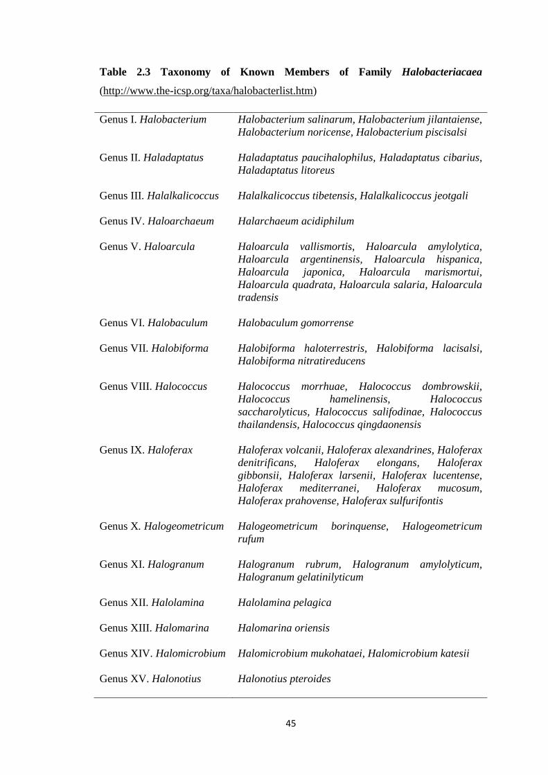
45
Table 2.3 Taxonomy of Known Members of Family Halobacteriacaea
(http://www.the-icsp.org/taxa/halobacterlist.htm)
Genus I. Halobacterium
Halobacterium salinarum, Halobacterium jilantaiense,
Halobacterium noricense, Halobacterium piscisalsi
Genus II. Haladaptatus
Haladaptatus paucihalophilus, Haladaptatus cibarius,
Haladaptatus litoreus
Genus III. Halalkalicoccus
Halalkalicoccus tibetensis, Halalkalicoccus jeotgali
Genus IV. Haloarchaeum
Halarchaeum acidiphilum
Genus V. Haloarcula
Haloarcula vallismortis, Haloarcula amylolytica,
Haloarcula argentinensis, Haloarcula hispanica,
Haloarcula japonica, Haloarcula marismortui,
Haloarcula quadrata, Haloarcula salaria, Haloarcula
tradensis
Genus VI. Halobaculum
Halobaculum gomorrense
Genus VII. Halobiforma
Halobiforma haloterrestris, Halobiforma lacisalsi,
Halobiforma nitratireducens
Genus VIII. Halococcus
Halococcus morrhuae, Halococcus dombrowskii,
Halococcus hamelinensis, Halococcus
saccharolyticus, Halococcus salifodinae, Halococcus
thailandensis, Halococcus qingdaonensis
Genus IX. Haloferax
Haloferax volcanii, Haloferax alexandrines, Haloferax
denitrificans, Haloferax elongans, Haloferax
gibbonsii, Haloferax larsenii, Haloferax lucentense,
Haloferax mediterranei, Haloferax mucosum,
Haloferax prahovense, Haloferax sulfurifontis
Genus X. Halogeometricum
Halogeometricum borinquense, Halogeometricum
rufum
Genus XI. Halogranum
Halogranum rubrum, Halogranum amylolyticum,
Halogranum gelatinilyticum
Genus XII. Halolamina
Halolamina pelagica
Genus XIII. Halomarina
Halomarina oriensis
Genus XIV. Halomicrobium
Halomicrobium mukohataei, Halomicrobium katesii
Genus XV. Halonotius
Halonotius pteroides
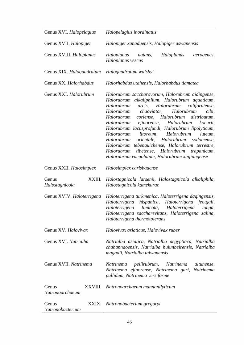
46
Genus XVI. Halopelagius
Halopelagius inordinatus
Genus XVII. Halopiger
Halopiger xanaduensis, Halopiger aswanensis
Genus XVIII. Haloplanus
Haloplanus natans, Haloplanus aerogenes,
Haloplanus vescus
Genus XIX. Haloquadratum
Haloquadratum walsbyi
Genus XX. Halorhabdus
Halorhabdus utahensis, Halorhabdus tiamatea
Genus XXI. Halorubrum
Halorubrum saccharovorum, Halorubrum aidingense,
Halorubrum alkaliphilum, Halorubrum aquaticum,
Halorubrum arcis, Halorubrum californiense,
Halorubrum chaoviator, Halorubrum cibi,
Halorubrum coriense, Halorubrum distributum,
Halorubrum ejinorense, Halorubrum kocurii,
Halorubrum lacusprofundi, Halorubrum lipolyticum,
Halorubrum litoreum, Halorubrum luteum,
Halorubrum orientale, Halorubrum sodomense,
Halorubrum tebenquichense, Halorubrum terrestre,
Halorubrum tibetense, Halorubrum trapanicum,
Halorubrum vacuolatum, Halorubrum xinjiangense
Genus XXII. Halosimplex
Halosimplex carlsbadense
Genus XXIII.
Halostagnicola
Halostagnicola larsenii, Halostagnicola alkaliphila,
Halostagnicola kamekurae
Genus XVIV. Haloterrigena
Haloterrigena turkmenica, Haloterrigena daqingensis,
Haloterrigena hispanica, Haloterrigena jeotgali,
Haloterrigena limicola, Haloterrigena longa,
Haloterrigena saccharevitans, Haloterrigena salina,
Haloterrigena thermotolerans
Genus XV. Halovivax
Halovivax asiaticus, Halovivax ruber
Genus XVI. Natrialba
Natrialba asiatica, Natrialba aegyptiaca, Natrialba
chahannaoensis, Natrialba hulunbeirensis, Natrialba
magadii, Natrialba taiwanensis
Genus XVII. Natrinema
Natrinema pellirubrum, Natrinema altunense,
Natrinema ejinorense, Natrinema gari, Natrinema
pallidum, Natrinema versiforme
Genus XXVIII.
Natronoarchaeum
Natronoarchaeum mannanilyticum
Genus XXIX.
Natronobacterium
Natronobacterium gregoryi
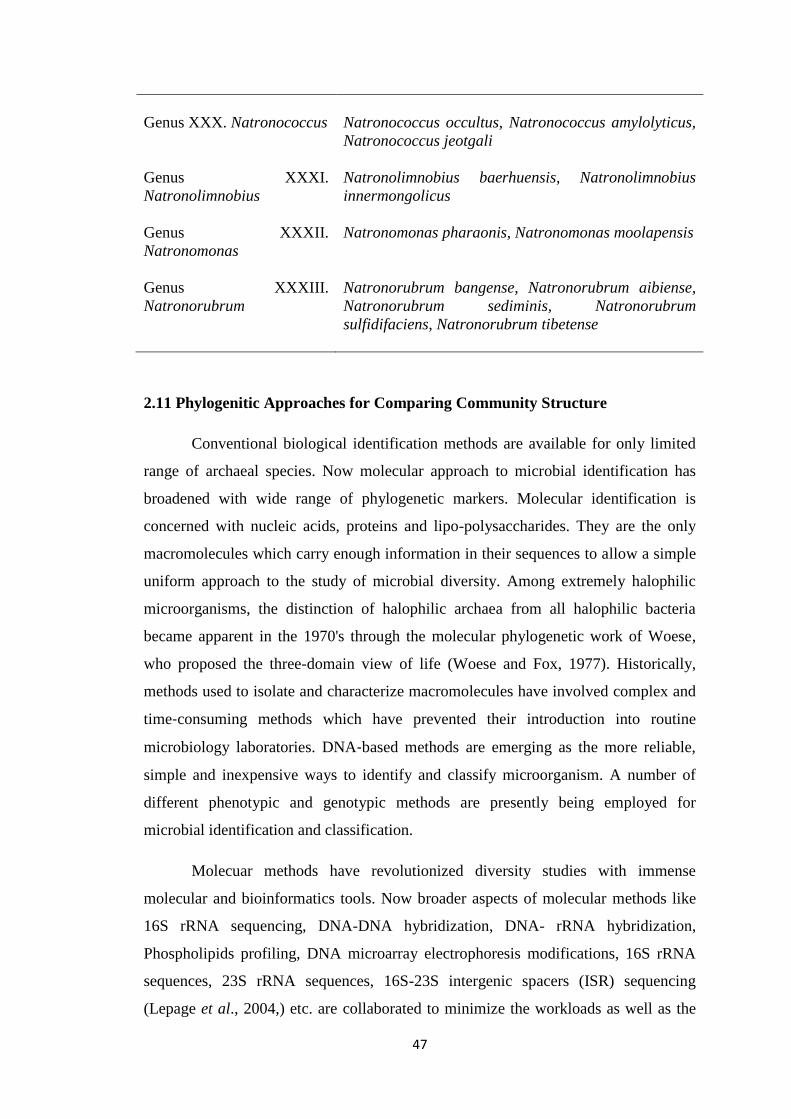
47
Genus XXX. Natronococcus
Natronococcus occultus, Natronococcus amylolyticus,
Natronococcus jeotgali
Genus XXXI.
Natronolimnobius
Natronolimnobius baerhuensis, Natronolimnobius
innermongolicus
Genus XXXII.
Natronomonas
Natronomonas pharaonis, Natronomonas moolapensis
Genus XXXIII.
Natronorubrum
Natronorubrum bangense, Natronorubrum aibiense,
Natronorubrum sediminis, Natronorubrum
sulfidifaciens, Natronorubrum tibetense
2.11 Phylogenitic Approaches for Comparing Community Structure
Conventional biological identification methods are available for only limited
range of archaeal species. Now molecular approach to microbial identification has
broadened with wide range of phylogenetic markers. Molecular identification is
concerned with nucleic acids, proteins and lipo-polysaccharides. They are the only
macromolecules which carry enough information in their sequences to allow a simple
uniform approach to the study of microbial diversity. Among extremely halophilic
microorganisms, the distinction of halophilic archaea from all halophilic bacteria
became apparent in the 1970's through the molecular phylogenetic work of Woese,
who proposed the three-domain view of life (Woese and Fox, 1977). Historically,
methods used to isolate and characterize macromolecules have involved complex and
time‐consuming methods which have prevented their introduction into routine
microbiology laboratories. DNA‐based methods are emerging as the more reliable,
simple and inexpensive ways to identify and classify microorganism. A number of
different phenotypic and genotypic methods are presently being employed for
microbial identification and classification.
Molecuar methods have revolutionized diversity studies with immense
molecular and bioinformatics tools. Now broader aspects of molecular methods like
16S rRNA sequencing, DNA-DNA hybridization, DNA- rRNA hybridization,
Phospholipids profiling, DNA microarray electrophoresis modifications, 16S rRNA
sequences, 23S rRNA sequences, 16S-23S intergenic spacers (ISR) sequencing
(Lepage et al., 2004,) etc. are collaborated to minimize the workloads as well as the
48
accuracy of the analysis. The minimal standards for characterization of halophiles also
include characterization of polar lipids, G+C content along with other characters
(Oran et al., 1997). The full genome sequence of the model halphilic organism reveals
some of the general characters of halophillic archaea.
2.11.1 Molecular Techniques to Characterise Microbial Diversity
Molecular approaches used to characterise the microbial community in the
environment involves extraction of community genomic DNA and then amplification
of the 16S rRNA gene by Polymerase Chain Reaction (PCR) using universal primers
designed against conserved regions near the ends of the gene. This mixture of PCR
products is then subject to a range of molecular approaches. Other dominant genetic
markers used in phylogenetic studies that clearly defined and separated domains are
supported by genes for several highly conserved proteins: the elongation factor Tu (1
alpha); heat shock protein Hsp60; and RNA polymerase subunits, aspartyl–, leucyl–,
phenylalanyl–, tyrosyl–, and tryptophanyl– tRNA synthetases. The rRNA-based
bacterial phylum concept is, at least, partly supported by all alternative markers. If
multiple approch is using in phylogenetic trees, dynamic structures that may change
their local topologies as new data become available.
Electrophoretic analysis provides a genetic fingerprint of the whole
community by differential migration of the mixed PCR products by electroporation
through agarose or polyacrylamide. This can be dependent on size amplified
ribosomal DNA restriction analysis (ARDRA), terminal restriction fragment length
polymorphism (t-RFLP), ribosomal intergenic spacer analysis (RISA), random
amplified polymorphic DNA (RAPD) or sequence denaturing gradient gel
electrophoresis (DGGE), temperature gradient gel electrophoresis (TGGE). The
methodology of these techniques has been reviewed elsewhere (Ranjard et al., 2000).
However, these techniques are generally only useful in looking at changes in
microbial community over a period of time by taking several fingerprint profiles at
certain time intervals. In order to characterise the microbial community, bands must
still be extracted and sequenced.
The limitations of some of these techniques have been discovered by others
(Rees, 2002). However, there are drawbacks to PCR-based methods. Templates with
high mole% G+C content may be discriminated against during PCR due to the low
49
efficiency of strands separation. The fidelity of DNA polymerases used during PCR
may also vary which results in miss-incorporation of nucleotides and may lead to
assumptions of novel taxa present in the sample (Head et al., 1998). In addition,
studies have shown that a proportion of amplified sequences from a mixed community
form chimeras during PCR (Kopczynski et al., 1994). They are formed when DNA
synthesis begins at one end of a sequence but is interrupted and continues on another
template sharing some degree of localised homology to the original. This results in
two or more fragments from different genes joining, which is introduced into the PCR
as a full length template to be amplified. It has become increasingly important that
chimeras are recognised and removed from diversity studies as novel lineages could
be erroneously assigned and give misleading impressions of biodiversity (Hugenholtzt
and Huber, 2003).
These sequences could be deposited in public databases inducing the quality
of such repositories. Furthermore, primers are not completely universal. In fact, a
previous study showed that alternative bacterial primers designed against a different
section of the 16S rRNA gene sequence was able to amplify a proportion of the
microbial community in sputum samples that was not obtained with the universal 16S
rRNA primer, 27-917b. Therefore, the widespread use of these 'universal' 16S rRNA
primers can lead to a significant part of the microbial community.
Therefore, in the present studies, continuous sampling, isolation, and
identification using molecular methodology was employed on different cultivated
organisms from Rann ecoregion. There is little information available about the
diversity of halophilic archaea in this largest single hyper saline region in India. As it
is the first report of haloarcheal community in Rann ecosystem, there were potentially
many novel archaeal lineages that remain to be discovered. Traditional cultivation
methods that mimic the natural conditions of Rann of Kutch minimizes the failure to
cultivate most microorganisms (Kaeberlein et al., 2002). Successful approaches are
those that mimic the natural environment, i.e., novel halophiles were isolated from the
Red Sea by using media close to the composition of the Red Sea brine (Eder et al.,
2001). Similarly cultivation methods are standardized according to physiology of the
Rann ecosystem.