2011 - cardoso-cruz - ejn - place cells - final
TRANSCRIPT

8/2/2019 2011 - Cardoso-Cruz - EJN - Place Cells - Final
http://slidepdf.com/reader/full/2011-cardoso-cruz-ejn-place-cells-final 1/10
NEUROSYSTEMS
Instability of spatial encoding by CA1 hippocampal placecells after peripheral nerve injury
Helder Cardoso-Cruz,1,2 Deolinda Lima1,2 and Vasco Galhardo1,2
1Instituto de Biologia Molecular e Celular (IBMC), Grupo de Morfofisiologia do Sistema Somatosensitivo, Universidade do Porto,
4150-180 Porto, Portugal2Departamento de Biologia Experimental, Faculdade de Medicina, Universidade do Porto, Porto, Portugal
Keywords : multichannel recording, neuropathy, pain, rat
AbstractSeveral authors have shown that the hippocampus responds to painful stimulation and suggested that prolonged painful conditions
could lead to abnormal hippocampal functioning. The aim of the present study was to evaluate whether the induction of persistent
peripheral neuropathic pain would affect basic hippocampal processing such as the spatial encoding performed by CA1 place cells.
These place cells fire preferentially in a certain spatial position in the environment, and this spatial mapping remains stable across
multiple experimental sessions even when the animal is removed from the testing environment. To address the effect of prolonged
pain on the stability of place cell encoding, we chronically implanted arrays of electrodes in the CA1 hippocampal region of adult rats
and recorded the multichannel neuronal activity during a simple food-reinforced alternation task in a U-shaped runway. The activity of
place cells was followed over a 3-week period before and after the establishment of an animal model of neuropathy, spared nerve
injury. Our results show that the nerve injury increased the number of place fields encoded per cell and the mapping size of the place
fields. In addition, there was an increase in in-field coherence while the amount of spatial information content that a single spike
conveyed about the animal location decreased over time. Other measures of spatial tuning (in-field firing rate, firing peak and number
of spikes) were unchanged between the experimental groups. These results demonstrate that the functioning of spatial place cells is
altered during neuropathic pain conditions.
Introduction
It has long been known that the hippocampus is crucial for learning
and memory. One special feature of pyramidal complex-spike neurons
found in CA1 and CA3 areas of the hippocampus is that they encode
the animal location in the environment (Fox & Ranck, 1981). These
place cells increase their firing rate when an animal is in a particular
position within its environment (place field; O’ Keefe & Dostrovsky,
1971; O’ Keefe & Nadel, 1978), and this place field is usually stable
across repeated visits to the same environment, even when the animal
is removed from it for extended time periods (Muller et al., 1987;
Thompson & Best, 1990). Several studies have shown that changes inthe spatial features or motivational content of the testing environment
may disrupt the place field stability (Kobayashi et al., 1997; Markus
et al., 1995; Moita et al., 2004; Muller & Kubie, 1987; Wood et al.,
2000). On the other hand it has been shown that place field instability
could also result from pharmacological manipulations of hippocampal
functioning (Dragoi et al., 2003; Kentros et al., 1998; Rotenberg
et al., 2000) or from direct lesioning of the hippocampus or
hippocampal-connected areas (Leutgeb & Mizumori, 1999; Liu et al.,
2003; McNaughton et al., 1989; Mizumori et al., 1994; Muir &
Bilkey, 2001).
In addition to spatial information, the hippocampus has repeatedly
been described as being involved in the regulation of several
behavioural aspects of the adaptation to aversive situations, including
pain (Khanna, 1997; Khanna & Sinclair, 1992; Soleimannejad et al.,
2006). Hippocampal neurons respond to noxious stimulation
(Delgado, 1955; Halgren et al., 1978; Khanna, 1997; Soleimannejad
et al., 2006; Tai et al., 2006; Wei et al., 2000; Zheng & Khanna,
2008), and human imaging studies have shown its activation by acutenoxious stimulation (Bingel et al., 2002; Ploghaus et al., 2001, 2000).
Moreover, partial hippocampectomy has been used (albeit with
moderated success) as a treatment for human chronic pain syndromes
(Gol & Faibish, 1967), while the inactivation of hippocampal synaptic
transmission attenuated nociceptive behaviour in the rat formalin
model (Khanna, 1997; McKenna & Melzack, 1992, 2001).
Despite all the knowledge gathered on how the hippocampus affects
pain processing, little is known about how pain affects hippocampal
functioning. It has been shown that chronic pain changes c-fos
expression (Carter et al., 2011; Ceccarelli et al., 2003) and long-term
potentiation (Kodama et al., 2007; Ren et al., 2011), and causes
changes in hippocampal volume (Lutz et al., 2008; Younger et al.,
2010), but it is still unknown whether pain affects crucial hippocampal
functions such as the generation of spatial maps. Therefore, our goal
Correspondence: Vasco Galhardo, 1Instituto de Biologia Molecular e Celular (IBMC),
as above.
E-mail: [email protected]
Received 2 February 2011, revised 13 March 2011, accepted 5 April 2011
European Journal of Neuroscience, Vol. 33, pp. 2255–2264, 2011 doi:10.1111/j.1460-9568.2011.07721.x
ª 2011 The Authors. European Journal of Neuroscience ª 2011 Federation of European Neuroscience Societies and Blackwell Publishing Ltd
European Journal of Neuroscience

8/2/2019 2011 - Cardoso-Cruz - EJN - Place Cells - Final
http://slidepdf.com/reader/full/2011-cardoso-cruz-ejn-place-cells-final 2/10
was to verify whether long-term neuropathic pain disrupts the stability
of CA1 hippocampal place fields in the absence of spatial or
motivational changes introduced in the familiar testing environment,
or alterations in the performance capability of the animal. We recorded
neuronal activity of freely moving rats in a simple spatial alternation
task, performed before and after the induction of the spared nerve
injury (SNI) model of neuropathic pain (Decosterd & Woolf, 2000).
Materials and methods
Subjects
Twelve adult male Sprague–Dawley rats weighing between 275 and
325 g were used in this study. The rats were maintained on a 12-h
light–dark cycle, and both training and recording sessions ran at
approximately the same time each day, during the light portion of the
cycle. All procedures and experiments adhered to the guidelines of the
Committee for Research and Ethical Issues of IASP (Zimmermann,
1983) and with the Ethical Guidelines for Animal Experimentation of
the European Community Directive Number 86 ⁄ 609 ⁄ ECC of 24
November of 1986, and were approved by national and local boards.
Behavioral arena
The testing environment consisted of a U-shaped arena with three
sections that were 72 cm long and 15 cm wide, and with opaque walls
30 cm high (Fig. 1); this arena was similar to an elevated runway
previously used in a place cell stability study (Tropp et al., 2005). Rats
were trained to run an alternation schedule on the arena to receive food
reinforcements at both ends of the runway; one chocolate-flavoured
pellet was automatically delivered by a pellet dispenser (Coulbourn
Instruments, Whitehall, PA, USA), depending on previous visit to the
opposite end of the runway. Control of pellet dispensers was fully
automated using the OpenControl software adapted to this particular task (Aguiar et al., 2007). We preferred the use of this simple task in a
linear runway instead of a more complex bifurcating arena that
requires navigational decision-making because pain is known to affect
both decision-making and spatial navigation in working memory-
dependent tasks (Pais-Vieira et al., 2009a; Hu et al., 2010; our own
unpublished results), and any perturbations of the navigational
performance of the animals may lead to spontaneous place field
remapping.
Experimental schedule
During the training period, the rats were given two 15-min sessions
per day to learn the alternation schedule until they performed 80correct runway alternations within a period of 4 consecutive days
(Tropp et al., 2005). After reaching this performance criterion the
animals were surgically implanted unilaterally with a multielectrode
array for single-unit recording (see details below).
After 7 days of recovery from the implantation surgery, 12 animals
(n = 6 in both sham and nerve-lesioned groups) were recorded over 4
consecutive days (hereafter named baseline or control period) while
performing the runway alternation task in two daily sessions of
15 min. The detailed results from this control period sessions are
presented in Table S1 as Supporting Information. On the following
day the animals were briefly anesthetized and either surgically
subjected to the SNI model of neuropathic pain (Decosterd & Woolf,
2000) or to a sham intervention with the same extent of skin incision
and muscle dissection, performed in the left hindpaw contralateral to
the location of the intracranial recording electrodes. Both groups of
animals were then recorded for a period of 3 weeks (with recordings
performed on days 1, 2, 3, 7, 10, 15 and 21 after SNI or sham
intervention). The sensory threshold for noxious stimulation was
measured using von Frey filaments (Somedic, Sweden) as previously
described (Chaplan et al., 1994). Von Frey testing was always
performed after the second recording session of the day (with at least a minimum interval of 1 h after replacing the animals in their home
cage). Testing was performed in an elevated chamber with a thin
metallic mesh floor that allowed easy access to the plantar surface of
the left hindpaw; filament series were run from the thinnest to the
widest to detect the filament to which the animal withdrew the paw in
at least six of 10 successive applications; we then performed another
two series of 10 stimulations using the same filament (2-min intervals
between sessions) and averaged the number of positive responses
evoked by the three series.
Multielectrode implantation
For the surgical implantation of the intracranial multielectrode arraythe animals were anesthetized with an intramuscular injection of a
mixture of xylazine and ketamine (10 and 60 mg ⁄ kg, respectively).
Anesthesia was maintained with small additional injections of
ketamine (one-third of the initial dosage). Oxygen was supplied
during the procedure via a face mask. The depth of anesthesia and
paralysis of the musculature was assessed by regularly testing the
corneal blink, hindpaw withdrawal and tail-pinch reflexes. After the
anesthesia induction, each animal received a single dose of atropine
sulphate (0.02 mg ⁄ kg, subcutaneous) and 1 mL of serum (sucrose 2%
w ⁄ v in NaCl 0.9% w ⁄ v, subcutaneous) every hour throughout the
surgery. Core body temperature was measured with a rectal thermom-
eter and maintained at 37 °C by means of a homeothermic blanket
system. The animal’s head was shaved and cleaned using a triple
application of alcohol (70%, v ⁄ v) and Betadine. A midline subcuta-
neous injection of 0.3 mL of 1% lignocaine (B Braun, Melsungen,
Germany) was applied to the scalp for local analgesia. Anesthetized
animals were secured in a stereotaxic frame using ear bars, and a
midline incision was made caudally to the animal’s eyes and ending
between ears. The connective tissue was blunt-dissected, and the top
of the skull was exposed and cleaned using hydrogen peroxide. After
exposure of the scalp, holes were bored for fixation of four screws
used for securing the array and for grounding. One craniotomy
(3 · 3 mm) was made for implantation of the multielectrode.
Each multielectrode array consisted of eight filaments of isonel-
coated tungsten wire (35 lm diameter; California Fine Wire Company,
Grover Beach, CA, USA) with impedances varying between 0.5 and
0.7 MX at 1 kHz. The multielectrode arrays were built in a 4 · 2architecture, interspaced with 250 lm between rows and 400 lm
between columns (Silva et al., 2010). The arrays were rostrocaudally
oriented and mounted in the holder of a hydraulic micropositioner (FHC
Inc, Bowdoin,ME, USA) and slowly driven (50 lm ⁄ min) into theright
hippocampal CA1 region after dura mater removal, while monitoring
the neuronal activity. The following coordinates in mm relative to
bregma (Paxinos & Watson, 1998) were used to center the arrays: )3.2
rostrocaudal, +2.2 mediolateral and )2.6 dorsoventral. After the
electrodes were advanced to the correct position, the craniotomy was
sealed with a layer of agar (4% in saline) and the array was cemented to
the skull screws using dental acrylic. At the end of the implantation the
animal was transferred to a recovery cage. The analgesic carprofen
(7.5 mg ⁄ kg; Rimadyl, Pfizer Animal Health, Lisbon, Portugal) and the
antibiotic amoxicillin (6 mg ⁄ kg; Clamoxyl, Pfizer Animal Health,
2256 H. Cardoso-Cruz et al.
ª 2011 The Authors. European Journal of Neuroscience ª 2011 Federation of European Neuroscience Societies and Blackwell Publishing Ltd European Journal of Neuroscience, 33, 2255–2264

8/2/2019 2011 - Cardoso-Cruz - EJN - Place Cells - Final
http://slidepdf.com/reader/full/2011-cardoso-cruz-ejn-place-cells-final 3/10
Lisbon, Portugal) was administered subcutaneously every 24 h for
3 days while the animal recovered from surgery. Rats were allowed to
recover for 1 week before the task re-training sessions began.
Neural recordings
All neural activity signals from the implanted microelectrodes were
recorded and processed using a Multineuron Acquisition Processor
system (16-MAP; Plexon Inc., Dallas, TX, USA). Voltage–time
threshold windows were used to identify single-unit waveforms. The
differentiated neural signals were preamplified (10 000–25 000 ·),
and digitized at 40 kHz. Up to two neuronal action potentials per
recording channel were sorted online (SortClient 2008; Plexon Inc.)
and validated by offline analysis (Offline Sorter 2.8.; Plexon Inc.)
according to the following cumulative criteria: voltage thresholds
> 2 SD of amplitude distributions; signal-to-noise ratio > 3; fewer
than 1% of interspike intervals > 1.2 ms; and stability of the
waveform shape as determined by a waveform matching template
A C
B
Fig. 1. Illustration of activity of a place cell during the testing environment navigation before (baseline) and after nerve injury (SNI). (A) Normalized firing ratemaps. The bright red pixels correspond to regions where the cell fired at a higher rate, and the dark blue to a lower rate. (B) Correspondent encoded place fields(green pixels). (C) Photograph of the U-shaped testing environment. The rats were trained to alternate between feeders A and B in the maze for food reinforcement dispensed by pellet feeders located one at the end of each arm. The feeders dispensed a single food pellet when the animal performed a correct alternation.
Pain-induced instability of hippocampal place cells 2257
ª 2011 The Authors. European Journal of Neuroscience ª 2011 Federation of European Neuroscience Societies and Blackwell Publishing Ltd European Journal of Neuroscience, 33, 2255–2264
8/2/2019 2011 - Cardoso-Cruz - EJN - Place Cells - Final
http://slidepdf.com/reader/full/2011-cardoso-cruz-ejn-place-cells-final 4/10
algorithm and principal component analysis. In addition, the
Waveform Tracker software (Plexon Inc.) was used to verify that
the recorded units were stable across experimental sessions. This
software ensures that the same neuron is recorded in consecutive
sessions through the analysis of the projection of the waveform of
each recorded unit onto its three-dimensional principal components
analysis space. An overhead video tracking system (CinePlex; PlexonInc.) was used to provide information about the animal location on the
runway and synchronize the video recordings with the acquired
neuronal data.
Data analysis
Neural ensemble data were processed offline using NeuroExplorer 4
(NEX; Plexon Inc.) and exported to our own MatLab R14 routines for
additional analysis (MathWorks, Natick, MA, USA). Statistical
comparisons of the experimental groups were performed by
repeated-measures two-way anova and, when appropriate, a post
hoc analysis was carried out using the Bonferroni test (Prism 5.0;
GraphPad, San Diego, CA, USA). The level of significance was set as
at 5%. Results are expressed as mean ± SEM.
Classification of cells
Putative pyramidal complex-spike ‘place cells’ were classified as
such if they had a spike width (peak-to-trough) > 450 ls a nd a
signal-to-noise ratio > 3 : 1. These cells were characterized by
complex bursts wherein they often fired two to five spikes with an
interspike interval of approximately 5 ms. Complex bursts were
identified with an autocorrelation function (Neuroexplorer; Plexon
Inc.) that calculated the time between all spike pairs and distin-
guished between the firing patterns of place cells and those of theta
cells (putative interneurons; Markus et al., 1994). Data from
recorded theta cells were not included in the present study. Placecells recorded in the testing environment typically presented a mean
firing rate < 5 Hz and a maximal in-field firing rate < 30 Hz.
Because most of the examined parameters are affected by low firing
rates, cells that had firing rates < 0.1 Hz were not analyzed.
Furthermore, because it was important to ensure that the same unit
was being recorded in repeated sessions, only units that exhibited
stable spike amplitudes and consistent waveforms within and
between recording sessions were included in this study.
Firing properties
Firing rate maps were prepared for each cell by dividing the recording
environment into a 32 · 32-bin array, each bin corresponding to20 · 20 pixels of video resolution. Average firing rate for each bin
was calculated for each neuron by dividing the number of recorded
spikes by the time spent in that bin. Place fields were designated as an
area of 5–20 bins sharing adjacent edges, with a firing rate > 2 SD
above of the average cell firing rate over the entire arena (Muller &
Kubie, 1989). Place field size was defined as the number of bins
sharing adjacent edges that comprised the place field.
Basic firing properties were calculated: (i) the average firing rate of
the unit on testing environment; (ii) the average firing rate within the
place field; (iii) the maximum peak of firing rate within the place field;
(iv) spatial discrimination capability, the ratio between the mean firing
rate inside the place field and the mean firing rate outside the place
field; and (v) the coherence of the firing inside the place field, this
coherence measure being a computed autocorrelation between the rate
for each bin of the place field and the average rate of all in-field bins
(Neuroexplorer; Plexon Inc.).
In order to provide a measure of spatial firing that does not require
characterization of the place field, the data obtained from the recording
sessions were analyzed using a measure of spatial information content
measure (I, or specificity; Skaggs & McNaughton, 1996; Skaggs
et al., 1993). Specificity of the place field was calculated in terms of the amount of spatial information content (in bits) that a single spike
conveyed about the animal’s location and was calculated using the
equation I = R P i ( Ri ⁄ R).log2( Ri ⁄ R), where i is the bin number, P i is
the probability for occupancy of bin i, Ri is the mean firing rate for
bin i and R is the overall mean firing rate. A value of 0 indicates that
no spatial information is conveyed, while a typical place cell generates
a value close to 1 bit of information per spike.
The averaged position of a place field on the runway was defined by
calculating the centre of mass (centroid) of the firing rate distribution
within the place field boundaries (Mehta et al., 1997). To assess place
field expansion we calculated the size (in bins) of each field. In
addition, to assess shifts in the place field position, we calculated the
linear distance (in pixels) between field centroids of the current and
previous recording sessions.
Histology
At the end of all experiments, the rats were deeply anesthetized with a
mixture of ketamine and xylazine, and the recording site was marked
by injecting DC current (10–20lA for 10–20 s) through one
microelectrode, marking the area below the electrode tips. Afterwards
animals were perfused through the heart with 0.01 m phosphate buffer
(pH = 7.2) in 0.9% saline solution, followed by 4% paraformalde-
hyde. Brains were removed and post-fixed in 4% paraformaldehyde
for 4 h and stored in 30% sucrose before they were frozen and
sectioned into 60-lm slices. Sections were stained with Cresyl violet
for microscopic identification of the recording site (shown inSupporting Information Fig. S1).
Results
Mechanical hypersensitivity after SNI
All SNI animals developed mechanical allodynia as indicated by the
significant decrease in the mechanical force needed to evoke paw
withdrawal to Von Frey stimulation in the hindpaw ipsilateral to the
lesion, but not in the contralateral hindpaw (Bonferroni, P < 0.001). In
the SHAM group, no statistical differences were noted between before
and after surgery sessions (Fig. 2A).
Behavioral task activity
No evident variation was found in rat activity across training sessions,
and no signs of satiation were observed in the 15-min duration of each
session. anova revealed no significant differences in the number of
correct alternations (from feeder A to feeder B) between SNI and
SHAM animals (anova-RM, F 7,70 = 1.33, P = 0.2421; Fig. 2B).
However, there was a within-group time effect across pre- and post-
surgery recording sessions (anova-RM, F 7,70 = 8.33, P < 0.0001),
probably due to the decrease in correct alternations that occurred in
both groups in the first 3 days after surgery.
These results show that the induction of chronic pain did not cause a
significant change in task performance between control and pain
groups; this is technically crucial for establishing that eventual
alterations in hippocampal spatial encoding did not result from
2258 H. Cardoso-Cruz et al.
ª 2011 The Authors. European Journal of Neuroscience ª 2011 Federation of European Neuroscience Societies and Blackwell Publishing Ltd European Journal of Neuroscience, 33, 2255–2264

8/2/2019 2011 - Cardoso-Cruz - EJN - Place Cells - Final
http://slidepdf.com/reader/full/2011-cardoso-cruz-ejn-place-cells-final 5/10
changes in either locomotion or reward motivation, which are known
to induce place cell instability by themselves.
Neuronal activity during task performance
We used the Waveform Tracker software (Plexon Inc.) to ensure that
individual neural recordings were correctly identified across experi-
ments; only place cells that remained stable throughout the recording
sessions in terms of waveform shape were considered in the present
study (Supporting Information Fig. S1). Therefore, a total of 117
validated neurons were recorded from the CA1 region in six nerve-
injured rats (SNI; n = 58 cells) and six control SHAM rats (n = 59
cells). Some cells were excluded from the final analysis because they
did not exhibit the criterion properties of pyramidal complex cells
described earlier (18 neurons, presumably theta cells). Consequently,
49 ⁄ 58 (84.48%) of cells from the SNI group and 50 ⁄ 59 (84.75%) cells
from the SHAM group were determined to be place cells (Fig. 2C).
The mean firing rate was calculated for each place cell for the
duration of the behavioural task. A two-factor repeated-measures
anova was used to compare the firing rates of cells from SNI and
SHAM animals in pre- and post-surgery recording sessions (Fig. 2D),
and revealed no significant differences in mean firing rate between the
two experimental groups (anova-RM, F 7,70 = 0.99, P = 0.4466) and
across the recording sessions (anova-RM, F 7,70 = 1.03, P = 0.4225).
Encoding of place fields
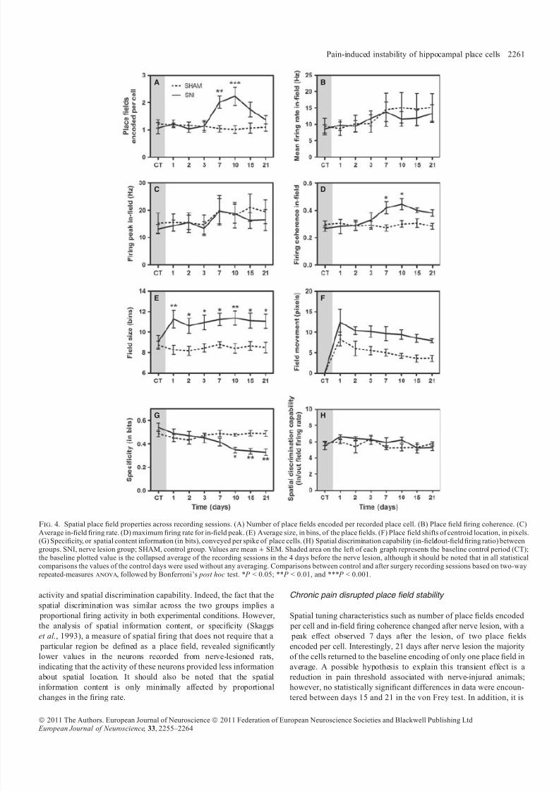
CA1 pyramidal cells of the SNI group showed a significant increase in
the number of encoded place fields. This increase was observed on a
cell-by-cell basis and did not affect all cells simultaneously recorded in
the same animal (Fig. 3). Repeated-measures anova revealed that
there were differences between the two groups ( F 7,70 = 5.87,
P < 0.0001) and across time ( F 7,70 = 3.27, P = 0.0460). Moreover,
post hoc analysis showed that the number of place fields in the SNI
group was larger than in SHAM control animals (Bonferroni, P < 0.01
for day 7 after SNI and P < 0.001 for day 10 after SNI; Fig. 4A).
A two-factor repeated-measures anova was used to compare the in-
field firing activity of cells from SHAM and lesion animals in pre- and
post-surgery recording sessions. There was no significant group effect
for the place cells’ in-field mean firing rate ( F 7,70 = 1.03, P = 0.4201)
or in-field peak of firing ( F 7,70 = 0.66, P = 0.7059; Fig. 4B and C).
However, over the recording sessions there was a significant effect of
time in the in-field mean firing rate ( F 7,70 = 6.99, P < 0.0001) and in-
field peak of firing ( F 7,70 = 4.26, P = 0.0006).The recordings in the SNI and SHAM groups show that the internal
place field coherence of place cells was affected after peripheral nerve
injury with a significant increase after 7 days of lesion (Fig. 4D).
Analysis revealed that there were differences between groups (anova-
RM, F 7,70 = 5.45, P < 0.0001) and across the recording sessions
(anova-RM, F 7,70 = 4.74, P = 0.0002). Post hoc analysis showed
that values of field coherence after nerve injury were greater than those
observed in the SHAM group (days 7 and 10; Bonferroni, P < 0.05).
There were also differences in the field size between the two
experimental groups (anova-RM, F 7,70 = 3.57, P = 0.0025) and
across the recording sessions (anova-RM, F 7,70 = 2.63, P = 0.0179;
Fig. 4E). Post hoc analysis of field size revealed a significant increase
for SNI group after nerve injury. However, no group interaction was
found for field centroid movement (anova-RM, F 7,70 = 1.38,
A B
C D
Fig. 2. Behavioural performance in the runway alternation task. (A) Level of sensitivity to mechanical stimulation evaluated using von Frey filaments. A largedecrease was observed in the threshold required to induce a paw response in the SNI group. (B) Number of correct alternations on the U-shaped maze. Similar levelsof behavioural task activity were observed for the two experimental groups. (C) The proportion of recorded neurons that were classified as place cells showed a similar distribution in the two experimental groups. Note that only neurons classified as place cells were considered in the analyses of the present study. (D) Averagefiring rate per place cell during the entire session remained unchanged after surgery. SNI, nerve lesion group; SHAM, control group. Values are mean ± SEM.Shaded area on the left of each graph represents the baseline control period (CT); the baseline plotted value is the collapsed average of the recording sessions in the4 days before the nerve lesion, although it should be noted that in all statistical comparisons the values of the control days were used without any averaging.Comparisons between control and after surgery recording sessions based on two-way repeated-measures anova, followed by Bonferroni’s post hoc test.
Pain-induced instability of hippocampal place cells 2259
ª 2011 The Authors. European Journal of Neuroscience ª 2011 Federation of European Neuroscience Societies and Blackwell Publishing Ltd European Journal of Neuroscience, 33, 2255–2264

8/2/2019 2011 - Cardoso-Cruz - EJN - Place Cells - Final
http://slidepdf.com/reader/full/2011-cardoso-cruz-ejn-place-cells-final 6/10
P = 0.2275), although both groups presented changes over time
(anova-RM, F 7,70 = 13.10, P < 0.0001; Fig. 4F).
Spatial information content The information content (bits ⁄ spike) of cells recorded from peripheral
nerve injury animals (SNI group) was significantly lower than that of
cells from SHAM control animals (Fig. 4G). anova-RM analysis
revealed there was a difference between groups ( F 7,70 = 16.06,
P < 0.0001) and across the recording sessions ( F 7,70 = 10.44,
P < 0.0001). Post hoc analysis revealed a significant decrease in
information content encoded by the cells of the SNI group 10 days
after nerve injury when compared with SHAM group (Bonferroni,
P < 0.05).
Spatial discrimination capability
Spatial discrimination capability, or the ratio between the mean firingrate inside the place field and the mean firing rate outside the place
field, is shown in Fig. 4H. The results indicate that spatial discrim-
ination did not differ between experimental groups (anova-RM,
F 7,70 = 1.13, P = 0.3565) or over recording sessions (anova-RM,
F 7,70 = 1.79, P = 0.1037).
Discussion
The aim of the present study was to address for the first time whether
the onset of an animal model of the chronic neuropathic pain condition
affects the spatial encoding properties of hippocampal CA1 pyramidal
cells. This type of cell is considered to be crucial for the continuously
updated representation of space and individual position. However, this
dynamic information is only one of several features stored in the
hippocampal network (Eichenbaum et al., 1999; Leutgeb et al., 2005),
and for this reason it is of primary interest to determine what factors,
including pain, may contribute to disruption of the stability of place
cells.
Our results show that pain causes instability in hippocampal placefield encoding in the absence of changes in overall task performance.
As intended, the onset of the pain model caused a transient reduction
in task performance only in the days immediately following the
surgery and this effect was equally observed in both control and pain
animals. We specifically used this simple alternation task because it is
not cognitively challenging and our results are in agreement with
previous reports showing that pain has no impact on performance in
simple spatial and nonspatial memory tasks (Apkarian et al., 2004;
LaBuda & Fuchs, 2000; Leite-Almeida et al., 2009), although pain-
related memory deficits may be observed in more complex memory
tasks (Dick & Rashiq, 2007; Millecamps et al., 2004; Leite-Almeida
et al., 2009). Moreover, the transient effect in performance (Fig. 2B)
is not temporally correlated with the peak of place field instability
(Fig. 4A), suggesting that the late onset instability of CA1 place fieldsis not caused by motor impairment or reduced motivation for task
completion.
It is important to note that, although the task used in this study is not
strictly hippocampus-dependent, several single-unit recording studies
have also used tasks which are not hippocampus-dependent, such as
forced-choice tasks, to examine place field characteristics, showing
that place cells present environment re-mapping even on nonhippo-
campal tasks (Markus et al., 1995; Muller & Kubie, 1987; Ranck,
1973).
Chronic pain changed CA1 place cell activity
Basic firing properties of CA1 place cells remained stable after
peripheral nerve lesion. These properties included mean firing rate
Fig. 3. Differential effect of pain over the remapping of place fields. The top two rows show the normalized firing rate per bin of two simultaneously recordedhippocampal place cells during the alternation task in the U-shaped runway. Note the similarity in the spatial firing activity of neuron 1 across recording sessions, andthe encoding instability of neuron 2 in the days following the nerve lesion. The bottom row shows the time spent in each bin during the entire recording session, anddemonstrates that the place field locations are commonly unrelated to the movement of the animal. Bright red represents bins with higher firing rate (top two rows) or where the animal spent more time (bottom row), and dark blue represent bins with lower firing rates (top two rows) or where the animal spent less time during thesession (bottom row). The shaded plot on the left of each row represents the baseline control period; the baseline plotted values are the collapsed average of theactivity in the 4 days before the nerve lesion.
2260 H. Cardoso-Cruz et al.
ª 2011 The Authors. European Journal of Neuroscience ª 2011 Federation of European Neuroscience Societies and Blackwell Publishing Ltd European Journal of Neuroscience, 33, 2255–2264
8/2/2019 2011 - Cardoso-Cruz - EJN - Place Cells - Final
http://slidepdf.com/reader/full/2011-cardoso-cruz-ejn-place-cells-final 7/10
activity and spatial discrimination capability. Indeed, the fact that the
spatial discrimination was similar across the two groups implies a
proportional firing activity in both experimental conditions. However,
the analysis of spatial information content, or specificity (Skaggs
et al., 1993), a measure of spatial firing that does not require that a
particular region be defined as a place field, revealed significantly
lower values in the neurons recorded from nerve-lesioned rats,
indicating that the activity of these neurons provided less information
about spatial location. It should also be noted that the spatial
information content is only minimally affected by proportional
changes in the firing rate.
Chronic pain disrupted place field stability
Spatial tuning characteristics such as number of place fields encoded
per cell and in-field firing coherence changed after nerve lesion, with a
peak effect observed 7 days after the lesion, of two place fields
encoded per cell. Interestingly, 21 days after nerve lesion the majority
of the cells returned to the baseline encoding of only one place field in
average. A possible hypothesis to explain this transient effect is a
reduction in pain threshold associated with nerve-injured animals;
however, no statistically significant differences in data were encoun-
tered between days 15 and 21 in the von Frey test. In addition, it is
A B
C D
E F
G H
Fig. 4. Spatial place field properties across recording sessions. (A) Number of place fields encoded per recorded place cell. (B) Place field firing coherence. (C)
Average in-field firing rate. (D) maximum firing rate for in-field peak. (E) Average size, in bins, of the place fields. (F) Place field shifts of centroid location, in pixels.(G) Specificity, or spatial content information (in bits), conveyed per spike of place cells. (H) Spatial discrimination capability (in-field⁄ out-field firing ratio) betweengroups. SNI, nerve lesion group; SHAM, control group. Values are mean ± SEM. Shaded area on the left of each graph represents the baseline control period (CT);the baseline plotted value is the collapsed average of the recording sessions in the 4 days before the nerve lesion, although it should be noted that in all statisticalcomparisons the values of the control days were used without any averaging. Comparisons between control and after surgery recording sessions based on two-wayrepeated-measures anova, followed by Bonferroni’s post hoc test. * P < 0.05; ** P < 0.01, and *** P < 0.001.
Pain-induced instability of hippocampal place cells 2261
ª 2011 The Authors. European Journal of Neuroscience ª 2011 Federation of European Neuroscience Societies and Blackwell Publishing Ltd European Journal of Neuroscience, 33, 2255–2264

8/2/2019 2011 - Cardoso-Cruz - EJN - Place Cells - Final
http://slidepdf.com/reader/full/2011-cardoso-cruz-ejn-place-cells-final 8/10
important to note that the second place field occupied a location which
was not adjacent to the first one; this suggests that pain-induced field
remapping and field expansion are different phenomena. It has been
reported that place fields may be modified during a single recording
session, and this may represent the within-session continuous
acquisition of novel spatial information (Mehta et al., 1997, 2000).
This hypothesis does not apply to our results because we only startedthe control recording sessions after several training sessions, and
during all control recording sessions the properties of place field
remained stable (see Supporting Information Table S1).
Mechanisms of place field instability
Apart from studies involving lesioning of the hippocampus or
hippocampus-connected areas (McNaughton et al., 1989; Muir &
Bilkey, 2001), to our best knowledge no previous studies have shown
disruption of place fields in the absence of changes to the testing
environment. Stressful stimuli were shown to alter the in-field firing
rate stability of place cells but not the stability of the field’s location
within a familiar environment (Kim et al., 2007), although the data presented by the authors suggest a shift in pre- vs. post-stress location.
A similar study has shown that the stability of place fields in a familiar
environment does not change across the estrous cycle (Tropp et al.,
2005).
Several studies have shown that place field instability is accompa-
nied by an overall increase in the size of preexisting place fields, and it
has been reported that this size expansion is diminished in aged rats
(Barnes et al., 1997; Shen et al., 1997) and abolished by selective
blockade of NMDA receptors (Ekstrom et al., 2001). Moreover,
NMDA-dependent long-term potentiation processes are important for
maintaining the stability of place fields (Kentros et al., 1998; Shapiro
& Eichenbaum, 1999).
Only incomplete and sometimes conflicting data exist on the
molecular mechanisms of interplay between pain and hippocampal plasticity; it has been shown that chronic neuropathy reduces CA1
long-term potentiation (Kodama et al., 2007; Ren et al., 2011) while
the opposite effect has been described after acute peripheral injection
of formalin (Zhao et al., 2009). In addition, recent studies have shown
that chronic pain reduces the hippocampal levels of BDNF (Duric &
McCarson, 2005; Hu et al., 2010; Al-Amin et al., 2011), which is
known to be a key regulator of hippocampal synaptic plasticity
(Minichiello, 2009). It must be noted that to our best knowledge no
direct connection has been demonstrated between neurotrophin levels
in the hippocampus and the stability of place cells, but it is expected
that the modulation of molecules important for synaptic plasticity also
leads to changes in the circuitry of hippocampal spatial encoding.
Finally, it has been proposed that hippocampal remapping mayresult from memory interference between concurrent sets of experi-
ences (Colgin et al., 2008). This is in agreement with the idea that
evoked or spontaneous pain perception causes an interference with
ongoing cognitive functions (Seminowicz & Davis, 2007; Moriarty
et al., 2011), disrupting the attentional processes that are crucial for
learning and memory (Boyette-Davis et al., 2008; Pais-Vieira et al.,
2009b).
Conclusion
In summary, our data suggest that peripheral nerve injury (SNI)
induces a relative instability of hippocampal CA1 place cells’ spatial
features. The present data indicate that nerve lesion induces a clear
reduction in the speciality measure, indicating that place cells provided
less information about spatial location after lesion. Our findings also
demonstrate place field disturbances, namely in the number, size and
in-field firing coherence. These changes are probably caused by
hippocampal structural adaptive mechanisms that occur during the
onset of the painful condition, which may disturb the mnemonic
processes that rely on the integration and consolidation of spatial
reference memory.
Supporting Information
Additional supporting information may be found in the online version
of this article:
Fig. S1. Stability of waveform shapes of two hippocampal place
cells simultaneously recorded from the same channel (yellow and
green) across experimental sessions (A). Note that the waveform
shape of each place cell remained stable throughout the recording
sessions. Only units with a > 3 : 1 signal to noise rate were
considered. (B) Illustration of the Unit A firing activity recorded
from a rat running on the U-shaped task encoding a spatial place
field (area with peak of firing). Maximum firing rate is indicated by
red and occupancy with no firing by blue. (C) Offline analysis of 3-
D PC cluster stability from the channel shown across the whole
recording sessions using the WaveTracker software (Plexon Inc.,
Dallas, TX, USA). In this view (D), 2-D PC clusters are projected as
function of time ( Z -axis). Stability across time and absence of
overlap between units isolated from the same channel were used as
extra-selection criteria. (E) Location of implanted multielectrode
arrays for nine rats used in this study. The black dots indicate the
location of the centre of the array in CA1 region.
Table S1. Statistical summary for SHAM and SNI-group comparison
across all measurements during the eight recording sessions of the
control period.
Please note: As a service to our authors and readers, this journal
provides supporting information supplied by the authors. Suchmaterials are peer-reviewed and may be re-organized for online
delivery, but are not copy-edited or typeset by Wiley-Blackwell.
Technical support issues arising from supporting information (other
than missing files) should be addressed to the authors.
Acknowledgements
This work was supported by grants from the Portuguese Foundation
for Science and Technology – FCT: FCT SFRH ⁄ 42500 ⁄ 2007, FCT
PTDC ⁄ SAU-NEU ⁄ 100733 ⁄ 2008; and BIAL Foundation: BIAL Pro-
ject 126 ⁄ 08.
Abbreviations
SNI, spared nerve injury.
Conflict of interest
The authors do not have any conflicts of interest.
References
Aguiar, P., Mendonca, L. & Galhardo, V. (2007) OpenControl: a freeopensource software for video tracking and automated control of behavioralmazes. J. Neurosci. Methods, 166, 66–72.
Al-Amin, H., Sarkis, R., Atweh, S., Jabbur, S. & Saade, N. (2011) Chronicdizocilpine or apomorphine and development of neuropathy in two animal
2262 H. Cardoso-Cruz et al.
ª 2011 The Authors. European Journal of Neuroscience ª 2011 Federation of European Neuroscience Societies and Blackwell Publishing Ltd European Journal of Neuroscience, 33, 2255–2264
8/2/2019 2011 - Cardoso-Cruz - EJN - Place Cells - Final
http://slidepdf.com/reader/full/2011-cardoso-cruz-ejn-place-cells-final 9/10
models II: effects on brain cytokines and neurotrophins. Exp. Neurol., 228,30–40.
Apkarian, A.V., Sosa, Y., Krauss, B.R., Thomas, P.S., Fredrickson, B.E., Levy,R.E., Harden, R.N. & Chialvo, D.R. (2004) Chronic pain patients areimpaired on an emotional decision-making task. Pain, 108, 129–136.
Barnes, C.A., Suster, M.S., Shen, J.M. & McNaughton, B.L. (1997)Multistability of cognitive maps in the hippocampus of old rats. Nature,388, 272–275.
Bingel, U., Quante, M., Knab, R., Bromm, B., Weiller, C. & Buchel, C. (2002)Subcortical structures involved in pain processing: evidence from single-trialfMR1. Pain, 99, 313–321.
Boyette-Davis, J.A., Thompson, C.D. & Fuchs, P.N. (2008) Alterations inattentional mechanisms in response to acute inflammatory pain and morphineadministration. Neuroscience, 151, 558–563.
Carter, J.L., Lubahn, C., Lorton, D., Osredkar, T., Der, T.C., Schaller, J.,Evelsizer, S., Flowers, S., Ruff, N., Reese, B. & Bellinger, D.L. (2011)Adjuvant-induced arthritis induces c-Fos chronically in neurons in thehippocampus. J. Neuroimmunol., 230, 85–94.
Ceccarelli, I., Scaramuzzino, A., Massafra, C. & Aloisi, A.M. (2003) The
behavioral and neuronal effects induced by repetitive nociceptive stimulationare affected by gonadal hormones in male rats. Pain, 104, 35–47.
Chaplan, S.R., Bach, F.W., Pogrel, J.W., Chung, J.M. & Yaksh, T.L. (1994)Quantitative assessment of tactile allodynia in the rat paw. J. Neurosci.
Methods, 53, 55–63.
Colgin, L.L., Moser, E.I. & Moser, M.B. (2008) Understanding memorythrough hippocampal remapping. Trends Neurosci., 31, 469–477.Decosterd, I. & Woolf, C.J. (2000) Spared nerve injury: an animal model of
persistent peripheral neuropathic pain. Pain, 87, 149–158.Delgado, J.M.R. (1955) Cerebral structures involved in transmission and
elaboration of noxious stimulation. J. Neurophysiol., 18, 261–275.Dick, B.D. & Rashiq, S. (2007) Disruption of attention and working memory
traces in individuals with chronic pain. Anesth. Analg., 104, 1223–1229.
Dragoi, G., Harris, K.D. & Buzsaki, G. (2003) Place representation withinhippocampal networks is modified by long-term potentiation. Neuron, 39,843–853.
Duric, V. & McCarson, K.E. (2005) Hippocampal neurokinin-1 receptor and brain-derived neurotrophic factor gene expression is decreased in rat modelsof pain and stress. Neuroscience, 133, 999–1006.
Eichenbaum, H., Dudchenko, P., Wood, E., Shapiro, M. & Tanila, H. (1999)The hippocampus, memory, and place cells: is it spatial memory or a memory space? Neuron, 23, 209–226.
Ekstrom, A.D., Meltzer, J., McNaughton, B.L. & Barnes, C.A. (2001) NMDAreceptor antagonism blocks experience-dependent expansion of hippocampal‘‘place fields’’. Neuron, 31, 631–638.
Fox, S.E. & Ranck, J.B. Jr (1981) Electrophysiological characteristics of
hippocampal complex-spikecells and thetacells. Exp.Brain Res.,41,399–410.Gol, A. & Faibish, G.M. (1967) Effects of human hippocampal ablation.
J. Neurosurg., 26, 390–398.Halgren, E., Walter, R.D., Cherlow, D.G. & Crandall, P.H. (1978) Mental
phenomena evoked by electrical-stimulation of human hippocampal forma-tion and amygdala. Brain, 101, 83–117.
Hu, Y., Yang, J., Hu, Y., Wang, Y. & Li, W. (2010) Amitriptyline rather thanlornoxicam ameliorates neuropathic pain-induced deficits in abilities of spatial learning and memory. Eur. J. Anaesthesiol., 27, 162–168.
Kentros, C., Hargreaves, E., Hawkins, R.D., Kandel, E.R., Shapiro, M. &Muller, R.V. (1998) Abolition of long-term stability of new hippocampal
place cell maps by NMDA receptor blockade. Science, 280, 2121–2126.Khanna, S. (1997) Dorsal hippocampus field CA1 pyramidal cell responses to a
persistent versus an acute nociceptive stimulus and their septal modulation. Neuroscience, 77, 713–721.
Khanna, S. & Sinclair, J. (1992) Responses in the CA1 region of the rat hippocampus to a noxious stimulus. Exp. Neurol., 117, 28–35.
Kim, J., Lee, H., Welday, A., Song, E., Cho, J., Sharp, P., Jung, M. & Blair, H.(2007) Stress-induced alterations in hippocampal plasticity, place cells, andspatial memory. Proc. Natl. Acad. Sci. USA, 104, 18297–18302.
Kobayashi, T., Nishijo, H., Fukuda, M., Bures, J. & Ono, T. (1997) Task-dependent representations in rat hippocampal place neurons. J. Neurophys-iol., 78, 597–613.
Kodama, D., Ono, H. & Tanabe, M. (2007) Altered hippocampal long-term potentiation after peripheral nerve injury in mice. Eur. J. Pharmacol., 574,127–132.
LaBuda, C.J. & Fuchs, P.N. (2000) A behavioral test paradigm to measure theaversive quality of inflammatory and neuropathic pain in rats. Exp. Neurol.,163, 490–494.
Leite-Almeida, H., Almeida-Torres, L., Mesquita, A.R., Pertovaara, A., Sousa, N., Cerqueira, J.J. & Almeida, A. (2009) The impact of age on emotional andcognitive behaviours triggered by experimental neuropathy in rats. Pain,144, 57–65.
Leutgeb, S. & Mizumori, S.J.Y. (1999) Excitotoxic septal lesions result inspatial memory deficits and altered flexibility of hippocampal single-unit representations. J. Neurosci., 19, 6661–6672.
Leutgeb, S., Letugeb, J.K., Barnes, C.A., Moser, E.I., McNaughton, B.L. &
Moser, M.B. (2005) Independent codes for spatial and episodic memory inhippocampal neuronal ensembles. Science, 309, 619–623.
Liu, X., Muller, R.U., Huang, L.T., Kubie, J.L., Rotenberg, A., Rivard, B.,Cilio, M.R. & Holmes, G.L. (2003) Seizure-induced changes in place cell
physiology: relationship to spatial memory. J. Neurosci., 23, 11505–11515.Lutz, J., Jager, L., de Quervain, D., Krauseneck, T., Padberg, F., Wichnalek, M.,
Beyer, A., Stahl, R., Zirngibl, B., Morhard, D., Reiser, M. & Schelling, G.(2008) White and gray matter abnormalities in the brain of patients withfibromyalgia: a diffusion-tensor and volumetric imaging study. Arthritis
Rheum., 58, 3960–3969.Markus, E.J., Barnes, C.A., McNaughton, B.L., Gladden, V.L. & Skaggs, W.E.
(1994) Spatial information content and reliability of hippocampal CA1neurons: effects of visual input. Hippocampus, 4, 410–421.
Markus, E.J., Qin, Y.L., Leonard, B., Skaggs, W.E., McNaughton, B.L. &Barnes, C.A. (1995) Interactions between location and task affect the spatialand directional firing of hippocampal neurons. J. Neurosci., 15, 7079–7094.
McKenna, J.E. & Melzack, R. (1992) Analgesia produced by lidocainemicroinjection into the dentate gyrus. Pain, 49, 105–112.McKenna, J.E. & Melzack, R. (2001) Blocking NMDA receptors in the
hippocampal dentate gyrus with AP5 produces analgesia in the formalin paintest. Exp. Neurol., 172, 92–99.
McNaughton, B.L., Barnes, C.A., Meltzer, J. & Sutherland, R.J. (1989)Hippocampal granule cells are necessary for normal spatial learning but not for spatially-selective pyramidal cell discharge. Exp. Brain Res., 76, 485–
496.Mehta, M.R., Barnes, C.A. & McNaughton, B.L. (1997) Experience-depen-
dent, asymmetric expansion of hippocampal place fields. Proc. Natl. Acad.Sci. U S A, 94, 8918–8921.
Mehta, M.R., Quirk, M.C. & Wilson, M.A. (2000) Experience-dependent asymmetric shape of hippocampal receptive fields. Neuron, 25, 707–715.
Millecamps, M., Etienne, M., Jourdan, D., Eschalier, A. & Ardid, D. (2004)Decrease in non-selective, non-sustained attention induced by a chronicvisceral inflammatory state as a new pain evaluation in rats. Pain, 109, 214–
224.Minichiello, L. (2009) TrkB signalling pathways in LTP and learning. Nat. Rev.
Neurosci., 10, 850–860.Mizumori, S., Miya, D. & Ward, K. (1994) Reversible inactivation of the lateral
dorsal thalamus disrupts hippocampal place representation and impairsspatial learning. Brain Res., 644, 168–174.
Moita, M.A., Rosis, S., Zhou, Y., LeDoux, J.E. & Blair, H.T. (2004) Puttingfear in its place: remapping of hippocampal place cells during fear conditioning. J. Neurosci., 24, 7015–7023.
Moriarty, O., McGuireb, B.E. & Finn, D.P. (2011) The effect of pain oncognitive function: a review of clinical and preclinical research. Prog.
Neurobiol., 93, 385–404.Muir, G. & Bilkey, D. (2001) Instability in the place field location of
hippocampal place cells after lesions centered on the perirhinal cortex. J. Neurosci., 21, 4016–4025.
Muller, R.U. & Kubie, J.L. (1987) The effects of changes in the environment onthe spatial firing of hippocampal complex-spike cells. J. Neurosci., 7, 1951– 1968.
Muller, R.U. & Kubie, J.L. (1989) The firing of hippocampal placecells predicts the future position of freely moving rats. J. Neurosci., 9,4101–4110.
Muller, R.U., Kubie, J.L. & Ranck, J.B. Jr (1987) Spatial firing patterns of hippocampal complex-spike cells in a fixed environment. J. Neurosci., 7,1935–1950.
O’ Keefe, J. & Dostrovsky, J. (1971) Hippocampus as a spatial map – preliminary evidencefrom unitactivity in freely-moving rat. Brain Res., 34, 5.
O’ Keefe, J. & Nadel, L. (1978) The hippocampus as a cognitive map.Clarendon, Oxford.
Pais-Vieira, M., Mendes-Pinto, M.M., Lima, D. & Galhardo, V. (2009a)Cognitive impairment of prefrontal-dependent decision-making in rats after the onset of chronic pain. Neuroscience, 161, 671–679.
Pais-Vieira, M., Lima, D. & Galhardo, V. (2009b) Sustained attention deficits inrats with chronic inflammatory pain. Neurosci. Lett., 463, 98–102.
Pain-induced instability of hippocampal place cells 2263
ª 2011 The Authors. European Journal of Neuroscience ª 2011 Federation of European Neuroscience Societies and Blackwell Publishing Ltd European Journal of Neuroscience, 33, 2255–2264

8/2/2019 2011 - Cardoso-Cruz - EJN - Place Cells - Final
http://slidepdf.com/reader/full/2011-cardoso-cruz-ejn-place-cells-final 10/10
Paxinos, G. & Watson, C. (1998) The Rat Brain in Stereotaxic Coordinates.Academic Press, San Diego.
Ploghaus, A., Tracey, I., Clare, S., Gati, J.S., Rawlins, J.N.P. & Matthews, P.M.(2000) Learning about pain: the neural substrate of the prediction error for aversive events. Proc. Natl Acad. Sci. USA, 97, 9281–9286.
Ploghaus, A., Narain, C., Beckmann, C.F., Clare, S., Bantick, S., Wise, R.,Matthews, P.M., Rawlins, J.N.P. & Tracey, I. (2001) Exacerbation of pain byanxiety is associated with activity in a hippocampal network. J. Neurosci.,
21, 9896–9903.Ranck, J.B. Jr (1973) Studies on single neurons in dorsal hippocampal
formation and septum in unrestrained rats. I. Behavioral correlates and firingrepertoires. Exp. Neurol., 41, 461–531.
Ren, W.J., Liu, Y., Zhou, L.J., Li, W., Zhong, Y., Pang, R.P., Xin, W.J., Wei,X.H., Wang, J., Zhu, H.Q., Wu, C.Y., Qin, Z.H., Liu, G. & Liu, X.G. (2011)Peripheral nerve injury leads to working memory deficits and dysfunction of the hippocampus by upregulation of TNF-a in rodents. Neuropsychophar-macology, 36, 979–992.
Rotenberg, A., Abel, T., Hawkins, R.D., Kandel, E.R. & Muller, R.U.(2000) Parallel instabilities of long-term potentiation, place cells, and
learning caused by decreased protein kinase A activity. J. Neurosci., 20,8096–8102.
Seminowicz, D.A. & Davis, K.D. (2007) Interactions of pain intensity andcognitive load: the brain stays on task. Cereb. Cortex, 17, 1412–1422.
Shapiro, M.L. & Eichenbaum, H. (1999) Hippocampus as a memory map:
synaptic plasticity and memory encoding by hippocampal neurons. Hippo-campus, 9, 365–384.Shen, J., Barnes, C.A., McNaughton, B.L., Skaggs, W.E. & Weaver, K.L.
(1997) The effect of aging on experience-dependent plasticity of hippocam- pal place cells. J. Neurosci., 17, 6769–6782.
Silva, A., Cardoso-Cruz, H., Silva, F., Galhardo, V. & Antunes, L. (2010)Comparison of anesthetic depth indexes based on thalamocortical local field
potentials in rats. Anesthesiology, 112, 355–363.
Skaggs, W.E. & McNaughton, B.L. (1996) Replay of neuronal firing sequencesin rat hippocampus during sleep following spatial experience. Science, 271,1870–1873.
Skaggs, W.E., McNaughton, B.L., Gothard, K.M. & Markus, E.J. (1993) AnInformation-theoretic approach to deciphering the hippocampal code. InHanson, S.J., Cowan, J.D. & Giles, C.L. (Eds), Advances in neural information processing . Morgan Kaufmann, San Mateo, pp. 1030–1037.
Soleimannejad, E., Semnanian, S., Fathollahi, Y. & Naghdi, N. (2006)Microinjection of ritanserin into the dorsal hippocampal CA1 and dentategyrus decrease nociceptive behavior in adult male rat. Behav. Brain Res.,
168, 221–225.
Tai, S.K., Huang, F.D., Moochhala, S. & Khanna, S. (2006) Hippocampal theta state in relation to formalin nociception. Pain, 121, 29–42.
Thompson, L.T. & Best, P.J. (1990) Long-term stability of the place-fieldactivity of single units recorded from the dorsal hippocampus of freely
behaving rats. Brain Res., 509, 299–308.Tropp, J., Figueiredo, C.M. & Markus, E.J. (2005) Stability of hippocam-
pal place cell activity across the rat estrous cycle. Hippocampus, 15,154–165.
Wei, F., Xu, Z.C., Qu, Z., Milbrandt, J. & Zhuo, M. (2000) Role of EGR1 inhippocampal synaptic enhancement induced by tetanic stimulation andamputation. J. Cell Biol., 149, 1325–1333.
Wood, E.R., Dudchenko, P.A., Robitsek, R.J. & Eichenbaum, H. (2000)Hippocampal neurons encode information about different types of memoryepisodes occurring in the same location. Neuron, 27, 623–633.
Younger, J.W., Shen, Y.F., Goddard, G. & Mackey, S.C. (2010) Chronicmyofascial temporomandibular pain is associated with neural abnormalities
in the trigeminal and limbic systems. Pain,149
, 222–228.Zhao, X.Y., Liu, M.G., Yuan, D.L., Wang, Y., He, Y., Wang, D.D., Chen, X.F.,Zhang, F.K., Li, H., He, X.S. & Chen, J. (2009) Nociception-induced spatialand temporal plasticity of synaptic connection and function in thehippocampal formation of rats: a multi-electrode array recording.Mol. Pain,5, 55.
Zheng, F. & Khanna, S. (2008) Intra-hippocampal tonic inhibition influencesformalin pain-induced pyramidal cell suppression, but not excitation in
dorsal field CA1 of rat. Brain Res. Bull., 77, 374–381.Zimmermann, M. (1983) Ethical guidelines for investigations of experimental
pain in conscious animals. Pain, 16, 109–110.
2264 H. Cardoso-Cruz et al.
ª 2011 The Authors. European Journal of Neuroscience ª 2011 Federation of European Neuroscience Societies and Blackwell Publishing Ltd European Journal of Neuroscience, 33, 2255–2264