5.1 5.2 membrane structure, synthesis, and transport...
TRANSCRIPT
98 CHAPTER 5
5.1 Membrane StructureLearning Outcomes:
1. Describe the fluid-mosaic model of membrane structure.2. Identify the three different types of membrane proteins.3. Explain the technique of freeze-fracture electron microscopy.
An important biological principle is that structure determines func-tion. Throughout this chapter, we will see how the structure of cel-lular membranes enables them to compartmentalize the cell while selectively importing and exporting vital substances. The two pri-mary components of membranes are phospholipids, which form the basic matrix of a membrane, and proteins, which are embedded in the membrane or loosely attached to its surface. A third component is carbohydrate, which may be attached to membrane lipids and pro-teins. In this section, we will be mainly concerned with the organiza-tion of these components to form a biological membrane and how they are important in the overall function of membranes.
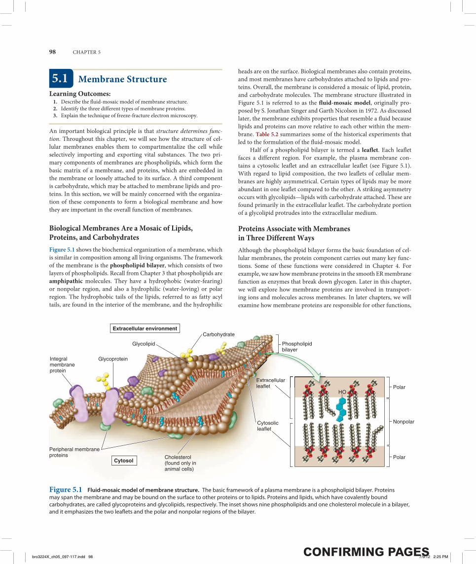
Biological Membranes Are a Mosaic of Lipids, Proteins, and CarbohydratesFigure 5.1 shows the biochemical organization of a membrane, which is similar in composition among all living organisms. The framework of the membrane is the phospholipid bilayer, which consists of two layers of phospholipids. Recall from Chapter 3 that phospholipids are amphipathic molecules. They have a hydrophobic (water-fearing) or nonpolar region, and also a hydrophilic (water-loving) or polar region. The hydrophobic tails of the lipids, referred to as fatty acyl tails, are found in the interior of the membrane, and the hydrophilic
heads are on the surface. Biological membranes also contain proteins, and most membranes have carbohydrates attached to lipids and pro-teins. Overall, the membrane is considered a mosaic of lipid, protein, and carbohydrate molecules. The membrane structure illustrated in Figure 5.1 is referred to as the fluid-mosaic model, originally pro-posed by S. Jonathan Singer and Garth Nicolson in 1972. As discussed later, the membrane exhibits properties that resemble a fluid because lipids and proteins can move relative to each other within the mem-brane. Table 5.2 summarizes some of the historical experiments that led to the formulation of the fluid-mosaic model.
Half of a phospholipid bilayer is termed a leaflet. Each leaflet faces a different region. For example, the plasma membrane con-tains a cytosolic leaflet and an extracellular leaflet (see Figure 5.1). With regard to lipid composition, the two leaflets of cellular mem-branes are highly asymmetrical. Certain types of lipids may be more abundant in one leaflet compared to the other. A striking asymmetry occurs with glycolipids—lipids with carbohydrate attached. These are found primarily in the extracellular leaflet. The carbohydrate portion of a glycolipid protrudes into the extracellular medium.
Proteins Associate with Membranes in Three Different WaysAlthough the phospholipid bilayer forms the basic foundation of cel-lular membranes, the protein component carries out many key func-tions. Some of these functions were considered in Chapter 4. For example, we saw how membrane proteins in the smooth ER membrane function as enzymes that break down glycogen. Later in this chapter, we will explore how membrane proteins are involved in transport-ing ions and molecules across membranes. In later chapters, we will examine how membrane proteins are responsible for other functions,
Extracellular environment
Cytosol
Integral membraneprotein
Glycoprotein
Glycolipid
Carbohydrate
Extracellularleaflet
Phospholipidbilayer
Cytosolicleaflet
Peripheral membraneproteins Cholesterol
(found only inanimal cells)
Polar
Nonpolar
Polar
HO
EEEExExExxttttrraacellulareeaaaaflflet
y
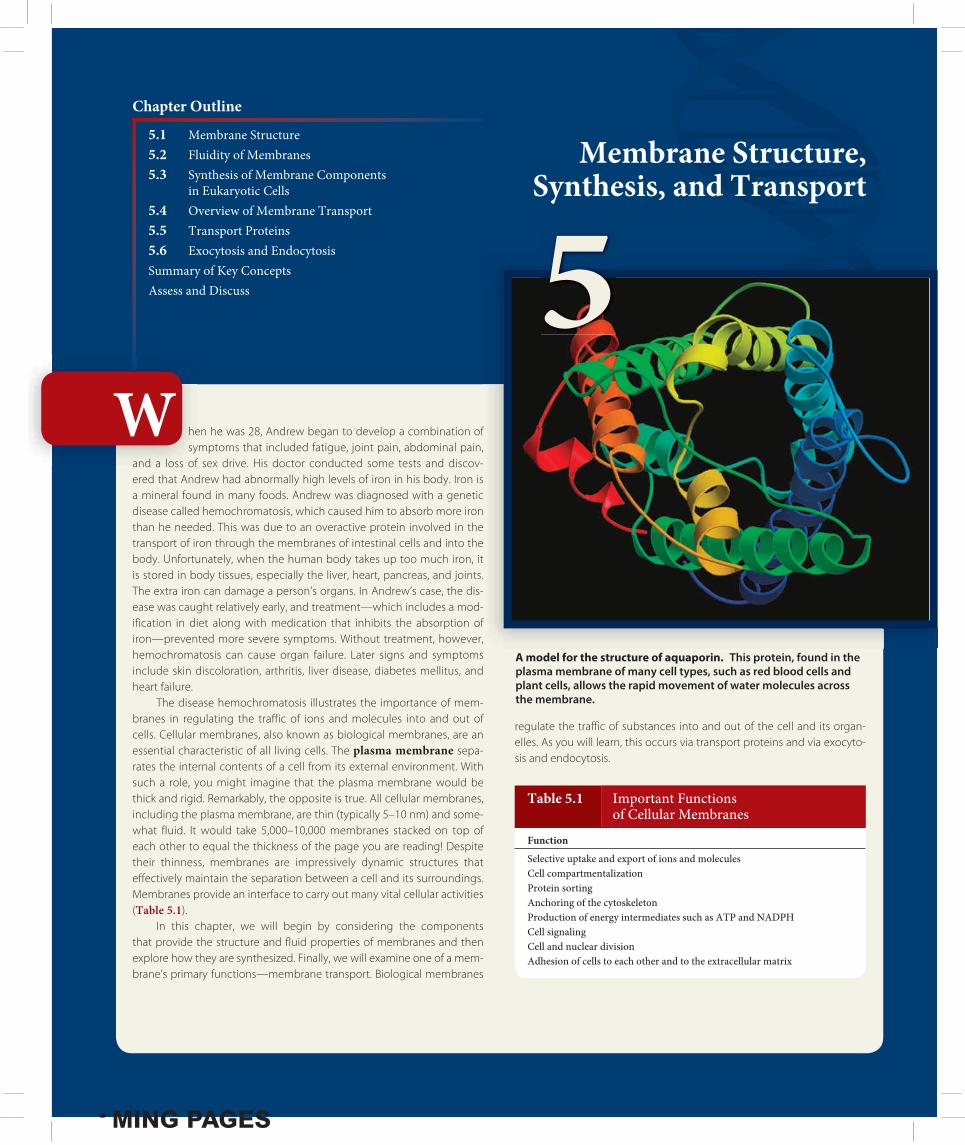
Figure 5.1 Fluid-mosaic model of membrane structure. The basic framework of a plasma membrane is a phospholipid bilayer. Proteins may span the membrane and may be bound on the surface to other proteins or to lipids. Proteins and lipids, which have covalently bound carbohydrates, are called glycoproteins and glycolipids, respectively. The inset shows nine phospholipids and one cholesterol molecule in a bilayer, and it emphasizes the two leaflets and the polar and nonpolar regions of the bilayer.
bro3224X_ch05_097-117.indd 98bro3224X_ch05_097-117.indd 98 7/9/12 2:25 PM7/9/12 2:25 PM
MEMBRANE STRUCTURE, SYNTHESIS, AND TRANSPORT 99
including ATP synthesis (Chapter 7), photosynthesis (Chapter 8), cell signaling (Chapter 9), and cell-to-cell adhesion (Chapter 10).
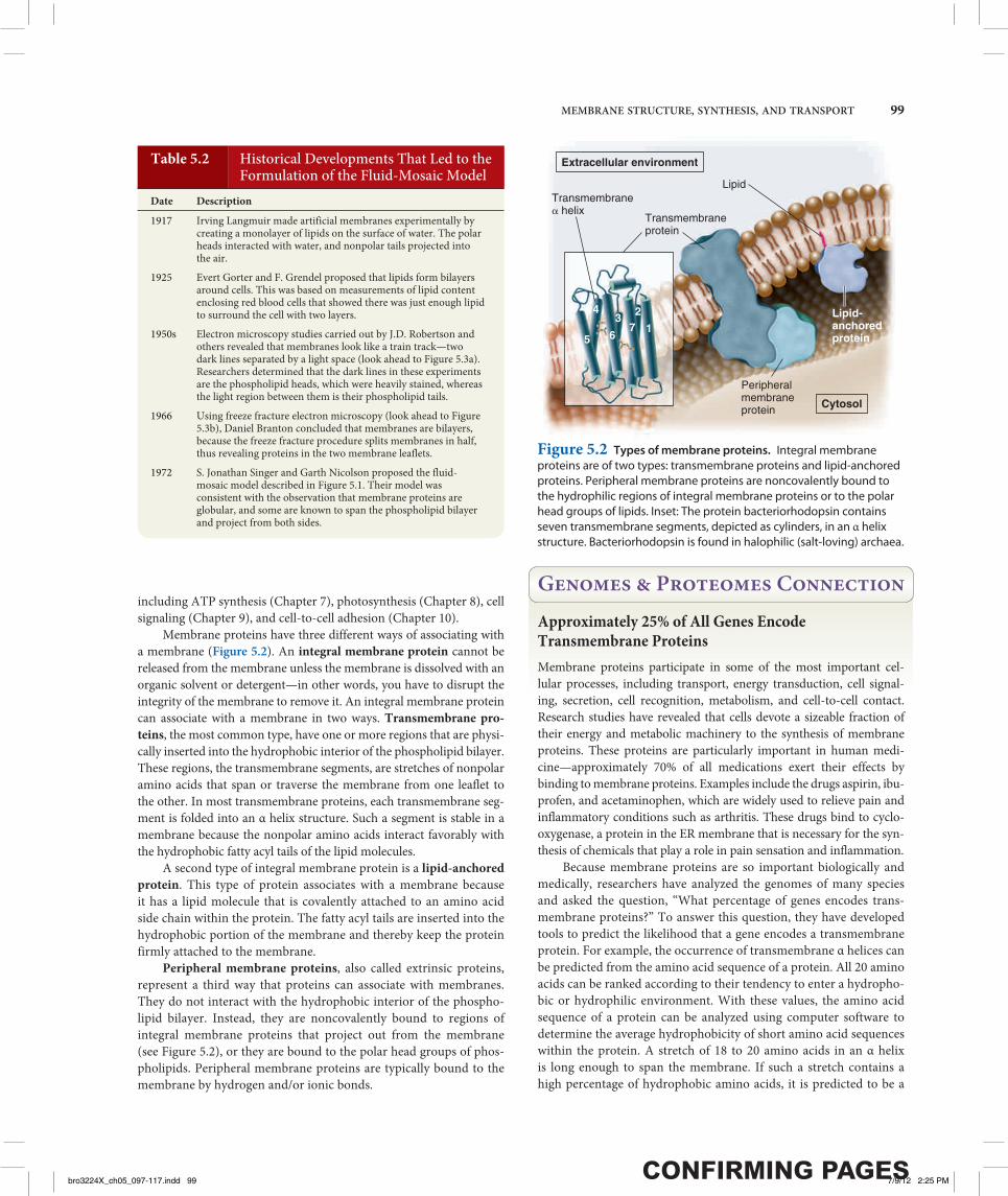
Membrane proteins have three different ways of associating with a membrane (Figure 5.2). An integral membrane protein cannot be released from the membrane unless the membrane is dissolved with an organic solvent or detergent—in other words, you have to disrupt the integrity of the membrane to remove it. An integral membrane protein can associate with a membrane in two ways. Transmembrane pro-teins, the most common type, have one or more regions that are physi-cally inserted into the hydrophobic interior of the phospholipid bilayer. These regions, the transmembrane segments, are stretches of nonpolar amino acids that span or traverse the membrane from one leaflet to the other. In most transmembrane proteins, each transmembrane seg-ment is folded into an α helix structure. Such a segment is stable in a membrane because the nonpolar amino acids interact favorably with the hydrophobic fatty acyl tails of the lipid molecules.
A second type of integral membrane protein is a lipid-anchored protein. This type of protein associates with a membrane because it has a lipid molecule that is covalently attached to an amino acid side chain within the protein. The fatty acyl tails are inserted into the hydrophobic portion of the membrane and thereby keep the protein firmly attached to the membrane.
Peripheral membrane proteins, also called extrinsic proteins, represent a third way that proteins can associate with membranes. They do not interact with the hydrophobic interior of the phospho-lipid bilayer. Instead, they are noncovalently bound to regions of integral membrane proteins that project out from the membrane (see Figure 5.2), or they are bound to the polar head groups of phos-pholipids. Peripheral membrane proteins are typically bound to the membrane by hydrogen and/or ionic bonds.
Genomes & Proteomes ConnectionApproximately 25% of All Genes Encode Transmembrane ProteinsMembrane proteins participate in some of the most important cel-lular processes, including transport, energy transduction, cell signal-ing, secretion, cell recognition, metabolism, and cell-to-cell contact. Research studies have revealed that cells devote a sizeable fraction of their energy and metabolic machinery to the synthesis of membrane proteins. These proteins are particularly important in human medi-cine—approximately 70% of all medications exert their effects by binding to membrane proteins. Examples include the drugs aspirin, ibu-profen, and acetaminophen, which are widely used to relieve pain and inflammatory conditions such as arthritis. These drugs bind to cyclo-oxygenase, a protein in the ER membrane that is necessary for the syn-thesis of chemicals that play a role in pain sensation and inflammation. Because membrane proteins are so important biologically and medically, researchers have analyzed the genomes of many species and asked the question, “What percentage of genes encodes trans-membrane proteins?” To answer this question, they have developed tools to predict the likelihood that a gene encodes a transmembrane protein. For example, the occurrence of transmembrane α helices can be predicted from the amino acid sequence of a protein. All 20 amino acids can be ranked according to their tendency to enter a hydropho-bic or hydrophilic environment. With these values, the amino acid sequence of a protein can be analyzed using computer software to determine the average hydrophobicity of short amino acid sequences within the protein. A stretch of 18 to 20 amino acids in an α helix is long enough to span the membrane. If such a stretch contains a high percentage of hydrophobic amino acids, it is predicted to be a
Table 5.2 Historical Developments That Led to the Formulation of the Fluid-Mosaic Model
Date Description
1917 Irving Langmuir made artificial membranes experimentally by creating a monolayer of lipids on the surface of water. The polar heads interacted with water, and nonpolar tails projected into the air.
1925 Evert Gorter and F. Grendel proposed that lipids form bilayers around cells. This was based on measurements of lipid content enclosing red blood cells that showed there was just enough lipid to surround the cell with two layers.
1950s Electron microscopy studies carried out by J.D. Robertson and others revealed that membranes look like a train track—two dark lines separated by a light space (look ahead to Figure 5.3a). Researchers determined that the dark lines in these experiments are the phospholipid heads, which were heavily stained, whereas the light region between them is their phospholipid tails.
1966 Using freeze fracture electron microscopy (look ahead to Figure 5.3b), Daniel Branton concluded that membranes are bilayers, because the freeze fracture procedure splits membranes in half, thus revealing proteins in the two membrane leaflets.
1972 S. Jonathan Singer and Garth Nicolson proposed the fluid-mosaic model described in Figure 5.1. Their model was consistent with the observation that membrane proteins are globular, and some are known to span the phospholipid bilayer and project from both sides.
Transmembraneprotein
Lipid
Lipid-anchoredprotein
Peripheralmembraneprotein
Transmembrane� helix
172
6
34
5
Extracellular environment
Cytosol
Figure 5.2 Types of membrane proteins. Integral membrane proteins are of two types: transmembrane proteins and lipid-anchored proteins. Peripheral membrane proteins are noncovalently bound to the hydrophilic regions of integral membrane proteins or to the polar head groups of lipids. Inset: The protein bacteriorhodopsin contains seven transmembrane segments, depicted as cylinders, in an α helix structure. Bacteriorhodopsin is found in halophilic (salt-loving) archaea.
bro3224X_ch05_097-117.indd 99bro3224X_ch05_097-117.indd 99 7/9/12 2:25 PM7/9/12 2:25 PM
100 CHAPTER 5
transmembrane α helix. However, such computer predictions must eventually be verified by experimentation. Using a computer approach, many research groups have attempted to calculate the percentage of genes that encode trans-membrane proteins in various species. Table 5.3 shows the results of one such study. The estimated percentage of transmembrane proteins is substantial: 20–30% of all genes may encode transmembrane pro-teins. This trend is found throughout all domains of life, including archaea, bacteria, and eukaryotes. For example, about 30% of human genes encode transmembrane proteins. With a genome size of 20,000 to 25,000 different genes, the total number of genes that encode dif-ferent transmembrane proteins is estimated to be 6,000 to 7,500. The functions of many of them have yet to be determined. Identifying their functions will help researchers gain a better understanding of human biology. Likewise, medical researchers and pharmaceutical companies are interested in the identification of new transmembrane proteins that could be targets for effective new medications.
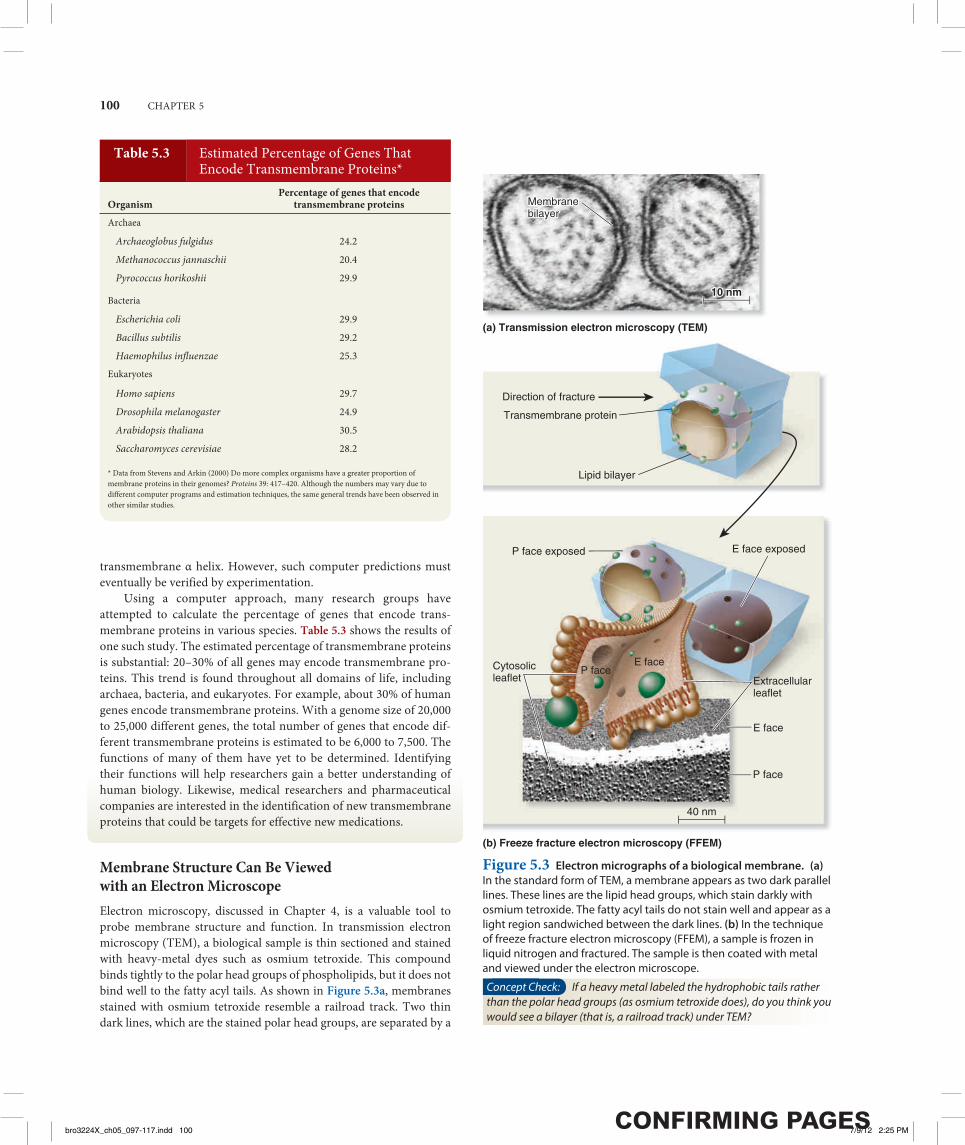
Membrane Structure Can Be Viewed with an Electron MicroscopeElectron microscopy, discussed in Chapter 4, is a valuable tool to probe membrane structure and function. In transmission electron microscopy (TEM), a biological sample is thin sectioned and stained with heavy-metal dyes such as osmium tetroxide. This compound binds tightly to the polar head groups of phospholipids, but it does not bind well to the fatty acyl tails. As shown in Figure 5.3a, membranes stained with osmium tetroxide resemble a railroad track. Two thin dark lines, which are the stained polar head groups, are separated by a
Table 5.3 Estimated Percentage of Genes That Encode Transmembrane Proteins*
OrganismPercentage of genes that encode
transmembrane proteinsArchaea
Archaeoglobus fulgidus 24.2
Methanococcus jannaschii 20.4
Pyrococcus horikoshii 29.9
Bacteria
Escherichia coli 29.9
Bacillus subtilis 29.2
Haemophilus influenzae 25.3Eukaryotes
Homo sapiens 29.7
Drosophila melanogaster 24.9
Arabidopsis thaliana 30.5
Saccharomyces cerevisiae 28.2
* Data from Stevens and Arkin (2000) Do more complex organisms have a greater proportion of membrane proteins in their genomes? Proteins 39: 417–420. Although the numbers may vary due to different computer programs and estimation techniques, the same general trends have been observed in other similar studies.
Membranebilayer
(b) Freeze fracture electron microscopy (FFEM)
Transmembrane protein
Lipid bilayer
Direction of fracture
P face exposed
P faceE face
E face exposed
E face
P face
Cytosolicleaflet Extracellular
leaflet
40 nm
(a) Transmission electron microscopy (TEM)
10 nm10 nm
Membranebilayer
Figure 5.3 Electron micrographs of a biological membrane. (a) In the standard form of TEM, a membrane appears as two dark parallel lines. These lines are the lipid head groups, which stain darkly with osmium tetroxide. The fatty acyl tails do not stain well and appear as a light region sandwiched between the dark lines. (b) In the technique of freeze fracture electron microscopy (FFEM), a sample is frozen in liquid nitrogen and fractured. The sample is then coated with metal and viewed under the electron microscope.
Concept Check: If a heavy metal labeled the hydrophobic tails rather than the polar head groups (as osmium tetroxide does), do you think you would see a bilayer (that is, a railroad track) under TEM?
bro3224X_ch05_097-117.indd 100bro3224X_ch05_097-117.indd 100 7/9/12 2:25 PM7/9/12 2:25 PM
MEMBRANE STRUCTURE, SYNTHESIS, AND TRANSPORT 101
uniform light space about 2 nm thick. This railroad track morphology is seen when cell membranes are subjected to electron microscopy.
A specialized form of TEM, freeze fracture electron microscopy (FFEM), is used to analyze the interiors of phospholipid bilayers. Russell Steere invented this method in 1957. In FFEM, a sample is frozen in liquid nitrogen and split with a knife (Figure 5.3b). The knife does not actually cut through the bilayer, but it fractures the frozen sample. Due to the weakness of the central membrane region, the leaflets separate into a P face (the protoplasmic face that was next to the cytosol) and the E face (the extracellular face). Most transmem-brane proteins do not break in half. They remain embedded within one of the leaflets, usually in the P face. The samples, which are under a vacuum, are then sprayed with a heavy metal such as platinum, which coats the sample and reveals architectural features within each leaflet. When viewed with an electron microscope, membrane pro-teins are visible as bumps that provide significant three-dimensional detail about their form and shape.
5.2 Fluidity of MembranesLearning Outcomes:
1. Describe the fluidity of membranes. 2. Analyze how membrane fluidity is affected by lipid composition.
Let’s now turn our attention to the dynamic properties of mem-branes. Although a membrane provides a critical interface between a cell and its environment, it is not a solid, rigid structure. Rather, bio-logical membranes exhibit properties of fluidity, which means that individual molecules remain in close association yet have the ability to readily move within the membrane. In this section, we will exam-ine the fluid properties of biological membranes.
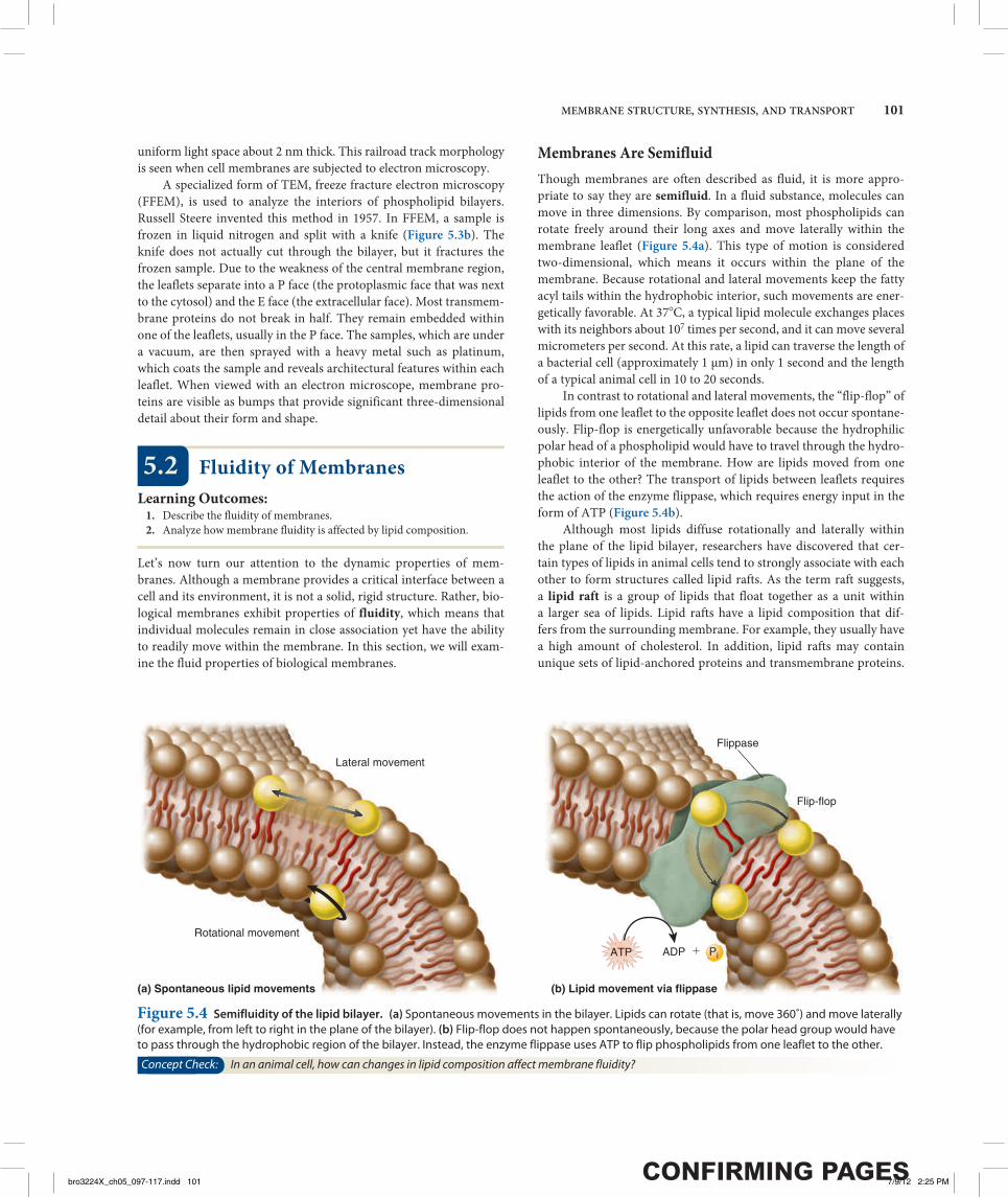
Membranes Are SemifluidThough membranes are often described as fluid, it is more appro-priate to say they are semifluid. In a fluid substance, molecules can move in three dimensions. By comparison, most phospholipids can rotate freely around their long axes and move laterally within the membrane leaflet (Figure 5.4a). This type of motion is considered two-dimensional, which means it occurs within the plane of the membrane. Because rotational and lateral movements keep the fatty acyl tails within the hydrophobic interior, such movements are ener-getically favorable. At 378C, a typical lipid molecule exchanges places with its neighbors about 107 times per second, and it can move several micrometers per second. At this rate, a lipid can traverse the length of a bacterial cell (approximately 1 μm) in only 1 second and the length of a typical animal cell in 10 to 20 seconds.
In contrast to rotational and lateral movements, the “flip-flop” of lipids from one leaflet to the opposite leaflet does not occur spontane-ously. Flip-flop is energetically unfavorable because the hydrophilic polar head of a phospholipid would have to travel through the hydro-phobic interior of the membrane. How are lipids moved from one leaflet to the other? The transport of lipids between leaflets requires the action of the enzyme flippase, which requires energy input in the form of ATP (Figure 5.4b).
Although most lipids diffuse rotationally and laterally within the plane of the lipid bilayer, researchers have discovered that cer-tain types of lipids in animal cells tend to strongly associate with each other to form structures called lipid rafts. As the term raft suggests, a lipid raft is a group of lipids that float together as a unit within a larger sea of lipids. Lipid rafts have a lipid composition that dif-fers from the surrounding membrane. For example, they usually have a high amount of cholesterol. In addition, lipid rafts may contain unique sets of lipid-anchored proteins and transmembrane proteins.
(a) Spontaneous lipid movements (b) Lipid movement via flippase
Lateral movement
Rotational movement
Flip-flop
Flippase
ATP ADP � Pi
Figure 5.4 Semifluidity of the lipid bilayer. (a) Spontaneous movements in the bilayer. Lipids can rotate (that is, move 360˚) and move laterally (for example, from left to right in the plane of the bilayer). (b) Flip-flop does not happen spontaneously, because the polar head group would have to pass through the hydrophobic region of the bilayer. Instead, the enzyme flippase uses ATP to flip phospholipids from one leaflet to the other.
Concept Check: In an animal cell, how can changes in lipid composition affect membrane fluidity?
bro3224X_ch05_097-117.indd 101bro3224X_ch05_097-117.indd 101 7/9/12 2:25 PM7/9/12 2:25 PM
102 CHAPTER 5
The functional importance of lipid rafts is the subject of a large amount of current research. Lipid rafts may play an important role in endocytosis (discussed later in this chapter) and cell signaling.
Lipid Composition Affects Membrane FluidityThe biochemical properties of phospholipids affect the fluidity of the phospholipid bilayer. One key factor is the length of fatty acyl tails, which range from 14 to 24 carbon atoms, with 18 to 20 carbons being the most common. Shorter acyl tails are less likely to interact with each other, which makes the membrane more fluid.
A second important factor is the presence of double bonds in the acyl tails. When a double bond is present, the lipid is said to be unsaturated with respect to the number of hydrogens that are bound to the carbon atoms (refer back to Figure 3.10). A double bond cre-ates a kink in the fatty acyl tail (see inset to Figure 5.1), making it more difficult for neighboring tails to interact and making the bilayer more fluid. As described in Chapter 3, unsaturated lipids tend to be more liquid than saturated lipids, which often form solids at room temperature (refer back to Figure 3.11).
A third factor affecting fluidity is the presence of cholesterol, a short and rigid molecule produced by animal cells (see inset to Figure 5.1). Plant cell membranes contain phytosterols that resemble choles-terol in their chemical structure. Cholesterol tends to stabilize mem-branes; its effects depend on temperature. At higher temperatures, such as those observed in mammals that maintain a constant body temperature, cholesterol makes the membrane less fluid. At lower temperatures, such as icy water, cholesterol has the opposite effect. It makes the membrane more fluid and prevents it from freezing.
An optimal level of bilayer fluidity is essential for normal cell function, growth, and division. If a membrane is too fluid, which may occur at higher temperatures, it can become leaky. However, if a membrane becomes too solid, which may occur at lower tempera-tures, the functioning of membrane proteins will be inhibited. How can organisms cope with changes in temperature? The cells of many species adapt to changes in temperature by altering the lipid composi-tion of their membranes. For example, when the water temperature drops, the cells of certain fish will incorporate more cholesterol in their membranes, making the membrane more fluid. If a plant cell is exposed to high temperatures for many hours or days, it will alter its lipid composition to have longer fatty acyl tails and fewer double bonds, which will make the membrane less fluid.
Many Transmembrane Proteins Can Rotate and Move Laterally, But Some Are Restricted in Their MovementLike lipids, many transmembrane proteins may rotate and laterally move throughout the plane of a membrane. Because transmembrane proteins are larger than lipids, they move within the membrane at a much slower rate. Flip-flop of transmembrane proteins does not occur, because the proteins also contain hydrophilic regions that project out from the phospholipid bilayer, and it would be energeti-cally unfavorable for the hydrophilic regions of membrane proteins to pass through the hydrophobic portion of the phospholipid bilayer.
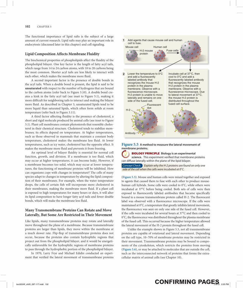
In 1970, Larry Frye and Michael Edidin conducted an experi-ment that verified the lateral movement of transmembrane proteins
(Figure 5.5). Mouse and human cells were mixed together and exposed to agents that caused them to fuse with each other to produce mouse-human cell hybrids. Some cells were cooled to 08C, while others were incubated at 378C before being cooled. Both sets of cells were then exposed to fluorescently labeled antibodies that became specifically bound to a mouse transmembrane protein called H-2. The fluorescent label was observed with a fluorescence microscope. If the cells were maintained at 08C, a temperature that greatly inhibits lateral movement, the fluorescence was seen on only one side of the fused cell. However, if the cells were incubated for several hours at 378C and then cooled to 08C, the fluorescence was distributed throughout the plasma membrane of the fused cell. This occurred because the higher temperature allowed the lateral movement of the H-2 protein throughout the fused cell.
Unlike the example shown in Figure 5.5, not all transmembrane proteins are capable of rotational and lateral movement. Depending on the cell type, 10–70% of membrane proteins may be restricted in their movement. Transmembrane proteins may be bound to compo-nents of the cytoskeleton, which restricts the proteins from moving (Figure 5.6), or may be attached to molecules that are outside the cell, such as the interconnected network of proteins that forms the extra-cellular matrix of animal cells (see Chapter 10).
H-2 mouseprotein
Mouse cell Human cell
Add agents that cause mouse cell and humancell to fuse.
1
2
Fluorescent dye
Antibody
H-2
Fuse cells
Lower the temperature to 0�C and add a fluorescently labeled antibody that recognizes the mouse H-2 protein in the plasma membrane. Observe with a fluorescence microscope. H-2 protein is unable to move laterally and remains on one side of the fused cell.
Incubate cell at 37�C, then cool to 0�C and add a fluorescently labeled antibody that recognizes the mouse H-2 protein in the plasma membrane. Observe with a fluorescence microscope. Due to lateral movement at 37�C, the mouse H-2 protein is distributed throughout the fused cell surface.
Figure 5.5 A method to measure the lateral movement of membrane proteins.
BIOLOGY PRINCIPLE Biology is an experimental science. This experiment verifi ed that membrane proteins
can diff use laterally within the plane of the lipid bilayer.
Concept Check: Explain why the H-2 proteins are found on only one side of the cell when the cells were incubated at 08C.
bro3224X_ch05_097-117.indd 102bro3224X_ch05_097-117.indd 102 7/9/12 2:25 PM7/9/12 2:25 PM

MEMBRANE STRUCTURE, SYNTHESIS, AND TRANSPORT 103
5.3 Synthesis of Membrane Components in Eukaryotic Cells
Learning Outcomes:1. Outline the synthesis of lipids at the ER membrane.2. Explain how transmembrane proteins are inserted into the ER
membrane.3. Describe the process of glycosylation and its functional consequences.
As we have seen, cellular membranes are composed of lipids, proteins, and carbohydrates. Most of the membrane components of eukaryotic cells are made at the endoplasmic reticulum (ER). In this section, we will begin by considering how phospholipids are synthesized at the ER membrane. We will then examine the process by which trans-membrane proteins are inserted into the ER membrane and explore how some proteins are glycosylated.
Lipid Synthesis Occurs at the ER MembraneIn eukaryotic cells, the cytosol and endomembrane system work together to synthesize most lipids. This process occurs at the cytosolic leaflet of the smooth ER membrane. Figure 5.7 shows a simplified pathway for the synthesis of phospholipids. The building blocks for a phospholipid are two fatty acids, each with an acyl tail, one glyc-erol molecule, one phosphate, and a polar head group. These building blocks are made via enzymes in the cytosol, or they are taken into cells from food. To begin the process of phospholipid synthesis, the fatty acids are activated by attachment to an organic molecule called
coenzyme A (CoA). This activation promotes the bonding of the two fatty acids to a glycerol-phosphate molecule, and the resulting molecule is inserted into the cytosolic leaflet of the ER membrane. The phosphate is removed from glycerol, and then a polar molecule already linked to phosphate is attached to glycerol. In the example shown in Figure 5.7, the polar head group contains choline, but many other types are possible. Phospholipids are initially inserted into the cytosolic leaflet. Flippases in the ER membrane transfer some of the newly made lipids to the other leaflet so similar amounts of lipids are found in both leaflets.
The lipids made in the ER membrane are transferred to other membranes in the cell by a variety of mechanisms. Phospholipids in the ER can diffuse laterally to the nuclear envelope. In addition, lipids are transported via vesicles to the Golgi, lysosomes, vacuoles, or plasma membrane. A third mode of lipid transfer involves lipid exchange proteins, which extract a lipid from one membrane, dif-fuse through the cell, and insert the lipid into another membrane. Such transfer can occur between any two membranes, even between the endomembrane system and semiautonomous organelles. For example, lipid exchange proteins transfer lipids between the ER and mitochondria. In addition, chloroplasts and mitochondria synthesize certain types of lipids that are transferred from these organelles to other cellular membranes via lipid exchange proteins.
Most Transmembrane Proteins Are First Inserted into the ER MembraneIn Chapter 4, we learned that eukaryotic proteins contain sorting signals that direct them to their proper destination (see Figure 4.28). With the exception of proteins destined for semiautonomous organ-elles, most transmembrane proteins contain an ER signal sequence that directs them to the ER membrane. If a polypeptide also contains a stretch of 20 amino acids that are mostly hydrophobic and form an α helix, this region will become a transmembrane segment. In the example shown in Figure 5.8, the polypeptide contains one such sequence. After the ER signal sequence is removed by signal pepti-dase (refer back to Figure 4.29), a membrane protein with a single transmembrane segment is the result. Other polypeptides may con-tain more than one transmembrane segment. Each time a polypeptide sequence contains a stretch of 20 hydrophobic amino acids that forms an α helix, an additional transmembrane segment is synthesized into the membrane. From the ER, membrane proteins can be transferred via vesicles to other regions of the cell, such as the Golgi, lysosomes, vacuoles, or plasma membrane.
Glycosylation of Proteins Occurs in the ER and Golgi ApparatusGlycosylation refers to the process of covalently attaching a carbo-hydrate to a lipid or protein. When a carbohydrate is attached to a lipid, a glycolipid is created, whereas attachment of a carbohydrate to a protein produces a glycoprotein.
What is the function of glycosylation? Though the roles of car-bohydrate in cell structure and function are not entirely understood, some functional consequences of glycosylation have emerged. Glyco-lipids and glycoproteins often play a role in cell surface recognition.
Cytoskeletal filament
Linker protein
Fiber in the extracellularmatrix (ECM)
Plasma membrane
Cytosol
Extracellular matrix
Figure 5.6 Attachment of transmembrane proteins to the cytoskeleton and extracellular matrix of an animal cell. Some transmembrane proteins have regions that extend into the cytosol and are anchored to large cytoskeletal filaments via linker proteins. Being bound to these large filaments restricts the movement of these proteins. Similarly, some transmembrane proteins are bound to large, immobile fibers in the extracellular matrix, which restricts their movements.
BioConnections: Look ahead to Figure 10.8. Discuss how transmembrane proteins are important in the binding of cells to each other and the binding of cells to the extracellular matrix.
bro3224X_ch05_097-117.indd 103bro3224X_ch05_097-117.indd 103 7/9/12 2:25 PM7/9/12 2:25 PM
104 CHAPTER 5
When glycolipids and glycoproteins are found in the plasma mem-brane, the carbohydrate portion is located in the extracellular region. During embryonic development in animals, significant cell move-ment occurs. Layers of cells slide over each other to create body structures such as the spinal cord and internal organs. The proper migration of individual cells and cell layers relies on the recognition of cell types via the carbohydrates on their cell surfaces.
Carbohydrates often have a protective effect. The carbohydrate-rich zone on the surface of certain animal cells shields the cell from mechanical and physical damage. Similarly, the carbohydrate portion of glycosylated proteins protects them from the harsh conditions of the extracellular environment and degradation by extracellular prote-ases, which are enzymes that digest proteins.
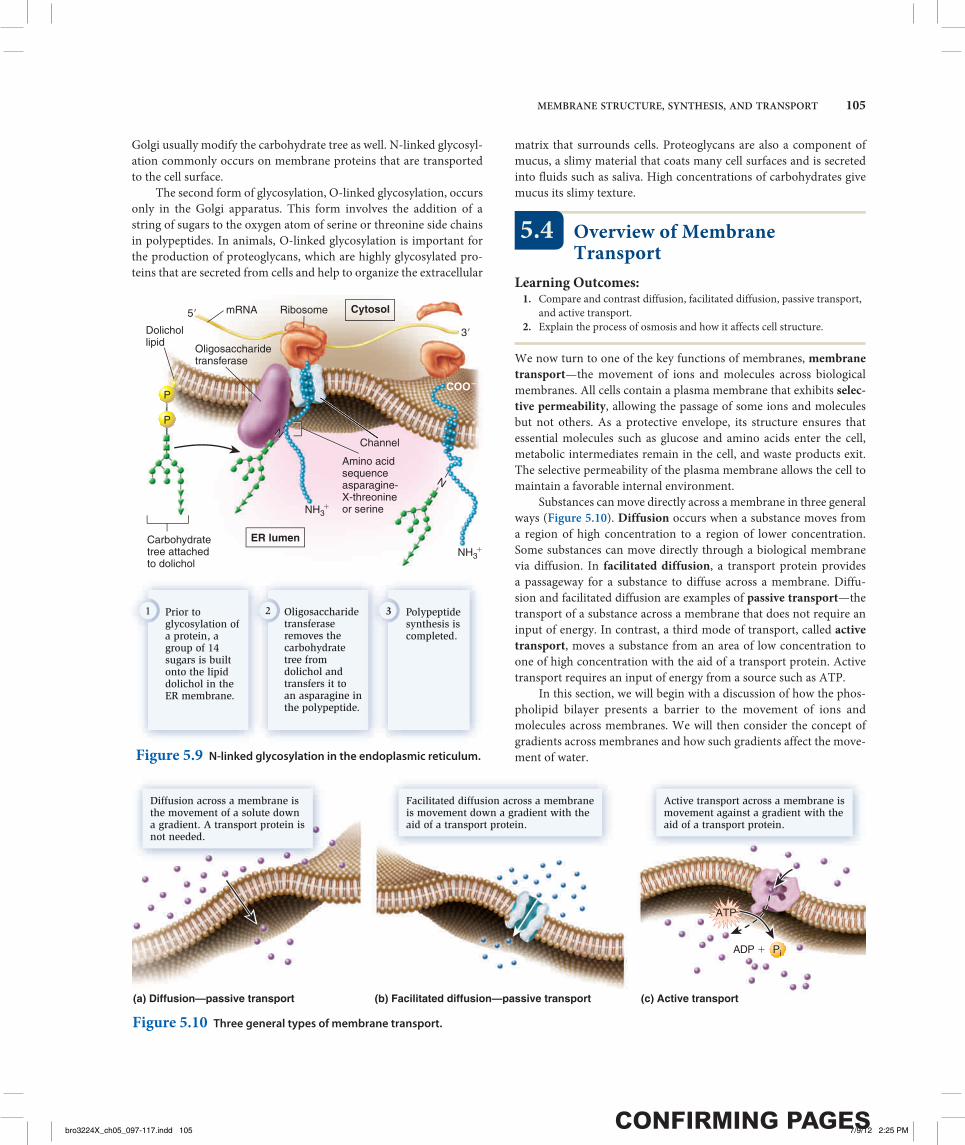
Two forms of protein glycosylation occur in eukaryotes: N-linked and O-linked. N-linked glycosylation, which also occurs in archaea, involves the attachment of a carbohydrate to the amino acid asparagine in a polypeptide chain. It is called N-linked because the carbohydrate attaches to a nitrogen atom of the asparagine side chain. For this to occur, a group of 14 sugar molecules are built onto a lipid called dolichol, which is found in the ER membrane. This carbohy-drate tree is then transferred to an asparagine as a polypeptide is syn-thesized into the ER lumen through a channel protein (Figure 5.9). It attaches only to asparagines occurring in the sequence asparagine–X–threonine or asparagine–X–serine, where X can be any amino acid except proline. An enzyme in the ER, oligosaccharide transferase, recognizes this sequence and transfers the carbohydrate tree from dolichol to the asparagine. Following this initial glycosylation step, the carbohydrate tree is further modified as other enzymes in the ER attach additional sugars or remove sugars. After a glycosylated pro-tein is transferred to the Golgi by vesicle transport, enzymes in the
In the cytosol, fatty acids are activated by the attachment of a CoA molecule.
1
Glycerol-phosphate
2 activatedmolecules
Acyltransferase
Phosphatase
Cholinephosphotransferase
Flippase
2 fattyacids
y The activated fatty acids bond to glycerol-phosphate and are inserted into the cytosolic leaflet of the ER membrane via acyl transferase.
2 Flippases transfer some of the phospholipids to the other leaflet.
5d
The phosphate is removed by a phosphatase enzyme.
3
�
Cholineheadgroup
ER lumen
Pi
P
P
CO O
OH
C
OH CoA CoA
CO OCO
CH2
O
CO OC
CH2 CH
P
OH
CH2
OH
CH2 CH
Cytosol
5
ChoCholinlineeee
A choline already linked to phosphateis attached via choline phosphotransferase.
4
Figure 5.7 A simplified pathway for the synthesis of membrane phospholipids at the ER membrane. Note: Phosphate is abbreviated P when it is attached to an organic molecule and Pi when it is unattached. The subscript i refers to the inorganic form of phosphate.
Concept Check: How are phospholipids transferred to the leaflet of the ER membrane that faces the ER lumen?
Transmembranesegment with20 hydrophobicamino acids
Signalpeptidase
mRNARibosome
Cytosol
5� 3�
ERmembrane
ER lumen
CleavedER signalsequence
ChannelNH3
�
NH3�
NH3�
COO�COO�
A protein begins synthesis into the ER, and the ER signal sequence is cleaved.
1 Polypeptide synthesis continues, and a hydrophobic transmembrane segment is made as the polypeptide is being threaded through the channel.
2 Polypeptide synthesis is completed,and the transmembrane segment remains in the membrane.
3
Figure 5.8 Insertion of membrane proteins into the ER membrane.
Concept Check: What structural feature of a protein causes a region to form a transmembrane segment?
bro3224X_ch05_097-117.indd 104bro3224X_ch05_097-117.indd 104 7/9/12 2:25 PM7/9/12 2:25 PM
MEMBRANE STRUCTURE, SYNTHESIS, AND TRANSPORT 105
Golgi usually modify the carbohydrate tree as well. N-linked glycosyl-ation commonly occurs on membrane proteins that are transported to the cell surface.
The second form of glycosylation, O-linked glycosylation, occurs only in the Golgi apparatus. This form involves the addition of a string of sugars to the oxygen atom of serine or threonine side chains in polypeptides. In animals, O-linked glycosylation is important for the production of proteoglycans, which are highly glycosylated pro-teins that are secreted from cells and help to organize the extracellular
matrix that surrounds cells. Proteoglycans are also a component of mucus, a slimy material that coats many cell surfaces and is secreted into fluids such as saliva. High concentrations of carbohydrates give mucus its slimy texture.
5.4 Overview of Membrane Transport
Learning Outcomes:1. Compare and contrast diffusion, facilitated diffusion, passive transport,
and active transport.2. Explain the process of osmosis and how it affects cell structure.
We now turn to one of the key functions of membranes, membrane transport—the movement of ions and molecules across biological membranes. All cells contain a plasma membrane that exhibits selec-tive permeability, allowing the passage of some ions and molecules but not others. As a protective envelope, its structure ensures that essential molecules such as glucose and amino acids enter the cell, metabolic intermediates remain in the cell, and waste products exit. The selective permeability of the plasma membrane allows the cell to maintain a favorable internal environment.
Substances can move directly across a membrane in three general ways (Figure 5.10). Diffusion occurs when a substance moves from a region of high concentration to a region of lower concentration. Some substances can move directly through a biological membrane via diffusion. In facilitated diffusion, a transport protein provides a passageway for a substance to diffuse across a membrane. Diffu-sion and facilitated diffusion are examples of passive transport—the transport of a substance across a membrane that does not require an input of energy. In contrast, a third mode of transport, called active transport, moves a substance from an area of low concentration to one of high concentration with the aid of a transport protein. Active transport requires an input of energy from a source such as ATP.
In this section, we will begin with a discussion of how the phos-pholipid bilayer presents a barrier to the movement of ions and molecules across membranes. We will then consider the concept of gradients across membranes and how such gradients affect the move-ment of water.
N
Amino acidsequence asparagine-X-threonineor serine
5�
3�
Carbohydratetree attachedto dolichol
Dolichollipid
Oligosaccharidetransferase
mRNA Ribosome
Channel
Prior to glycosylation of a protein, a group of 14 sugars is builtonto the lipid dolichol in the ER membrane.
1 Oligosaccharide transferase removes the carbohydrate tree from dolichol and transfers it to an asparagine in the polypeptide.
2 Polypeptide synthesis is completed.
3
NN
NH3�
NH3�
P
PCOO�
Cytosol
ER lumen
Figure 5.9 N-linked glycosylation in the endoplasmic reticulum.
(b) Facilitated diffusion—passive transport (c) Active transport(a) Diffusion—passive transport
Diffusion across a membrane is the movement of a solute down a gradient. A transport protein is not needed.
Facilitated diffusion across a membrane is movement down a gradient with the aid of a transport protein.
Active transport across a membrane is movement against a gradient with the aid of a transport protein.
ATP
ADP � Pi
Figure 5.10 Three general types of membrane transport.
bro3224X_ch05_097-117.indd 105bro3224X_ch05_097-117.indd 105 7/9/12 2:25 PM7/9/12 2:25 PM
106 CHAPTER 5
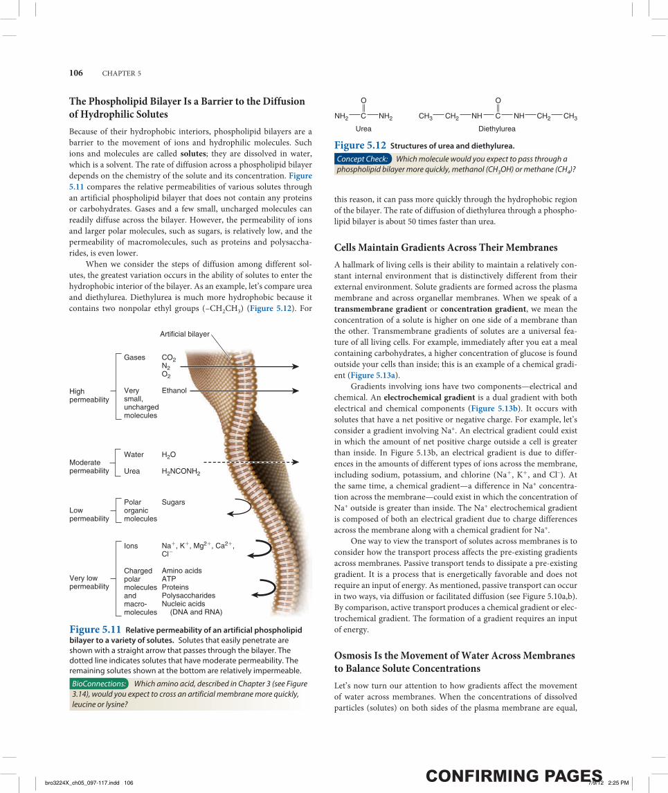
The Phospholipid Bilayer Is a Barrier to the Diffusion of Hydrophilic SolutesBecause of their hydrophobic interiors, phospholipid bilayers are a barrier to the movement of ions and hydrophilic molecules. Such ions and molecules are called solutes; they are dissolved in water, which is a solvent. The rate of diffusion across a phospholipid bilayer depends on the chemistry of the solute and its concentration. Figure 5.11 compares the relative permeabilities of various solutes through an artificial phospholipid bilayer that does not contain any proteins or carbohydrates. Gases and a few small, uncharged molecules can readily diffuse across the bilayer. However, the permeability of ions and larger polar molecules, such as sugars, is relatively low, and the permeability of macromolecules, such as proteins and polysaccha-rides, is even lower.
When we consider the steps of diffusion among different sol-utes, the greatest variation occurs in the ability of solutes to enter the hydrophobic interior of the bilayer. As an example, let’s compare urea and diethylurea. Diethylurea is much more hydrophobic because it contains two nonpolar ethyl groups (–CH2CH3) (Figure 5.12). For
this reason, it can pass more quickly through the hydrophobic region of the bilayer. The rate of diffusion of diethylurea through a phospho-lipid bilayer is about 50 times faster than urea.
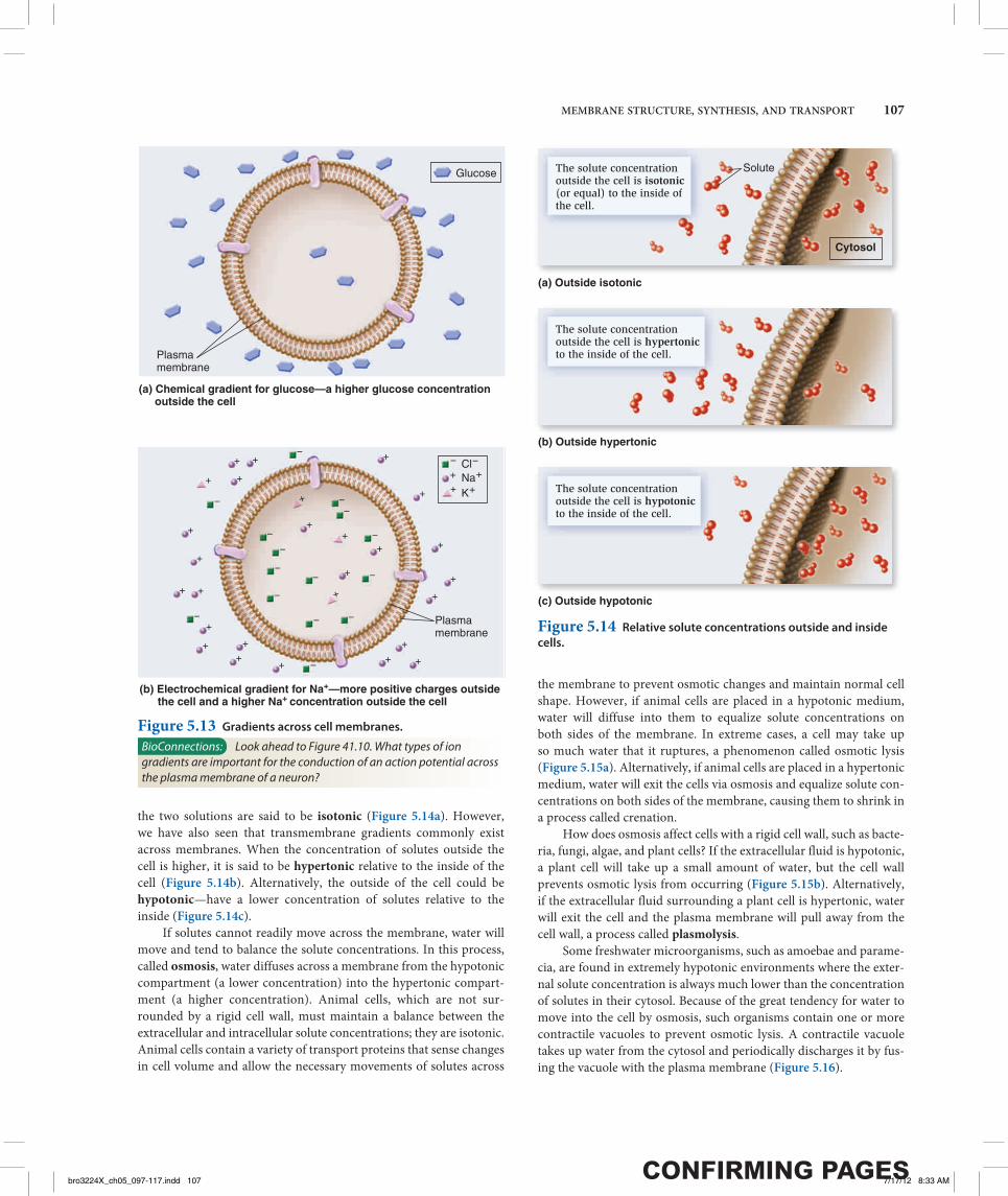
Cells Maintain Gradients Across Their MembranesA hallmark of living cells is their ability to maintain a relatively con-stant internal environment that is distinctively different from their external environment. Solute gradients are formed across the plasma membrane and across organellar membranes. When we speak of a transmembrane gradient or concentration gradient, we mean the concentration of a solute is higher on one side of a membrane than the other. Transmembrane gradients of solutes are a universal fea-ture of all living cells. For example, immediately after you eat a meal containing carbohydrates, a higher concentration of glucose is found outside your cells than inside; this is an example of a chemical gradi-ent (Figure 5.13a).
Gradients involving ions have two components—electrical and chemical. An electrochemical gradient is a dual gradient with both electrical and chemical components (Figure 5.13b). It occurs with solutes that have a net positive or negative charge. For example, let’s consider a gradient involving Na+. An electrical gradient could exist in which the amount of net positive charge outside a cell is greater than inside. In Figure 5.13b, an electrical gradient is due to differ-ences in the amounts of different types of ions across the membrane, including sodium, potassium, and chlorine (Na+, K+, and Cl–). At the same time, a chemical gradient—a difference in Na+ concentra-tion across the membrane—could exist in which the concentration of Na+ outside is greater than inside. The Na+ electrochemical gradient is composed of both an electrical gradient due to charge differences across the membrane along with a chemical gradient for Na+.
One way to view the transport of solutes across membranes is to consider how the transport process affects the pre-existing gradients across membranes. Passive transport tends to dissipate a pre-existing gradient. It is a process that is energetically favorable and does not require an input of energy. As mentioned, passive transport can occur in two ways, via diffusion or facilitated diffusion (see Figure 5.10a,b). By comparison, active transport produces a chemical gradient or elec-trochemical gradient. The formation of a gradient requires an input of energy.
Osmosis Is the Movement of Water Across Membranes to Balance Solute ConcentrationsLet’s now turn our attention to how gradients affect the movement of water across membranes. When the concentrations of dissolved particles (solutes) on both sides of the plasma membrane are equal,
High permeability
Moderatepermeability
Low permeability
Very low permeability
Gases
Verysmall,unchargedmolecules
CO2N2O2
Ethanol
Water
Urea
H2O
H2NCONH2
Polar organicmolecules
Sugars
Ions
Charged polar moleculesandmacro-molecules
Na�, K�, Mg2�, Ca2�,Cl�
Amino acidsATPProteinsPolysaccharidesNucleic acids
(DNA and RNA)
Artificial bilayer
Figure 5.11 Relative permeability of an artificial phospholipid bilayer to a variety of solutes. Solutes that easily penetrate are shown with a straight arrow that passes through the bilayer. The dotted line indicates solutes that have moderate permeability. The remaining solutes shown at the bottom are relatively impermeable.
BioConnections: Which amino acid, described in Chapter 3 (see Figure 3.14), would you expect to cross an artificial membrane more quickly, leucine or lysine?
Urea Diethylurea
NH2 NH2 CH3 CH3CH2 CH2NH NH
O
C
O
C
Figure 5.12 Structures of urea and diethylurea.
Concept Check: Which molecule would you expect to pass through a phospholipid bilayer more quickly, methanol (CH3OH) or methane (CH4)?
bro3224X_ch05_097-117.indd 106bro3224X_ch05_097-117.indd 106 7/9/12 2:25 PM7/9/12 2:25 PM
MEMBRANE STRUCTURE, SYNTHESIS, AND TRANSPORT 107
the two solutions are said to be isotonic (Figure 5.14a). However, we have also seen that transmembrane gradients commonly exist across membranes. When the concentration of solutes outside the cell is higher, it is said to be hypertonic relative to the inside of the cell ( Figure 5.14b). Alternatively, the outside of the cell could be hypotonic—have a lower concentration of solutes relative to the inside (Figure 5.14c).
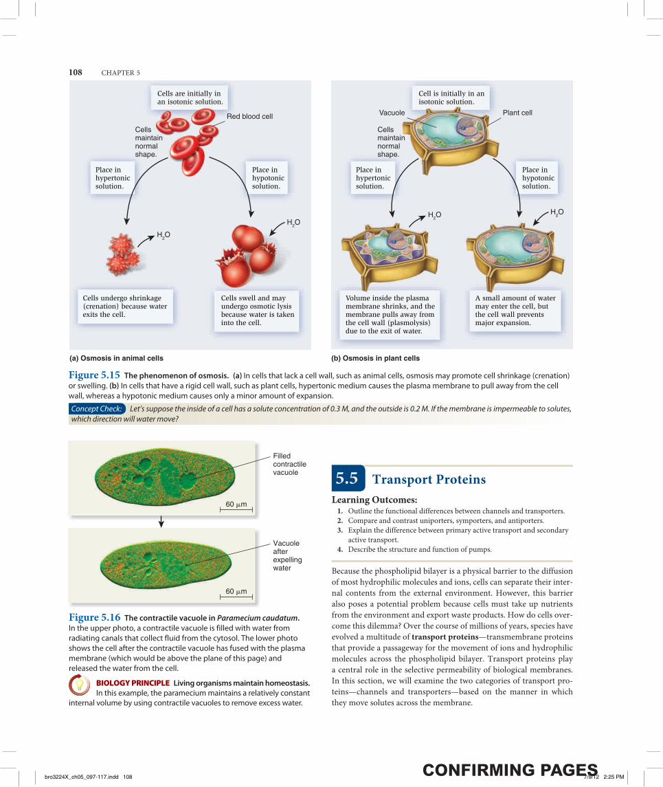
If solutes cannot readily move across the membrane, water will move and tend to balance the solute concentrations. In this process, called osmosis, water diffuses across a membrane from the hypotonic compartment (a lower concentration) into the hypertonic compart-ment (a higher concentration). Animal cells, which are not sur-rounded by a rigid cell wall, must maintain a balance between the extracellular and intracellular solute concentrations; they are isotonic. Animal cells contain a variety of transport proteins that sense changes in cell volume and allow the necessary movements of solutes across
the membrane to prevent osmotic changes and maintain normal cell shape. However, if animal cells are placed in a hypotonic medium, water will diffuse into them to equalize solute concentrations on both sides of the membrane. In extreme cases, a cell may take up so much water that it ruptures, a phenomenon called osmotic lysis ( Figure 5.15a). Alternatively, if animal cells are placed in a hypertonic medium, water will exit the cells via osmosis and equalize solute con-centrations on both sides of the membrane, causing them to shrink in a process called crenation.
How does osmosis affect cells with a rigid cell wall, such as bacte-ria, fungi, algae, and plant cells? If the extracellular fluid is hypotonic, a plant cell will take up a small amount of water, but the cell wall prevents osmotic lysis from occurring (Figure 5.15b). Alternatively, if the extracellular fluid surrounding a plant cell is hypertonic, water will exit the cell and the plasma membrane will pull away from the cell wall, a process called plasmolysis.
Some freshwater microorganisms, such as amoebae and parame-cia, are found in extremely hypotonic environments where the exter-nal solute concentration is always much lower than the concentration of solutes in their cytosol. Because of the great tendency for water to move into the cell by osmosis, such organisms contain one or more contractile vacuoles to prevent osmotic lysis. A contractile vacuole takes up water from the cytosol and periodically discharges it by fus-ing the vacuole with the plasma membrane (Figure 5.16).
(a) Chemical gradient for glucose—a higher glucose concentration outside the cell
Plasmamembrane
Plasmamembrane
K�
Na�Cl�
(b) Electrochemical gradient for Na+—more positive charges outside the cell and a higher Na+ concentration outside the cell
Glucose
�
�
�
�
�
��
�
��
�
�
�
�
�
�
�
�
� �
�
�
��
�
�
�
�
��
�
�
�
�
��
�
�
�
�
�
�
�
�
�
Figure 5.13 Gradients across cell membranes.
BioConnections: Look ahead to Figure 41.10. What types of ion gradients are important for the conduction of an action potential across the plasma membrane of a neuron?
The solute concentration outside the cell is isotonic (or equal) to the inside ofthe cell.
The solute concentration outside the cell is hypertonicto the inside of the cell.
The solute concentration outside the cell is hypotonicto the inside of the cell.
Solute
Cytosol
(a) Outside isotonic
(b) Outside hypertonic
(c) Outside hypotonic
Figure 5.14 Relative solute concentrations outside and inside cells.
bro3224X_ch05_097-117.indd 107bro3224X_ch05_097-117.indd 107 7/17/12 8:33 AM7/17/12 8:33 AM
108 CHAPTER 5
5.5 Transport ProteinsLearning Outcomes:
1. Outline the functional differences between channels and transporters.2. Compare and contrast uniporters, symporters, and antiporters.3. Explain the difference between primary active transport and secondary
active transport.4. Describe the structure and function of pumps.
Because the phospholipid bilayer is a physical barrier to the diffusion of most hydrophilic molecules and ions, cells can separate their inter-nal contents from the external environment. However, this barrier also poses a potential problem because cells must take up nutrients from the environment and export waste products. How do cells over-come this dilemma? Over the course of millions of years, species have evolved a multitude of transport proteins—transmembrane proteins that provide a passageway for the movement of ions and hydrophilic molecules across the phospholipid bilayer. Transport proteins play a central role in the selective permeability of biological membranes. In this section, we will examine the two categories of transport pro-teins—channels and transporters—based on the manner in which they move solutes across the membrane.
Cells are initially in an isotonic solution.
Cells undergo shrinkage (crenation) because water exits the cell.
Cells swell and may undergo osmotic lysisbecause water is taken into the cell.
Cell is initially in an isotonic solution.
Place in hypertonicsolution.
Place in hypotonicsolution.
Place in hypertonicsolution.
Place in hypotonicsolution.
Cells maintainnormal shape.
Cells maintainnormal shape.
Plant cellVacuole
Volume inside the plasmamembrane shrinks, and the membrane pulls away from the cell wall (plasmolysis) due to the exit of water.
A small amount of water may enter the cell, but the cell wall prevents major expansion.
Red blood cell
(a) Osmosis in animal cells (b) Osmosis in plant cells
H2O
H2OH2O
H2O
Figure 5.15 The phenomenon of osmosis. (a) In cells that lack a cell wall, such as animal cells, osmosis may promote cell shrinkage (crenation) or swelling. (b) In cells that have a rigid cell wall, such as plant cells, hypertonic medium causes the plasma membrane to pull away from the cell wall, whereas a hypotonic medium causes only a minor amount of expansion.
Concept Check: Let’s suppose the inside of a cell has a solute concentration of 0.3 M, and the outside is 0.2 M. If the membrane is impermeable to solutes, which direction will water move?
Filledcontractilevacuole
Vacuoleafterexpellingwater
60 �m
60 �m
Figure 5.16 The contractile vacuole in Paramecium caudatum. In the upper photo, a contractile vacuole is filled with water from radiating canals that collect fluid from the cytosol. The lower photo shows the cell after the contractile vacuole has fused with the plasma membrane (which would be above the plane of this page) and released the water from the cell.
BIOLOGY PRINCIPLE Living organisms maintain homeostasis. In this example, the paramecium maintains a relatively constant
internal volume by using contractile vacuoles to remove excess water.
bro3224X_ch05_097-117.indd 108bro3224X_ch05_097-117.indd 108 7/9/12 2:25 PM7/9/12 2:25 PM
MEMBRANE STRUCTURE, SYNTHESIS, AND TRANSPORT 109
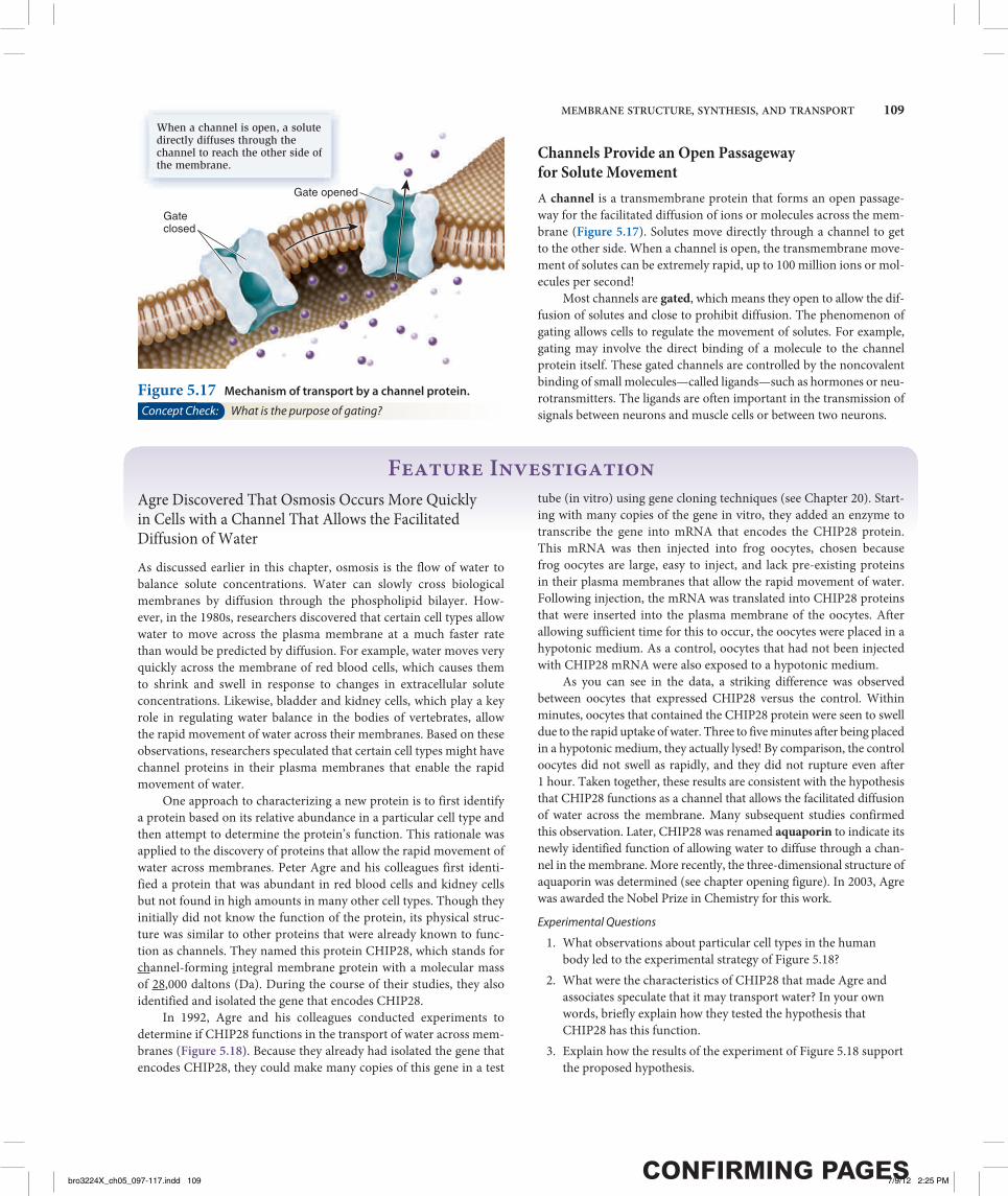
Channels Provide an Open Passageway for Solute MovementA channel is a transmembrane protein that forms an open passage-way for the facilitated diffusion of ions or molecules across the mem-brane (Figure 5.17). Solutes move directly through a channel to get to the other side. When a channel is open, the transmembrane move-ment of solutes can be extremely rapid, up to 100 million ions or mol-ecules per second!
Most channels are gated, which means they open to allow the dif-fusion of solutes and close to prohibit diffusion. The phenomenon of gating allows cells to regulate the movement of solutes. For example, gating may involve the direct binding of a molecule to the channel protein itself. These gated channels are controlled by the noncovalent binding of small molecules—called ligands—such as hormones or neu-rotransmitters. The ligands are often important in the transmission of signals between neurons and muscle cells or between two neurons.
When a channel is open, a solute directly diffuses through the channel to reach the other side of the membrane.
Gateclosed
Gate opened
Figure 5.17 Mechanism of transport by a channel protein.
Concept Check: What is the purpose of gating?
Feature InvestigationAgre Discovered That Osmosis Occurs More Quickly in Cells with a Channel That Allows the Facilitated Diffusion of WaterAs discussed earlier in this chapter, osmosis is the flow of water to balance solute concentrations. Water can slowly cross biological membranes by diffusion through the phospholipid bilayer. How-ever, in the 1980s, researchers discovered that certain cell types allow water to move across the plasma membrane at a much faster rate than would be predicted by diffusion. For example, water moves very quickly across the membrane of red blood cells, which causes them to shrink and swell in response to changes in extracellular solute concentrations. Likewise, bladder and kidney cells, which play a key role in regulating water balance in the bodies of vertebrates, allow the rapid movement of water across their membranes. Based on these observations, researchers speculated that certain cell types might have channel proteins in their plasma membranes that enable the rapid movement of water. One approach to characterizing a new protein is to first identify a protein based on its relative abundance in a particular cell type and then attempt to determine the protein’s function. This rationale was applied to the discovery of proteins that allow the rapid movement of water across membranes. Peter Agre and his colleagues first identi-fied a protein that was abundant in red blood cells and kidney cells but not found in high amounts in many other cell types. Though they initially did not know the function of the protein, its physical struc-ture was similar to other proteins that were already known to func-tion as channels. They named this protein CHIP28, which stands for channel-forming integral membrane protein with a molecular mass of 28,000 daltons (Da). During the course of their studies, they also identified and isolated the gene that encodes CHIP28. In 1992, Agre and his colleagues conducted experiments to determine if CHIP28 functions in the transport of water across mem-branes (Figure 5.18). Because they already had isolated the gene that encodes CHIP28, they could make many copies of this gene in a test
tube (in vitro) using gene cloning techniques (see Chapter 20). Start-ing with many copies of the gene in vitro, they added an enzyme to transcribe the gene into mRNA that encodes the CHIP28 protein. This mRNA was then injected into frog oocytes, chosen because frog oocytes are large, easy to inject, and lack pre-existing proteins in their plasma membranes that allow the rapid movement of water. Following injection, the mRNA was translated into CHIP28 proteins that were inserted into the plasma membrane of the oocytes. After allowing sufficient time for this to occur, the oocytes were placed in a hypotonic medium. As a control, oocytes that had not been injected with CHIP28 mRNA were also exposed to a hypotonic medium. As you can see in the data, a striking difference was observed between oocytes that expressed CHIP28 versus the control. Within minutes, oocytes that contained the CHIP28 protein were seen to swell due to the rapid uptake of water. Three to five minutes after being placed in a hypotonic medium, they actually lysed! By comparison, the control oocytes did not swell as rapidly, and they did not rupture even after 1 hour. Taken together, these results are consistent with the hypothesis that CHIP28 functions as a channel that allows the facilitated diffusion of water across the membrane. Many subsequent studies confirmed this observation. Later, CHIP28 was renamed aquaporin to indicate its newly identified function of allowing water to diffuse through a chan-nel in the membrane. More recently, the three-dimensional structure of aquaporin was determined (see chapter opening figure). In 2003, Agre was awarded the Nobel Prize in Chemistry for this work.
Experimental Questions
1. What observations about particular cell types in the human body led to the experimental strategy of Figure 5.18?
2. What were the characteristics of CHIP28 that made Agre and associates speculate that it may transport water? In your own words, briefly explain how they tested the hypothesis that CHIP28 has this function.
3. Explain how the results of the experiment of Figure 5.18 support the proposed hypothesis.
bro3224X_ch05_097-117.indd 109bro3224X_ch05_097-117.indd 109 7/9/12 2:25 PM7/9/12 2:25 PM

110 CHAPTER 5
Figure 5.18 The discovery of water channels (aquaporins) by Agre.
Place oocytes into a hypotonic medium and observe under a light microscope. As a control, also place oocytes that have not been injected with CHIP28 mRNA into a hypotonic medium and observe by microscopy.
Oocyte rupturingOocyte
Control CHIP28
SOURCE Preston, G.M., Carroll, T.P., Guggino, W.B., and Agre, P. 1992. Appearance of water channels in Xenopus oocytes expressing red cell CHIP28 protein. Science 256:385–387.
CONCLUSION The CHIP28 protein, now called aquaporin, allows the rapid movement of water across the membrane.
Inject the CHIP28 mRNA into frog eggs (oocytes). Wait several hours to allow time for the mRNA to be translated into CHIP28 protein at the ER membrane and then moved via vesicles to the plasma membrane.
Add an enzyme (RNA polymerase) and nucleotides to a test tube that contains many copies of the CHIP28 gene. This results in the synthesis of many copies of CHIP28 mRNA.
HYPOTHESIS CHIP28 may function as a water channel.
KEY MATERIALS Prior to this work, a protein called CHIP28 was identified that is abundant in red blood cells and kidney cells. The gene that encodes this protein was cloned, which means that many copies of the gene were made in a test tube.
Experimental level Conceptual level
1
2
3
4 THE DATA
Enzymesand nucleotides
CHIP28 DNA
RNA polymerase CHIP28 mRNA
Frog oocyte CHIP28 protein
CHIP28 protein
Ribosome
Control
CHIP28 mRNA
CHIP28 protein is inserted into the plasma membrane.
Nucleus Cytosol
Control CHIP28
3–5 minutes
5
6
bro3224X_ch05_097-117.indd 110bro3224X_ch05_097-117.indd 110 7/9/12 2:25 PM7/9/12 2:25 PM

MEMBRANE STRUCTURE, SYNTHESIS, AND TRANSPORT 111
Transporters Bind Their Solutes and Undergo Conformational ChangesLet’s now turn our attention to a second category of transport pro-teins known as transporters.* These transmembrane proteins bind their solutes in a hydrophilic pocket and undergo a conformational change that switches the exposure of the pocket from one side of the membrane to the other side (Figure 5.19). For example, in 1995, American biologist Robert Brooker and colleagues proposed that a transporter called lactose permease, which is found in the bacterium E. coli, has a hydrophilic pocket that binds lactose. They further pro-posed that the two halves of the transporter protein come together at an interface that moves in such a way that the lactose-binding site alternates between an outwardly accessible pocket and an inwardly accessible pocket, as shown in Figure 5.19. This idea was later con-firmed by studies that determined the structure of the lactose perme-ase and related transporters.
Transporters provide the principal pathway for the uptake of organic molecules, such as sugars, amino acids, and nucleotides. In animals, they also allow cells to take up certain hormones and neu-rotransmitters. In addition, many transporters play a key role in export. Waste products of cellular metabolism must be released from cells before they reach toxic levels. For example, a transporter removes lactic acid, a by-product of muscle cells during exercise. Other trans-porters, which are involved with ion transport, play an important role in regulating internal pH and controlling cell volume. Transporters tend to be much slower than channels. Their rate of transport is typi-cally 100 to 1,000 ions or molecules per second.
Transporters are named according to the number of solutes they bind and the direction in which they transport those solutes ( Figure 5.20). Uniporters bind a single ion or molecule and transport
it across the membrane. Symporters bind two or more ions or mole-cules and transport them in the same direction. Antiporters bind two or more ions or molecules and transport them in opposite directions.
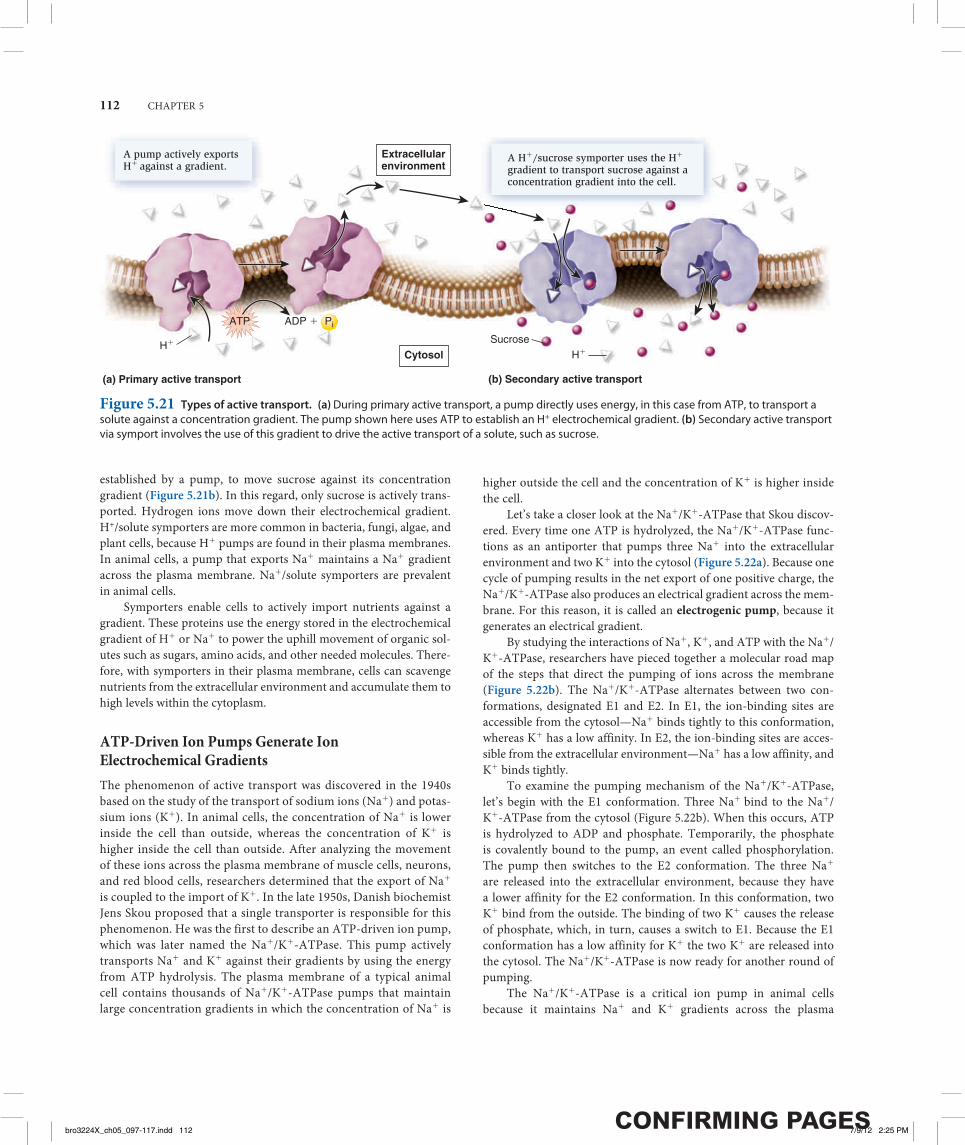
Active Transport Is the Movement of Solutes Against a GradientAs mentioned, active transport is the movement of a solute across a membrane against its concentration gradient—that is, from a region of low concentration to higher concentration. Active transport is energetically unfavorable and requires an input of energy. Primary active transport involves the functioning of a pump—a type of transporter that directly uses energy to transport a solute against a concentration gradient. Figure 5.21a shows a pump that uses ATP to transport H+ against a gradient. Such a pump can establish an H+ electrochemical gradient across a membrane.
Secondary active transport involves the use of a pre-existing gradient to drive the active transport of another solute. For exam-ple, an H+/sucrose symporter uses an H+ electrochemical gradient,
Hydrophilic pocket
Solute
For transport to occur, a solute binds in a hydrophilic pocket exposed on one side of the membrane. The transporter then undergoes a conformational change that switches the exposure of the pocket to the other side of the membrane, where the solute is then released.
Conformational change
Figure 5.19 Mechanism of transport by a transporter, also called a carrier.
BIOLOGY PRINCIPLE Structure determines function. Two structural features—a hydrophilic pocket and the ability to
wobble back and forth between two conformations—allow transporters to move ions and molecules across the membrane.
* Transporters are also called carriers. However, this term is misleading because transporters do not physically carry their solutes across the membrane.
Two solutes move inopposite directions.
A single solute moves in one direction.
Two solutes move in thesame direction.
(a) Uniporter
(b) Symporter
(c) Antiporter
Figure 5.20 Types of transporters based on the direction of transport.
bro3224X_ch05_097-117.indd 111bro3224X_ch05_097-117.indd 111 7/9/12 2:25 PM7/9/12 2:25 PM
112 CHAPTER 5
established by a pump, to move sucrose against its concentration gradient (Figure 5.21b). In this regard, only sucrose is actively trans-ported. Hydrogen ions move down their electrochemical gradient. H+/solute symporters are more common in bacteria, fungi, algae, and plant cells, because H+ pumps are found in their plasma membranes. In animal cells, a pump that exports Na+ maintains a Na+ gradient across the plasma membrane. Na+/solute symporters are prevalent in animal cells.
Symporters enable cells to actively import nutrients against a gradient. These proteins use the energy stored in the electrochemical gradient of H+ or Na+ to power the uphill movement of organic sol-utes such as sugars, amino acids, and other needed molecules. There-fore, with symporters in their plasma membrane, cells can scavenge nutrients from the extracellular environment and accumulate them to high levels within the cytoplasm.
ATP-Driven Ion Pumps Generate Ion Electrochemical GradientsThe phenomenon of active transport was discovered in the 1940s based on the study of the transport of sodium ions (Na+) and potas-sium ions (K+). In animal cells, the concentration of Na+ is lower inside the cell than outside, whereas the concentration of K+ is higher inside the cell than outside. After analyzing the movement of these ions across the plasma membrane of muscle cells, neurons, and red blood cells, researchers determined that the export of Na+
is coupled to the import of K+. In the late 1950s, Danish biochemist Jens Skou proposed that a single transporter is responsible for this phenomenon. He was the first to describe an ATP-driven ion pump, which was later named the Na+/K+-ATPase. This pump actively transports Na+ and K+ against their gradients by using the energy from ATP hydrolysis. The plasma membrane of a typical animal cell contains thousands of Na+/K+-ATPase pumps that maintain large concentration gradients in which the concentration of Na+ is
higher outside the cell and the concentration of K+ is higher inside the cell.
Let’s take a closer look at the Na+/K+-ATPase that Skou discov-ered. Every time one ATP is hydrolyzed, the Na+/K+-ATPase func-tions as an antiporter that pumps three Na+ into the extracellular environment and two K+ into the cytosol (Figure 5.22a). Because one cycle of pumping results in the net export of one positive charge, the Na+/K+-ATPase also produces an electrical gradient across the mem-brane. For this reason, it is called an electrogenic pump, because it generates an electrical gradient.
By studying the interactions of Na+, K+, and ATP with the Na+/K+-ATPase, researchers have pieced together a molecular road map of the steps that direct the pumping of ions across the membrane (Figure 5.22b). The Na+/K+-ATPase alternates between two con-formations, designated E1 and E2. In E1, the ion-binding sites are accessible from the cytosol—Na+ binds tightly to this conformation, whereas K+ has a low affinity. In E2, the ion-binding sites are acces-sible from the extracellular environment—Na+ has a low affinity, and K+ binds tightly.
To examine the pumping mechanism of the Na+/K+-ATPase, let’s begin with the E1 conformation. Three Na+ bind to the Na+/K+-ATPase from the cytosol (Figure 5.22b). When this occurs, ATP is hydrolyzed to ADP and phosphate. Temporarily, the phosphate is covalently bound to the pump, an event called phosphorylation. The pump then switches to the E2 conformation. The three Na+ are released into the extracellular environment, because they have a lower affinity for the E2 conformation. In this conformation, two K+ bind from the outside. The binding of two K+ causes the release of phosphate, which, in turn, causes a switch to E1. Because the E1 conformation has a low affinity for K+ the two K+ are released into the cytosol. The Na+/K+-ATPase is now ready for another round of pumping.
The Na+/K+-ATPase is a critical ion pump in animal cells because it maintains Na+ and K+ gradients across the plasma
A pump actively exports H� against a gradient.
Extracellularenvironment
A H�/sucrose symporter uses the H�
gradient to transport sucrose against aconcentration gradient into the cell.
(a) Primary active transport (b) Secondary active transport
ATP ADP �
H� Sucrose
H�Cytosol
Pi
Figure 5.21 Types of active transport. (a) During primary active transport, a pump directly uses energy, in this case from ATP, to transport a solute against a concentration gradient. The pump shown here uses ATP to establish an H+ electrochemical gradient. (b) Secondary active transport via symport involves the use of this gradient to drive the active transport of a solute, such as sucrose.
bro3224X_ch05_097-117.indd 112bro3224X_ch05_097-117.indd 112 7/9/12 2:25 PM7/9/12 2:25 PM

MEMBRANE STRUCTURE, SYNTHESIS, AND TRANSPORT 113
5.6 Exocytosis and EndocytosisLearning Outcome:
1. Describe the steps in exocytosis and endocytosis.
We have seen that most small substances are transported via membrane proteins such as channels and transporters, which provide a passageway for the movement of ions and molecules directly across the membrane. Eukaryotic cells have two other mechanisms, exocytosis and endocyto-sis, to transport larger molecules such as proteins and polysaccharides, and even very large particles. Both mechanisms involve the packaging of the transported substance, sometimes called the cargo, into a mem-brane vesicle or vacuole. Table 5.5 describes some examples.
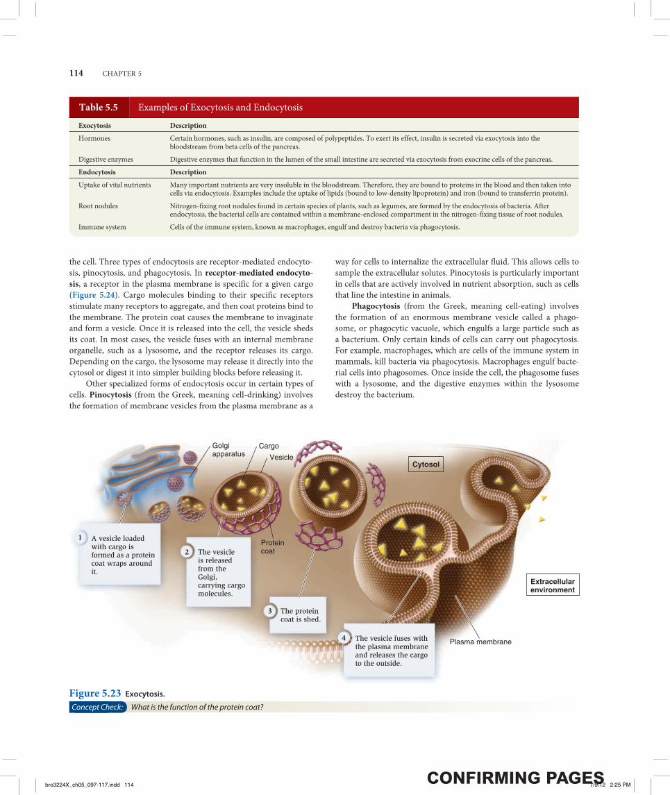
Exocytosis During exocytosis, material inside the cell is packaged into vesicles and then excreted into the extracellular environment (Figure 5.23). These vesicles are usually derived from the Golgi appa-ratus. As the vesicles form, a specific cargo is loaded into their inte-rior. The budding process involves the formation of a protein coat around the emerging vesicle. The assembly of coat proteins on the surface of the Golgi membrane causes the bud to form. Eventually, the bud separates from the membrane to form a vesicle. After the vesicle is released, the coat is shed. Finally, the vesicle fuses with the plasma membrane and releases the cargo into the extracellular environment.
Endocytosis During endocytosis, the plasma membrane invagi-nates, or folds inward, to form a vesicle that brings substances into
3 Na�
Na�/K�-ATPase
2 K�
ATPADP � Pi
High [Na�]Low [K�]
Low [Na�]
Nerve cell
(a) Active transport by the Na�/ K�-ATPase (b) Mechanism of pumping
3 Na� bind from cytosol. ATP is hydrolyzed. ADPis released and phosphate(P) is covalently attachedto the pump, switching itto the E2 conformation.
3 Na� are released outside of the cell.
2 K� bind fromoutside of the cell.
Phosphate (Pi) is released, and the pump switchesto the E1 conformation.2 K� are released intocytosol. The processrepeats.
E1
E2
E2
E1
ADPATP P
Pi
3 Na�
2 K�
1 2 43
Extracellular environment
Extracellular environment
CytosolCytosolHigh [K�]
2 K�
3 Na�
Figure 5.22 Structure and function of the Na+/K+-ATPase. (a) Active transport by the Na+/K+-ATPase. Each time this protein hydrolyzes one ATP molecule, it pumps out three Na+ and pumps in two K+. (b) Pumping mechanism. This figure illustrates the protein conformational changes between E1 and E2. As this occurs, ATP is hydrolyzed to ADP and phosphate. During the process, phosphate is covalently attached to the protein but is released after two K+ bind.
Concept Check: If a cell contained ATP and Na+, but K+ were missing from the extracellular medium, how far through these steps could the Na+/K+-ATPase proceed?
Table 5.4 Important Functions of Ion Electrochemical Gradients
Function Description
Transport of ions and molecules
Symporters and antiporters use H+ and Na+ gradients to take up nutrients and export waste products (see Figure 5.21).
Production of energy intermediates
In the mitochondrion and chloroplast, H+ gradients are used to synthesize ATP.
Osmotic regulation Animal cells control their internal volume by regulating ion gradients between the cytosol and extracellular fluid.
Neuronal signaling Na+ and K+ gradients are involved in conducting action potentials, the signals transmitted by neurons.
Muscle contraction Ca2+ gradients regulate the ability of muscle fibers to contract.
Bacterial swimming H+ gradients drive the rotation of bacterial flagella.
membrane. Many other types of ion pumps are also found in the plasma membrane and in organellar membranes. Ion pumps play the primary role in the formation and maintenance of ion gradients that drive many important cellular processes (Table 5.4). ATP is com-monly the source of energy to drive ion pumps, and cells typically use a substantial portion of their ATP to keep them working. For exam-ple, neurons use up to 70% of their ATP just to operate ion pumps!
bro3224X_ch05_097-117.indd 113bro3224X_ch05_097-117.indd 113 7/9/12 2:25 PM7/9/12 2:25 PM
114 CHAPTER 5
the cell. Three types of endocytosis are receptor-mediated endocyto-sis, pinocytosis, and phagocytosis. In receptor-mediated endocyto-sis, a receptor in the plasma membrane is specific for a given cargo (Figure 5.24). Cargo molecules binding to their specific receptors stimulate many receptors to aggregate, and then coat proteins bind to the membrane. The protein coat causes the membrane to invaginate and form a vesicle. Once it is released into the cell, the vesicle sheds its coat. In most cases, the vesicle fuses with an internal membrane organelle, such as a lysosome, and the receptor releases its cargo. Depending on the cargo, the lysosome may release it directly into the cytosol or digest it into simpler building blocks before releasing it.
Other specialized forms of endocytosis occur in certain types of cells. Pinocytosis (from the Greek, meaning cell-drinking) involves the formation of membrane vesicles from the plasma membrane as a
way for cells to internalize the extracellular fluid. This allows cells to sample the extracellular solutes. Pinocytosis is particularly important in cells that are actively involved in nutrient absorption, such as cells that line the intestine in animals.
Phagocytosis (from the Greek, meaning cell-eating) involves the formation of an enormous membrane vesicle called a phago-some, or phagocytic vacuole, which engulfs a large particle such as a bacterium. Only certain kinds of cells can carry out phagocytosis. For example, macrophages, which are cells of the immune system in mammals, kill bacteria via phagocytosis. Macrophages engulf bacte-rial cells into phagosomes. Once inside the cell, the phagosome fuses with a lysosome, and the digestive enzymes within the lysosome destroy the bacterium.
Table 5.5 Examples of Exocytosis and Endocytosis
Exocytosis Description
Hormones Certain hormones, such as insulin, are composed of polypeptides. To exert its effect, insulin is secreted via exocytosis into the bloodstream from beta cells of the pancreas.
Digestive enzymes Digestive enzymes that function in the lumen of the small intestine are secreted via exocytosis from exocrine cells of the pancreas.
Endocytosis Description
Uptake of vital nutrients Many important nutrients are very insoluble in the bloodstream. Therefore, they are bound to proteins in the blood and then taken into cells via endocytosis. Examples include the uptake of lipids (bound to low-density lipoprotein) and iron (bound to transferrin protein).
Root nodules Nitrogen-fixing root nodules found in certain species of plants, such as legumes, are formed by the endocytosis of bacteria. After endocytosis, the bacterial cells are contained within a membrane-enclosed compartment in the nitrogen-fixing tissue of root nodules.
Immune system Cells of the immune system, known as macrophages, engulf and destroy bacteria via phagocytosis.
Cytosol
A vesicle loaded with cargo is formed as a protein coat wraps around it.
1
The vesicle is released from the Golgi, carrying cargo molecules.
2
The vesicle fuses with the plasma membrane and releases the cargo to the outside.
4
The protein coat is shed.
3
Plasma membrane
Golgiapparatus
Protein coat
Vesicle
Cargo
Extracellularenvironment
Figure 5.23 Exocytosis.
Concept Check: What is the function of the protein coat?
bro3224X_ch05_097-117.indd 114bro3224X_ch05_097-117.indd 114 7/9/12 2:25 PM7/9/12 2:25 PM

MEMBRANE STRUCTURE, SYNTHESIS, AND TRANSPORT 115
Summary of Key Concepts
5.1 Membrane Structure• A plasma membrane separates a cell from its surroundings. The
plasma membrane and organellar membranes provide interfaces for carrying out vital cellular activities (Table 5.1).
• The accepted model of membranes is the fluid-mosaic model, and its basic framework is the phospholipid bilayer. Cellular membranes also contain proteins, and most membranes have attached carbohydrates (Figure 5.1, Table 5.2).
• The three main types of membrane proteins are transmembrane proteins, lipid-anchored proteins, and peripheral membrane proteins. Transmembrane proteins and lipid-anchored proteins are classified as integral membrane proteins. Researchers are working to identify new membrane proteins and their functions because these proteins are important biologically and medically (Figure 5.2, Table 5.3).
• Electron microscopy is a valuable tool for studying membrane structure and function. Freeze fracture electron microscopy (FFEM) is used to analyze the interiors of phospholipid bilayers (Figure 5.3).
5.2 Fluidity of Membranes• Bilayer semifluidity is essential for normal cell function, growth,
and division. Lipids and many proteins can move rotationally and laterally, but the flip-flop of lipids from one leaflet to the opposite does not occur spontaneously. Some membrane proteins are restricted in their movements (Figures 5.4, 5.5, 5.6).
• The chemical properties of phospholipids—such as tail length and the presence of double bonds—and the amount of cholesterol affect the fluidity of membranes.
5.3 Synthesis of Membrane Components in Eukaryotic Cells
• In eukaryotic cells, most membrane phospholipids are synthesized at the cytosolic leaflet of the smooth ER membrane. Flippases move some phospholipids to the other leaflet (Figure 5.7).
• Most transmembrane proteins are first inserted into the ER membrane (Figure 5.8).
• Glycosylation of proteins occurs in the ER and Golgi apparatus (Figure 5.9).
5.4 Overview of Membrane Transport• Biological membranes exhibit selective permeability. Diffusion
occurs when a solute moves from a region of high concentration to a region of lower concentration. Passive transport of a solute across a membrane can occur via diffusion or facilitated diffusion. Active transport is the movement of a substance against a gradient (Figure 5.10).
• The phospholipid bilayer is relatively impermeable to many substances (Figures 5.11, 5.12).
• Living cells maintain an internal environment that is separated from their external environment. Transmembrane gradients are established across the plasma membrane and across organellar membranes (Figure 5.13, Table 5.4).
• In the process of osmosis, water diffuses through a membrane from a solution that is hypotonic (lower concentration of dissolved particles) into a solution that is hypertonic (higher concentration of dissolved particles). Solutions with identical concentrations are isotonic. Some cells have contractile vacuoles to eliminate excess water (Figures 5.14, 5.15, 5.16).
Cargo binds to receptor and receptors aggregate. The receptors cause coat proteins to bind to the surrounding membrane. The plasma membrane invaginates as coat proteins cause a vesicle to form.
1The vesicle is released in the cell.
2
The protein coat is shed.
3 The vesicle fuses with an internal organelle such as a lysosome.
4
Cargo is released into the cytosol.
Cytosol
Extracellularenvironment
5
Lysosome
Receptor
CargoInvagination
Coat protein
Figure 5.24 Receptor-mediated endocytosis.
bro3224X_ch05_097-117.indd 115bro3224X_ch05_097-117.indd 115 7/9/12 2:25 PM7/9/12 2:25 PM
116 CHAPTER 5
5.5 Transport Proteins• Two classes of transport proteins are channels and transporters.• Channels form an open passageway for the direct diffusion of solutes
across the membrane. One example is aquaporin, which allows the movement of water. Most channels are gated, which allows cells to regulate the movement of solutes (Figures 5.17, 5.18).
• Transporters, which tend to be slower than channels, bind their solutes in a hydrophilic pocket and undergo a conformational change that switches the exposure of the pocket to the other side of the membrane. They can be uniporters, symporters, or antiporters (Figures 5.19, 5.20).
• Primary active transport involves pumps that directly use energy to generate a solute gradient. Secondary active transport uses a pre-existing gradient (Figure 5.21).
• The Na+/K+-ATPase is an electrogenic pump that uses energy in the form of ATP to transport ions across the membrane (Figure 5.22, Table 5.4).
5.6 Exocytosis and Endocytosis• In eukaryotes, exocytosis and endocytosis are used to transport
large molecules and particles. Exocytosis is a process in which material inside the cell is packaged into vesicles and excreted into the extracellular environment. During endocytosis, the plasma membrane folds inward to form a vesicle that brings substances into the cell. Receptor-mediated endocytosis, pinocytosis, and phagocytosis are forms of endocytosis (Figures 5.23, 5.24, Table 5.5).
Assess and Discuss
Test Yourself 1. Which of the following statements best describes the chemical
composition of biological membranes?a. Biological membranes are bilayers of proteins with associated lipids
and carbohydrates.b. Biological membranes are composed of two layers—one layer of
phospholipids and one layer of proteins.c. Biological membranes are bilayers of phospholipids with associated
proteins and carbohydrates.d. Biological membranes are composed of equal numbers of
phospholipids, proteins, and carbohydrates.e. Biological membranes are composed of lipids with proteins attached
to the outer surface. 2. Which of the following events in a biological membrane would not be
energetically favorable and therefore not occur spontaneously?a. the rotation of phospholipidsb. the lateral movement of phospholipidsc. the flip-flop of phospholipids to the opposite leafletd. the rotation of membrane proteinse. the lateral movement of membrane proteins
3. Let’s suppose an insect, which doesn’t maintain a constant body temperature, was exposed to a shift in temperature from 608F to 808F. Which of the following types of membrane changes would be the most beneficial to help this animal cope with the temperature shift?a. increase the number of double bonds in the fatty acyl tails of
phospholipidsb. increase the length of the fatty acyl tails of phospholipidsc. decrease the amount of cholesterol in the membraned. decrease the amount of carbohydrate attached to membrane
proteinse. decrease the amount of carbohydrate attached to phospholipids
4. Carbohydrates of the plasma membranea. are bonded to a protein or lipid.b. are located on the outer surface of the plasma membrane.c. can function as cell markers for recognition by other cells.d. all of the abovee. a and c only
5. A transmembrane protein in the plasma membrane is glycosylated at two sites in the polypeptide sequence. One site is Asn—Val—Ser and the other site is Asn—Gly—Thr. Where in this protein would you expect these two sites to be found?a. in transmembrane segmentsb. in hydrophilic regions that project into the extracellular
environmentc. in hydrophilic regions that project into the cytosold. could be anywheree. b and c only
6. The tendency for Na+ to move into the cell could be due toa. the higher numbers of Na+ outside the cell, resulting in a chemical
concentration gradient.b. the net negative charge inside the cell attracting the positively
charged Na+.c. the attractive force of K+ inside the cell pulling Na+ into the cell.d. all of the above.e. a and b only.
7. Let’s suppose the solute concentration inside the cells of a plant is 0.3 M and outside is 0.2 M. If we assume that the solutes do not readily cross the membrane, which of the following statements best describes what will happen?a. The plant cells will lose water, and the plant will wilt.b. The plant cells will lose water, which will result in a higher turgor
pressure.c. The plant cells will take up a lot of water and undergo osmotic lysis.d. The plant cells will take up a little water and have a higher turgor
pressure.e. Both a and b are correct.
8. What features of a membrane are major contributors to its selective permeability?a. phospholipid bilayerb. transport proteinsc. glycolipids on the outer surface of the membraned. peripheral membrane proteinse. both a and b
bro3224X_ch05_097-117.indd 116bro3224X_ch05_097-117.indd 116 7/9/12 2:25 PM7/9/12 2:25 PM
MEMBRANE STRUCTURE, SYNTHESIS, AND TRANSPORT 117
9. What is the name given to the process in which solutes are moved across a membrane against their concentration gradient?a. diffusionb. facilitated diffusionc. osmosisd. passive diffusione. active transport
10. Large particles or large volumes of fluid can be brought into the cell bya. facilitated diffusion.b. active transport.c. endocytosis.d. exocytosis.e. all of the above.
Conceptual Questions 1. With your textbook closed, draw and describe the fluid-mosaic model
of membrane structure. 2. Describe two different ways that integral membrane proteins associate
with a membrane. How do peripheral membrane proteins associate with a membrane?
3. A principle of biology is that living organisms interact with their environment. Discuss how lipid bilayers, channels, and transporters influence the ability of cells to interact with their environment.
Collaborative Questions 1. Proteins in the plasma membrane are often the target of medicines.
Discuss why you think this is the case. How would you determine experimentally that a specific membrane protein was the target of a drug?
2. With regard to bringing solutes into the cell across the plasma membrane, discuss the advantages and disadvantages of diffusion, facilitated diffusion, active transport, and endocytosis.
Online Resourcewww.brookerbiology.com
Stay a step ahead in your studies with animations that bring concepts to life and practice tests to assess your understanding. Your instructor may also recommend the interactive eBook, individualized learning tools, and more.
bro3224X_ch05_097-117.indd 117bro3224X_ch05_097-117.indd 117 7/9/12 2:25 PM7/9/12 2:25 PM