7/17/2019 - aapm
TRANSCRIPT

7/17/2019
1
Image Guidance in Head and Neck Surgery: Current State and Future
Opportunities
Joseph Paydarfar, MD FACS
Associate Professor of Surgery - Otolaryngology
Chief, Section of Otolaryngology, Audiology, and Maxillofacial Surgery
THE ABILITY TO RESECT TUMORS OF THE HEAD AND NECK REQUIRES ADEQUATE
ACCESS AND VISUALIZATION
TRADITIONAL SURGICAL APPROACHES FOR LARYNGEAL AND PHARYNGEAL RESECTION
Trans-mandibular
Trans-cervical
Excellent exposure
Critical structures are identified prior to
tumor resection
Can palpate and assess depth of tumorAdva
nta
ges
Increased operative time
Increased morbidity
Dra
wbac
ks
“Outside-In”
7/17/2019
2
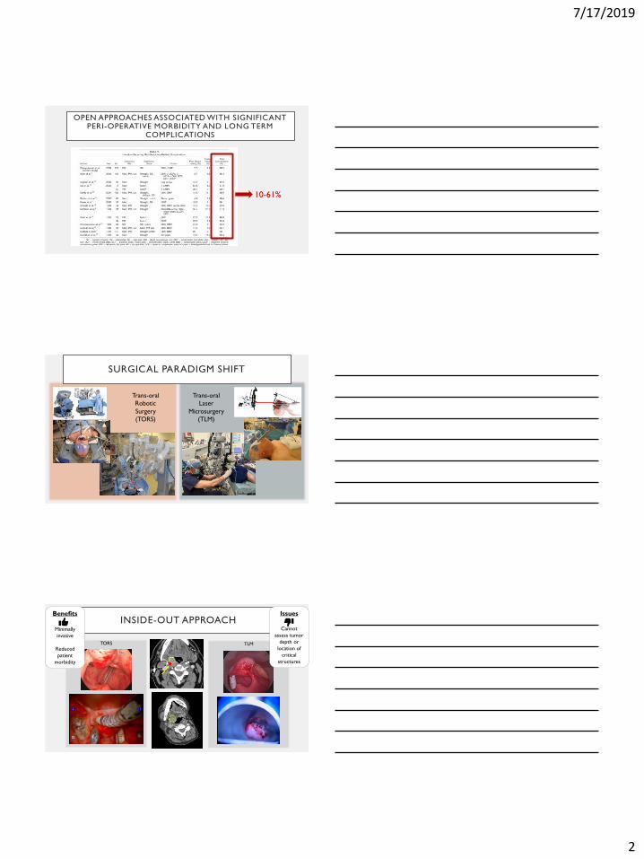
OPEN APPROACHES ASSOCIATED WITH SIGNIFICANT PERI-OPERATIVE MORBIDITY AND LONG TERM
COMPLICATIONS
10-61%
SURGICAL PARADIGM SHIFT
Trans-oral
Robotic
Surgery
(TORS)
Trans-oral
Laser
Microsurgery
(TLM)
INSIDE-OUT APPROACH
TORS TLM
Benefits
Minimally
invasive
Reduced
patient
morbidity
Issues
Cannot
assess tumor
depth or
location of
critical
structures

7/17/2019
3

7/17/2019
4
COULD INTRAOPERATIVE
IMAGING IMPROVE SAFETY AND EFFICACY?
• Unique Dartmouth imaging resource:
• Center for Surgical Innovation (CSI)
• 2 Operating Rooms
• 1 Procedure Room
• Intra-operative CT and MRI systems
• Intra-operative navigation
• Animal and human use
RETRACTORS AND SCOPES
USED ARE METAL AND NOT CT/MRI COMPATIBLE
3D PRINTED CT/MRI COMPATIBLE LARYNGOSCOPY SYSTEM
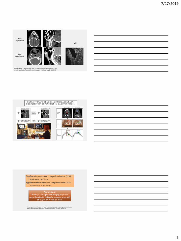
7/17/2019
5
Our
Laryngoscope
Metal
Laryngoscope
MRI
CT
Paydarfar JA, Wu X, Halter RJ. MRI- and CT-Compatible Polymer Laryngoscope: A Step
toward Image-Guided Transoral Surgery. Otolaryngol -- Head Neck Surg. 2016;610:1-3.
CURRENT STATE OF LOCALIZATION ACCURACY: LOCALIZATION EXPERIMENT IN CADAVER MODEL
Significant improvement in target localization (21%)
12.8±9.9 versus 10±7.5 mm
Significant reduction in task completion time (25%)
22 minutes down to 16 minutes
Conclusions:
Although intraoperative imaging improves
target localization, clinically surgeons were still
off target by 10 mm or more
P. Kahng, X. Wu, N. Ramesh, D. Pastel, R. Halter, J. Paydarfar, “Improving target localization
during trans-oral surgery with use of intraoperative imaging,” IJCARS, Feb. 2019.

7/17/2019
6
IMAGE GUIDED SURGICAL
NAVIGATION IN TORS/TLM?
• Successfully used in sinus
and skull base surgery,
neurosurgery, orthopedic
spine
• Actively researched in GI,
GU, thoracic surgery, others
IMAGE GUIDED SURGICAL NAVIGATION IMPROVES SAFETY SINUS SURGERY
• Systematic Review
• IGS vs non-IGS
• Major complications significantly less in IGS group:
• Entry into any area outside sinuses (eye, brain)
• Post-op bleeding requiring surgical intervention
• Abort procedure for any reason
Otolaryngology–Head and Neck Surgery 149(1) 17–29
USE OF IMAGE GUIDANCE DURING ENDOSCOPIC SINUS SURGERY
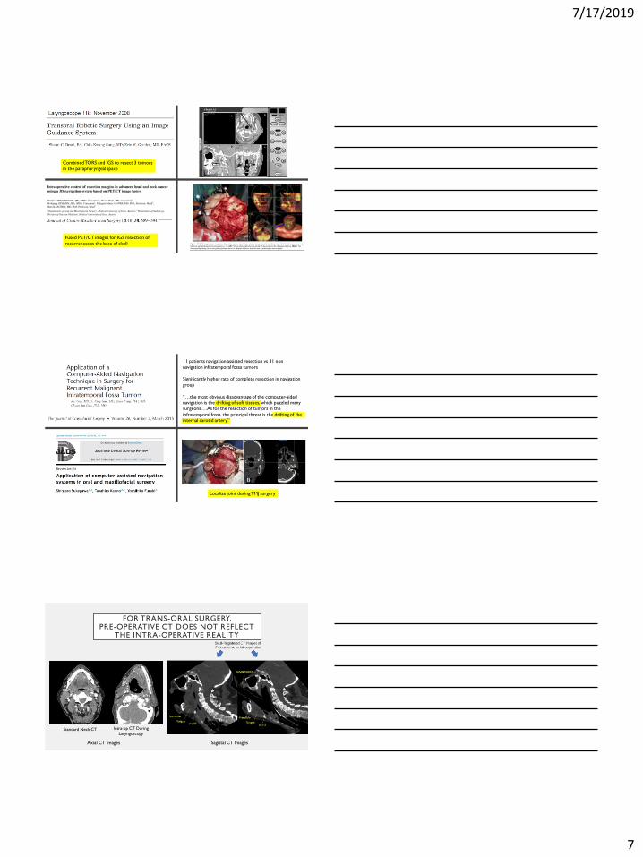
7/17/2019
7
Combined TORS and IGS to resect 3 tumors
in the parapharyngeal space
Fused PET/CT images for IGS resection of
recurrences at the base of skull
Localize joint during TMJ surgery
11 patients navigation assisted resection vs 31 non
navigation infratemporal fossa tumors
Significantly higher rate of complete resection in navigation
group
“…the most obvious disadvantage of the computer-aided
navigation is the drifting of soft tissues, which puzzled many
surgeons….As for the resection of tumors in the
infratemporal fossa, the principal threat is the drifting of the
internal carotid artery.”
FOR TRANS-ORAL SURGERY, PRE-OPERATIVE CT DOES NOT REFLECT
THE INTRA-OPERATIVE REALITY
Standard Neck CT Intra-op CT During
Laryngoscopy
Axial CT Images Sagittal CT Images

7/17/2019
8
PROOF OF CONCEPT: SURGICAL NAVIGATION WITH INTRAOPERATIVE IMAGING TO IMPROVE
LOCALIZATION ACCURACY
Paydarfar JA, Wu X, Halter RJ. Initial experience with image‐guided surgical
navigation in transoral surgery. Head & Neck. 2018;1–10
HIGH LEVEL OF REGISTRATION
ACCURACY (<= 1 MM)
7/17/2019
9
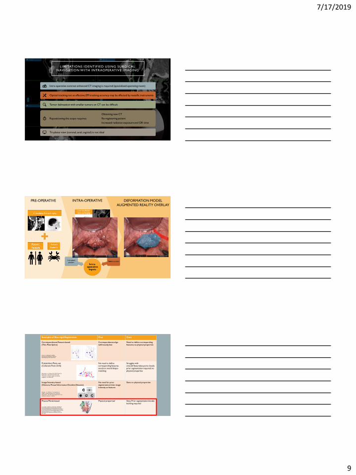
LIMITATIONS IDENTIFIED USING SURGICAL NAVIGATION WITH INTRAOPERATIVE IMAGING
Intra-operative contrast enhanced CT imaging is required (specialized operating room)
Optical tracking not as effective; EM tracking accuracy may be affected by metallic instruments
Tumor delineation with smaller tumors on CT can be difficult
Repositioning the scope requires:
Obtaining new CT
Re-registering patient
Increased radiation exposure and OR time
Tri-planar view (coronal, axial, sagittal) is not ideal
PRE-OPERATIVE INTRA-OPERATIVE DEFORMATION MODEL
AUGMENTED REALITY OVERLAY
Examples of Non-rigid Registration Pros Cons
Correspondences/Feature-based:
(Thin-Plate Spline)
Correspondences align
well/exactly; fast
Need to define corresponding
features; no physical properties
Probabilistic/Point-set:
(Coherent Point Drift)
No need to define
corresponding features;
excels at overall shape-
matching
Struggles with
smooth/featureless point-clouds;
prior segmentation required; no
physical properties
Image/Intensity-based:
(Demons, Mutual Information/Gradient Descent)
No need for prior
segmentation; Uses image
intensity as features
Slow; no physical properties
Physics/Model-based Physical properties! Slow; Prior segmentation/model-
building required
Donato, G., & Belongie, S.J. (2002).
Approximation Methods for Thin Plate
Spline Mappings and Principal Warps.
Myronenko, A., & Song, X.B. (2010). Point Set
Registration: Coherent Point Drift. IEEE
Transactions on Pattern Analysis and Machine
Intelligence, 32, 2262-2275.
Pennec, X., Cathier, P., & Ayache, N. (1999). Understanding the "Demon's Algorithm": 3D Non-rigid Registration by Gradient Descent. MICCAI.
J. E. Lloyd, I. Stavness , and S. Fels, “ArtiSynth: A Fast Interactive Biomechanical Modeling Toolkit Combining Multibody and Finite Element Simulation,” in Soft Tissue Biomechanical Modeling for Computer Assisted Surgery, vol. 11, Y. Payan, Ed. Berlin, Heidelberg: Springer Berlin Heidelberg, 2012, pp. 355–394.

7/17/2019
10
TLE improved from
11.2±5.0 mm vs. 5.8±2.5 with IGS
Patient placed
under general
anesthesia
Fiducial markers are
placed along the
tongue
Pre-laryngoscopy
contrast CT scan is
obtained
CT compatible
laryngoscope is
introduced and
placed in
suspension
Intra-operative
contrast CT scan is
obtained
PROTOCOL TO STUDY UPPER AERODIGESTIVE TRACT DEFORMATION
Patients undergoing staging or
operative laryngoscopy
No dentition (reduce artifact)
10 without prior treatment
5 with prior radiation

7/17/2019
11
Mandible Displacement Hyoid Displacement Tongue Deformation
QUANTIFY DEFORMATION DURING LARYNGOSCOPY
X. Wu, J. A. Paydarfar, and R. J. Halter, “Quantifying
Anatomic Deformations During Laryngoscopy,” Ann. Biomed. Eng., vol. 46, no. 6, pp. 912–925, 2018.
X. Wu, J. Paydarfar, and R. Halter, “Intraoperative deformation during laryngoscopy of
irradiated and non-irradiated patients,” in Medical Imaging 2018: Image-Guided
Procedures, Robotic Interventions, and Modeling, 2018, vol. 10576, p. 45.
C. Rees, X. Wu, D. Pastel , R. Halter, J. Paydarfar, “Prior radiation exposure alters
airway deformation during laryngoscopy,” The Triological Society Sections
Meeting, Coronado, CA, Jan. 2019.
PRIOR RADIATION SIGNIFICANTLY AFFECTS DEFORMATION
Differences in translation and rotation of
mandible and hyoid bone
Reduced pharyngeal airway volume
VISUALIZE TISSUE AND TUMOR DEFORMATION DURING LARYNGOSCOPY

7/17/2019
12
QUANTIFY FORCES GENERATED DURING OPERATIVE LARYNGOSCOPY
Avg peak force of 50 lbf recorded
on scope!
10 lbf on chest!
A. Ponukumati, X. Wu, P. Kahng, J. Skinner, J. Paydarfar, R. Halter, “A System for
Characterizing Intraoperative Force Distribution during Operative Laryngoscopy,”
IEEE Transactions of Biomedical Engineering, Submitted Mar. 2019.
SUMMARY OF WORK COMPLETED TO DATE

7/17/2019
13
F.R .A.N.K. : FUNCTIONAL REFERENCE ANATOMY KNOWLEDGE
• Collaboration with University of British
Columbia
• Hybrid model: Combines both FEM and multi-
body methods
• Generic template of head and neck structures
• Patient-specific model created by registering
template to segmented CT images
P. Anderson, S. Fels, N. M. Harandi, A. Ho, S. Moisik, C. A. Sánchez, I. Stavness, and
K. Tang, “FRANK: A Hybrid 3D Biomechanical Model of the Head and Neck,” in
Biomechanics of Living Organs: Hyperelastic Constitutive Laws for Finite Element
Modeling, 2017, pp. 413–447.
J. E. Lloyd, I. Stavness, and S. Fels, “ArtiSynth: A Fast Interactive Biomechanical
Modeling Toolkit Combining Multibody and Finite Element Simulation,” in Soft Tissue
Biomechanical Modeling for Computer Assisted Surgery, vol. 11, Y. Payan, Ed. Berlin,
Heidelberg: Springer Berlin Heidelberg, 2012, pp. 355–394.
39
Deformation
Modeling and Deformable
Registration in
TOS
7 With University of British Columbia
Real Tracked Trajectory
TLE
9.7±3.0mm

7/17/2019
14
FUTURE DIRECTIONS
Pressure and position data to drive dynamic model in cadaver and clinical studies
Develop CT/MRI compatible retractors for TORS applications
Incorporate EM tracking with robotic instrumentation
Examine the use of lower resolution intra-operative imaging (O-Arm) in cadaver model
ACKNOWLEDGEMENTS
• Thayer Co-PI:• Ryan Halter PhD
• DHMC Radiology:• David Pastel MD
• Thayer graduate student:• Xiaotian (Dennis) Wu
• Geisel Medical students:• Peter Kahng
• Aravind Ponukumati
• Christiaan Rees PhD
• Michael Sramek
• Otolaryngology resident:• Eric Eisen MD
• Undergraduate students:• Jiyoo Chang
• Erick Quintanilla
• CSI:• Michael Pearl
• Michaela Whitty
• John Pieffer
• DHMC Engineering:• Doug McKenney
• Machine Shop:• Pete Fontaine
• Jason Downs
• Kevin Baron
Grant #: UL1TR001086
2018 DOS Faculty
Development Award