9781461455370 c1
TRANSCRIPT
17G.S. Everly and J.M. Lating, A Clinical Guide to the Treatment of the Human Stress Response, DOI 10.1007/978-1-4614-5538-7_2, © Springer Science+Business Media New York 2013
In the fi rst chapter, we provided the following working de fi nition of the stress response: “Stress is a physiological response that serves as a mechanism of media-tion linking any given stressor to its target-organ effect.” By viewing the phenome-nology of stress within the context of a “linking” mechanism, we can answer one of the most critical questions in psychosomatic medicine, that is, through what mecha-nisms can stressor stimuli, such as life events, lead to disease and dysfunction? The response to that query will be addressed within the next two chapters.
This chapter describes, within the boundaries of historical reviews and foundations, current fi ndings and speculation, the anatomical and physiological foundations of the human stress response by (1) addressing basic neuroanatomical structures and (2) trac-ing the psychophysiological effector mechanisms that actually represent the stress response, as currently de fi ned. To assist in the pedagogical process, a basic model of the human stress response is constructed to serve as a unifying thread for better under-standing of not only the phenomenology of human stress but also its measurement and treatment. Chapter 3 will pursue the logical extension by reviewing several models of pathogenesis, that is, the process by which stress arousal leads to disease.
Neurological Foundations
In order to understand the stress response, we must fi rst understand its foundations, which reside in the structure and function of the human nervous systems.
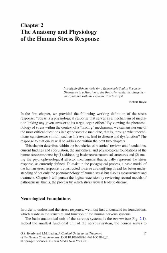
The basic anatomical unit of the nervous systems is the neuron (see Fig. 2.1 ). Indeed the smallest functional unit of the nervous system, the neuron serves to
Chapter 2 The Anatomy and Physiology of the Human Stress Response
It is highly dishonorable for a Reasonable Soul to live in so Divinely built a Mansion as the Body she resides in, altogether unacquainted with the exquisite structure of it.
Robert Boyle
18 2 The Anatomy and Physiology of the Human Stress Response
conduct sensory, motor, and regulatory signals throughout the body. The neuron consists of three basic units: (1) the dendrites and their outermost membranes—the postsynaptic dendritic membranes; (2) the neural cell body , which contains the nucleus of the cell; and (3) the axon , with its branching projections called the telo-dendria and their end points, the presynaptic membranes.
Neural Transmission
An incoming signal is fi rst received by the postsynaptic membranes of the dendrites. Chemical (metabotropic) or electrical (ionotropic) processes are initiated upon stimu-lation of the postsynaptic dendritic membranes, which cause the neuron to conduct the incoming signal through the dendrites and the cell body. Finally, a neural impulse relayed to the axon travels down the axon until it reaches the telodendria and ulti-mately the presynaptic membranes. It is the task of the presynaptic membrane to relay the signal to the subsequent postsynaptic membrane of the next neuron. This is not easily achieved, however, because the neurons do not actually touch one another. Rather, there exists a space between neurons called the synaptic cleft.
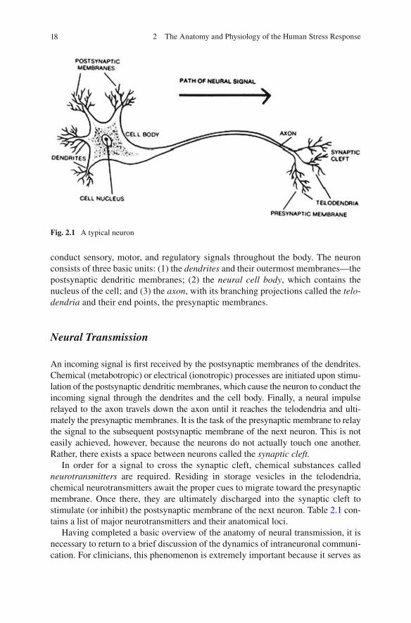
In order for a signal to cross the synaptic cleft, chemical substances called neurotransmitters are required. Residing in storage vesicles in the telodendria, chemical neurotransmitters await the proper cues to migrate toward the presynaptic membrane. Once there, they are ultimately discharged into the synaptic cleft to stimulate (or inhibit) the postsynaptic membrane of the next neuron. Table 2.1 con-tains a list of major neurotransmitters and their anatomical loci.
Having completed a basic overview of the anatomy of neural transmission, it is necessary to return to a brief discussion of the dynamics of intraneuronal communi-cation. For clinicians, this phenomenon is extremely important because it serves as
Fig. 2.1 A typical neuron
19Neurological Foundations
the basis for electrophysiological events such as electromyography, electrocardiog-raphy, and electroencephalography.
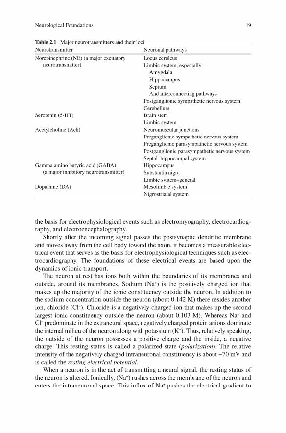
Shortly after the incoming signal passes the postsynaptic dendritic membrane and moves away from the cell body toward the axon, it becomes a measurable elec-trical event that serves as the basis for electrophysiological techniques such as elec-trocardiography. The foundations of these electrical events are based upon the dynamics of ionic transport.
The neuron at rest has ions both within the boundaries of its membranes and outside, around its membranes. Sodium (Na + ) is the positively charged ion that makes up the majority of the ionic constituency outside the neuron. In addition to the sodium concentration outside the neuron (about 0.142 M) there resides another ion, chloride (Cl − ). Chloride is a negatively charged ion that makes up the second largest ionic constituency outside the neuron (about 0.103 M). Whereas Na + and Cl − predominate in the extraneural space, negatively charged protein anions dominate the internal milieu of the neuron along with potassium (K + ). Thus, relatively speaking, the outside of the neuron possesses a positive charge and the inside, a negative charge. This resting status is called a polarized state ( polarization ). The relative intensity of the negatively charged intraneuronal constituency is about −70 mV and is called the resting electrical potential.
When a neuron is in the act of transmitting a neural signal, the resting status of the neuron is altered. Ionically, (Na + ) rushes across the membrane of the neuron and enters the intraneuronal space. This in fl ux of Na+ pushes the electrical gradient to
Table 2.1 Major neurotransmitters and their loci
Neurotransmitter Neuronal pathways
Norepinephrine (NE) (a major excitatory neurotransmitter)
Locus ceruleus Limbic system, especially Amygdala Hippocampus Septum And interconnecting pathways Postganglionic sympathetic nervous system Cerebellum
Serotonin (5-HT) Brain stem Limbic system
Acetylcholine (Ach) Neuromuscular junctions Preganglionic sympathetic nervous system Preganglionic parasympathetic nervous system Postganglionic parasympathetic nervous system Septal–hippocampal system
Gamma amino butyric acid (GABA) (a major inhibitory neurotransmitter)
Hippocampus Substantia nigra Limbic system–general
Dopamine (DA) Mesolimbic system Nigrostriatal system
20 2 The Anatomy and Physiology of the Human Stress Response
about +50 mV (from the resting −70 mV). This process of sodium ion in fl ux is called depolarization and represents the actual fi ring, or discharge, of the neuron. Depolarization lasts about 1.5 ms. Depolarization moves longitudinally along the axon as a wave of ionic in fl ux. After 1.5 ms, however, the neuron begins to repolarize. Repolarization occurs as K + and Na + are pumped out of the neuron and any remaining Na + is assimilated into the neuron itself. The result of repolarization is the return of the +50 mV to a resting −70 mV, ready for subsequent discharge. This process is shown in Fig. 2.2 .
Fig. 2.2 The electrochemical neural impulse
21Neurological Foundations
Basic Neuroanatomy
From the preceding discussion of basic neural transmission, the next step to be undertaken is an analysis of the fundamental anatomical structures involved in the human stress response.
The nervous systems, the functional structures within which millions upon mil-lions of neurons reside, may be classi fi ed from either an anatomical or a functional perspective. For the sake of parsimony, we describe the nervous systems from an anatomical perspective.
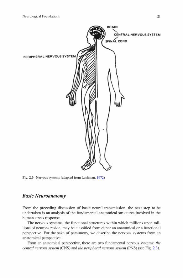
From an anatomical perspective, there are two fundamental nervous systems: the central nervous system (CNS) and the peripheral nervous system (PNS) (see Fig. 2.3 ).
Fig. 2.3 Nervous systems (adapted from Lachman, 1972 )
22 2 The Anatomy and Physiology of the Human Stress Response
Table 2.2 The human nervous systems
The central nervous system (CNS) Brain Spinal cord
The peripheral nervous systems (PNS) The somatic branch The autonomic branches (ANS)
Sympathetic (SNS) Parasympathetic (PSNS)
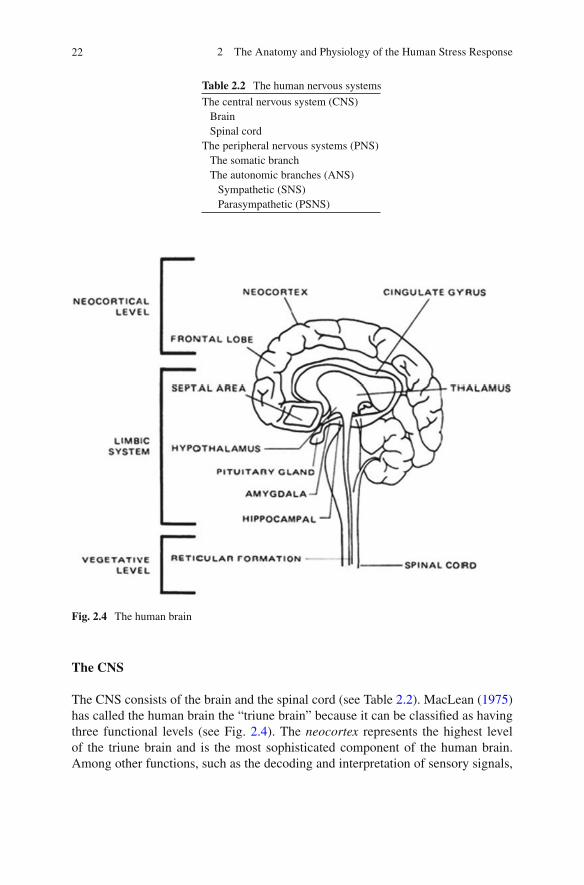
Fig. 2.4 The human brain
The CNS
The CNS consists of the brain and the spinal cord (see Table 2.2 ). MacLean ( 1975 ) has called the human brain the “triune brain” because it can be classi fi ed as having three functional levels (see Fig. 2.4 ). The neocortex represents the highest level of the triune brain and is the most sophisticated component of the human brain. Among other functions, such as the decoding and interpretation of sensory signals,
23Neurological Foundations
communications, and gross control of motor (musculoskeletal) behaviors, the neocortex (primarily the frontal lobe ) presides over imagination, logic, decision making, memory, problem solving, planning, and apprehension.
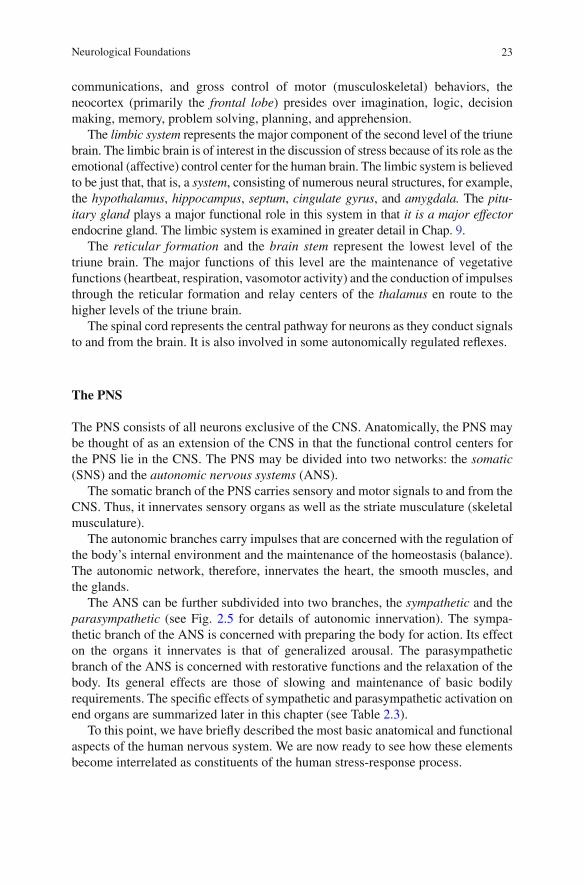
The limbic system represents the major component of the second level of the triune brain. The limbic brain is of interest in the discussion of stress because of its role as the emotional (affective) control center for the human brain. The limbic system is believed to be just that, that is, a system , consisting of numerous neural structures, for example, the hypothalamus , hippocampus , septum , cingulate gyrus , and amygdala. The pitu-itary gland plays a major functional role in this system in that it is a major effector endocrine gland. The limbic system is examined in greater detail in Chap. 9 .
The reticular formation and the brain stem represent the lowest level of the triune brain. The major functions of this level are the maintenance of vegetative functions (heartbeat, respiration, vasomotor activity) and the conduction of impulses through the reticular formation and relay centers of the thalamus en route to the higher levels of the triune brain.
The spinal cord represents the central pathway for neurons as they conduct signals to and from the brain. It is also involved in some autonomically regulated re fl exes.
The PNS
The PNS consists of all neurons exclusive of the CNS. Anatomically, the PNS may be thought of as an extension of the CNS in that the functional control centers for the PNS lie in the CNS. The PNS may be divided into two networks: the somatic (SNS) and the autonomic nervous systems (ANS).
The somatic branch of the PNS carries sensory and motor signals to and from the CNS. Thus, it innervates sensory organs as well as the striate musculature (skeletal musculature).
The autonomic branches carry impulses that are concerned with the regulation of the body’s internal environment and the maintenance of the homeostasis (balance). The autonomic network, therefore, innervates the heart, the smooth muscles, and the glands.
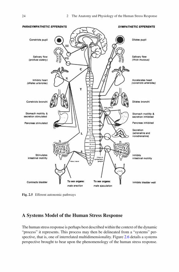
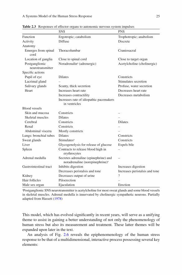
The ANS can be further subdivided into two branches, the sympathetic and the parasympathetic (see Fig. 2.5 for details of autonomic innervation). The sympa-thetic branch of the ANS is concerned with preparing the body for action. Its effect on the organs it innervates is that of generalized arousal. The parasympathetic branch of the ANS is concerned with restorative functions and the relaxation of the body. Its general effects are those of slowing and maintenance of basic bodily requirements. The speci fi c effects of sympathetic and parasympathetic activation on end organs are summarized later in this chapter (see Table 2.3 ).
To this point, we have brie fl y described the most basic anatomical and functional aspects of the human nervous system. We are now ready to see how these elements become interrelated as constituents of the human stress-response process.
24 2 The Anatomy and Physiology of the Human Stress Response
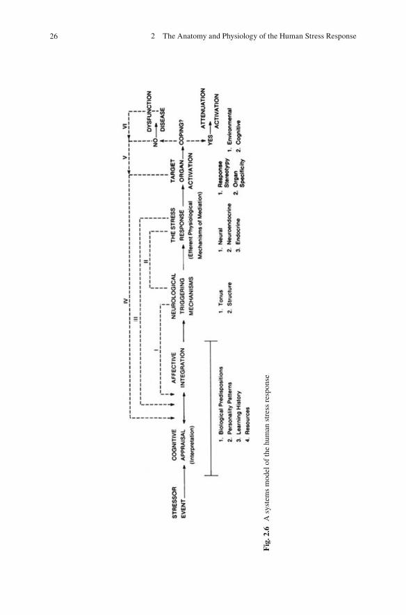
A Systems Model of the Human Stress Response
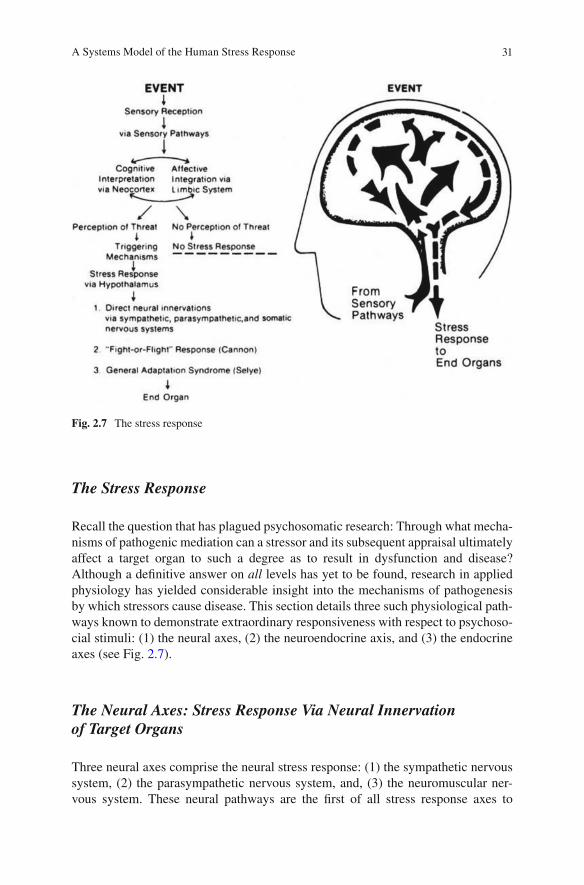
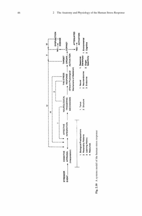
The human stress response is perhaps best described within the context of the dynamic “process” it represents. This process may then be delineated from a “systems” per-spective, that is, one of interrelated multidimensionality. Figure 2.6 details a systems perspective brought to bear upon the phenomenology of the human stress response.
Fig. 2.5 Efferent autonomic pathways
25A Systems Model of the Human Stress Response
This model, which has evolved signi fi cantly in recent years, will serve as a unifying theme to assist in gaining a better understanding of not only the phenomenology of human stress but also its measurement and treatment. These latter themes will be expanded upon later in the text.
An analysis of Fig. 2.6 reveals the epiphenomenology of the human stress response to be that of a multidimensional, interactive process possessing several key elements:
Table 2.3 Responses of effector organs to autonomic nervous system impulses SNS PNS
Function Ergotropic; catabolism Trophotropic; anabolism Activity Diffuse Discrete Anatomy
Emerges from spinal cord
Thoracolumbar Craniosacral
Location of ganglia Close to spinal cord Close to target organ Postganglionic
neurotransmitter Noradrenalin a (adrenergic) Acetylcholine (cholinergic)
Speci fi c actions Pupil of eye Dilates Constricts Lacrimal gland – Stimulates secretion Salivary glands Scanty, thick secretion Profuse, water secretion Heart Increases heart rate Decreases heart rate
Increases contractility Decreases metabolism Increases rate of idiopathic pacemakers
in ventricles Blood vessels
Skin and mucosa Constricts – Skeletal muscles Dilates – Cerebral Constricts Dilates Renal Constricts – Abdominal viscera Mostly constricts –
Lungs: bronchial tubes Dilates Constricts Sweat glands Stimulates a Constricts Liver Glycogenolysis for release of glucose Expels bile Spleen Contracts to release blood high in
erythrocytes –
Adrenal medulla Secretes adrenaline (epinephrine) and noradrenaline (norepinephrine) a
–
Gastrointestinal tract Inhibits digestion Increases digestion Decreases peristalsis and tone Increases peristalsis and tone
Kidney Decreases output of urine ? Hair follicles Piloerection – Male sex organ Ejaculation Erection
a Postganglionic SNS neurotransmitter is acetylcholine for most sweat glands and some blood vessels in skeletal muscles. Adrenal medulla is innervated by cholinergic sympathetic neurons. Partially adapted from Hassett ( 1978 )
26 2 The Anatomy and Physiology of the Human Stress Response
Fig
. 2.6
A
sys
tem
s m
odel
of
the
hum
an s
tres
s re
spon
se
27A Systems Model of the Human Stress Response
1. Stressor events (real or imagined). 2. Cognitive appraisal and affective integration. 3. Neurological triggering mechanisms (e.g., locus ceruleus, limbic nuclei, hypo-
thalamic nuclei). 4. The stress response (a physiological mechanism of mediation). 5. Target-organ activation. 6. Coping behavior.
A detailed analysis of each of these elements is appropriate at this point.
Stressor Events
Because Selye used the term stress to refer to a “response,” it was necessary to employ a word to delineate the stimulus for the stress response—that word is stressor. Stressor events, as noted earlier, fall in one of the two categories: (1) psychosocial stressors and (2) biogenic stressors (Girdano, Dusek, & Everly, 2009 ) .
Psychosocial stressors are either real or imagined environmental events that “set the stage” for the elicitation of the stress response. They cannot directly “cause” the stress response but must work through cognitive appraisal mechanics. Most stres-sors are, indeed, psychosocial stressors. For this reason, one may argue that “stressors, like beauty, reside in the eye of the beholder.”
Biogenic stressors, however, actually “cause” the elicitation of the stress response. Such stimuli bypass the higher cognitive appraisal mechanisms and work directly on affective and neurological triggering nuclei. Thus, by virtue of their biochemical properties, they directly initiate the stress response without the usual requisite cognitive–affective processing. Examples of such stimuli include the following:
Ginseng • Ginkgo biloba • Amphetamine • Phenylpropanolamine • Caffeine • Theobromine • Theophylline • Nicotine • Certain physical factors such as pain-evoking stimuli, extreme heat, and extreme • cold Guarana • Yohimbine •
As just mentioned, however, most stressors are not biogenic stressors. Therefore, in clinical practice, therapists will most likely be treating patients who are plagued by environmental events—real, imagined, anticipated, or recalled—that are per-ceived in such a manner as to lead to activation of the stress response. To better understand this process we move now to the second step in the model: the cognitive–affective integration stage.
28 2 The Anatomy and Physiology of the Human Stress Response
Cognitive–Affective Domain
Practically speaking, there is simply no such thing as “reality” without considering the human perspective that might be brought to bear upon it. The cognitive–affective domain is delineated within this model in order to capture that notion.
Cognitive appraisal refers to the process of cognitive interpretation, that is, the meanings that we assign to the world as it unfolds before us. Affective integration refers to the blending and coloring of felt emotion into the cognitive interpretation. The resultant cognitive–affective complex represents how the stressors are ultimately perceived. In effect, this critical integrated perception represents the determination of whether psychosocial stimuli become psychosocial stressors or not. Such a perceptual process, however, is uniquely individualized and vulnerable to biological predisposi-tions (Millon & Everly, 1985 ) , personality patterns (Millon, Grossman, Millon, Meagher, & Ramnath, 2004 ) , learning history (Lachman, 1972 ) , and available resources for coping (Lazarus, 2006 ; Lazarus & Folkman, 1984 ) .
Although Fig. 2.6 portrays a reciprocity between cognitive and affective mecha-nisms, it should be noted that there exists substantial evidence supporting the cogni-tive primacy hypothesis (see Chap. 8 ); that is, cognition determines affect (felt emotion) and thus assumes a superordinate role in the process of restructuring human behavior patterns. Let us explore this important notion further.
Perhaps the earliest recognition that cognition is superordinate to affect has been credited by Albert Ellis to the fi fth-century Greco-Roman philosopher Epictetus, who reportedly said, “Men are disturbed not by things, but by the views which they take of them.” The science of physiology follows in kind. Hans Selye, also known as the father of modern endocrinology, has summarized over 50 years of research into human stress with the conclusion, “It is not what happens to you that matters, but how you take it,” Similarly, the noted neurophysiologist Ernest Gellhorn (Gellhorn & Loofbourrow, 1963 ) recognized the preeminent role of the prefrontal lobe cognitive processes in felt and expressed emotion in his research spanning the 1950s, 1960s, and 1970s. In fl uential authors such as Arnold ( 1970, 1984 ) , Cassel ( 1974 ) , Lazarus ( 1966, 1982, 1991 ) , Meichenbaum ( 1985 ) , Meichenbaum and Jaremko ( 1983 ) , and Selye ( 1976 ) strongly support the cognitive primacy position as it relates to human stress.
More recently, Everly, Davy, Smith, Lating, and Nucifora ( 2011 ) [also see Everly, Smith, and Lating ( 2009 ) and Smith, Everly, and Johns ( 1992, 1993 ) ] assessed the role of cognitive processes in the determination of stress-related illness. “Stressors, like beauty, lie in the eye of the beholder,” is the assertion. Using a sample of 1,618 adults, Smith, Everly, and Johns ( 1993 ) employed structural modeling, exploratory, and con fi rmatory factor analyses to investigate the relative roles of environmental stressors compared to cognitive processes as predictors of physiological symp-toms of stress-related illness or dysfunction. The role of coping mechanisms was also investigated. The results of this investigation indicated that, consistent with the speculations of Epictetus and even Selye, environmental stressors exert their pathogenic effect only indirectly. Rather, environmental stressors act “through their
29A Systems Model of the Human Stress Response
ability to cause psychological discord. In fact, psychological discord had the stron-gest in fl uence on maladaptive coping behaviors and stress-related illness” (Smith et al., 1993 , p. 445). Psychological discord, as assessed by these authors, re fl ects the cognitive interpretations of the environmental events.
An extended physiological perspective may be of value at this point. If a given, nonsympathomimetic stimulus is to engender a stress response, it must fi rst be received by the receptors of the PNS. Once stimulated, these receptors send their impulses along the PNS toward the brain. According to Pen fi eld ( 1975 ) , once in the CNS, collateral neurons diverge from the main ascending pathways to the neocorti-cal targets and innervate the reticular formation. These collaterals diverge and pass through limbic constituents, but seldom are such afferent diversions suf fi cient to generate full-blown emotional reactions. Rather, such diversions may account for nonspeci fi c arousal (startle or defense re fl exes) or subtle affective coloration (“gut reactions”). Cognitive theorists do not regard these momentary acute, ontogeneti-cally primitive events as emotions (Lazarus, 1982 ) .
These divergent pathways ultimately reunite with the main ascending pathways and innervate the primary sensory and appraisal loci. Arnold ( 1984 ) has written that “the sheer experience of things around us cannot lead to action unless they are appraised for their effect on us” (p. 125). She has hypothesized the anatomical locus of such appraisal to be the cingulate gyrus and the limbic–prefrontal neocortical interface (see Aggleton, 1992 ) .
Arnold ( 1984 ) notes that the granular cells of the limbic–prefrontal interface contain relay centers that connect all sensory, motor, and association areas. She states:
These connections would enable the individual to appraise information from every modal-ity: smells, via relays from the posterior orbital cortex; movement and movement impulses, via relays from frontal and prefrontal cortex; somatic sensations can provide data via relays from parietal association areas; and things seen could be appraised over relays from occipi-tal association areas. Finally, something heard can be appraised as soon as relays from the auditory association area reach the hippocampal gyrus. (pp. 128–129)
As noted in Fig. 2.6 , appraisal is a function of any existing biological predisposi-tions, personality patterns, learning history, and available coping resources. Once appraisal is made, efferent impulses project so as to potentiate the stimulation of two major effector systems:
1. Impulses project back to the highly sensitive emotional anatomy in the limbic system (Arnold, 1984 ; Cullinan, Herman, Helmreich, & Watson, 1995 ; Gellhorn & Loufbourrow, 1963 ; Gevarter, 1978 ; Nauta, 1979 ) , especially the hippocampus (Reiman et al., 1986 ) , for the experience of stimulus-speci fi c felt emotion and the potential to trigger visceral effector mechanisms.
2. Impulses similarly project to the areas of the neocortex concerned with neuro-muscular behavior where, through pyramidal and extrapyramidal systems, mus-cle tone (tension) is increased and the intention to act can be potentially translated to actual overt motor activity (Gellhorn, 1964a, 1964b ) .
30 2 The Anatomy and Physiology of the Human Stress Response
Thus far, we have seen that psychosocial stimuli, once perceived, excite nonspeci fi c arousal and cognitive appraisal mechanisms. If the appraisal of the stimulus is ultimately one of threat, challenge, or aversion, then emotional arousal will likely result.
In most individuals, activation of the limbic centers for emotional arousal leads to expression of the felt emotion in the form of visceral activation and neuromuscu-lar activity. Such visceral and neuromuscular activation represents the multiaxial physiological mechanisms of mediation Selye called the “stress response.” Thus, in the fi nal analysis, it can be seen that physiological reactions to psychosocial stimuli result from the cognitive interpretations and emotional reactions to those stimuli, not the stimuli themselves. Stressors are, indeed, in the eye of the beholder!
Before turning to a discussion of the multiaxial nature of the stress response, we must fi rst discuss a mechanism that prefaces activation of the stress response axes. Research in the last several years has necessitated speci fi c consideration of mecha-nisms that serve to “trigger” the elicitation of the multiaxial stress response. These mechanisms are referred to as neurological triggering mechanisms.
Neurological Triggering Mechanisms
The next step in the model depicted in Fig. 2.6 is the neurological triggering mechanisms consisting of the locus ceruleus (LC), limbic system, and hypotha-lamic efferent triggering complex. Linked through ventral and dorsal adrenergic as well as serotonergic projections (among others), this complex appears to con-sist of the LC, the hippocampus, the septal–hippocampal–amygdaloid complexes, and the anterior and posterior hypothalamic nuclei (Nauta & Domesick, 1982 ; Reiman et al., 1986 ) . These structures appear to be the anatomical epicenters for the visceral and somatic efferent discharges in response to emotional arousal (Aggleton, 1992 ; Gellhorn, 1964a, 1964b, 1965, 1967 ; MacLean, 1949 ; Nauta, 1979 ; Redmond, 1979 ) ; that is, these structures appear to give rise to the multi-axial stress response. Indeed, these centers even seem capable of establishing an endogenously determined neurological tone that is potentially self-perpetuating (Gellhorn, 1967 ; Weil, 1974 ) . This notion of a positive feedback loop is initially depicted in Fig. 2.6 by the dotted line labeled I. Subsequent dotted lines are labeled with Roman numerals to show other feedback mechanisms that maintain what Gellhorn ( 1957 ) has called a state of “egotropic tuning,” what Everly (Everly & Benson, 1989 ) calls “limbic hypersensitivity” (discussed in Chap. 3 ), and what Weil ( 1974 ) has called a “charged arousal system.” Each of these terms is indica-tive of a predisposition for physiological arousal.
More speci fi cally, these terms describe a preferential pattern of SNS (and related arousal mechanism) responsiveness. Such a chronic tonic status may, over time, serve as the basis for a host of psychiatric and psychophysiological disorders (Gellhorn, 1967 ) . The mechanisms by which such neurological tone can exert an effect upon a given target organ is the subject of the next phase of the system’s model: the stress response—a physiological mechanism of mediation.
31A Systems Model of the Human Stress Response
The Stress Response
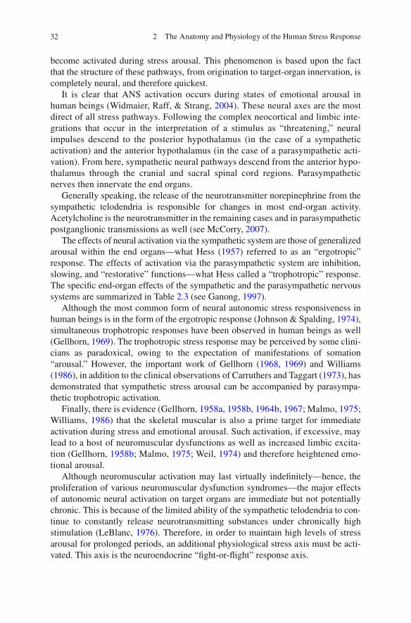
Recall the question that has plagued psychosomatic research: Through what mecha-nisms of pathogenic mediation can a stressor and its subsequent appraisal ultimately affect a target organ to such a degree as to result in dysfunction and disease? Although a de fi nitive answer on all levels has yet to be found, research in applied physiology has yielded considerable insight into the mechanisms of pathogenesis by which stressors cause disease. This section details three such physiological path-ways known to demonstrate extraordinary responsiveness with respect to psychoso-cial stimuli: (1) the neural axes, (2) the neuroendocrine axis, and (3) the endocrine axes (see Fig. 2.7 ).
The Neural Axes: Stress Response Via Neural Innervation of Target Organs
Three neural axes comprise the neural stress response: (1) the sympathetic nervous system, (2) the parasympathetic nervous system, and, (3) the neuromuscular ner-vous system. These neural pathways are the fi rst of all stress response axes to
Fig. 2.7 The stress response
32 2 The Anatomy and Physiology of the Human Stress Response
become activated during stress arousal. This phenomenon is based upon the fact that the structure of these pathways, from origination to target-organ innervation, is completely neural, and therefore quickest.
It is clear that ANS activation occurs during states of emotional arousal in human beings (Widmaier, Raff, & Strang, 2004 ) . These neural axes are the most direct of all stress pathways. Following the complex neocortical and limbic inte-grations that occur in the interpretation of a stimulus as “threatening,” neural impulses descend to the posterior hypothalamus (in the case of a sympathetic activation) and the anterior hypothalamus (in the case of a parasympathetic acti-vation). From here, sympathetic neural pathways descend from the anterior hypo-thalamus through the cranial and sacral spinal cord regions. Parasympathetic nerves then innervate the end organs.
Generally speaking, the release of the neurotransmitter norepinephrine from the sympathetic telodendria is responsible for changes in most end-organ activity. Acetylcholine is the neurotransmitter in the remaining cases and in parasympathetic postganglionic transmissions as well (see McCorry, 2007 ) .
The effects of neural activation via the sympathetic system are those of generalized arousal within the end organs—what Hess ( 1957 ) referred to as an “ergotropic” response. The effects of activation via the parasympathetic system are inhibition, slowing, and “restorative” functions—what Hess called a “trophotropic” response. The speci fi c end-organ effects of the sympathetic and the parasympathetic nervous systems are summarized in Table 2.3 (see Ganong, 1997 ) .
Although the most common form of neural autonomic stress responsiveness in human beings is in the form of the ergotropic response (Johnson & Spalding, 1974 ) , simultaneous trophotropic responses have been observed in human beings as well (Gellhorn, 1969 ) . The trophotropic stress response may be perceived by some clini-cians as paradoxical, owing to the expectation of manifestations of somation “arousal.” However, the important work of Gellhorn ( 1968, 1969 ) and Williams ( 1986 ) , in addition to the clinical observations of Carruthers and Taggart ( 1973 ) , has demonstrated that sympathetic stress arousal can be accompanied by parasympa-thetic trophotropic activation.
Finally, there is evidence (Gellhorn, 1958a, 1958b, 1964b, 1967 ; Malmo, 1975 ; Williams, 1986 ) that the skeletal muscular is also a prime target for immediate activation during stress and emotional arousal. Such activation, if excessive, may lead to a host of neuromuscular dysfunctions as well as increased limbic excita-tion (Gellhorn, 1958b ; Malmo, 1975 ; Weil, 1974 ) and therefore heightened emo-tional arousal.
Although neuromuscular activation may last virtually inde fi nitely—hence, the proliferation of various neuromuscular dysfunction syndromes—the major effects of autonomic neural activation on target organs are immediate but not potentially chronic. This is because of the limited ability of the sympathetic telodendria to con-tinue to constantly release neurotransmitting substances under chronically high stimulation (LeBlanc, 1976 ) . Therefore, in order to maintain high levels of stress arousal for prolonged periods, an additional physiological stress axis must be acti-vated. This axis is the neuroendocrine “ fi ght-or- fl ight” response axis.
33A Systems Model of the Human Stress Response
The “Fight-or-Flight” Response: The Neuroendocrine Axis
In 1926, the same year that Selye fi rst described the “syndrome of just being sick,” physiologist Walter Cannon fi rst wrote about a phenomenon that he termed homeo-stasis , described as the effort of the physiological systems within the body to actively maintain a level of functioning, within the limits of tolerance of the systems, in the face of ever-changing conditions. Homeostasis was the adaptational effort of the body to stay in balance. From his early efforts, it was clear that the work of Cannon was to parallel and augment that of Selye in terms of understanding the psychophys-iological stress response.
Cannon wrote extensively on one particular aspect of the ANS’s role in the stress response—the neuroendocrine process. He researched what he termed the “ fi ght-or- fl ight” response. The pivotal organ in this response is the adrenal medulla—thus giving this response both neural ANS and endocrine characteristics (Cannon, 1914, 1953 ; Cannon & Paz, 1911 ) .
The “ fi ght-or- fl ight” response is thought to be a mobilization of the body to pre-pare for muscular activity in response to a perceived threat. This mechanism allows the organism either to fi ght or to fl ee from the perceived threat (Cannon, 1953 ) .
Research has demonstrated that the homeostatic, neuroendocrine “ fi ght-or- fl ight” response can be activated in human beings by numerous and diverse psychological in fl uences, including varied psychosocial stimuli (Levi, 1972 ; Mason, 1968a, 1972 ) .
The dorsomedial amygdalar complex appears to represent the highest point of origination for the “ fi ght-or- fl ight” response as a functionally discrete psychophysi-ological axis (Lang, 1975 ; Roldan, Alvarez-Palaez, & de Molina, 1974 ) . From that point, the downward fl ow of neural impulses passes to the lateral and posterior hypothalamic regions (Roldan et al., 1974 ) . From here, neural impulses continue to descend through the thoracic spinal cord, converging at the celiac ganglion, then innervating the adrenal gland, or more speci fi cally, the adrenal medulla.
The adrenal gland in mammals consists of two functionally and histologically discrete constituents: the adrenal medulla and the adrenal cortex. The adrenal medulla consists of chromaf fi n cells (pheochromoblasts) that lie at the core, or cen-ter, of the adrenal gland (medulla means stalk). Chromaf fi n cells are responsible for the creation and secretion of adrenal medullary catecholamines. This process is referred to as catecholaminogenesis.
The hormonal output of the neuroendocrine stress-response axis is the secretion of the adrenal medullary catecholamines. There are two adrenal medullary cate-cholamines: norepinephrine (noradrenaline) and epinephrine (adrenaline). These two hormones are collectively referred to as adrenal medullary catecholamines because of their origin and the chemical nature; that is, these hormones are secreted by the two adrenal medullae that lie at the superior poles of the kidneys. Furthermore, the biochemical structure of these hormones is related to a group of organic com-pounds referred to as catechols (or pyrocatechols).
The adrenal medullary cells are divided into two types: A cells, which secrete epinephrine, and N cells, which secrete norepinephrine. About 80% of the
34 2 The Anatomy and Physiology of the Human Stress Response
medullary catecholamine activity in humans is accounted for by epinephrine (Harper, 1975 ; Mazeh, Paldor, & Chen, 2012 ) . It is critical to note at this juncture that norepinephrine is secreted by not only the adrenal medulla but also the adren-ergic neurons of the CNS and the SNS. The biosynthesis and actions are the same regardless of whether the norepinephrine originates in the medulla or in the adren-ergic neurons of the CNS or SNS.
Upon neural stimulation, the adrenal medulla releases the medullary cate-cholamines as just described. The effect of these medullary catecholamines is an increase in generalized adrenergic somatic activity in human beings (Folkow & Neil, 1971 ; Maranon, 1924 ; Wenger et al., 1960 ) . The effect, therefore, is function-ally identical to that of direct sympathetic innervation (see Table 2.3 ), except that the medullary catecholamines require a 20 to 30 second delay of onset for mea-surable effects and display a tenfold increase in effect duration (Usdin, Kretnansky, & Kopin, 1976 ) . Also, the catecholamines only prolong the adrenergic sympathetic response. Cholinergic responses, such as increased electrodermal activity and bron-chiole effects, are unaffected by medullary catecholamine release (Usdin et al).
The “ fi ght-or- fl ight” response has been somewhat reformulated by writers such as Schneiderman (McCabe & Schneiderman, 1984 ) , who view this system as an “active coping” system. This active coping system has been referred to as the “sym-pathoadrenomedullary system” (SAM).
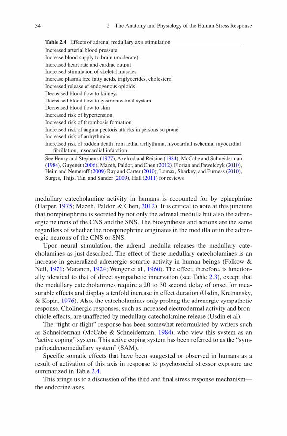
Speci fi c somatic effects that have been suggested or observed in humans as a result of activation of this axis in response to psychosocial stressor exposure are summarized in Table 2.4 .
This brings us to a discussion of the third and fi nal stress response mechanism—the endocrine axes.
Table 2.4 Effects of adrenal medullary axis stimulation
Increased arterial blood pressure Increase blood supply to brain (moderate) Increased heart rate and cardiac output Increased stimulation of skeletal muscles Increase plasma free fatty acids, triglycerides, cholesterol Increased release of endogenous opioids Decreased blood fl ow to kidneys Decreased blood fl ow to gastrointestinal system Decreased blood fl ow to skin Increased risk of hypertension Increased risk of thrombosis formation Increased risk of angina pectoris attacks in persons so prone Increased risk of arrhythmias Increased risk of sudden death from lethal arrhythmia, myocardial ischemia, myocardial
fi brillation, myocardial infarction
See Henry and Stephens ( 1977 ) , Axelrod and Reisine ( 1984 ) , McCabe and Schneiderman ( 1984 ) , Guyenet ( 2006 ) , Mazeh, Paldor, and Chen ( 2012 ) , Florian and Pawelczyk ( 2010 ) , Heim and Nemeroff ( 2009 ) Ray and Carter ( 2010 ), Lomax, Sharkey, and Furness ( 2010 ) , Surges, Thijs, Tan, and Sander ( 2009 ) , Hall ( 2011 ) for reviews
35A Systems Model of the Human Stress Response
Endocrine Axes
The most chronic and prolonged somatic responses to stress are the result of the endocrine axes (Mason, 1968b ) . Four well-established endocrine axes have been associated with the stress response:
1. The adrenal cortical axis. 2. The somatotropic axis. 3. The thyroid axis. 4. The posterior pituitary axis.
These axes not only represent the most chronic aspects of the stress response but also require greater intensity stimulation to activate (Levi, 1972 ) .
Reviews by Axelrod and Reisine ( 1984 ) , Levi ( 1972 ) , Mason ( 1968c, 1972 ) , Mason et al. ( 1995 ) , Selye ( 1976 ) , Yehuda, Giller, Levengood, Southwick, and Siever ( 1995 ) , and more recently by, Entringer, Kumsta, Hellhammer, Wadhwa, and Wust ( 2009 ) , and Foley and Kirschbaum ( 2010 ) , demonstrate that these axes can be activated in humans by numerous and diverse psychological stimuli, including varied psychosocial stimuli.
The Adrenal Cortical Axis
The septal–hippocampal complex appears to represent the highest point of origina-tion for the adrenal cortical axis as a physiologically discrete mechanism (Henry & Ely, 1976 ; Henry & Stephens, 1977 ) . From these points, neural impulses descend to the median eminence of the hypothalamus. The neurosecretory cells in the median eminence release corticotropin-releasing factor (CRF) into the hypothalamic–hypophyseal portal system (Rochefort, Rosenberger, & Saffran, 1959 ) . The CRF descends the infundibular stalk to the cells of the anterior pituitary. The chemo-phobes of the anterior pituitary are sensitive to the presence of CRF and respond by releasing adrenocorticotropic hormone (ACTH) in the systemic circulation. At the same time, the precursor to the various endogenous analgesic opioids (endorphins) is released. This precursor substance, beta lipotropin, yields the proliferation of endog-enous opioids during human stress (Rossier, Bloom, & Guillemin, 1980 ) .
ACTH is carried through the systemic circulation until it reaches its primary target organ: an endocrine gland, the adrenal cortex. The two adrenal cortices are wrapped around the two adrenal medullae (neuroendocrine axis) and sit at the supe-rior poles of the kidneys.
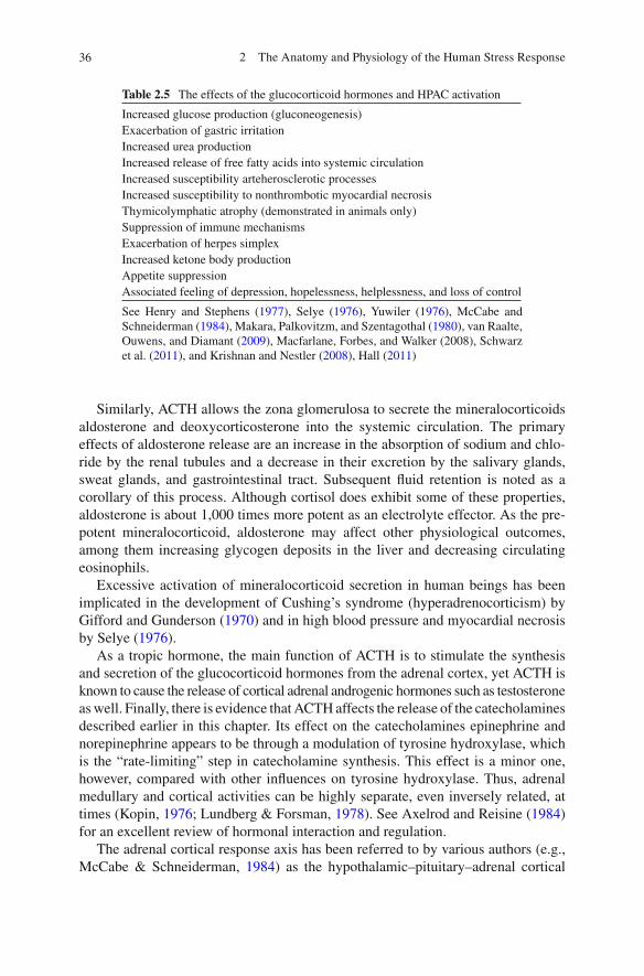
ACTH appears to act upon three discrete layers, or zona, of the adrenal cortex. It stimulates the cells of the zona reticularis and zona fasciculata to release the gluco-corticoids cortisol and corticosterone into the systemic circulation. The effects of the glucocorticoids in apparent response to stressful stimuli are summarized in Table 2.5 .
36 2 The Anatomy and Physiology of the Human Stress Response
Similarly, ACTH allows the zona glomerulosa to secrete the mineralocorticoids aldosterone and deoxycorticosterone into the systemic circulation. The primary effects of aldosterone release are an increase in the absorption of sodium and chlo-ride by the renal tubules and a decrease in their excretion by the salivary glands, sweat glands, and gastrointestinal tract. Subsequent fl uid retention is noted as a corollary of this process. Although cortisol does exhibit some of these properties, aldosterone is about 1,000 times more potent as an electrolyte effector. As the pre-potent mineralocorticoid, aldosterone may affect other physiological outcomes, among them increasing glycogen deposits in the liver and decreasing circulating eosinophils.
Excessive activation of mineralocorticoid secretion in human beings has been implicated in the development of Cushing’s syndrome (hyperadrenocorticism) by Gifford and Gunderson ( 1970 ) and in high blood pressure and myocardial necrosis by Selye ( 1976 ) .
As a tropic hormone, the main function of ACTH is to stimulate the synthesis and secretion of the glucocorticoid hormones from the adrenal cortex, yet ACTH is known to cause the release of cortical adrenal androgenic hormones such as testosterone as well. Finally, there is evidence that ACTH affects the release of the catecholamines described earlier in this chapter. Its effect on the catecholamines epinephrine and norepinephrine appears to be through a modulation of tyrosine hydroxylase, which is the “rate-limiting” step in catecholamine synthesis. This effect is a minor one, however, compared with other in fl uences on tyrosine hydroxylase. Thus, adrenal medullary and cortical activities can be highly separate, even inversely related, at times (Kopin, 1976 ; Lundberg & Forsman, 1978 ) . See Axelrod and Reisine ( 1984 ) for an excellent review of hormonal interaction and regulation.
The adrenal cortical response axis has been referred to by various authors (e.g., McCabe & Schneiderman, 1984 ) as the hypothalamic–pituitary–adrenal cortical
Table 2.5 The effects of the glucocorticoid hormones and HPAC activation
Increased glucose production (gluconeogenesis) Exacerbation of gastric irritation Increased urea production Increased release of free fatty acids into systemic circulation Increased susceptibility arteherosclerotic processes Increased susceptibility to nonthrombotic myocardial necrosis Thymicolymphatic atrophy (demonstrated in animals only) Suppression of immune mechanisms Exacerbation of herpes simplex Increased ketone body production Appetite suppression Associated feeling of depression, hopelessness, helplessness, and loss of control
See Henry and Stephens ( 1977 ) , Selye ( 1976 ) , Yuwiler ( 1976 ) , McCabe and Schneiderman ( 1984 ) , Makara, Palkovitzm, and Szentagothal ( 1980 ) , van Raalte, Ouwens, and Diamant ( 2009 ) , Macfarlane, Forbes, and Walker (2008), Schwarz et al. ( 2011 ) , and Krishnan and Nestler ( 2008 ) , Hall ( 2011 )
37A Systems Model of the Human Stress Response
system (HPAC). Activation of this system in the aggregate has been associated with the helplessness/hopelessness depression syndrome, passivity, the perception of no control, immunosuppression, and gastrointestinal symptomatology. Behaviorally, the HPAC system appears to be activated when active coping is not possible; thus, it has been called the “passive coping” system. Considering the HPAC system with respect to the SAM, Frankenhauser ( 1980 ) has concluded:
1. Effort without distress → activation of the SAM response system. 2. Distress without effort → activation of the HPAC response system. 3. Effort with distress → activation of both SAM and HPAC.
The most extreme variation of the human stress response is, arguably, posttrau-matic stress. The codi fi ed variant of this response is posttraumatic stress disorder (PTSD), the subject of a specialized review in Chap. 21 . Nevertheless, we believe it warrants mention in this discussion of physiological mechanisms because of com-plex and often contradictory fi ndings. In PTSD, both the adrenal medullary cate-cholamine axis and the HPAC pathways are implicated in PTSD. Given the aforementioned discussion, one would expect increased glucocorticoid secretion in PTSD given the intensity, chronicity, and overall severity of PTSD as a clinical syndrome. While enhanced cortisol secretion is, indeed, evidenced in PTSD patients, there is also evidence of decreased cortisol secretion. Yehuda et al. ( 1995 ) provide a useful review and reformulation of this issue. PTSD patients evidence enhanced CRF activity but lower overall cortisol levels in many instances. These authors summarize as follows:
The study of PTSD, whose de fi nition rests on being the sequelae of stress, represents an opportunity to express the effects of extreme stress… from a unique perspective. The fi ndings suggest that… individuals who suffer from PTSD show evidence of a highly sen-sitized HPA axis characterized by decreased basal cortisol levels, increased number of lym-phocyte glucocorticoid receptors, a greater suppression of cortisol to dexamethasone, and a more sensitized pituitary gland. (p. 362)
Thus, in summary, in addition to the more “classic” Selyean observation of increased cortisol as a constituent of extreme stress, PTSD may represent an exten-sion of the Selyean formulation characterized by an increase in CRF, a hypersensi-tized pituitary, and a resultant down-regulation of the HPAC system via an enhanced negative feedback system. As Yehuda et al. ( 1995 ) note, “The fi ndings challenge us to regard the stress response as diversi fi ed and varied, rather than as conforming to a simple, unidirectional pattern” (pp. 362–363).
The Somatotropic Axis
The somatotropic axis appears to share the same basic physiological mechanisms from the septal–hippocampal complex through the hypothalamic–hypophyseal portal system as the previous axis, with the exception that somatotropin-releasing factor (SRF) stimulates the anterior pituitary within this axis. The anterior pitu-itary responds to the SRF by releasing growth hormone (somatotropic hormone)
38 2 The Anatomy and Physiology of the Human Stress Response
into the systemic circulation (see Makara, Palkovitzm, & Szentagothal, 1980 ; Selye, 1976 ) .
The role of growth hormone in stress is somewhat less clearly understood than that of the adrenal cortical axis. However, research has documented its release in response to psychological stimuli in human beings (Selye, 1976 ) , and certain effects are suspected. Selye ( 1956 ) has stated that growth hormone stimulates the release of the mineralocorticoids. Yuwiler ( 1976 ) , in his review of stress and endocrine func-tion, suggests that growth hormone produces a diabetic-like insulin-resistant effect, as well as mobilization of fats stored in the body. The effect is an increase in the concentration of free fatty acids and glucose in the blood.
The Thyroid Axis
The thyroid axis is now a well-established stress response mechanism. From the median eminence of the hypothalamus is released thyrotropin-releasing factor (TRF). The infundibular stalk carries the TRF to its target—the anterior pituitary. From here, the tropic thyroid-stimulating hormone (TSH) is released into the systemic circulation. TSH ultimately stimulates the thyroid gland to release two thyroid hormones: triiodothyronine (T3) and thyroxine (T4). Once secreted into the systemic circulation system, these hormones are bound to speci fi c plasma protein carriers, primarily thyroxin-binding globulin (TBG). A small amount of the thyroid hormones remains as “free” unbound hormones. About 0.4% of T4 and about 0.4% of T3 remain unbound. Proper evaluation of thyroid function is best based upon an assessment of free thyroid hormones. At the level of target-cell tissue, only free hormone is metabolically active. The T3 and T4 hormones serve to participate in a negative feedback loop, thus suppressing their own subse-quent secretion.
In humans, psychosocial stimuli have generally led to an increase in thyroidal activity (Levi, 1972 ; Makara et al., 1980 ; Yuwiler, 1976 ) . Levi has stated that the thyroid hormones have been shown to increase general metabolism, heart rate, heart contractility, peripheral vascular resistance (thereby increasing blood pressure), and the sensitivity of some tissues to catecholamines. Hypothyroidism has been linked to depressive episodes. Levi therefore concludes that the thyroid axis could play a signi fi cant role as a response axis in human stress. See Mason et al. ( 1995 ) for a comprehensive review.
The Posterior Pituitary Axis and Other Phenomena
Since the early 1930s, there has been speculation on the role of the posterior pitu-itary in the stress response. The posterior pituitary (neurohypophysis) receives neu-ral impulses from the supraoptic nuclei of the hypothalamus. Stimulation from these nuclei results in the release of the hormones vasopressin (antidiuretic hormone, or ADH) and oxytocin into the systemic circulation.
39A Systems Model of the Human Stress Response
ADH affects the human organism by increasing the permeability of the collecting ducts that lie subsequent to the distal ascending tubules within the glomerular struc-tures of the kidneys. The end result is water retention.
Corson and Corson ( 1971 ) , in their review of psychosocial in fl uences on renal function, note several studies that report signi fi cant amounts of water retention in apparent response to psychological in fl uences in human beings. Although there seems to be agreement that water retention can be psychogenically induced, there is little agreement on the speci fi c mechanism. Corson and Corson ( 1971 ) report studies that point to the release of elevated amounts of ADH in response to stressful episodes. On the other hand, some studies conclude that the antidiuretic effect is due to decreased renal blood fl ow. Some human participants even responded with a diuretic response to psychosocial stimuli.
Nevertheless, Makara et al. ( 1980 ) , in their review of 25 years of research, found ample evidence for the increased responsiveness of ADH during the stress response. ADH is now seen as one of the wide range of diverse, stress-responsive hormones.
Oxytocin, the other major hormone found in the posterior pituitary axis, is syn-thesized in the same nuclei as ADH, but in different cells. Its role in the human stress response is currently unclear but may be involved in psychogenic labor con-tractions (Omer & Everly, 1988 ) and premature birth, as well as the stress response, particularly for women (Taylor, 2006 ) .
Various investigations have shown that both interstitial cell-stimulating hormone (Sowers, Carlson, Brautbar, & Hershman, 1977 ) , also known as luteinizing hor-mone, and testosterone (Williams, 1986 ) have been shown to be responsive to the presentation of various stressors.
Finally, the hormone prolactin has clearly shown responsiveness to psychosocial stimulation as well (see Makara et al., 1980 , and more recently, Zimmermann et al., 2009 ) . The role of prolactin in disease or dysfunction phenomena, however, has not been well established. Attempts to link prolactin with premenstrual dysfunction have yet to yield a clear line of evidence. The speci fi c role of prolactin in stress-related disease needs further elucidation.
The “General Adaptation Syndrome”
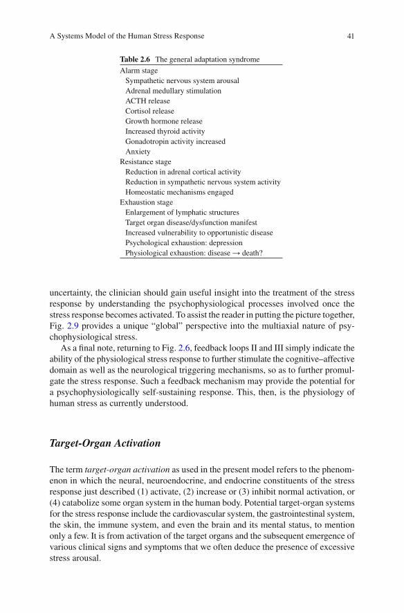
As a means of integrating his psychoendocrinological research, Hans Seyle ( 1956 ) proposed an integrative model for the stress response, known as the “General Adaptation Syndrome” (GAS).
The GAS is a tri-phasic phenomenon. The fi rst phase Selye refers to as the “alarm” phase, representing a generalized somatic shock, or “call to arms” of the body’s defense mechanisms. The second phase is called the “stage of resistance,” in which there is a dramatic reduction in most alarm stage processes and the body fi ghts to reestablish and maintain homeostasis. Stages 1 and 2 can be repeated throughout one’s life. Should the stressor persist, however, eventually the “adap-tive energy,” that is, the adaptive mechanisms in the second stage, may become depleted. At this point, the body enters the third and fi nal stage, the “stage of
40 2 The Anatomy and Physiology of the Human Stress Response
exhaustion,” which, when applied to a target organ, is indicative of the exhaustion of that organ, and the symptoms of disease and dysfunction become manifest. When the fi nal stage is applied to the entire body, life itself may be in jeopardy. The three stages of the GAS are detailed in Table 2.6 .
The Stress Response: A Summary
In this section, we have presented a unifying perspective from which to view the complex psychophysiological processes that have come to be known as the stress response. The intention was to provide clinicians with an understandable interpreta-tion of the complexities of the stress-response process that they often fi nd them-selves treating. Because effective treatment of the stress phenomenon is related to comprehension of the nature of the problem (Miller, 1978, 1979 ) , it is our hope that this discussion will prove useful for the clinician.
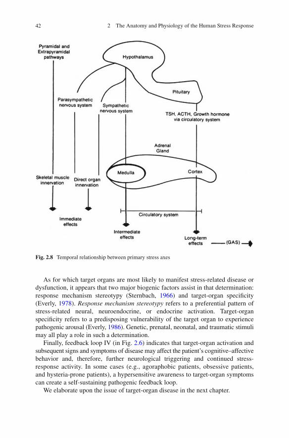
The unifying thread throughout this discussion has been the temporal sequencing of the stress-response process. We have shown that the most immediate response to a stressful stimulus occurs via the direct neural innervations of end organs. The inter-mediate stress effects are due to the neuroendocrine “ fi ght-or- fl ight” axis. The reaction time of this axis is reduced by its utilization of systemic circulation as a transport mechanism. However, its effects range from intermediate to chronic in duration and may overlap with the last stress-response system to respond to a stimulus—the endo-crine axes. The endocrine axes are the fi nal pathways to react to stressful stimuli, owing primarily to the almost total reliance on the circulatory system for transporta-tion, as well as the fact that a higher intensity stimulus is needed to activate this axis. The GAS provides an additional schema to extend the endocrine response axis in the adaptation of the organism to the presence of a chronic stressor [see Selye ( 1956 ) , for a discussion of diseases of adaptation]. Figure 2.8 summarizes the sequential activa-tion of the stress-response axes.
It is important to understand that there is a potential for the activation of each of these axes to overlap. The most common axes to be simultaneously active are the neuroendocrine and endocrine axes—both of which have potential for chronic responsivity (Mason, 1968a, 1968c ) .
On the other hand, it is clear that all mechanisms and axes detailed cannot pos-sibly discharge each and every time a person is faced with a stressor. Perhaps clear-est of all is the fact that each sympathetic and parasympathetic effect is not manifest to all stressors. Therefore, what determines which stress-response mechanisms will be activated by which stressors in which individuals? The answer to this question is currently unknown. However, some evidence suggests the existence of a psy-chophysiological predisposition for some individuals to undergo stress-response pattern speci fi city (see Sternbach, 1966 ) . We expand on this topic in Chap. 3 .
These, then, are the stress-response axes and the various mechanisms that work within each. They represent the potential response patterns result each time the human organism is exposed to a stressor. As to when each responds and why, we are unsure at this time. Current speculations are reviewed in Chap. 3 . Despite this
41A Systems Model of the Human Stress Response
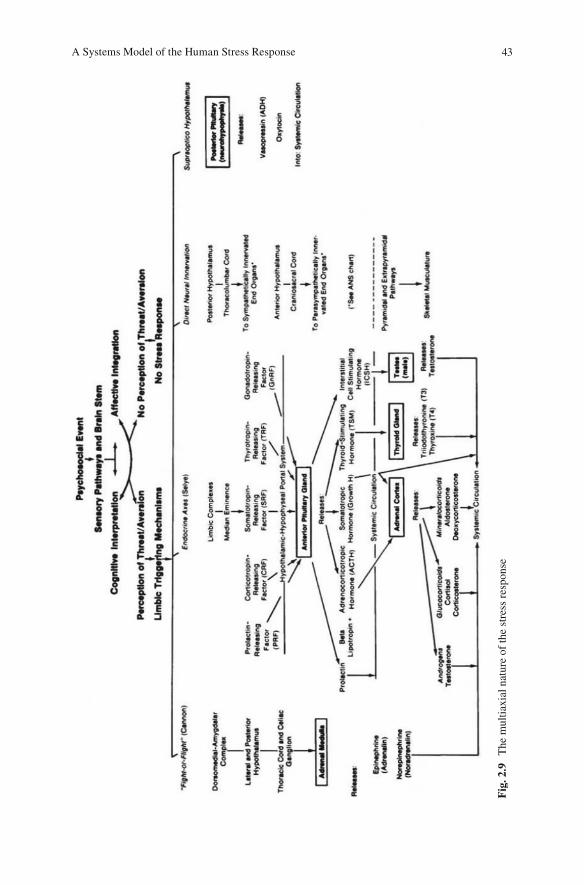
uncertainty, the clinician should gain useful insight into the treatment of the stress response by understanding the psychophysiological processes involved once the stress response becomes activated. To assist the reader in putting the picture together, Fig. 2.9 provides a unique “global” perspective into the multiaxial nature of psy-chophysiological stress.
As a fi nal note, returning to Fig. 2.6 , feedback loops II and III simply indicate the ability of the physiological stress response to further stimulate the cognitive–affective domain as well as the neurological triggering mechanisms, so as to further promul-gate the stress response. Such a feedback mechanism may provide the potential for a psychophysiologically self-sustaining response. This, then, is the physiology of human stress as currently understood.
Target-Organ Activation
The term target-organ activation as used in the present model refers to the phenom-enon in which the neural, neuroendocrine, and endocrine constituents of the stress response just described (1) activate, (2) increase or (3) inhibit normal activation, or (4) catabolize some organ system in the human body. Potential target-organ systems for the stress response include the cardiovascular system, the gastrointestinal system, the skin, the immune system, and even the brain and its mental status, to mention only a few. It is from activation of the target organs and the subsequent emergence of various clinical signs and symptoms that we often deduce the presence of excessive stress arousal.
Table 2.6 The general adaptation syndrome
Alarm stage Sympathetic nervous system arousal Adrenal medullary stimulation ACTH release Cortisol release Growth hormone release Increased thyroid activity Gonadotropin activity increased Anxiety
Resistance stage Reduction in adrenal cortical activity Reduction in sympathetic nervous system activity Homeostatic mechanisms engaged
Exhaustion stage Enlargement of lymphatic structures Target organ disease/dysfunction manifest Increased vulnerability to opportunistic disease Psychological exhaustion: depression Physiological exhaustion: disease → death?
42 2 The Anatomy and Physiology of the Human Stress Response
As for which target organs are most likely to manifest stress-related disease or dysfunction, it appears that two major biogenic factors assist in that determination: response mechanism stereotypy (Sternbach, 1966 ) and target-organ speci fi city (Everly, 1978 ) . Response mechanism stereotypy refers to a preferential pattern of stress-related neural, neuroendocrine, or endocrine activation. Target-organ speci fi city refers to a predisposing vulnerability of the target organ to experience pathogenic arousal (Everly, 1986 ) . Genetic, prenatal, neonatal, and traumatic stimuli may all play a role in such a determination.
Finally, feedback loop IV (in Fig. 2.6 ) indicates that target-organ activation and subsequent signs and symptoms of disease may affect the patient’s cognitive–affective behavior and, therefore, further neurological triggering and continued stress-response activity. In some cases (e.g., agoraphobic patients, obsessive patients, and hysteria-prone patients), a hypersensitive awareness to target-organ symptoms can create a self-sustaining pathogenic feedback loop.
We elaborate upon the issue of target-organ disease in the next chapter.
Fig. 2.8 Temporal relationship between primary stress axes
43A Systems Model of the Human Stress Response
Fig
. 2.9
T
he m
ultia
xial
nat
ure
of th
e st
ress
res
pons
e
44 2 The Anatomy and Physiology of the Human Stress Response
Coping
The preceding two sections went into great detail in an attempt to describe what many phenomenologists have called the “missing link” in psychosomatic phenomena, that is, the physiological mechanisms of mediation by which cognitive–affective discord could result in physical disease and dysfunction. It is an understanding of these physiological mechanisms of mediation that allows us to see stress-related disorders as the quintessential intertwining of “mind and body” as opposed to some anomaly of hysteria. Yet we know that the manifestations of human stress are highly varied and individualistic. Whereas biological predisposition certainly plays a role in this process, a major factor in determining the impact of stress on the patient is his or her perceived ability to cope.
Coping is de fi ned as: efforts, both action-oriented and intrapsychic, to manage (that is, master, tolerate, reduce, minimize) environmental and internal demands, and con fl icts among them, which tax or exceed a person’s resources. Coping can occur prior to a stressful confrontation, in which case it is called anticipatory coping, as well as in reaction to a present or past confrontation with harm. (Cohen & Lazarus, 1979 , p. 219)
More recently, coping has been de fi ned as “constantly changing cognitive and behavioral efforts to manage speci fi c … demands that are appraised as taxing or exceeding the resources of the person” (Lazarus & Folkman, 1984 , p. 141).
From the perspective of the current model (Fig. 2.6 ), coping may be thought of as environmental or cognitive tactics designed to attenuate the stress response. The present model views coping as residing subsequent to the physiological stress response and target-organ activation. Thus, coping is seen as an attempt to reestab-lish homeostasis. Anticipatory coping, as mentioned by Lazarus and other theorists, is subsumed, in the present model, in the complex interactions of the cognitive–affective domain.
To further re fi ne the notion of coping, we suggest that coping strategies can be either adaptive or maladaptive (Girdano et al., 2009 ) . Adaptive coping strategies reduce stress while at the same time promoting long-term health (e.g., exercise, relaxation, proper nutrition). Maladaptive coping strategies, on the other hand, do indeed reduce stress in the short term but serve to erode health in the long term (alcohol/drug abuse, cigarette smoking, interpersonal withdrawal) (see Everly, 1979a ) .
Figure 2.6 re fl ects the belief that when coping is successful, extraordinary target-organ activation is reduced or eliminated and homeostasis is reestablished. If coping strategies are unsuccessful, target-organ activation is maintained and the chances of target-organ disease are increased.
Feedback loops V and VI once again re fl ect the interrelatedness of all compo-nents included in Fig. 2.6 .
The model depicted in Fig. 2.6 re fl ects an integration of recent research and critical thought concerning human stress. It is presented as nothing more than a pedagogical tool designed to facilitate the clinician’s understanding of the phenom-enology of the stress response. If it has sensitized the clinician to the major components
45Summary
of the stress response and shown their interrelatedness, it has served its purpose. This phenomenological model is used as a common reference in subsequent chapters to facilitate better understanding of the topics of measurement and treatment of the human stress response.
Summary
Our purpose has been to provide a somewhat detailed analysis of the psychophysi-ological nature of the human stress response. Let us review the main points of this chapter.
1. The nervous systems serve as the foundation of the stress response. The neuron is the smallest functional unit within any given nervous system. Communications between neurons, and therefore within nervous systems, are based upon electri-cal (ionic transport) and chemical (neurotransmitter mobilization) processes.
Nervous systems are anatomically arranged in the following schema:
(a) Central nervous system Brain • Spinal cord •
(b) Peripheral nervous systems Somatic (to skeletal musculature) • Autonomic (to glands, organs, viscera)•
Sympathetic – Parasympathetic –
2. Figure 2.6 , which represents an integrative epiphenomenological model of the stress response, is reproduced once again here as Fig. 2.10 for review purposes. Let us summarize its components.
3. Environmental events (stressors) may either “cause” the activation of the stress response (as in the case of sympathomimetic stressors) or, as is usually the case, simply “set the stage” for the mobilization of the stress response.
4. The cognitive–affective domain is the critical “causal” phase in most stress reac-tions. Stress, like beauty, appears to be in the eye of the beholder. One’s interpre-tation of the environmental event is what creates most stressors and subsequent stress responses.
5. The locus ceruleus, limbic complexes, and the hypothalamic nuclei trigger effer-ent neurological, neuroendocrine, and endocrine reactions in response to higher cognitive–affective interactions.
6. The actual stress response itself is the next step in the system’s analysis. Possessing at least three major efferent axes—neurological, neuroendocrine, and endocrine—this “physiological mechanism of mediation” represents numerous combinations and permutations of efferent activity directed toward numerous and diverse target organs (see Fig. 2.9 ).
46 2 The Anatomy and Physiology of the Human Stress Response
Fig
. 2.1
0 A
sys
tem
s m
odel
of
the
hum
an s
tres
s re
spon
se
47References
The most rapid of the physiological stress axes are the neurological axes. They consist of mobilization of the sympathetic, parasympathetic, and neuro-muscular nervous systems. The neuroendocrine axis, sometimes called the sympathoadrenomedullary system (SAM), but better known as Cannon’s “ fi ght-or- fl ight” response, is next to be mobilized. Activation leads to the extraordi-nary release of epinephrine and norepinephrine. Finally, the endocrine axes, researched primarily by Hans Selye, are potential response mechanisms. Consisting of the adrenal cortical axis (HPAC system), the somatotropic axis, the thyroid axis, and the posterior pituitary axis, these axes play a major role in chronic disease and dysfunction. Selye’s notion of the General Adaptation Syndrome is an attempt to unify these axes (see Table 2.6 ).
7. As a result of the stress response axes being extraordinarily mobilized, target-organ activation is realized.
8. The fi nal step before pathogenic target-organ activation is coping. Here, the patient has the opportunity to act environmentally or cognitively, or both, so as to reduce or mitigate the overall amplitude and level of activation that reaches the target organs.
9. Should stress arousal be excessive in either acute intensity or chronicity, target-organ dysfunction and/or pathology will result.
10. As a fi nal note, remember that the aforementioned axes are always activated at some level of functioning. Inclusion in this chapter simply re fl ects their poten-tial for pathogenic arousal in response to stressor stimuli and thus their aggre-gate designation as the physiological mechanisms of the stress response.
11. In summary, this chapter was designed to provide the reader with a reasonable approximation of the mechanisms that serve to link the stressor stimulus with target-organ activation. Chapter 3 extends this examination into the link between stress arousal and subsequent disease.
References
Aggleton, J. P. (Ed.). (1992). The Amygdala . New York, NY: Wiley-Liss. Arnold, M. (1970). Feelings and emotions . New York, NY: Academic. Arnold, M. (1984). Memory and the brain . Hillsdale, NJ: Erlbaum. Axelrod, J., & Reisine, T. (1984). Stress hormones. Science, 224 , 452–459. Cannon, W. B. (1914). The emergency function of the adrenal medulla in pain and in the major
emotions. American Journal of Physiology, 33 , 356–372. Cannon, W. B. (1953). Bodily changes in pain, hunger, fear, and rage . Boston, MA: Branford. Cannon, W. B., & Paz, D. (1911). Emotional stimulation of adrenal secretion. American Journal of
Physiology, 28 , 64–70. Carruthers, M., & Taggart, P. (1973). Vagotonicity of violence. British Medical Journal, 3 ,
384–389. Cassel, J. (1974). Psychosocial processes and stress: The behavioral sciences and preventive med-
icine . Washington, DC: Public Health Service. Cohen, F., & Lazarus, R. S. (1979). Coping with the stresses of illness. In G. Stone, F. Cohen, &
N. Adler (Eds.), Health psychology (pp. 217–254). San Francisco, CA: Jossey-Bass.
48 2 The Anatomy and Physiology of the Human Stress Response
Corson, S., & Corson, E. (1971). Psychosocial in fl uences on renal function: Implications for human pathophysiology. In L. Levi (Ed.), Society, stress, and disease (Vol. 1, pp. 338–351). New York, NY: Oxford University Press.
Cullinan, W., Herman, J. P., Helmreich, D., & Watson, S. (1995). A neuroanatomy of stress. In M. J. Friedman, D. Charney, & A. Deutch (Eds.), Neurobiological and clinical consequences of stress (pp. 3–26). Philadelphia, PA: Lippincott-Raven.
Entringer, S., Kumsta, R., Hellhammer, D. H., Wadhwa, P. D., & Wust, S. (2009). Prenatal expo-sure to maternal psychosocial stress and HPA axis regulation in young adults. Hormones and Behavior, 55 , 292–298.
Everly, G. S., Jr. (1978). The Organ Speci fi city Score as a measure of psychophysiological stress reactivity . Unpublished doctoral dissertation, University of Maryland, College Park.
Everly, G. S., Jr. (1979a). Strategies for coping with stress: An assessment scale . Washington, DC: Of fi ce of Health Promotion, Department of Health and Human Services.
Everly, G. S., Jr. (1986). A “biopsychosocial analysis” of psychosomatic disease. In T. Millon & G. Kierman (Eds.), Contemporary directions in psychopathology (pp. 535–551). New York, NY: Guilford.
Everly, G. S., Jr., & Benson, H. (1989). Disorders of arousal and the relaxation response. International Journal of Psychosomatics, 36 , 15–21.
Everly, G. S., Jr., Davy, J. A., Smith, K. J., Lating, J. M., & Nucifora, F. C., Jr. (2011). A de fi ning aspect of human resilience in the workplace: A structural modeling approach. Disaster Medicine and Public Health Preparedness, 5 (2), 98–105.
Everly, G. S., Jr., Smith, K. J., & Lating, J. M. (2009). A rationale for cognitively-based resilience and psychological fi rst aid (PFA) training: A structural modeling analysis. International Journal of Emergency Mental Health, 11 (4), 249–262.
Florian, J. P., & Pawelczyk, J. A. (2010). Non-esteri fi ed fatty acids increase arterial pressure via central sympathetic activation in humans. Clinical Science, 118 , 61–69.
Foley, P., & Kirschbaum, C. (2010). Human hypothalamus-pituitary-adrenal axis responses to acute psychosocial stress in laboratory settings. Neuroscience and Biobehavioral Reviews, 35 , 91–96.
Folkow, B., & Neil, E. (1971). Circulation . London: Oxford University Press. Frankenhaeuser, M. (1980). Psychoneuroendocrine approaches to the study of stressful person-
environment transactions. In H. Selye (Ed.), Selye’s guide to stress research (pp. 46–70). New York, NY: Van Nostrand Reinhold.
Ganong, W. F. (1997). Review of medical physiology (18th ed.). Stamford, CT: Appleton & Lange.
Gellhorn, E. (1957). Autonomic imbalance and the hypothalamus . Minneapolis, MN: University of Minnesota Press.
Gellhorn, E. (1958a). The physiological basis of neuromuscular relaxation. Archives of Internal Medicine, 102 , 392–399.
Gellhorn, E. (1958b). The in fl uence of curare on hypothalamic excitability and the electroencepha-logram. Electroencephalography and Clinical Neurophysiology, 10 , 697–703.
Gellhorn, E. (1964a). Motion and emotion. Psychological Review, 71 , 457–472. Gellhorn, E. (1964b). Sympathetic reactivity in hypertension. Acta Neurovegetative, 26 , 35–44. Gellhorn, E. (1965). The neurophysiological basis of anxiety. Perspectives in Biology and Medicine,
8 , 488–515. Gellhorn, E. (1967). Principles of autonomic-somatic integrations . Minneapolis, MN: University
of Minnesota Press. Gellhorn, E. (1968). Central nervous system tuning and its implications for neuropsychiatry.
Journal of Nervous and Mental Disease, 147 , 148–162. Gellhorn, E. (1969). Further studies on the physiology and pathophysiology of the tuning of the
central nervous system. Psychosomatics, 10 , 94–104. Gellhorn, E., & Loofburrow, G. (1963). Emotions and emotional disorders . New York, NY: Harper
& Row.
49References
Gevarter, W. (1978). Psychotherapy and the brain. Unpublished paper, NASA, Washington, DC. Gifford, S., & Gunderson, J. G. (1970). Cushing’s disease as a psychosomatic disorder: A selective
review. Perspectives in Biology and Medicine, 13 , 169–221. Girdano, D., Dusek, D., & Everly, G. (2009). Controlling stress and tension . San Francisco, CA:
Pearson Benjamin Cummings. Guyenet, P. G. (2006). The sympathetic control of blood pressure. Nature Reviews/Neuroscience,
7 , 335–346. Hall, J. E. (2011). Guyton and Hall Textbook of Medical Physiology (12th ed.). Philadelphia, PA:
Saunders Elsevier. Harper, H. A. (1975). Review of physiological chemistry . Los Altos, CA: Lange. Hassett, J. (1978). A primer of psychophysiology . San Francisco, CA: W. H. Freeman. Heim, C. & Nemeroff, C. B. (2009). Neurobiology of posttraumatic stress disorder. CNS Spectrum,
14(1) (Suppl 1) , 13–24. Henry, J. P., & Ely, D. (1976). Biologic correlates of psychosomatic illness. In R. Grenen & S.
Galay (Eds.), Biological foundations of psychiatry (pp. 945–986). New York, NY: Raven Press.
Henry, J. P., & Stephens, P. (1977). Stress, health, and the social environment . New York, NY: Springer-Verlag.
Hess, W. (1957). The functional organization of the diencephalon . New York, NY: Grune &Stratton.
Johnson, R. H., & Spalding, J. M. (1974). Disorders of the autonomic nervous system . Philadelphia: Davis.
Kopin, L. (1976). Catecholamines, adrenal hormones, and stress. Hospital Practice, 11 , 49–55. Krishnan, V., & Nestler, E. J. (2008). The molecular neurobiology of depression. Nature, 455 (16),
894–902. Lachman, S. (1972). Psychosomatic disorders: A behavioristic interpretation . New York: Wiley. Lang, I. M. (1975). Limbic involvement in the vagosympathetic arterial pressor response of the rat .
Unpublished master’s thesis, Temple University, Philadelphia. Lazarus, R. S. (1966). Psychological stress and the coping process . New York: McGraw-Hill. Lazarus, R. S. (1982). Thoughts on the relations between emotions and cognition. American
Psychologist, 37 , 1019–1024. Lazarus, R. S. (1991). Emotion and adaptation . New York: Oxford University Press. Lazarus, R. S. (2006). Stress and Emotion: A New Synthesis . New York, NY: Springer Publishing
Company, Inc. Lazarus, R. S., & Folkman, S. (1984). Stress, appraisal, and coping . New York: Springer. Le Blanc, J. (1976, July). The role of catecholamines in adaptation to chronic and acute stress .
Paper presented at the proceedings of the International Symposium on Catecholamines and Stress, Bratislava, Czechoslovakia.
Levi, L. (1972). Psychosocial stimuli, psychophysiological reactions and disease. Acta Medico Scandinavica (entire Suppl. 528) .
Lomax, A. E., Sharkey, K. A., & Furness, J. B. (2010). The participation of the sympathetic inner-vation of the gastrointestinal tract in disease states. Neurogastroenterology & Motility, 22 , 7–18.
Lundberg, U., & Forsman, L. (1978). Adrenal medullary and adrenal cortical responses to under-stimulation and overstimulation (Report No. 541). Stockholm: Department of Psychology, University of Stockholm.
Macfarlane, D. P., Forbes, S., & Walker, B. R. (2008). Glucocoticoids and fatty acid metabolism in humans: fuelling fat redistribution in the metabolic syndrome. Journal of Endocrinology, 197 , 189–204.
MacLean, P. D. (1949). Psychosomatic disease and the “visceral brain. Psychosomatic Medicine, 11 , 338–353.
MacLean, P. D. (1975). On the evolution of three mentalities. Man-Environment System, 5 , 213–994.
50 2 The Anatomy and Physiology of the Human Stress Response
Makara, G., Palkovits, M., & Szentagothal, J. (1980). The endocrine hypothalamus and the hormonal response to stress. In H. Selye (Ed.), Selye’s guide to stress research (pp. 280–337). New York: Van Nostrand Reinhold.
Malmo, R. B. (1975). On emotions, needs, and our archaic brain . New York: Holt, Rinehart & Winston.
Maranon, G. (1924). Contribution a 1’etude de 1’action emotive de 1’ademaline. Revue Francais d’Endrocrinologie, 2 , 301–325.
Mason, J. W. (1968a). A review of psychendocrine research on the sympathetic–adrenal medullary system. Psychosomatic Medicine, 30 , 631–653.
Mason, J. W. (1968b). Organization of psychoendocrine mechanisms. Psychosomatic Medicine, 30 (entire P. 2).
Mason, J. W. (1968c). A review of psychoendocrine research on the pituitary–adrenal–cortical system. Psychosomatic Medicine, 30 , 576–607.
Mason, J. W. (1972). Organization of psychoendocrine mechanisms: A review and reconsideration of research. In N. Green fi eld & R. Sternbach (Eds.), Handbook of psychophysiology (pp. 3–76). New York: Holt, Rinehart & Winston.
Mason, J. W., Wang, S., Yehuda, R., Bremner, J. D., Riney, S., Lubin, H., & Charney, D. (1995). Some approaches to the study of clinical implications of thyroid alterations in post-traumatic stress disorder. In M. J. Friedman, D. Charney, & A. Deutch (Eds.), Neurobiological, and clini-cal consequences of stress, (pp. 367–380). Philadelphia: Lippincott-Raven.
Mazeh, H., Paldor, I., & Chen, H. (2012). The endocrine system: Pituitary and adrenal glands. ACS Surgery: Principles and Practice , 1–13
McCabe, P., & Schneiderman, N. (1984). Psychophysiologic reactions to stress. In N. Schneiderman & J. Tapp (Eds.), Behavioral medicine (pp. 3–32). Hillsdale, NJ: Erlbaum.
McCorry, L. K., PhD. (2007). Physiology of the autonomic nervous system. American Journal of Pharmaceutical Education, 71 (4), 1–78.
Meichenbaum, D. (1985). Stress innoculation training . New York: Plenum Press. Meichenbaum, D., & Jaremko, M. (1983). Stress reduction and prevention . New York: Plenum
Press. Miller, N. E. (1978). Biofeedback and visceral learning. Annual Review of Psychology, 29 ,
373–404. Miller, N. E. (1979). General discussion and a review of recent results with paralyzed patients. In
R. Gatchel & K. Price (Eds.), Clinical applications of biofeedback (pp. 215–225). Oxford, UK: Pergamon Press.
Millon, T., & Everly, G. S., Jr. (1985). Personality and its disorders . New York: Wiley. Millon, T., Grossman, S., Millon, C., Meagher, S., & Ramnath, R. (2004). Personality disorders in
modern life . Hoboken, NJ: John Wiley & Sons, Inc. Nauta, W. (1979). Expanding borders of the limbic system concept. In T. Rasmussen & R. Marino
(Eds.), Functional neurosurgery (pp. 7–23). New York: Raven Press. Nauta, W., & Domesick, V. (1982). Neural associations of the limbic system. In A. Beckman (Ed.),
Neural substrates of behavior (pp. 3–29). New York: Spectrum. Omer, H., & Everly, G. S., Jr. (1988). Psychological in fl uences on pre-term labor. American
Journal of Psychiatry, 145 (12), 1507–1513. Pen fi eld, W. (1975). The mystery of the mind . Princeton, NJ: Princeton University Press. Ray, C. A. & Carter, J. R. (2010). Effects of aerobic exercise training on sympathetic and renal
responses to mental stress in humans. American Journal of Physiology – Heart and Circulatory Physiology, 298, H229–H234.
Redmond, D. E. (1979). New and old evidence for the involvement of a brain norepinephrine sys-tem in anxiety. In W. Fann, I. Karacan, A. Pikomey, & R. Williams (Eds.), Phenomenology and treatment of anxiety (pp. 153–204). New York: Spectrum.
Reiman, E., Raichle, M. E., Robins, E., Butler, F. K., Herscovitch, P., Fox, P., & Perlmutter, J. (1986). The application of positron emission tomography to the study of panic disorder. American Journal of Psychiatry, 143 , 469–477.
51References
Rochefort, G. J., Rosenberger, J., & Saffran, M. (1959). Depletion of pituitary corticotropin by various stresses and by neurohypophyseal preparations. Journal of Physiology, 146 , 105–116.
Roldan, E., Alvarez-Pelaez, P., & de Molina, F. (1974). Electrographic study of the amygdaloid defense response. Physiology and Behavior, 13 , 779–787.
Rossier, J., Bloom, F., & Guillemin, R. (1980). In H. Selye (Ed.), Selye’s guide to stress research (pp. 187–207). New York: Van Nostrand Reinhold.
Schwarz, N. A., Rigby, B. R., La Bounty, P., Shelmadine, B., & Bowden, R. G. (2011). A review of weight control strategies and their effects on the regulation of hormonal balance. Journal of Nutrition and Metabolism, 2011 , 1–15.
Selye, H. (1956). The stress of life . New York: McGraw-Hill. Selye, H. (1976). Stress in health and disease . Boston: Butterworth. Smith, K. J., Everly, G. S., & Johns, T. (1992, December). A structural modeling analysis of the
mediating role of cognitive-affective arousal in the relationship between job stressors and ill-ness among accountants. Paper presented to the Second APA/NIOSH Conference on Occupational Stress, Washington, DC.
Smith, K. J., Everly, G. S., & Johns, T. (1993). The role of stress arousal in the dynamics of the stressor-to-illness process among accountants. Contemporary Accounting Research, 9 , 432–449.
Sowers, J. R., Carlson, H. E., Brautbar, N., & Hershman, J. M. (1977). Effect of dexamethasone on prolactin and TSH responses to TRH and metoclopramide in man. The Journal of Clinical Ednocrinology and Metabolism, 44 (2), 237–241.
Sternbach, R. (1966). Principles of psychophysiology: An introductory text and readings . Oxford, England: Academic Press.
Surges, R., Thijs, R. D., Tan, H. L., & Sander, J. W. (2009). Sudden unexpected death in epilepsy: risk factors and potential pathomechanisms. Nature Reviews/Neurology, 5 , 492–504.
Taylor, S. E. (2006). Tend and befriend: Biobehavioral bases of af fi liation under stress. Current Directions in Psychological Science, 15 (6), 273–277.
Usdin, E., Kretnansky, R., & Kopin, L. (1976). Catecholamines and stress . Oxford, UK: Pergamon Press.
van Raalte, D. H., Ouwens, D. M., & Diamant, M. (2009). Novel insights into glucocorticoid-mediated diabetogenic effects: towards expansion of therapeutic options? European Journal of Clinical Investigation, 39 (2), 81–93.
Weil, J. (1974). A neurophysiological model of emotional and intentional behavior . Spring fi eld, IL: Charles C. Thomas.
Wenger, M. A., Clemens, T., Darsie, M. L., Engel, B. T., Estess, F. M., & Sonnenschien, R. R. (1960). Autonomic response patterns during intravenous infusion of epinephrme and norepi-nephrine. Psychosomatic Medicine, 22 , 294–307.
Widmaier, E. P., Raff, H., & Strang, K. T. (2004). Vander, Sherman, Luciano’s Human Physiology: The Mechanisms of Body Function . New York, NY: McGraw-Hill.
Williams, R. B. (1986). Patterns of reactivity and stress. In K. Matthews, R. R. Williams, S. B. Manuck, B. Faulkner, T. Dembroski, T. Detre, & S. M. Weiss (Eds.), Handbook of stress, reactivity, and cardiovascular disease (pp. 109–125). New York: Wiley.
Yehuda, R., Giller, E., Levengood, R., Southwick, S., & Siever, L. (1995). Hypothalamic– pituitary–adrenal-functioning in post-traumatic stress disorder. In M. J. Friedman, D. Charney, & A. Deutch (Eds.), Neurobiological, and clinical consequences of stress (pp. 351–366). Philadelphia: Lippincott-Raven.
Yuwiler, A. (1976). Stress, anxiety and endocrine function. In R. Grenell & S. Gabay (Eds.), Biological foundations of psychiatry (pp. 889–943). New York: Raven Press.
Zimmermann, U. S., Buchmann, A. F., Spring, C., Uhr, M., Holsboer, F., & Wittchen, H.-U. (2009). Ethanol administration dampens the prolactin response to psychosocial stress exposure in sons of alcohol-dependent fathers. Psychoneuroendocrinology, 34 , 996–1003.

http://www.springer.com/978-1-4614-5537-0