a 3′-untranslated region kras variant and triple-negative breast cancer: a case-control and...
TRANSCRIPT

A 3′-untranslated region KRAS variant and triple-negative breastcancer: a case-control and genetic analysis
Trupti Paranjape*, Helen Heneghan*, Robert Lindner, Florence K Keane, Aaron Hoffman,Antoinette Hollestelle, Jemima Dorairaj, Kimberly Geyda, Cory Pelletier, Sunitha Nallur,John W M Martens, Maartje J Hooning, Michael Kerin, Daniel Zelterman, Yong Zhu, DavidTuck, Lyndsay Harris, Nicola Miller, Frank Slack, and Joanne Weidhaas(T Paranjape PhD, C Pelletier PhD, S Nallur BS, J Weidhaas PhD), Department of Pathology(R Lindner BSc, D Tuck MD), Yale Medical School (F K Keane BA), Department of PublicHealth (A Hoffman PhD, Prof D Zelterman PhD, Y Zhu PhD), Department of Medical Oncology(K Geyda PhD, L Harris MD), and Department of Molecular, Cellular, and Developmental Biology(Prof F Slack PhD), Yale University, New Haven, CT, USA; Department of Surgery, ClinicalScience Institute, National University of Ireland, Galway, Ireland (H Heneghan MD, J Dorairaj BA,M Kerin MD, N Miller PhD); Institute of Pharmacy and Molecular Biotechnology, University ofHeidelberg, Heidelberg, Germany (R Lindner); Department of Epidemiology, Tulane Universityand Tulane Cancer Center, New Orleans, LA, USA (A Hoffman); and Department of MedicalOncology, Josephine Nefkens Institute and Daniel den Hoed Cancer Center, Erasmus UniversityMedical Center, Rotterdam, Netherlands (A Hollestelle PhD, J W M Martens PhD, M J HooningMD)
SummaryBackground—We previously identified a functional variant in a let-7 microRNA (miRNA)complementary site in the 3′-untranslated region of the KRAS oncogene (rs61764370) which isassociated with cancer. We aimed to investigate the association of this KRAS variant with breastcancer and tumour biology.
Methods—We assessed frequency distributions of the KRAS variant in 415 patients withhistologically confirmed breast cancer and 457 controls from Connecticut, USA (study group 1)and association of this variant with breast-cancer subtypes in 690 Irish women with knownoestrogen receptor (ER), progesterone receptor (PR), and HER2 statuses, and 360 controls (studygroup 2). We pooled data for study groups 1 and 2 with a cohort of 140 women with triple-negative breast cancer and 113 controls to assess the association of the KRAS variant with triple-negative breast cancer risk, and genome-wide mRNA and specific miRNA expression in patientswith triple-negative breast cancer.
Correspondence to: Dr Joanne Weidhaas, Department of Therapeutic Radiology, Yale University, New Haven, CT 06880, [email protected].*Authors contributed equally
Contributors TP participated in study design, experimentation, writing, preparation of figures, and statistical analysis. HHparticipated in study design, experimentation, and writing. RL did the analysis of mRNA and microRNA expression. FKK didstatistical analysis. AHof did experiments and statistical analysis. AHol did data analysis. JD, KG, CP, and SN did experiments.JWMM was involved in sample collection and writing. MJH provided data and participated in writing the report. MK participatedclinically in sample provision and edited the manuscript. DZ provided statistical analysis. YZ provided samples, experiments, andstatistical analysis. DT participated in mRNA expression analysis and edited the manuscript. LH supplied samples and edited themanuscript. NM supervised experimental work and manuscript editing. FS assisted in study design and writing. JW designed thestudy, and participated in analysis, writing, and oversight of the project.
Conflicts of interest FS and JW have patented intellectual property surrounding the KRAS variant through Yale University (NewHaven, CT, USA), and founded a company that has licensed this intellectual property from Yale University. All other authors declaredno conflicts of interest.
NIH Public AccessAuthor ManuscriptLancet Oncol. Author manuscript; available in PMC 2012 November 04.
Published in final edited form as:Lancet Oncol. 2011 April ; 12(4): 377–386. doi:10.1016/S1470-2045(11)70044-4.
$waterm
ark-text$w
atermark-text
$waterm
ark-text
Findings—Although frequency distributions of the KRAS variant in study group 1 did not differbetween all genotyped individuals, eight (33%) of 24 premenopausal women with ER/PR-negativecancer had the KRAS variant, compared with 27 (13%) of 201 premenopausal controls (p=0·015).In study group 2, the KRAS variant was significantly enriched in women with triple-negativebreast cancer (19 [21%] of 90 cases) compared with 64 (13%) of 478 for luminal A, 13 (15%) of87 for luminal B, and two (6%) of 35 for HER2-positive subgroups (p=0·044). Multivariateanalysis in the pooled study groups showed that the KRAS variant was associated with triple-negative breast cancer in premenopausal women (odds ratio 2·307, 95% CI 1·261–4·219,p=0·0067). Gene-expression analysis of triple-negative breast-cancer tumours suggested thatKRAS-variant positive tumours have significantly altered gene expression, and are enriched forthe luminal progenitor and BRCA1 deficiency signatures. miRNA analysis suggested reducedlevels of let-7 miRNA species in KRAS-variant tumours.
Interpretation—The KRAS variant might be a genetic marker for development of triple-negative breast cancer in premenopausal women, and altered gene and miRNA expressionsignatures should enable molecular and biological stratification of patients with this subgroup ofbreast cancer.
Funding—US National Institutes of Health.
IntroductionThe heterogeneity of breast cancer is shown in the variable risk factors, treatment responses,and outcomes of patients. Breast tumours are classified into oestrogen-receptor (ER)positive and/or progesterone-receptor (PR) positive, HER2 (ERBB2) amplified, and triple-negative tumours (ie, ER/PR negative and HER2 negative).1 Gene expression and receptorprofiling further classifies breast cancer into four biological subgroups: luminal A (ER and/or PR receptor positive, HER2 negative), luminal B (ER and/or PR receptor positive, HER2positive), HER2 positive (ER/PR negative, HER2 positive), and basal-like tumours (triple-negative breast cancer).1
Triple-negative breast cancer is the most aggressive subgroup, with the poorest cause-specific survival at 5 years.2 Transcriptional profiling studies suggest there is furtherheterogeneity within triple-negative breast cancers and these tumours can be categorised intotwo broad subgroups: triple-negative tumours that express epidermal growth factor receptor(EGFR) or cytokeratin (CK) 5/6 and are therefore termed basal-like, and triple-negativetumours that do not express EGFR or CK5/6. Basal-like triple-negative tumours are markedby a younger age of onset than are non-basal-like forms and low expression of BRCA1; thebasal-like phenotype is common in carriers of the BRCA1 mutation.3 An aberrant luminalprogenitor cell population (that might be ER positive) could be the target for transformationin BRCA1-associated basal tumours.4 Although prognostic gene-expression markers arehighly divergent, several modules such as DNA repair deficiency, signatures of immuneresponse, or transition from epithelium to mesenchyme are commonly noted in a subset ofthese tumours.5 Identification of the drivers of these transcriptional modules is a promisingapproach for discovery of specific and personalised therapies.
Association of the triple-negative breast cancer phenotype with young age of onset and anabsence of association with known risks or reproductive factors6 supports the notion thatthere are genetic risks for development of this cancer.7 Unfortunately, few genetic markersof such increased risk exist. Although BRCA1 mutations are often associated with triple-negative tumours, these mutations are rare and account for only 10–15% of patients withtriple-negative breast cancer, dependent on ethnic background and family history.8,9
Paranjape et al. Page 2
Lancet Oncol. Author manuscript; available in PMC 2012 November 04.
$waterm
ark-text$w
atermark-text
$waterm
ark-text

MicroRNAs (miRNAs) are a novel class of small non-coding RNAs that regulate geneexpression by base pairing with sequences within the 3′-untranslated region (UTR), 5′-UTR, and coding sequence regions of target mRNAs, causing mRNA cleavage ortranslational repression.10,11 miRNAs are misregulated in every cancer studied so farincluding breast cancer, in which certain miRNA changes (specifically reduced let-7) arefound in breast tumour-initiating cells, suggesting that low let-7 expression allows self-renewal and proliferation of these cells12 and probably increases risk of breast cancer.
Because miRNAs are global gene regulators, inherited variations in miRNAs are associatedwith increased cancer risk. Evidence is accumulating that polymorphisms disrupting miRNAcoding sequences13 or 3′-UTR miRNA binding sites are strong predictors of cancer risk,including breast cancer.14,15 However, none of the previously identified miRNA-alteringpolymorphisms has been associated with triple-negative breast cancer, or with altered geneor miRNA expression in tumours.
We previously identified a novel germline polymorphism (rs61764370) in a let-7 miRNAcomplementary site within the 3′-UTR of the KRAS oncogene, which is referred to here asthe KRAS variant. We showed that the KRAS variant is associated with low concentrationsof let-7 in tumours and altered KRAS regulation in lung cancer.16 Other groups reported thatthe KRAS variant predicts poor cancer specific outcome in head and neck cancer17 andaltered drug response in colon cancer,18,19 suggesting that this variant has biologicalrelevance. Recently we showed that the KRAS variant is enriched in ovarian cancer and ismost frequently associated with patients from families with hereditary breast and ovariancancer.20 On the basis of this evidence, we aimed to assess the role of the KRAS variant inbreast-cancer risk and tumour biology.
MethodsStudy populations
In this case-control study and genetic analysis, we assessed data from four cohorts (figure1). To assess frequency distributions of the KRAS-variant genotype, we assessed individualsfrom the Yale Breast Cancer Study (study group 1), who were enrolled in a breast cancercase-control study in Connecticut, USA; the study was approved by the Yale institutionalreview board as previously described.13 Briefly, patients were aged 30–80 years and hadincident, histologically confirmed breast cancer and no history of cancer (other than non-melanoma skin cancer). ER and PR statuses were established for all cases but HER2 statuseswere not known and not obtainable. Controls were recruited either from Yale–New HavenHospital (New Haven, CT, USA) or Tolland County, CT, USA. Controls from the Yale–New Haven Hospital underwent breast-related surgery for histologically confirmed benignbreast diseases. Controls from Tolland County were identified either through random-digitdialling (for individuals aged <65 years) or through the Health Care Finance Administrationfiles (≥65 years). Informed consent and data for family histories of cancer, reproductivehistory, demographic factors, and blood sample were obtained from all participants. 415cases and 457 controls had DNA samples available for this study, which were obtainedbetween 1990 and 1999.
To define the association of the KRAS variant with receptor status and breast cancersubtype, we assessed a cohort of 690 Irish women diagnosed with breast cancer withcomplete receptor status and subtype classification. Patients from this cohort (study group 2)had histologically confirmed breast cancer and were recruited from the west of Ireland afterappropriate ethical approval from the Galway University Hospital (Galway, Ireland) ethicscommittee. Informed consent and a detailed family history of breast cancer or ovariancancer, and a blood sample were obtained from all cases. We included 710 cases of breast
Paranjape et al. Page 3
Lancet Oncol. Author manuscript; available in PMC 2012 November 04.
$waterm
ark-text$w
atermark-text
$waterm
ark-text

cancer of all stages and histological types, apart from preinvasive carcinomas. ER, PR, andHER2 statuses were established for all samples by use of standard histopathological analysisand immuno histochemistry, and confirmed by fluorescence in-situ hybridisation for HER2positivity. Although gene-expression analysis was not done, these samples were classified asluminal A, luminal B, HER2, or triple-negative breast cancer by receptor status (seewebappendix p 1). 690 of 710 patients had complete information and were assessed in thisstudy. The 360 controls in this cohort were healthy women from the same geographical area,and were mainly older than 60 years, with no selfreported personal history of any cancer andno family history of breast cancer or ovarian cancer. Cases and controls were mainlyrecruited from July, 2006, to July, 2010.
To establish whether the KRAS variant predicted an increased risk of development of triple-negative breast cancer, we did a pooled analysis of a cohort of patients with triple-negativebreast cancer and controls from Yale (study group 3) and patients with triple-negative breastcancer and controls from study group 2 and controls from study group 1. Patients in studygroup 3 were receiving treatment either at Yale–New Haven Hospital or at the BridgeportHospital (Bridgeport, CT, USA). After approval by the Yale Human InvestigationCommittee, tissue or saliva specimens were obtained from 156 patients. Complete data wereavailable for 140 patients who were diagnosed in 1990–2007 and were included in thisstudy. 130 cases of triple-negative breast cancer had samples of tumour available before anytreatment for gene and miRNA-expression analysis, 78 of whom were also genotyped forthe KRAS variant. 113 controls in this cohort were healthy women who presented to theYale–New Haven Hospital and who had no personal history of cancer apart from non-melanoma skin cancer and were recruited between 2000 and 2007. We obtained clinicalinformation, age, ethnic origin, and family history for all cases and controls. Webappendix p2 summarises basic information for the aforementioned three cohorts.
To assess association of the KRAS variant with BRCA mutations in ER-negative tumours,we analysed BRCA1-mutation carriers with breast cancer and known KRAS-variant statusfrom our previous study of the Rotterdam population. The Rotterdam population has beendescribed21 but, briefly, consisted of Dutch patients with breast cancer and documentedBRCA1 mutations who were identified by investigators at the Erasmus University throughthe Rotterdam Family Clinic (Rotterdam, Netherlands).
ProceduresFor KRAS-variant genotyping assays, we genotyped DNA from all samples for the KRASvariant with a custom TaqMan SNP genotyping assay (Applied Biosystems, Carlsbad, CA,USA). On the basis of a previous study,16 we regarded samples that were heterozygous orhomozygous for the variant G allele as positive for the KRAS variant.
For gene-expression analysis, we measured genome-wide mRNA expression in 78 patientsfrom the Yale triple-negative cohort who were also tested for the KRAS variant. We isolatedtotal RNA from tissue specimen with the RecoverAll total nucleic acid isolation kit (AppliedBiosystems) and hybridised to the whole-genome DASL assay (HumanRef-8 version 3.0,Illumina, San Diego, CA, USA). Data preprocessing and statistical analysis were done withthe lumi package in Bioconductor/R software. Gene-expression data from three whole-genome DASL runs were combined and processed together. Samples with less than 30%detectable probes and probes that were detectable in less than 10% of the samples werediscarded before quantile-normalisation. 74 samples and 18345 probes remained afterfiltering.
Paranjape et al. Page 4
Lancet Oncol. Author manuscript; available in PMC 2012 November 04.
$waterm
ark-text$w
atermark-text
$waterm
ark-text

For miRNA analysis, we produced arrays with the Multiplex RT and TaqMan low densityarray human miRNA panel–real-time PCR system (Applied Biosystems) as per themanufacturer’s protocol.22 We examined expression levels of miRNAs of interest.
Statistical analysisGenotype distributions of all cases and controls were tested for Hardy-Weinberg equilibriumand were found to be in equilibrium. We did unconditional logistic regression to estimate therelative risk associated with every genotype. Controls were adjusted for age (continuous)and ethnic origin (white, black, Hispanic, or other). The population was stratified bymenopausal status (estimated by age ≤51 years or >51 years), and separate risk estimateswere obtained by ER and PR statuses with multinomial logistic regression with a three-leveloutcome variable coded as 0 for controls, 1 for cases with ER-positive and/or PR-positivetumours, and 2 for ER/PR-negative tumours. We did tests for interaction with a Wald χ2,comparing the parameter estimates obtained for every genotype in cases of ER-positive and/or PR-positive disease compared with ER/PR-negative disease.
Patients in study group 2 were stratified according to subtypes of breast cancer and a χ2 testwas done with GraphPad Prism4 software to calculate p values, odds ratios (ORs), and 95%CI. The dominant model was used for all genetic association analysis because of the lowfrequency of the KRAS variant.
We compared categorical variables (eg, ethnic origin, stage, and study site) between studygroups with a χ2 test or two-sided Fisher’s exact test, and continuous variables (eg, age)with a t test. We calculated ORs and 95% CI for the KRAS variant in controls and cases oftriple-negative breast cancer with an unconditional logistic regression model with a binaryoutcome variable. Multivariate logistic regression analyses with a binary outcome variablecoded as controls and cases included variables such as KRAS-variant status, age, ethnicorigin, and study site. The population was also stratified by age group, and separate logisticregression analyses were done for patients aged 51 years or younger (premenopausal group)or older than 51 years (postmenopausal group). Statistical analyses were done with SASversion 9.1.3.
Pathway activation was measured as correspondence with previously published expressionsignatures and axes derived from principal component analysis of the expression set.Principal component analysis was used to separate biological from technical sources ofinformation in the gene-expression dataset. Every component was characterised bycorrespondence to RNA quality, the structure of a batch effect, and biological annotations ofthe contributing probes (ie, probes with expression profiles that have high absoluteprojection values for the specified component). Signatures of gene expression are providedas lists of genes and their changes in expression in a specific condition. Such signatures areespecially valuable for noisy data because they require coordinated differential expression ofmultiple probes, typically in the order of 100. Because mRNA was extracted from formalin-fixed, paraffin-embedded (FFPE) blocks that were up to 20 years old, analysis of the data setwith a signature approach was justified.23 We calculated signature scores as Pearsoncorrelation between the respective signature vector of gene contributions and a sample’sexpression profile for these genes. Association of the KRAS variant with the outcomesdescribed by the respective signature was analysed by a paired Kolmogorov-Smirnov testbetween signatures scores of KRAS variant and wild-type samples. Differential geneexpression was assessed with a linear model, taking into account technical batch artifacts asan offset. Model fitting and empirical Bayesian error moderation of the fold changes wereperformed with the LIMMA package for R.24
Paranjape et al. Page 5
Lancet Oncol. Author manuscript; available in PMC 2012 November 04.
$waterm
ark-text$w
atermark-text
$waterm
ark-text
We analysed miRNA expression in eight batches of 46 miRNAs and two endogenouscontrols. miRNA expression was normalised on the basis of the geometric mean of allexpressed samples: a miRNA was judged to have been expressed if threshold fluorescencewas detected after fewer than 35 cycles and when the geometric mean cycle number of allexpressed miRNAs was subtracted. miRNAs that were not expressed in more than twothirds of all samples were removed, followed by scale-normalisation in all remainingthreshold-cycle values.
Role of the funding sourceThere was no funding source for this study. The corresponding author had full access to allthe data in the study and had final responsibility for the decision to submit for publication.
ResultsOverall, frequency distributions of the KRAS-variant genotype did not differ between casesand controls who were genotyped from study group 1 (figure 1, table 1). However, theKRAS variant was significantly associated with breast cancer in premenopausal patientswith ER/PR-negative tumours (table 1). This association was not observed forpostmenopausal women. Eight (33%) of 24 premenopausal women with ER/PR-negativecancer had the KRAS variant, compared with 27 (13%) of 201 controls and four (9%) of 44premenopausal women with cancer that was positive for ER and/or PR (webappendix p 10).Thus, the KRAS variant might be a genetic marker of increased risk of development ofreceptor-negative breast cancer for premenopausal women.
In study group 2, 478 women had luminal A breast cancer, 87 had luminal B disease, 90 hadtriple-negative disease, and 35 had HER2-positive disease. 98 (14%) of 690 breast-cancercases from this cohort had the KRAS variant, but prevalence varied between the breastcancer subtypes: the KRAS variant was significantly enriched in women with triple-negativebreast cancer (19 [21%] of 90 cases) compared with 64 (13%) of 478 for luminal A, 13(15%) of 87 for luminal B, and two (6%) of 35 for HER2-positive subgroups (p=0·044;figure 2). This association with triple-negative breast cancer was also noted in womenyounger than 51 years (p=0·033, figure 2).
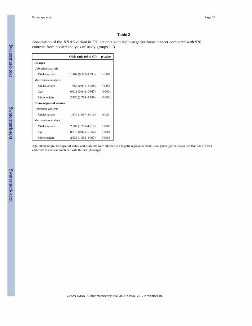
By comparison of cases of triple-negative breast cancer from groups 2 and 3 and controlsacross all three cohorts (n=1160), we did not note a significant difference between cases orbetween controls for the prevalence of the KRAS variant (webappendix p 3). However, therewere significantly more non-white women in the controls from study groups 1 and 3 thanthere were in the study group 2, which allowed assessment of the association of the KRASvariant in non-white women with triple-negative breast cancer in the multivariate analysis.After controlling for age, ethnic origin, and study site, the KRAS variant did not predict anincreased risk of development of triple-negative breast cancer for all women in multivariateanalysis (table 2, webappendix p 4). However, the KRAS variant was associated with asignificantly increased risk of development of triple-negative breast cancer in the 361premenopausal women in this pooled group in multivariate analysis (table 2, webappendixpp 5–6).
Because BRCA1 coding sequence mutations are associated with risk of triple-negativebreast cancer, and because we noted an apparent enrichment of the KRAS variant in BRCA1mutation-carriers with breast cancer,21 we aimed to establish whether the association of theKRAS variant with premenopausal triple-negative breast cancer was due only to itsassociation with carriers of BRCA1 mutation. Of 36 women with triple-negative breastcancer from cohort 2 and 3 who were BRCA tested, 25 (69%) were BRCA negative and 11(31%) were BRCA positive. Of these patients, eight (32%) BRCA-negative women
Paranjape et al. Page 6
Lancet Oncol. Author manuscript; available in PMC 2012 November 04.
$waterm
ark-text$w
atermark-text
$waterm
ark-text
harboured the KRAS variant compared with three (27%) women who were BRCA positive.These findings suggest that the KRAS variant is associated with an independent group ofpatients with triple-negative breast cancer without BRCA mutations.
Although we did not note an association between KRAS-variant status and ER or PRnegative statuses in the Rotterdam population cohort,21,23 we had not consideredmenopausal status. In this study, we did not note an enrichment of the KRAS variant in 126premenopausal BRCA1-mutation carriers who had ER/PR-negative breast cancer comparedwith all 268 BRCA1-mutation-carriers from the Rotterdam cohort (21·8% vs 23·5%,p=0·95). These findings again support the notion that association of the KRAS variant withpremenopausal triple-negative breast cancer is independent of its association with BRCA1mutations.
However, to further assess potential biological interaction between the KRAS variant andaltered BRCA1 expression in triple-negative disease, we appraised BRCA1 expressionlevels in 74 triple-negative tumours from study group 3 (figure 1). We noted that thosepatients with the KRAS variant had significantly reduced BRCA1 expression compared withKRAS-variant-negative triple-negative tumours (p=0·06 for probe 1 [ILMN_2311089] andp=0·01 for probe 2 [ILMN_1738027], figure 3). Furthermore, the KRAS variant wassignificantly associated with a gene expression signature of decreased BRCA1 activity(p=0·04).25 These findings suggest that, although the KRAS variant is not restricted topatients with triple-negative breast cancer with known BRCA1 mutations, there might besome biological interaction between the KRAS variant, altered BRCA1 expression orfunctionality, and development of triple-negative breast cancer.
We compared signalling pathways in triple-negative breast-cancer tumours that wereKRAS-variant positive with those that were KRAS-variant negative from patients in studygroup 3. Although analysis of KRAS mRNA did not vary by KRAS-variant status, thisfinding agrees with the other publications about the effect of miRNA binding to the KRAS3′-UTR.16,26 However, we noted an increase in both an NRAS mutation27 and a MAP-kinase activation signature28 (table 3) in tumours with the KRAS variant. This supports thenotion that the KRAS variant alters gene expression of canonical RAS pathways, and is toour knowledge the first in-vivo evidence that the KRAS variant leads to continued altereddownstream gene expression in tumours with which it is associated.
Because we had previously noted altered concentrations of let-7 miRNA in lung tumourswith the KRAS variant, we examined let-7 concentrations in triple-negative breast cancertumours with the KRAS variant. Consistent with our previous findings, we noted lowerconcentrations of all let-7 miRNA family members in KRAS-variant-associated tumours(figure 4).
To establish how the KRAS variant integrates with known gene-expression signatures oftriple-negative breast cancer, we assessed known signatures that are differentially expressedin such tumours. We found that KRAS-variant tumours have several features of triple-negative and basal-like tumour biology, including decreased oestrogen signalling in a maincomponent derived from our expression set (p=0·04). Furthermore, KRAS-variant tumourshave a luminal progenitor signature (p=0·04), which has been suggested4 as a candidateprogenitor for basal-like breast cancer (table 3, webappendix p 11). Within the luminalprogenitor and the BRCA mutation-like signatures, markers of cell adhesion, tissueinvasion, proliferation, and angiogenesis (such as α5 integrin, DUSP6, and aurora kinase B)were differentially regulated (webappendix p 7). This finding is in agreement with the slightenrichment by functional annotations that we noted in three of 41 genes for wound healing(p=0·02), three of 151 genes for glycan expression (p=0·05), and four of 148 genes for MEK
Paranjape et al. Page 7
Lancet Oncol. Author manuscript; available in PMC 2012 November 04.
$waterm
ark-text$w
atermark-text
$waterm
ark-text
activation (p=0·009) on the basis of the differentially expressed genes in a linear modelcomparing KRAS variant versus non-variant for the dataset (figure 5, webappendix pp 8–9).
DiscussionOur data suggest that a germline polymorphism in the KRAS 3′-UTR (the KRAS variant) isa genetic marker of increased risk of development of triple-negative breast cancer inpremenopausal women. Because study group 1 was small and only assessed patients withknown ER and PR statuses, we validated this association in larger case-control groups withfull receptor status. Most importantly, we show that patients with triple-negative breastcancer who have the KRAS variant have tumours with distinct gene-expression patternscompared with patients without this variant, suggesting that the mutation might drivespecific pathways that influence tumour biology and could modify tumour development.The KRAS variant could ultimately be of value in subclassifying tumours into meaningfulbiological subgroups to both predict prognosis and help to direct treatment in the future(panel).
The finding of reduced let-7 concentrations in triple-negative breast cancer tumours that areassociated with the KRAS variant, as has been reported in lung cancer, is notable. Studiessuggest that KRAS overexpression, through NFκB, can lead to induction of LIN-28 (anegative regulator of let-7) and lowering of let-7 expression.29–31 These conclusions suggesta potential mechanism whereby let-7 is lowered in premalignant tissue and, ultimately,tumours associated with the KRAS variant. Furthermore, let-7 regulates proliferation ofbreast-like stem cells,12 and low let-7 concentrations could allow expansion of this group ofcells, potentially increasing breast-cancer risk in women with the KRAS variant. Theassociation we noted of the KRAS variant with triple-negative breast cancer risk only inpremenopausal women suggests a meaningful interaction between the KRAS variant andhormonal exposure. Such associations and potential mechanisms need additional validationin large cohorts and tumour-initiation models.
Although more than half of breast tumours that carriers of the BRCA1 mutation develop aretriple-negative subtype,32 BRCA1 mutations are rare and thus only account for about 10–15% of all cases of triple-negative disease.8,9 Up to 23% of premenopausal patients withtriple-negative breast cancer have the KRAS variant, without an apparent significantenrichment in BRCA mutation carriers in these cohorts or in young ER/PR-negativeBRCA1-mutation carriers.23 However, the KRAS variant is associated with a BRCA1mutation-like gene-expression signature, supporting the notion that there might be increasedoncogenic risk in the presence of the KRAS variant and high KRAS expression and lowBRCA1 expression, either through mutation or other mechanisms.
We previously showed the KRAS variant affects the regulation of KRAS expression invitro, promoting high KRAS concentrations.16 The KRAS oncogene is an importantupstream mediator of the MAPK pathway, and its overexpression can lead to increasedactivation of the RAF/MEK/MAPK pathway, thereby promoting tumorigenesis. We showedhere that patients with the KRAS variant and triple-negative breast cancer show activation ofthe MAPK pathway (table 3). Oh and colleagues33 reported that hyperactivation of MAPKin breast cancer cells decreases ERα expression leading to a negative phenotype, which is inagreement with our finding that the KRAS variant is associated with even lower oestrogensignalling in these histologically ER-negative tumours. MAPK activation has beenimplicated in oestrogen-independent tumour growth and insensitivity to anti-oestrogentreatment,34 and might be a mechanism by which the KRAS variant drives the developmentof triple-negative breast cancer more than other breast cancer subtypes. The role of the
Paranjape et al. Page 8
Lancet Oncol. Author manuscript; available in PMC 2012 November 04.
$waterm
ark-text$w
atermark-text
$waterm
ark-text

KRAS variant in tumorigenesis and its specific association with triple-negative breast cancerremains to be delineated.
The KRAS variant is a biomarker of poor outcome in several cancers, including head andneck cancer,17 and is a biomarker of poor response to targeted therapies in colon cancer.18
Our finding that patients with the KRAS variant and triple-negative breast cancer have aluminal progenitor signature and differential expression of angiogenic and metastaticmarkers within the signature suggests that tumours harbouring the KRAS variant might bean aggressive subgroup of this cancer. Follow-up studies will be necessary to establish theeffect of the KRAS variant on outcome in patients with triple-negative breast cancer andpatients with breast cancer in general.
Our study suggests that the KRAS variant is associated with tumours that maintain uniquegene-expression patterns. Although investigations remain to be done to establish themechanisms of development of triple-negative breast cancer in women who are KRAS-variant positive, our findings give insight into crucial steps and pathways required fortransformation and tumour development in these women. We believe our results aremeaningful steps towards understanding of the mechanisms of gain of function miRNA-disrupting polymorphisms in cancer biology, which seem to be distinct in function frompreviously discovered genetic markers of cancer risk.
Supplementary MaterialRefer to Web version on PubMed Central for supplementary material.
AcknowledgmentsWe thank Neal Fischbach and the Cancer Genetic Counselling Shared Resource at the Yale Cancer Center (NewHaven, CT, USA) for contributions of samples to the study. TP was supported by a Yale Center for ClinicalInvestigation (YCCI) grant made possible by Clinical and Translational Science Awards (CTSA) grant numberUL1 RR024139 from the National Centre for Research Resources (NCRR), a component of the US NationalInstitutes of Health (NIH), and US NIH roadmap for Medical Research. FS and JW were supported by the USNational Cancer Institute (CA131301). JW was supported by a K08 grant [CA124484]. HH was supported by aHealth Research Board Clinician Scientist Fellowship and an Irish Higher Surgical Training Group TravellingScholarship. JD was supported by the National Breast Cancer Research Institute in Galway, Ireland. DZ hasreceived royalties for books published by John Wiley and Sons, Oxford University Press, Cambridge UniversityPress, and the SAS institute, and payment for service on a data monitoring committee from BristolMyersSquibb.
References1. Sørlie T, Perou CM, Tibshirani R, et al. Gene expression patterns of breast carcinomas distinguish
tumor subclasses with clinical implications. Proc Natl Acad Sci USA. 2001; 98:10869–74.[PubMed: 11553815]
2. Haffty BG, Yang Q, Reiss M, et al. Locoregional relapse and distant metastasis in conservativelymanaged triple-negative early-stage breast cancer. J Clin Oncol. 2006; 24:5652–57. [PubMed:17116942]
3. Rakha EA, Ellis IO. Triple-negative/basal-like breast cancer: review. Pathology. 2009; 41:40–47.[PubMed: 19089739]
4. Lim E, Vaillant F, Wu D, et al. Aberrant luminal progenitors as the candidate target population forbasal tumor development in BRCA1 mutation carriers. Nat Med. 2009; 15:907–13. [PubMed:19648928]
5. Bild AH, Parker JS, Gustafson AM, et al. An integration of complementary strategies for gene-expression analysis to reveal novel therapeutic opportunities for breast cancer. Breast Cancer Res.2009; 11:R55. [PubMed: 19638211]
Paranjape et al. Page 9
Lancet Oncol. Author manuscript; available in PMC 2012 November 04.
$waterm
ark-text$w
atermark-text
$waterm
ark-text

6. Yang XR, Sherman ME, Rimm DL, et al. Differences in risk factors for breast cancer molecularsubtypes in a population-based study. Cancer Epidemiol Biomarkers Prev. 2007; 16:439–43.[PubMed: 17372238]
7. Bauer KR, Brown M, Cress RD, Parise CA, Caggiano V. Descriptive analysis of estrogen receptor(ER)-negative, progesterone receptor (PR)-negative, and HER2-negative invasive breast cancer, theso-called triple-negative phenotype: a population-based study from the California cancer registry.Cancer. 2007; 109:1721–28. [PubMed: 17387718]
8. Young SR, Pilarski RT, Donenberg T, et al. The prevalence of BRCA1 mutations among youngwomen with triple-negative breast cancer. BMC Cancer. 2009; 9:86. [PubMed: 19298662]
9. Nanda R, Schumm LP, Cummings S, et al. Genetic testing in an ethnically diverse cohort of high-risk women: a comparative analysis of BRCA1 and BRCA2 mutations in American families ofEuropean and African ancestry. JAMA. 2005; 294:1925–33. [PubMed: 16234499]
10. He L, Thomson JM, Hemann MT, et al. A microRNA polycistron as a potential human oncogene.Nature. 2005; 435:828–33. [PubMed: 15944707]
11. Esquela-Kerscher A, Slack FJ. Oncomirs—microRNAs with a role in cancer. Nat Rev Cancer.2006; 6:259–69. [PubMed: 16557279]
12. Yu F, Yao H, Zhu P, et al. Let-7 regulates self renewal and tumorigenicity of breast cancer cells.Cell. 2007; 131:1109–23. [PubMed: 18083101]
13. Hoffman A, Zheng T, Yi C, et al. MicroRNA miR-196a-2 and breast cancer: a genetic andepigenetic association study and functional analysis. Cancer Res. 2009; 69:5970–77. [PubMed:19567675]
14. Pongsavee M, Yamkamon V, Dakeng S, et al. The BRCA1 3′UTR: 5711+421T/T_5711+1286T/Tgenotype is a possible breast and ovarian cancer risk factor. Genet Test Mol Biomarkers. 2009;13:307–17. [PubMed: 19405875]
15. Tchatchou S, Jung A, Hemminki K, et al. A variant affecting a putative miRNA target site inestrogen receptor (ESR) 1 is associated with breast cancer risk in premenopausal women.Carcinogenesis. 2009; 30:59–64. [PubMed: 19028706]
16. Chin L, Ratner E, Leng S, et al. A SNP in a let-7 microRNA complementary site in the KRAS3′UTR increases non-small cell cancer risk. Cancer Res. 2008; 68:8535–40. [PubMed: 18922928]
17. Christensen BC, Moyer BJ, Avissar M, et al. A let-7 microRNA binding site polymorphism in theKRAS 3′UTR is associatied with reduced survival in oral cancers. Carcinogenesis. 2009;30:1003–07. [PubMed: 19380522]
18. Graziano F, Canestrari E, Loupakis F, et al. Genetic modulation of the Let-7 microRNA binding toKRAS 3′-untranslated region and survival of metastatic colorectal cancer patients treated withsalvage cetuximab-irinotecan. Pharmacogenomics J. 2010; 10:458–64. [PubMed: 20177422]
19. Zhang W, Winder T, Ning Y, et al. A let-7 microRNA-binding site polymorphism in 3′-untranslated region of KRAS gene predicts response in wild-type KRAS patients with metastaticcolorectal cancer treated with cetuximab monotherapy. Ann Oncol. 2011; 22:104–09. [PubMed:20603437]
20. Ratner E, Lu L, Boeke M, et al. A KRAS-variant in ovarian cancer acts as a genetic marker ofcancer risk. Cancer Res. 2010; 70:6509–15. [PubMed: 20647319]
21. Hollestelle A, Pelletier C, Hooning M, et al. Prevalence of the variant allele rs61764370 T>G inthe 3′UTR of KRAS among Dutch BRCA1, BRCA2 and non-BRCA1/BRCA2 breast cancerfamlies. Breast Cancer Res Treat. 2010 published online July 30. DOI:10.1007/s10549-010-1080-z.
22. [accessed Jan 1, 2008] miRNA profiling. http://www.appliedbiosystems.com/absite/us/en/home/applications-technologies/real-time-pcr/mirna-profiling.html
23. Kibriya M, Jasmine F, Roy S, Paul-Brutus R, Argos M, Ahsan H. Analyses and interpretation ofwhole-genome gene expression from formalin-fixed paraffin-embedded tissue: an illustration withbreast cancer tissues. BMC Genomics. 2010; 11:622. [PubMed: 21059268]
24. Smyth, GK. Limma: linear models for microarray data. In: Gentleman, R.; Carey, V.; Huber, W.;Irizarry, R.; Dudoit, S., editors. Bioinformatics and computational biology solutions using R andbioconductor. Springer; New York, USA: 2005. p. 397-420.
Paranjape et al. Page 10
Lancet Oncol. Author manuscript; available in PMC 2012 November 04.
$waterm
ark-text$w
atermark-text
$waterm
ark-text

25. van’t Veer LJ, Dai H, van de Vijver MJ, et al. Gene expression profiling predicts clinical outcomeof breast cancer. Nature. 2002; 415:530–36. [PubMed: 11823860]
26. Johnson SM, Grosshans H, Shingara J, et al. RAS is regulated by the let-7 microRNA family. Cell.2005; 120:635–47. [PubMed: 15766527]
27. Croonquist PA, Linden MA, Zhao F, Van Ness BG. Gene profiling of a myeloma cell line revealssimilarities and unique signatures among IL-6 response, N-ras-activating mutations, and coculturewith bone marrow stromal cells. Blood. 2003; 102:2581–92. [PubMed: 12791645]
28. Creighton CJ, Hilger AM, Murthy S, Rae JM, Chinnaiyan AM, El-Ashry D. Activation ofmitogen-activated protein kinase in estrogen receptor α-positive breast cancer cells in vitroinduces an in vivo molecular phenotype of estrogen receptor α-negative human breast tumors.Cancer Res. 2006; 66:3903–11. [PubMed: 16585219]
29. Iliopoulos D, Hirsch H, Struhl K. An epigenetic switch involving NF-κB Lin28, let-7 microRNA,and IL6 links inflammation to cell transformation. Cell. 2009; 139:1–14.
30. Meylan E, Dooley A, Feldser D, et al. Requirement for NF-κB signalling in a mouse model of lungadenocarcinoma. Nature. 2009; 462:104–08. [PubMed: 19847165]
31. Barbie D, Tamayo P, Boehm J, et al. Systematic RNA interverence reveals that oncogenic KRAS-driven cancers require TBK1. Nature. 2009; 462:108–12. [PubMed: 19847166]
32. Atchley DP, Albarracin CT, Lopez A, et al. Clinical and pathologic characteristics of patients withBRCA-positive and BRCA-negative breast cancer. J Clin Oncol. 2008; 26:4282–88. [PubMed:18779615]
33. Oh AS, Lorant LA, Holloway JN, Miller DL, Kern FG, El-Ashry D. Hyperactivation of MAPKinduces loss of ERα expression in breast cancer cells. Mol Endocrinol. 2001; 15:1344–59.[PubMed: 11463858]
34. Santen RJ, Song RX, McPherson R, et al. The role of mitogen-activated protein (MAP) kinase inbreast cancer. J Steroid Biochem Mol Biol. 2002; 80:239–56. [PubMed: 11897507]
Paranjape et al. Page 11
Lancet Oncol. Author manuscript; available in PMC 2012 November 04.
$waterm
ark-text$w
atermark-text
$waterm
ark-text

Panel: Research in context
Systematic review
Examination of inherited variants in microRNAs (miRNA) and miRNA binding sites thatpredict cancer risk is a new and rapidly growing area of research. However, the effect ofthese miRNA disrupting variants on tumour biology has not been assessed. Because otherinvestigators have shown the potential of the KRAS variant to act as a biomarker of pooroutcome or poor response to targeted chemotherapy agents, we postulated that thisaltered biology may be noted in gene and miRNA differences in tumours. Our aim was tounderstand if miRNA disrupting variants, such as the KRAS variant, could both beassociated with tumour risk and tumour biology as notable in differences in gene andmiRNA expression.
Interpretation
Our study shows that altered tumour gene-expression patterns can be partly accounted forby inherited variants that disrupt miRNAs binding sites. This finding could explain howsuch variants can act as biomarkers of cancer outcome and response to therapy, andsuggests that such variants might be a simple way to subclassify tumours intobiologically relevant subgroups. Our conclusions provide evidence that baseline geneticdifferences between patients can predict genetic differences in their tumours, which is anexciting direction of study in oncology.
Paranjape et al. Page 12
Lancet Oncol. Author manuscript; available in PMC 2012 November 04.
$waterm
ark-text$w
atermark-text
$waterm
ark-text

Figure 1. Study groupsTNBC=triple-negative breast cancer. ER=oestrogen receptor. PR=progesterone receptor.
Paranjape et al. Page 13
Lancet Oncol. Author manuscript; available in PMC 2012 November 04.
$waterm
ark-text$w
atermark-text
$waterm
ark-text
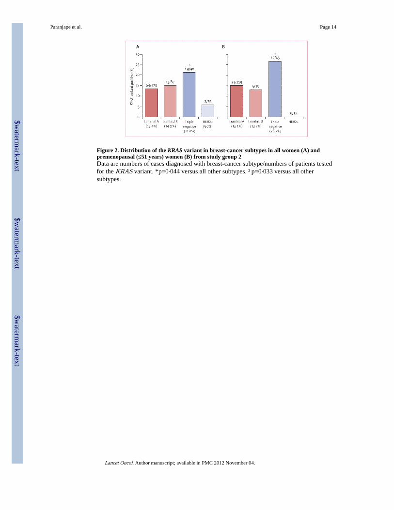
Figure 2. Distribution of the KRAS variant in breast-cancer subtypes in all women (A) andpremenopausal (≤51 years) women (B) from study group 2Data are numbers of cases diagnosed with breast-cancer subtype/numbers of patients testedfor the KRAS variant. *p=0·044 versus all other subtypes. †p=0·033 versus all othersubtypes.
Paranjape et al. Page 14
Lancet Oncol. Author manuscript; available in PMC 2012 November 04.
$waterm
ark-text$w
atermark-text
$waterm
ark-text

Figure 3. BRCA1 gene expression among the KRAS-variant positive and KRAS-variant negativecases of triple-negative breast cancerY-axes are in arbitrary units. (A) BRCA1 probe 1, p=0·06. (B) BRCA1 probe 2, p=0·01.
Paranjape et al. Page 15
Lancet Oncol. Author manuscript; available in PMC 2012 November 04.
$waterm
ark-text$w
atermark-text
$waterm
ark-text

Figure 4. Expression of let-7 family of microRNAs in the KRAS-variant positive versus KRAS-variant negative cases of triple-negative breast cancerY-axes are in arbitrary units.
Paranjape et al. Page 16
Lancet Oncol. Author manuscript; available in PMC 2012 November 04.
$waterm
ark-text$w
atermark-text
$waterm
ark-text
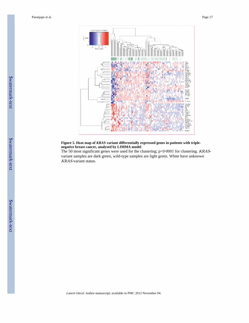
Figure 5. Heat map of KRAS variant differentially expressed genes in patients with triple-negative breast cancer, analysed by LIMMA modelThe 50 most significant genes were used for the clustering; p<0·0001 for clustering. KRAS-variant samples are dark green, wild-type samples are light green. White have unknownKRAS-variant status.
Paranjape et al. Page 17
Lancet Oncol. Author manuscript; available in PMC 2012 November 04.
$waterm
ark-text$w
atermark-text
$waterm
ark-text
$waterm
ark-text$w
atermark-text
$waterm
ark-text
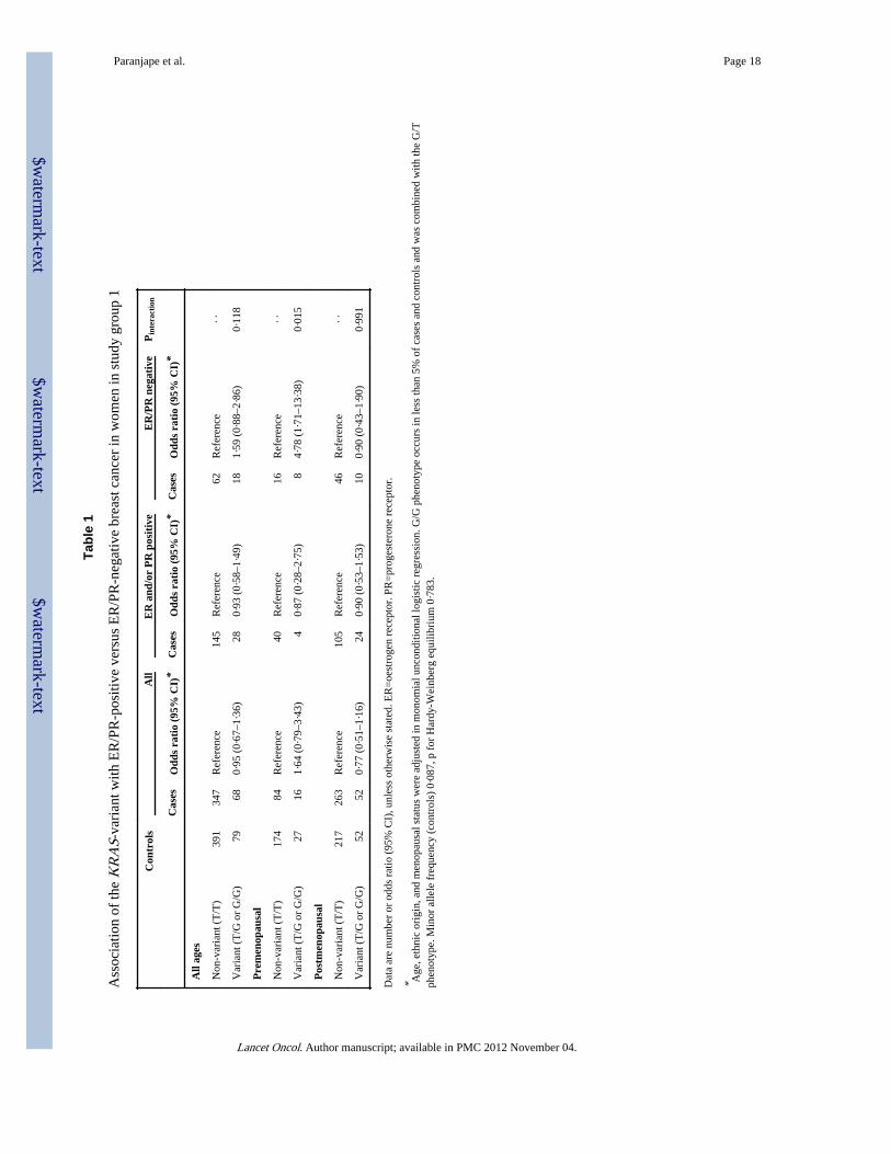
Paranjape et al. Page 18
Tabl
e 1
Ass
ocia
tion
of th
e K
RA
S-va
rian
t with
ER
/PR
-pos
itive
ver
sus
ER
/PR
-neg
ativ
e br
east
can
cer
in w
omen
in s
tudy
gro
up 1
Con
trol
sA
llE
R a
nd/o
r P
R p
osit
ive
ER
/PR
neg
ativ
eP
inte
ract
ion
Cas
esO
dds
rati
o (9
5% C
I)*
Cas
esO
dds
rati
o (9
5% C
I)*
Cas
esO
dds
rati
o (9
5% C
I)*
All
ages
Non
-var
iant
(T
/T)
391
347
Ref
eren
ce14
5R
efer
ence
62R
efer
ence
· ·
Var
iant
(T
/G o
r G
/G)
7968
0·95
(0·
67–1
·36)
280·
93 (
0·58
–1·4
9)18
1·59
(0·
88–2
·86)
0·11
8
Pre
men
opau
sal
Non
-var
iant
(T
/T)
174
84R
efer
ence
40R
efer
ence
16R
efer
ence
· ·
Var
iant
(T
/G o
r G
/G)
2716
1·64
(0·
79–3
·43)
40·
87 (
0·28
–2·7
5)8
4·78
(1·
71–1
3·38
)0·
015
Pos
tmen
opau
sal
Non
-var
iant
(T
/T)
217
263
Ref
eren
ce10
5R
efer
ence
46R
efer
ence
· ·
Var
iant
(T
/G o
r G
/G)
5252
0·77
(0·
51–1
·16)
240·
90 (
0·53
–1·5
3)10
0·90
(0·
43–1
·90)
0·99
1
Dat
a ar
e nu
mbe
r or
odd
s ra
tio (
95%
CI)
, unl
ess
othe
rwis
e st
ated
. ER
=oe
stro
gen
rece
ptor
. PR
=pr
oges
tero
ne r
ecep
tor.
* Age
, eth
nic
orig
in, a
nd m
enop
ausa
l sta
tus
wer
e ad
just
ed in
mon
omia
l unc
ondi
tiona
l log
istic
reg
ress
ion.
G/G
phe
noty
pe o
ccur
s in
less
than
5%
of
case
s an
d co
ntro
ls a
nd w
as c
ombi
ned
with
the
G/T
phen
otyp
e. M
inor
alle
le f
requ
ency
(co
ntro
ls)
0·08
7, p
for
Har
dy-W
einb
erg
equi
libri
um 0
·783
.
Lancet Oncol. Author manuscript; available in PMC 2012 November 04.
$waterm
ark-text$w
atermark-text
$waterm
ark-text
Paranjape et al. Page 19
Table 2
Association of the KRAS-variant in 230 patients with triple-negative breast cancer compared with 930controls from pooled analysis of study groups 1–3
Odds ratio (95% CI) p value
All ages
Univariate analysis
KRAS variant 1·162 (0·797–1·694) 0·4363
Multivariate analysis
KRAS variant 1·352 (0·901–2·028) 0·1455
Age 0·913 (0·942–0·967) <0·0001
Ethnic origin 2·536 (2·784–5·999) <0·0001
Premenopausal women
Univariate analysis
KRAS variant 1·879 (1·067–3·310) 0·029
Multivariate analysis
KRAS variant 2·307 (1·261–4·219) 0·0067
Age 0·913 (0·871–0·956) 0·0001
Ethnic origin 2·536 (1·582–4·067) 0·0001
Age, ethnic origin, menopausal status, and study site were adjusted in a logistic regression model. G/G phenotype occurs in less than 5% of casesand controls and was combined with the G/T phenotype.
Lancet Oncol. Author manuscript; available in PMC 2012 November 04.

$waterm
ark-text$w
atermark-text
$waterm
ark-text
Paranjape et al. Page 20
Table 3
Association of the KRAS-variant with pathway signatures in tumours of patients with triple-negative breastcancer and positive KRAS-variant status
Signature expression Kolmogorov-Smirnov pvalue
NRAS Upregulated 0·02
BRCA mutant-like Upregulated 0·04
Luminal progenitor Upregulated 0·04
MAPK (Creighton) Upregulated 0·06
PCA oestrogen Downregulated 0·04
Signature scores were computed as Pearson correlation between the signature vector of gene contributions and each sample’s expression profile forthese genes. The Kolmogorov-Smirnov test was used to analyse the association of the KRAS-variant with signature activation.
Lancet Oncol. Author manuscript; available in PMC 2012 November 04.