a basal forebrain site coordinates the modulation of endocrine
TRANSCRIPT
Systems/Circuits
A Basal Forebrain Site Coordinates the Modulation ofEndocrine and Behavioral Stress Responses via DivergentNeural Pathways
X Shane B. Johnson,1 X Eric B. Emmons,1 X Rachel M. Anderson,2 Ryan M. Glanz,2 Sara A. Romig-Martin,2
Nandakumar S. Narayanan,1,3 X Ryan T. LaLumiere,1,2 and X Jason J. Radley1,2
1Interdisciplinary Neuroscience Program, 2Department of Psychological and Brain Sciences, and 3Department of Neurology, Carver College of Medicine,University of Iowa, Iowa City, Iowa 52242
The bed nuclei of the stria terminalis (BST) are critically important for integrating stress-related signals between the limbic forebrain andhypothalamo-pituitary-adrenal (HPA) effector neurons in the paraventricular hypothalamus (PVH). Nevertheless, the circuitry under-lying BST control over the stress axis and its role in depression-related behaviors has remained obscure. Utilizing optogenetic approachesin rats, we have identified a novel role for the anteroventral subdivision of BST in the coordinated inhibition of both HPA output andpassive coping behaviors during acute inescapable (tail suspension, TS) stress. Follow-up experiments probed axonal pathways emanat-ing from the anteroventral BST which accounted for separable endocrine and behavioral functions subserved by this cell group. The PVHand ventrolateral periaqueductal gray were recipients of GABAergic outputs from the anteroventral BST that were necessary to restrainstress-induced HPA activation and passive coping behavior, respectively, during TS and forced swim tests. In contrast to other BSTsubdivisions implicated in anxiety-like responses, these results direct attention to the anteroventral BST as a nodal point in a stress-modulatory network for coordinating neuroendocrine and behavioral coping responses, wherein impairment could account for corefeatures of stress-related mood disorders.
Key words: bed nuclei of the stria terminalis; behavioral coping; corticosterone; HPA; paraventricular hypothalamus; periaqueductalgray area
IntroductionResponses to stress involve a stereotyped activation of auto-nomic, behavioral, and neuroendocrine (i.e., hypothalamo-
pituitary-adrenal, or HPA, axis) systems that play a vital role inadaptation. Conversely, dysregulation of neural systems thatcontrol stress-adaptive functions has been increasingly impli-cated in stress-related psychiatric illness (Sheline et al., 1996; Dre-vets et al., 1997; Videbech and Ravnkilde, 2004; MacQueen andFrodl, 2011; Price and Drevets, 2012). The bed nuclei of the striaterminalis (BST) and its subdivisions have been advanced as crit-ically important nodal points in integrating stress-related signals
Received March 30, 2016; revised June 30, 2016; accepted July 1, 2016.Author contributions: S.B.J., N.S.N., R.T.L., and J.J.R. designed research; S.B.J., E.B.E., R.M.A., R.M.G.,
S.A.R.-M., and J.J.R. performed research; S.B.J., E.B.E., N.S.N., and J.J.R. analyzed data; S.B.J., N.S.N., R.T.L.,and J.J.R. wrote the paper.
This work was supported by the National Institutes of Health (Grant MH-095972 to J.J.R., Grants DA-034684 and MH-104384 to R.T.L., and Grant NS-089470 to N.S.N.) and the Brain & Behavior Research Foun-dation (NARSAD Independent Investigator Grant to J.J.R.). We thank Dr. Karl Deisseroth for the materialtransfer agreement for the opsins.
The authors declare no competing financial interests.
Correspondence should be addressed to Jason Radley, Department of Psychology, University of Iowa, E11 Sea-shore Hall, Iowa City, IA 52242. E-mail: [email protected].
DOI:10.1523/JNEUROSCI.1185-16.2016Copyright © 2016 the authors 0270-6474/16/368687-13$15.00/0
Significance Statement
Dysregulation of the neural pathways modulating stress-adaptive behaviors is implicated in stress-related psychiatric illness.While aversive situations activate a network of limbic forebrain regions thought to mediate such changes, little is known abouthow this information is integrated to orchestrate complex stress responses. Here we identify novel roles for the anteroventral bednuclei of the stria terminalis in inhibiting both stress hormone output and passive coping behavior via divergent projections toregions of the hypothalamus and midbrain. Inhibition of these projections produced features observed with rodent models ofdepression, namely stress hormone hypersecretion and increased passive coping behavior, suggesting that dysfunction in thesenetworks may contribute to expression of pathological changes in stress-related disorders.
The Journal of Neuroscience, August 17, 2016 • 36(33):8687– 8699 • 8687

from the limbic forebrain to HPA effector neurons within theparaventricular nucleus of the hypothalamus (PVH). However,the existing body of evidence has led to conflicting interpreta-tions of the precise role of BST in modulating stress-inducedHPA output (Dunn, 1987; Casada and Dafny, 1991; Herman etal., 1994; Spencer et al., 2005; Choi et al., 2007; Choi et al., 2008;Radley et al., 2009; Radley and Sawchenko, 2011), highlightingthe need to use more sophisticated approaches to clarify circuitmechanisms underlying stress adaptation. Similarly, there is along-standing literature highlighting the role of BST in modulat-ing approach-avoidance (e.g., fear, anxiety-like) behaviors(Walker et al., 2003; Davis et al., 2010; Jennings et al., 2013; Kimet al., 2013), although little attention has been given to its capacityto modulate behavioral coping during inescapable challenges.Moreover, the circuitry accounting for BST modulation of be-havioral coping responses and its coordination with HPA activityduring stress is unknown.
Here, we used optogenetic approaches to identify a novel rolefor the anteroventral subdivision of BST (avBST) in coordinatingthe modulation of HPA activation and behavioral coping duringacute stress exposure. Photoexcitation and inhibition of avBSTduring the tail suspension (TS) test bidirectionally modulated(diminished and enhanced, respectively) central and hormonalindices of HPA activation. Photoinhibition of avBST cell bodiesalso increased immobility during TS, suggesting that avBST nor-mally modulates behavioral coping behaviors in response tostress challenges. Follow-up experiments probed axonal path-ways emanating from avBST. Notably, we found that GABAergicavBST inputs to PVH and the ventrolateral subdivision of theperiaqueductal gray area (vlPAG) were necessary for restrainingstress-induced HPA activation and passive coping behavior, re-spectively, during the TS and forced swim (FS) tests. These resultsdirect attention to the avBST as a stress-modulatory nodal pointof a circuit that coordinates neuroendocrine and behavioral ad-aptation, elucidating a novel site in a neural circuit wherein im-pairment could account for core features of stress-related mooddisorders.
Materials and MethodsAnimals and treatments. Adult male Sprague Dawley rats (225–250 g atthe time of arrival, Charles River) acclimated to an American Associationfor Laboratory Animal Care-approved vivarium on a 12 h light/darkcycle (lights on at 0600) with food and water available ad libitum for atleast 7 d before surgery. All procedures were approved by the Universityof Iowa Institutional Animal Care and Use Committee and were in ac-cordance with the National Institutes of Health’s Guide for the Care andUse of Laboratory Animals.
Surgeries. Rats were anesthetized with 4% isoflurane/oxygen, placed ina stereotaxic frame (Kopf Instruments), and maintained at 1.5–2% iso-flurane throughout surgery. Rats received preemptive analgesia (2 mg/kgmeloxicam) before an incision was made to expose the skull for craniot-omy, virus injection, and fiber-optic implantation. Bilateral injections ofadeno-associated virus (AAV, serotype 5; 0.5 �l/side) were directed atavBST [anteroposterior (AP): �0.10 mm; mediolateral (ML): 1.20 mm;dorsoventral (DV): �7.45 mm; all relative to bregma] via a 26 gaugeHamilton syringe. Rats were randomly assigned to groups receiving in-jections of AAV5 (University of North Carolina Vector Core) expressingenhanced yellow fluorescent protein (eYFP) alone (YFP controlgroups), ChannelRhodopsin2(E123A)-eYFP [ChR2(E123A)-eYFP;ChR2 groups), or enhanced archaerhodopsin 3.0-eYFP (eArch3.0-eYFP;Arch groups) under the control of a pan-neuronal promoter (humansynapsin-1, hSyn). avBST microinjection placements were based on pub-lished stereotaxic coordinates from us and others and were laterally bi-ased to avoid transduction of adjacent preoptic and parastrial regions(Radley et al., 2009, Radley and Sawchenko, 2011, 2015). After AAV
injection, fiber optics [200 �m diameter, 0.37 numerical aperture (NA);Thorlabs) housed in steel ferrules (Plastics One) were lowered into placeimmediately dorsal to avBST somata (AP: �0.10 mm; ML: 2.70 mm; DV:�6.95 mm; 10°), avBST terminal fields in PVH (AP: �1.55 mm; ML: 1.65mm; DV: �7.00 mm; 10°), or avBST terminal fields in vlPAG (AP: �7.45mm; ML: 1.75 mm; DV: �5.50 mm; 10°) and secured with dental cementand surgical screws. After surgery, rats were given 4 – 6 weeks for recoveryand to allow adequate time for opsin expression throughout somata andaxons. One week before stress experiments, animals were handled dailyby the experimenter and habituated to the testing room.
Hormone assays. Two days before acute stress exposure, rats were im-planted with indwelling jugular catheters based upon previous studies(Ericsson et al., 1994; Radley et al., 2006). Briefly, indwelling jugularcatheters (polyethylene PE 50) containing sterile heparin–saline (50U/ml) were implanted under isoflurane anesthesia. The sealed catheterwas positioned with its internal SILASTIC (Dow-Corning) tip at theatrium and was exteriorized in the nape of the neck in the interscapularregion. On the day of the experiment, at a time coinciding with the onsetof the diurnal trough (0600) in circadian corticosterone levels (CORT),rats were brought to the procedure room, where jugular catheters wereconnected to sterile, 1 ml syringes and flushed with heparinized saline.Dust caps were removed from the ferrules before the exposed fiber-opticinterfaces were cleaned and connected to optical fibers (200-�m-diameter fiber, 0.37 NA; ThorLabs) contained in leashes. After at least 90min of habituation, blood samples (�200 �l) were taken before (0 min)stress exposure to estimate basal adrenocorticotropic hormone (ACTH)and CORT levels. Additional samples were collected immediately afterTS (10 min) stress and at three subsequent time points (30, 60, and 90min after TS onset). After collection, each sample was immediatelyplaced in a chilled conical vial containing 15 �l of EDTA/ aprotinin,centrifuged for 20 min, and fractionated for storage at �80°C. PlasmaACTH was measured using a two-site radioimmunometric assay (MPBiomedicals) with rabbit antisera raised against ACTH-BSA with 125I-ACTH-BSA serving as a tracer. Intraassay and interassay coefficients ofvariation for ACTH radioimmunoassay were 3% and 7%, respectively,with a sensitivity of 1.5 pg/ml. Plasma corticosterone was measured with-out extraction using rabbit antisera raised against corticosterone-BSAwith 125I-corticosterone-BSA serving as a tracer (MP Biomedicals), withintraassay and interassay coefficients of 5% and 10% and a sensitivity of8 ng/ml.
Stressors and behavioral observations. The TS test has been used widelyin pharmacological and behavioral studies in mice (Steru et al., 1985), hasbeen adapted for use in rats (Izumi et al., 1997; Zhang et al., 2008; Yang etal., 2014; Paumier et al., 2015), and induces stress responses robustly(Brown et al., 1984; Strekalova et al., 2004; Stone et al., 2011). Rats weresuspended for 10 min by the tail such that their hindlimbs were elevatedbut forelimbs were allowed to touch the bottom of the cage (Chermat etal., 1986). Immobility tended to occur later in the stressor and was quan-tified as total time spent in a “passive coping” state wherein a total ab-sence of limb and head movement were observed. Bouts of immobilitywere counted only if they occurred after the first 1 min of stress and lastedfor at least 5 s.
The FS test was used as an additional test of active versus passivecoping behavioral responses to stress based on a previously describedprotocol (Porsolt et al., 1977; Detke and Lucki, 1996; Cryan et al., 2005).However, our test differed in that behavior was assessed over 10 min instress-naive rats (i.e., without a pretest) to avoid a potential ceiling effect.Animals that had previously received AAV-Arch and fiber-optic implan-tation were placed in a 25-cm-diameter chamber filled to a depth of 30cm with 25°C water and behavior was video recorded for 10 min concur-rent with 561 nm illumination. Animals were then dried and returned toa clean cage warmed by a lamp and heating pad. Immobility was laterscored as time spent in a passive (floating) as opposed to active copingstate (swimming, climbing). To assess locomotor activity, rats were videorecorded in a 38 � 38 cm square open-field chamber for 10 min concur-rent with 561 nm illumination. Later, velocity and distance traveled werequantified with EthoVision software (Noldus).
Illumination. Laser power was adjusted to deliver 10 mW at the tip ofthe implanted fiber optics, which, according to in vivo measurements, is
8688 • J. Neurosci., August 17, 2016 • 36(33):8687– 8699 Johnson et al. • Basal Forebrain Regulation of Stress

sufficient to activate opsins within a sphere (radius � 0.46 mm) belowthe termination of the fiber optic (Yizhar et al., 2011; Huff et al., 2013).During experiments, ChR2 animals received 5 ms pulses of 473 nm light(Opto Engine) at 20 Hz (Master-9 pulse generator), Arch animals re-ceived constant illumination with 561 nm light (Laser Century), and YFPcontrols received the same illumination as their respective ChR2 andArch counterparts. For all experiments, illumination began with TS andproceeded concurrently with the 10 min duration of stress exposure,except for one experiment that involved a 30 min stimulation interval(e.g., see Fig. 7). In the latter instance, our goal was to ascertain whetherstress-induced HPA activation could be more strongly modulated byextending the illumination period into the poststress recovery period.
Optrode recordings. In a separate experiment, rats that had previously(�4 weeks prior) received stereotaxic injections into avBST of bothAAV5-hSyn-ChR2(E123A)-eYFP and AAV5-hSyn-eArch3.0-eYFP orAAV5-hSyn-eYFP alone were anesthetized with 4% isoflurane. Next, asurgical level of anesthesia was attained with intraperitoneal injection ofketamine (100 mg/kg) and xylazine (10 mg/kg) and maintained withsupplementary injections of ketamine (30 mg/kg) as necessary. The scalpwas then retracted and the skull leveled between bregma and lambdabefore a craniotomy was made stereotaxically above the avBST of the lefthemisphere. Another small hole was drilled for insertion of the groundwire and two skull screws were implanted to secure the ground wire.
Recordings were collected using a combined microwire array and op-tical fiber, or “optrode” (MicroProbes for Life Science). The optrode waspositioned above avBST (AP �0.10, ML �1.20 mm relative to bregma)and slowly lowered (0.1 mm/min) into an initial location in the dorsal-most aspect of the structure (DV �7.2 mm, relative to dura). Singleneurons were identified online using an oscilloscope and audio monitorand neuronal recordings were made using a multielectrode recordingsystem (Plexon). After a pause to ensure that the recording was stable, thefollowing optogenetic protocol was initiated to determine whetheravBST neurons were modulated by illumination: 0:00 –15:00 min, nolaser on; 15:00 –30:00 min, 473 nm laser pulsed at 20 Hz with 10% dutycycle; 30:00 – 45:00 min, only 561 nm laser on. After the initial recording,the optrode was advanced ventrally in �0.3 mm increments for a total of4 recordings spanning �1 mm. After the recording session, the hardwarewas removed and animals were prepared for histology (see “Histologyand tissue processing” section).
The Plexon Off-Line Sorter program was used to analyze the signalsoffline and remove artifacts. Spike activity was analyzed for all cells thatfired at rates �0.1 Hz. Principal component analysis (PCA) and wave-form shape were used for spike sorting. Single units were identified ashaving the following: (1) consistent waveform shape, (2) separable clus-ters in PCA space, (3) average amplitude estimated at least three timeslarger than background activity, and (4) a consistent refractory period ofat least 2 ms in interspike interval histograms. Analysis of neuronal ac-tivity and quantitative analysis of basic firing properties were performedusing NeuroExplorer (Nex Technologies) and with custom routines forMATLAB (The MathWorks).
Histology and tissue processing. Rats were anesthetized with pentobar-bital (100 mg/kg, i.p.) and perfused at a rate of 55 ml/min through theascending aorta with 100 ml of 0.9% NaCl, followed by 900 ml of ice-cold4% paraformaldehyde (PFA). Brains were then harvested, postfixed in4% PFA at 4°C for 4 h, and then cryoprotected in a solution containing20% sucrose/KPBS overnight. The following day, brains were frozen ondry ice and sections were cut in the coronal plane in a one-in-five series at30 �m on a sliding microtome (Leica) and collected in a cryopreservativesolution before storage at �20°C.
Histochemistry. Fos protein was immunolocalized in free-floating sec-tions using a previously described avidin-biotin peroxidase method(Sawchenko et al., 1990; Radley et al., 2008) and primary antiserumraised against residues 4 –17 of rat Fos protein synthesized by J. Rivier(The Salk Institute). Endogenous peroxidase activity was neutralized by10 min of pretreatment with 0.3% H2O2 before incubating at 4°C for 48 hin primary antiserum (1:25k in PBS � 0.3% Triton X-100 � 3% normalgoat serum). Fos primary antiserum was reacted with Vectastain Elite(Vector Laboratories) reagents and the reaction product was developedusing a nickel-enhanced glucose oxidase method (Sawchenko et al.,
1990). GAD-65 was immunolocalized with a mouse monoclonal anti-body from the University of Iowa Developmental Studies HybridomaBank (1:100; GAD-6 deposited by David I. Gottlieb) and visualized withgoat anti-mouse Alexa Fluor 568 (1:500; Thermo Fisher). CRF was im-munolocalized with an anti-rat CRF-68 antibody raised in rabbit (Saw-chenko Laboratory, The Salk Institute) and then amplified and visualizedwith biotinylated goat anti-rabbit IgG and streptavidin conjugated AlexaFluor 633 (Thermo Fisher). YFP-expressing neurons and terminals werevisualized either with native fluorescence or immunofluorescence afterincubation in rabbit anti-GFP (1:25k; Thermo Fisher) and goat anti-rabbit Alexa Fluor 488 (1:500; Thermo Fisher).
Techniques for probe synthesis, hybridization, and autoradiographiclocalization of mRNA signal were adapted from Simmons et al. (1989). Insitu hybridization was performed using 35S-labeled sense (control) andantisense cRNA probes labeled to similar specific activities using a full-length probe for mRNA encoding the 67 kDa isoform of glutamic aciddecarboxylase (GAD67, Dr. A. Tobin, University of California, Los An-geles; Erlander et al., 1991). Probes for the vesicular glutamate trans-porter, types 1 and 2 (VGLUT1 and VGLUT2) were derived from mousecDNAs (Open Biosystems) bearing a high degree of homology to rat(VGLUT1: 96% homology to nucleotides 65– 437 of rat VGLUT1, Gen-Bank accession number BE950784; Dr. H. Chin, National Institute ofMental Health–National Institutes of Health, Bethesda, MD; VGLUT2:94% homology to nucleotides 545–1070 of rat VGLUT2, accession num-ber CB247147; Dr. R. Strausberg, National Institutes of Health, Bethesda,MD). Hybridization using antisense probes for VGLUT1 and VGLUT2yielded labeling that conformed with the reported distributions in ratbrain (Ziegler et al., 2012) and sense probes for each did not provideevidence of specific labeling. Isotopic hybridization histochemical local-ization GAD67 or VGLUT mRNA, was performed using a previouslydescribed protocol (Radley et al., 2009; Radley et al., 2013).
Quantitative histological analyses. High-resolution (100� objective,1.4 NA) z-stacks were collected with a Leica SP5 confocal microscope(pinhole set to 1 airy unit) to estimate the percentage colocalization ofGABAergic and glutamatergic synaptic markers with YFP puncta in PVHand vlPAG. Briefly, YFP-fluorescent puncta were identified visually andmarked within single sections using ImageJ. Markers representing thelocations of YFP puncta were then overlaid on corresponding GAD65 orvGlut fluorescent optical sections and colocalizations were noted. Colo-calization estimates reported below represent mean values [n � 3 ani-mals, 3 optical sections/animal/brain region (PVH, vlPAG)].
Stereological methods were used to quantify the number of Fos-immunoreactive neurons using a computer-assisted morphometry sys-tem (MBF Biosciences). For each analysis, boundaries defining theregions of interest were drawn at 10� using an adjacent series of Nissl-stained sections. Analyses of Fos-immunoreactive cells were performedon every fifth section, avoiding cells in the outermost plane of focus. Toprobe for possible treatment effects of stereotaxic and viral manipula-tions on regional anatomical volume, 3D estimates from cross-sectionalarea measurements were obtained using the Cavalieri method, but noeffects were observed.
Statistics. Grouped data from the immunohistochemical and behav-ioral experiments were analyzed using a factorial ANOVA, with optoge-netic treatment and stress status serving as factors, followed by post hocpairwise comparisons using Fisher’s least significant difference test.Group data from the hormone assays were compared with a mixed-design ANOVA with one within-group variable (time) and one between-group variable, followed by individual pairwise comparisons as above.Data are expressed as the mean SEM.
ResultsPhotoinhibition of avBST exaggerates stress responsesIn an initial series of experiments, we addressed the functionalrole of avBST in shaping endocrine and behavioral coping re-sponses by activating inhibitory opsins within neuronal somataconcurrent with stress exposure. A 4-week recovery period aftermicroinjection of either AAV-Arch or AAV-YFP into avBST wassufficient to produce robust expression of YFP within neuronal
Johnson et al. • Basal Forebrain Regulation of Stress J. Neurosci., August 17, 2016 • 36(33):8687– 8699 • 8689

somata (Fig. 1a). Rats bearing fiber-optic implantations directlyabove avBST for photoinhibition of neuronal somata were sub-jected to 10 min of TS with concurrent bilateral 561 nm laserillumination along with repeated blood sampling performed attime points before and after stress (Fig. 1b). One-way ANOVArevealed a main effect of treatment on Fos induction in the par-vicellular subdivision of the PVH (F(2,13) � 12.0; p 0.05). Eachgroup exposed to TS showed increases in the number of Fos-immunoreactive cells in PVH compared with the unstressed con-trol group (p 0.05 for each) and rats receiving photoinhibitionof Arch in avBST somata exhibited a further enhancement of Fosrelative to YFP controls receiving the same light stimulation (by56%, p 0.05; Fig. 1c–f). Ancillary experiments comparing Fosinduction in PVH in rats lacking viral injections with rats bearingAAV-YFP injections in avBST failed to reveal any effect of trans-gene expression on this index of stress-induced PVH activation(data not shown).
Comparison of HPA secretory responses were performed as afunction of photoinhibition of avBST cell bodies concurrent with10 min of TS stress exposure. Mixed-design ANOVA, with timeas a within-subjects factor and between-subjects as AAV-definedgroups, was performed on ACTH radioimmunoassay data andrevealed main effects of treatment (F(1,11) � 5.9; p 0.05), time(F(4,44) � 74.8; p 0.05), and a significant interaction betweenthese variables (F(4,44) � 3.8; p 0.05). Within-group measuresdemonstrated significant increases in peak ACTH levels (10 min,p 0.05) that returned to baseline by 60 min in all rats subjectedto TS, whereas ACTH levels were augmented in Arch rats relativeto YFP counterparts (at 30 min, p 0.05; Fig. 1g). Mixed-designANOVA for CORT radioimmunoassay data mirrored the trendsof ACTH release, although only main effects for time (F(4,44) �5.4; p 0.05) were noted. Nevertheless, Arch rats displayed sig-nificantly enhanced levels of plasma CORT at 30 min comparedwith the YFP control group (p 0.05; Fig. 1h). Examination ofintegrated ACTH and CORT responses over time (i.e., the areaunder the curve measurement) also revealed a significant in-crease in Arch animals compared with the YFP group (p 0.05)for ACTH but not for CORT (p � 0.2).
Immobility behavior was also quantified during TS in thesame groups of rats submitted to repeated blood sampling be-cause this provides a measure of the extent to which rats use activeversus passive coping strategies during this inescapable stressor.Moreover, assessment of immobility in this test has also beenwidely used as a measure of behavioral despair, largely basedupon its capacity to be modulated by antidepressants (Steru et al.,1985; Chermat et al., 1986; Paumier et al., 2015). Rats that re-ceived photoinhibition of avBST throughout 10 min of the TSprocedure spent significantly more time in an immobile statethan YFP controls receiving the same illumination (by 32%, p 0.05; Fig. 1i), implicating avBST in the modulation of both be-havioral coping and in constraining HPA activation during stressexposure.
We then confirmed photoinhibition of spiking in putativeArch-expressing avBST neurons with the same constant 561 nmillumination paradigm used in the preceding stress experiments.A combined microelectrode array and optical cannula (i.e., op-trode) was lowered into the avBST of rats (n � 2) that had pre-
Figure 1. Endocrine and behavioral consequences of avBST somata photoinhibition. a–e,Dark-field image (left) of a coronal section showing YFP-fluorescent neuron soma after AAVmicroinjection into avBST, with a schematic coronal section (b) illustrating fiber optic place-ment for the bilateral photoinhibition of neuronal somata therein. Example photomicrographsshowing Fos immunoperoxidase labeling in PVH from unstressed YFP control (c), as well as YFP(d) and Arch (e) rats subjected to TS, illustrating a marked increase in Fos immunoreactivityafter avBST somata photoinhibition. Scale bar, 200 �m (a, c–e). f, Quantification of Fos-immunoreactive nuclei reveals a significant induction as a result of stress exposure (*p 0.05)and further enhancement associated with photoinhibition of avBST neuron axons (†p 0.05).n � 3 YFP � control, n � 7 YFP � stress, n � 6 Arch � stress. g, Analysis of ACTH levelsbefore and after 10 min TS coincident with 561 nm illumination (light green shaded area) ofavBST somata. h, Plasma levels of CORT were significantly elevated in Arch animals 30 min afterthe onset of stress versus YFP controls (*p 0.05). Arch stimulation resulted in a prolonged
4
elevation of plasma titers of ACTH and CORT (at 30 min) after stress onset compared with YFPcontrol animals (*p 0.05). i, Photoinhibition of avBST somata was associated with a signifi-cant increase in immobility behavior during TS. n � 6 YFP, n � 6 Arch (f–i).
8690 • J. Neurosci., August 17, 2016 • 36(33):8687– 8699 Johnson et al. • Basal Forebrain Regulation of Stress

viously (�4 weeks prior) received microinjections of AAV-ChR2and AAV-Arch in the same brain structure (Fig. 2a). Recordingsthat were made before and concurrent with 561 nm illuminationshowed substantial suppression of firing rate in Arch-expressing(Fig. 2b–d), but not YFP-expressing, avBST neuronal somata(Fig. 2e–g).
Photoexcitation of avBST cell bodiesavBST receives extensive excitatory input from upstream pre-frontal cortical and hippocampal regions that are known to in-hibit stress-induced HPA activation (Herman et al., 1995; Radleyet al., 2009; Hill et al., 2011; Radley and Sawchenko, 2011), thuspositioning avBST as a gateway for limbic forebrain modulationof adaptive responses to stress. Therefore, we investigatedwhether optogenetically stimulating avBST somata abrogates en-docrine and behavioral responses to acute stress. Rats receivedbilateral microinjections of AAVs expressing ChR2 or YFP into,and fiber optic implantation above, avBST, followed by a 4-weekrecovery, and were then fitted with indwelling jugular catheters2 d before the blood collection experiment.
First, we investigated whether photoexcitation of avBST dur-ing stress promotes Fos induction in avBST and, in turn, reducesfunctional activation within PVH. Because the projections fromavBST to PVH are predominantly ipsilateral (Dong et al., 2001),we hypothesized that unilateral ChR2 activation in avBST wouldbe associated with increased Fos expression in avBST somatarelative to the unilluminated side and with decreased Fos expres-sion within its projection fields in the ipsilateral PVH (Fig. 3a–d).Consistent with this, stress induced Fos expression in avBST cellgroups compared with unstressed controls (F(1,5) � 17.2; p 0.05) and unilateral avBST photostimulation increased Fos ex-
pression ipsilaterally in both control and stress groups comparedwith nonstimulated counterparts (F(1,5) � 15.5; p 0.05; Fig. 3e).Repeated-measures ANOVA based on counts of Fos-immunoreactive nuclei revealed significant increases in PVH af-ter stress (F(1,5) � 9.3; p 0.05). Nevertheless, ChR2–avBSTstimulation produced decrements in stress-induced activationwithin the ipsilateral PVH (F(1,5) � 8.1; p 0.05), along with acorresponding significant interaction as a function of stress treat-ment and laterality (F(1,5) � 7.6; p 0.05; Fig. 3f).
In a separate experiment, we examined HPA secretory re-sponses to 10 min of TS in rats receiving concurrent bilateral 473nm laser illumination of avBST somata (20 Hz light pulses, 5 mspulse width). Mixed-design ANOVA was performed on ACTHradioimmunoassay data and revealed main effects of treatment(F(1,12) � 8.5; p 0.05) and time (F(4,48) � 65.2; p 0.05; Fig.4a). Analysis of CORT radioimmunoassay data revealed maineffects of treatment (F(1,12) � 4.5; p � 0.05) and time (F(4,48) �55.2; p 0.05). Between-group comparisons at individual timepoints revealed that CORT levels were significantly decreased inChR2 animals compared with YFP control animals at 10 and 90min (p 0.05 for each; Fig. 4b). Furthermore, area under thecurve analyses for stress hormonal activity patterns revealed de-creased integrated ACTH responses in ChR2 rats compared withthe YFP control group (p 0.05). However, analysis of immo-bility behavior during 10 min of TS in ChR2–avBST photostimu-lated rats failed to reveal any significant differences comparedwith controls (p � 0.4; Fig. 4c).
Ancillary experiments confirmed that photoexcitation of pu-tative ChR2-expressing avBST neurons increased neuronal activ-ity. Optrode recordings were taken from the avBST (n � 2) ofanimals that had previously received microinjections of AAV-
Figure 2. Neurophysiological confirmation of Arch-mediated photoinhibition. a, Epifluorescent image (left) displaying the microelectrode path (dashed line) along which neurophysiologicalactivity was recorded ventral to the anterior commissure (ac) to confirm suppression of neuronal activity with 561 nm light, with schematic diagram (right) illustrating optrode placement during therecording session. Scale bar, 200 �m. b–d, Example of activity over 15 min recording sessions in the absence (gray raster plot; b) and presence (green raster plot; c) of constant 561 nm illuminationin a putative Arch-expressing avBST unit, with summary histogram (d) illustrating suppression of neuronal firing (gray line, no laser session; green line, 561 nm laser session). e–g, In aYFP-microinjected animal, neuronal firing was unchanged between the unilluminated (e) and 561 nm illuminated periods (f), which are summarized in a histogram (g). Green bars indicatecontinuous laser illumination over 50 ms blocks.
Johnson et al. • Basal Forebrain Regulation of Stress J. Neurosci., August 17, 2016 • 36(33):8687– 8699 • 8691

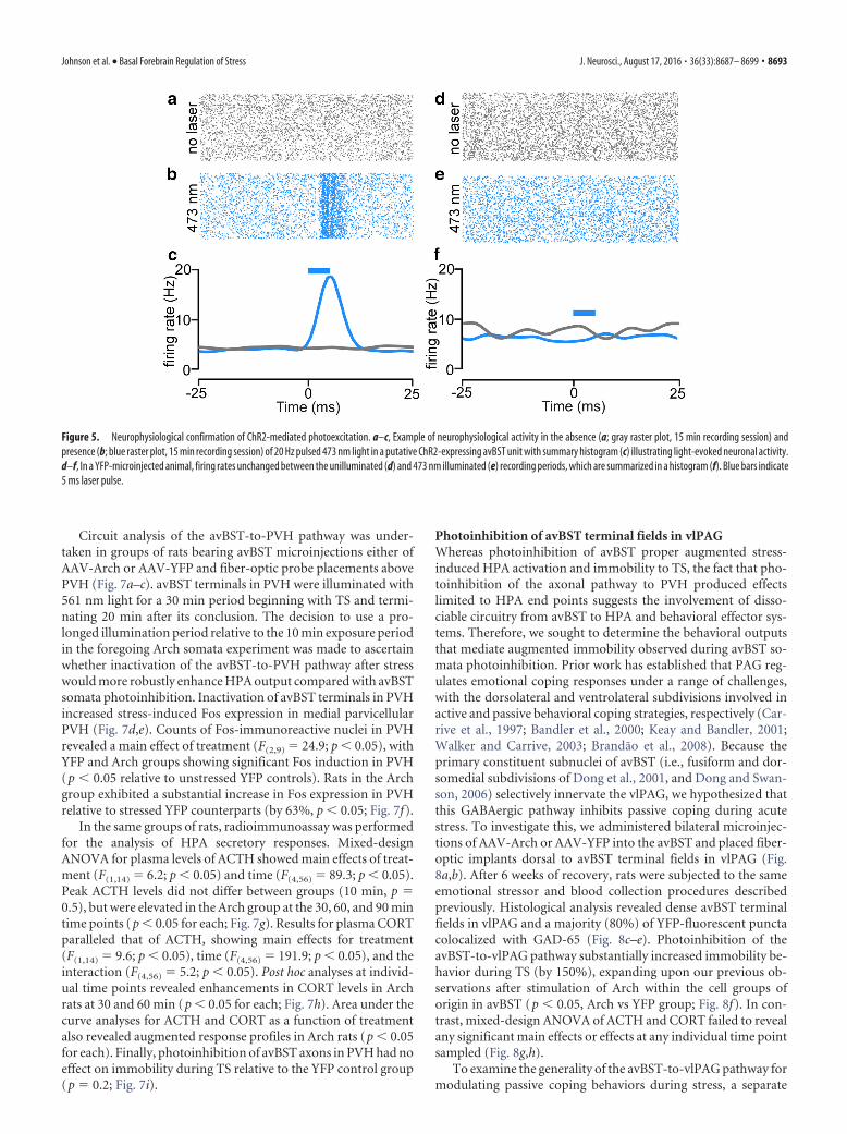
ChR2 and AAV-Arch in the avBST. Recording sessions lasted for15 min with identical illumination parameters to those used inthe preceding stress experiments (20 Hz, 5 ms pulse width, 473nm). Neurons in avBST displayed increased phasic firing rates inresponse to 473 nm light (Fig. 5a–c); however, we failed to ob-serve nonspecific effects of this illumination paradigm in optrodedata collected in an AAV-eYFP animal (Fig. 5d–f). These data,coupled with the functional experiments, provide confirmationof the efficacy of ChR2-mediated photoexcitation in this struc-ture and also suggest that avBST activity produces reliable down-stream reductions in PVH activation.
Photoinhibition of avBST terminal fields in PVHAlthough the foregoing experiments highlight the capacity ofavBST to modulate stress-induced HPA activation bidirection-ally, we sought to determine the circuit basis for this phenome-non by manipulating the axonal pathway from avBST to PVH.That avBST inhibition and excitation enhances and dampens
stress-induced HPA activation, respectively, suggests that this re-gion exerts inhibitory control over the stress axis. Nevertheless,whereas previous work suggests that the avBST-to-PVH pathwayis GABAergic, the complex cytoarchitecture and neuronal phe-notypes within this brain region have hindered attempts to re-solve this issue using conventional techniques (Dunn, 1987;Cullinan et al., 1993; Cecchi et al., 2002; Choi et al., 2007; Radleyet al., 2009; Radley and Sawchenko, 2011).
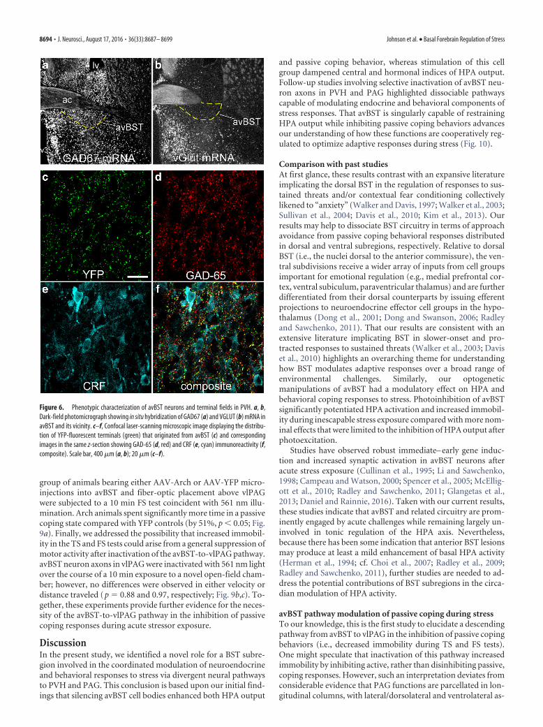
To characterize avBST cell and projection phenotypes, we firstevaluated the extent of GAD and VGLUT (i.e., a glutamatergic-specific neuronal marker) mRNA expression. Hybridization his-tochemistry confirmed that the vast majority of neurons in thisBST subdivision are GABAergic, with a scant proportion ofglutamatergic cells (Fig. 6a,b). Immunohistochemical analysisshowed that YFP-fluorescent terminals formed a dense plexusthroughout medial parvicellular PVH in rats bearing AAV injec-tions within avBST. Confocal laser-scanning microscopic analy-sis in PVH revealed extensive YFP-fluorescent puncta, most ofwhich (87%) colocalized with the GABA synthetic enzymeGAD-65 (Fig. 6c,d). Moreover, many colabeled YFP � GADpuncta resided in close apposition to CRF-immunoreactive se-cretory neurons (Fig. 6e,f). Furthermore, immunohistochemicalstaining for vGlut2 revealed no colocalization with YFP-positiveterminals originating in avBST (data not shown).
Figure 3. Changes in Fos induction after unilateral avBST somata photoexcitation. a, Sagit-tal diagram depicting bilateral AAV-ChR2 microinjection into, and fiber-optic placement above,avBST for unilateral avBST somata photoexcitation. b, Coronal view). c, Dark-field image illus-trating fiber-optic (blue outline) placement ventral to the anterior commissure (ac) and imme-diately dorsal to ChR2:YFP-expressing neurons in avBST. d, Brightfield photomicrographshowing Fos immunoperoxidase staining in PVH contralateral and ipsilateral (contra and ipsi,respectively) to the photoexcited side of avBST after TS. Scale bar, 200 �m (c, d). e, Counts ofFos-positive nuclei revealed significant increases within the photoexcited side of avBST in bothstressed and unstressed control animals compared with the contralateral side of unstressedanimals (*p 0.05). In stressed animals, illumination was associated with further increases inFos induction relative to the contralateral side (†p 0.05). f, Whereas stressed animals dis-played overall increases in Fos induction in PVH relative to controls (*p 0.05), the side of PVHipsilateral to avBST photoexcitation showed abrogated responses relative to the contralateralside (†p 0.05). n � 3 control, n � 5 stress (e, f).
Figure 4. Endocrine and behavioral consequences of bilateral avBST photoexcitation.Analysis of ACTH levels before (a) and after 10 min of TS concurrent with bilateral photo-excitation (light blue shaded area) of avBST somata failed to reveal any significant differ-ences for any individual time point, whereas CORT levels (b) were significantly reduced inChR2 animals immediately after TS (at 10 min) and at 90 min after its onset (*p 0.05).i, Immobility behavior during TS did not differ between ChR2 and YFP groups ( p � 0.4).n � 7 per group (a–c).
8692 • J. Neurosci., August 17, 2016 • 36(33):8687– 8699 Johnson et al. • Basal Forebrain Regulation of Stress
Circuit analysis of the avBST-to-PVH pathway was under-taken in groups of rats bearing avBST microinjections either ofAAV-Arch or AAV-YFP and fiber-optic probe placements abovePVH (Fig. 7a–c). avBST terminals in PVH were illuminated with561 nm light for a 30 min period beginning with TS and termi-nating 20 min after its conclusion. The decision to use a pro-longed illumination period relative to the 10 min exposure periodin the foregoing Arch somata experiment was made to ascertainwhether inactivation of the avBST-to-PVH pathway after stresswould more robustly enhance HPA output compared with avBSTsomata photoinhibition. Inactivation of avBST terminals in PVHincreased stress-induced Fos expression in medial parvicellularPVH (Fig. 7d,e). Counts of Fos-immunoreactive nuclei in PVHrevealed a main effect of treatment (F(2,9) � 24.9; p 0.05), withYFP and Arch groups showing significant Fos induction in PVH(p 0.05 relative to unstressed YFP controls). Rats in the Archgroup exhibited a substantial increase in Fos expression in PVHrelative to stressed YFP counterparts (by 63%, p 0.05; Fig. 7f).
In the same groups of rats, radioimmunoassay was performedfor the analysis of HPA secretory responses. Mixed-designANOVA for plasma levels of ACTH showed main effects of treat-ment (F(1,14) � 6.2; p 0.05) and time (F(4,56) � 89.3; p 0.05).Peak ACTH levels did not differ between groups (10 min, p �0.5), but were elevated in the Arch group at the 30, 60, and 90 mintime points (p 0.05 for each; Fig. 7g). Results for plasma CORTparalleled that of ACTH, showing main effects for treatment(F(1,14) � 9.6; p 0.05), time (F(4,56) � 191.9; p 0.05), and theinteraction (F(4,56) � 5.2; p 0.05). Post hoc analyses at individ-ual time points revealed enhancements in CORT levels in Archrats at 30 and 60 min (p 0.05 for each; Fig. 7h). Area under thecurve analyses for ACTH and CORT as a function of treatmentalso revealed augmented response profiles in Arch rats (p 0.05for each). Finally, photoinhibition of avBST axons in PVH had noeffect on immobility during TS relative to the YFP control group(p � 0.2; Fig. 7i).
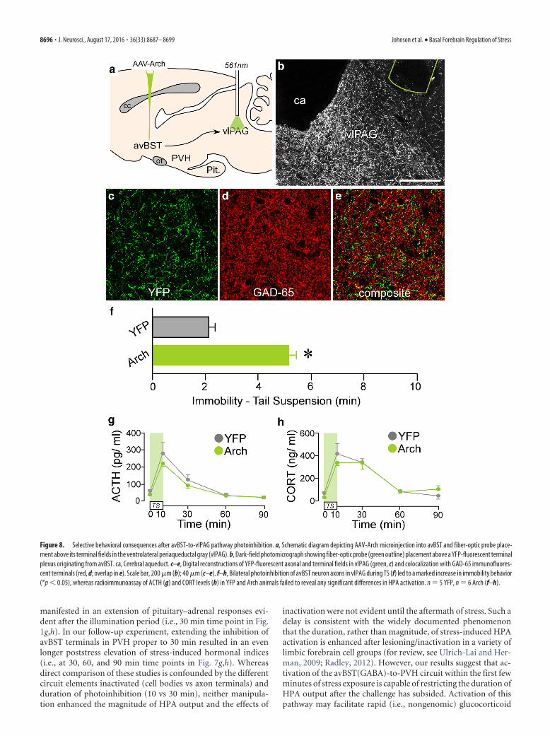
Photoinhibition of avBST terminal fields in vlPAGWhereas photoinhibition of avBST proper augmented stress-induced HPA activation and immobility to TS, the fact that pho-toinhibition of the axonal pathway to PVH produced effectslimited to HPA end points suggests the involvement of disso-ciable circuitry from avBST to HPA and behavioral effector sys-tems. Therefore, we sought to determine the behavioral outputsthat mediate augmented immobility observed during avBST so-mata photoinhibition. Prior work has established that PAG reg-ulates emotional coping responses under a range of challenges,with the dorsolateral and ventrolateral subdivisions involved inactive and passive behavioral coping strategies, respectively (Car-rive et al., 1997; Bandler et al., 2000; Keay and Bandler, 2001;Walker and Carrive, 2003; Brandao et al., 2008). Because theprimary constituent subnuclei of avBST (i.e., fusiform and dor-somedial subdivisions of Dong et al., 2001, and Dong and Swan-son, 2006) selectively innervate the vlPAG, we hypothesized thatthis GABAergic pathway inhibits passive coping during acutestress. To investigate this, we administered bilateral microinjec-tions of AAV-Arch or AAV-YFP into the avBST and placed fiber-optic implants dorsal to avBST terminal fields in vlPAG (Fig.8a,b). After 6 weeks of recovery, rats were subjected to the sameemotional stressor and blood collection procedures describedpreviously. Histological analysis revealed dense avBST terminalfields in vlPAG and a majority (80%) of YFP-fluorescent punctacolocalized with GAD-65 (Fig. 8c–e). Photoinhibition of theavBST-to-vlPAG pathway substantially increased immobility be-havior during TS (by 150%), expanding upon our previous ob-servations after stimulation of Arch within the cell groups oforigin in avBST (p 0.05, Arch vs YFP group; Fig. 8f). In con-trast, mixed-design ANOVA of ACTH and CORT failed to revealany significant main effects or effects at any individual time pointsampled (Fig. 8g,h).
To examine the generality of the avBST-to-vlPAG pathway formodulating passive coping behaviors during stress, a separate
Figure 5. Neurophysiological confirmation of ChR2-mediated photoexcitation. a–c, Example of neurophysiological activity in the absence (a; gray raster plot, 15 min recording session) andpresence (b; blue raster plot, 15 min recording session) of 20 Hz pulsed 473 nm light in a putative ChR2-expressing avBST unit with summary histogram (c) illustrating light-evoked neuronal activity.d–f, In a YFP-microinjected animal, firing rates unchanged between the unilluminated (d) and 473 nm illuminated (e) recording periods, which are summarized in a histogram (f). Blue bars indicate5 ms laser pulse.
Johnson et al. • Basal Forebrain Regulation of Stress J. Neurosci., August 17, 2016 • 36(33):8687– 8699 • 8693
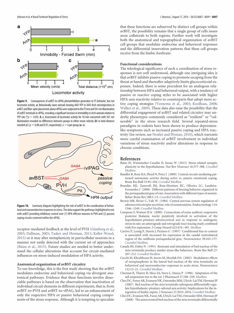
group of animals bearing either AAV-Arch or AAV-YFP micro-injections into avBST and fiber-optic placement above vlPAGwere subjected to a 10 min FS test coincident with 561 nm illu-mination. Arch animals spent significantly more time in a passivecoping state compared with YFP controls (by 51%, p 0.05; Fig.9a). Finally, we addressed the possibility that increased immobil-ity in the TS and FS tests could arise from a general suppression ofmotor activity after inactivation of the avBST-to-vlPAG pathway.avBST neuron axons in vlPAG were inactivated with 561 nm lightover the course of a 10 min exposure to a novel open-field cham-ber; however, no differences were observed in either velocity ordistance traveled (p � 0.88 and 0.97, respectively; Fig. 9b,c). To-gether, these experiments provide further evidence for the neces-sity of the avBST-to-vlPAG pathway in the inhibition of passivecoping responses during acute stressor exposure.
DiscussionIn the present study, we identified a novel role for a BST subre-gion involved in the coordinated modulation of neuroendocrineand behavioral responses to stress via divergent neural pathwaysto PVH and PAG. This conclusion is based upon our initial find-ings that silencing avBST cell bodies enhanced both HPA output
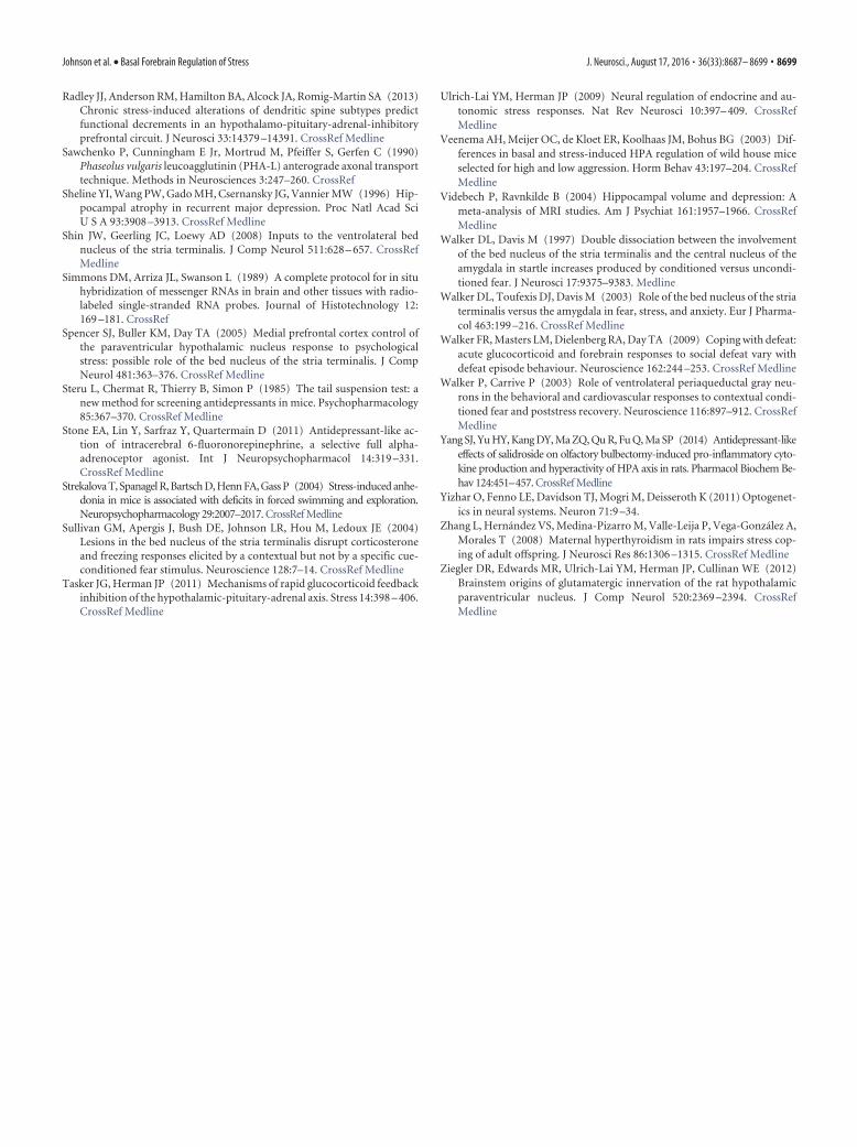
and passive coping behavior, whereas stimulation of this cellgroup dampened central and hormonal indices of HPA output.Follow-up studies involving selective inactivation of avBST neu-ron axons in PVH and PAG highlighted dissociable pathwayscapable of modulating endocrine and behavioral components ofstress responses. That avBST is singularly capable of restrainingHPA output while inhibiting passive coping behaviors advancesour understanding of how these functions are cooperatively reg-ulated to optimize adaptive responses during stress (Fig. 10).
Comparison with past studiesAt first glance, these results contrast with an expansive literatureimplicating the dorsal BST in the regulation of responses to sus-tained threats and/or contextual fear conditioning collectivelylikened to “anxiety” (Walker and Davis, 1997; Walker et al., 2003;Sullivan et al., 2004; Davis et al., 2010; Kim et al., 2013). Ourresults may help to dissociate BST circuitry in terms of approachavoidance from passive coping behavioral responses distributedin dorsal and ventral subregions, respectively. Relative to dorsalBST (i.e., the nuclei dorsal to the anterior commissure), the ven-tral subdivisions receive a wider array of inputs from cell groupsimportant for emotional regulation (e.g., medial prefrontal cor-tex, ventral subiculum, paraventricular thalamus) and are furtherdifferentiated from their dorsal counterparts by issuing efferentprojections to neuroendocrine effector cell groups in the hypo-thalamus (Dong et al., 2001; Dong and Swanson, 2006; Radleyand Sawchenko, 2011). That our results are consistent with anextensive literature implicating BST in slower-onset and pro-tracted responses to sustained threats (Walker et al., 2003; Daviset al., 2010) highlights an overarching theme for understandinghow BST modulates adaptive responses over a broad range ofenvironmental challenges. Similarly, our optogeneticmanipulations of avBST had a modulatory effect on HPA andbehavioral coping responses to stress. Photoinhibition of avBSTsignificantly potentiated HPA activation and increased immobil-ity during inescapable stress exposure compared with more nom-inal effects that were limited to the inhibition of HPA output afterphotoexcitation.
Studies have observed robust immediate– early gene induc-tion and increased synaptic activation in avBST neurons afteracute stress exposure (Cullinan et al., 1995; Li and Sawchenko,1998; Campeau and Watson, 2000; Spencer et al., 2005; McEllig-ott et al., 2010; Radley and Sawchenko, 2011; Glangetas et al.,2013; Daniel and Rainnie, 2016). Taken with our current results,these studies indicate that avBST and related circuitry are prom-inently engaged by acute challenges while remaining largely un-involved in tonic regulation of the HPA axis. Nevertheless,because there has been some indication that anterior BST lesionsmay produce at least a mild enhancement of basal HPA activity(Herman et al., 1994; cf. Choi et al., 2007; Radley et al., 2009;Radley and Sawchenko, 2011), further studies are needed to ad-dress the potential contributions of BST subregions in the circa-dian modulation of HPA activity.
avBST pathway modulation of passive coping during stressTo our knowledge, this is the first study to elucidate a descendingpathway from avBST to vlPAG in the inhibition of passive copingbehaviors (i.e., decreased immobility during TS and FS tests).One might speculate that inactivation of this pathway increasedimmobility by inhibiting active, rather than disinhibiting passive,coping responses. However, such an interpretation deviates fromconsiderable evidence that PAG functions are parcellated in lon-gitudinal columns, with lateral/dorsolateral and ventrolateral as-
Figure 6. Phenotypic characterization of avBST neurons and terminal fields in PVH. a, b,Dark-field photomicrograph showing in situ hybridization of GAD67 (a) and VGLUT (b) mRNA inavBST and its vicinity. c–f, Confocal laser-scanning microscopic image displaying the distribu-tion of YFP-fluorescent terminals (green) that originated from avBST (c) and correspondingimages in the same z-section showing GAD-65 (d, red) and CRF (e, cyan) immunoreactivity (f,composite). Scale bar, 400 �m (a, b); 20 �m (c–f).
8694 • J. Neurosci., August 17, 2016 • 36(33):8687– 8699 Johnson et al. • Basal Forebrain Regulation of Stress

pects mediating active and passive coping responses to stress,respectively (Keay and Bandler, 2001). Because most of theefferents from avBST to PAG are GABAergic and terminate pref-erentially within the ventrolateral subdivision, inactivating thispathway likely disengages the inhibitory influence on passivecoping that is normally provided during acute stress. Evidence forexclusivity of the avBST-to-vlPAG pathway in modulating onlypassive coping behavior derives largely from our observation thatinhibition of avBST cell bodies augmented both HPA output andimmobility, whereas inhibition of the avBST axonal pathway invlPAG increased only immobility, during TS. Although the vl-PAG has been shown to send projections to PVH proper (Floyd etal., 1996; Ziegler et al., 2012), the lack of any precedent for thispathway’s involvement in stress-induced HPA modulation andour data argue against any bidirectional or closed circuit relation-ship between behavioral coping and endocrine modulationthrough pathways involving avBST.
The increase in immobility that we observed after inactivationof avBST neuron axons in vlPAG was apparent in both TS and FStests. The involvement of the avBST-to-vlPAG pathway in mod-ulating behavioral coping responses merits further considerationof its role in depression-like behaviors because these tests are usedwidely to screen antidepressant drug efficacy by the assessment oftheir ability to decrease immobility (Porsolt et al., 1977; Steru etal., 1985; Chermat et al., 1986). From this perspective, activationof the avBST-to-vlPAG pathway during inescapable challengeshas the potential to protect against a depression-related behav-ioral end point or, conversely, that dysregulation could contrib-ute to pathological features related to mood disorders. Indeed, ithas been known for several decades that the serotonin-containingdorsal and median raphe nuclei innervate avBST (Kohler andSteinbusch, 1982; Molliver, 1987; Shin et al., 2008); however, thefunctional role of this pathway remains to be examined critically.
Characteristics of avBST modulation of HPA activationPhotoinhibition of avBST terminal fields in PVH augmentedcentral (Fos induction in PVH) and hormonal (plasma ACTHand CORT) indices of stress-induced HPA output, supportingour anatomical observations that this pathway is GABAergic.These results also extend the foregoing radioimmunometric ex-periments in which stimulation and inhibition of avBST, respec-tively, dampened and augmented stress-induced HPA activation.Our data clarify previous attempts to sort out the role of avBST instress regulation—an issue hampered by cell-type heterogeneityand complex connectivity patterns in this anatomical brain re-gion (Dunn, 1987; Herman et al., 1994; Cecchi et al., 2002; Choiet al., 2007; Radley and Sawchenko, 2011; Daniel and Rainnie,2016; Lebow and Chen, 2016). Whereas recent work indicatesthat avBST itself contains a heterogeneous group of excitatoryand inhibitory neurons (Jennings et al., 2013), our results do notsupport any such differentiation with respect to avBST efferentsto PVH (Fig. 3g,h).
One lingering question concerns the temporal relationshipbetween avBST activation and alteration of PVH/ HPA outputduring stress. In our initial experiments, avBST somata photoin-hibition for 10 min given concurrent with the stress challenge
Figure 7. Selective consequences of avBST-to-PVH pathway photoinhibition on HPA output.a, b, Sagittal schematic diagram depicting AAV-Arch microinjection into avBST (a) and bilateralfiber optic probe implantation dorsal to avBST terminal fields in PVH (right, coronal view; b). c,Dark-field photomicrograph depicting fiber-optic termination (green outline) above YFP-fluorescent avBST terminals in the medial parvicellular (mp) and posterior magnocellular (pm)PVH. d, e, Photomicrographs showing immunoperoxidase labeling of Fos in PVH from a YFP ratsubjected to TS and the marked increase in immunoreactivity after avBST terminal photoinhi-bition. Scale bar, 200 �m (c–e). f, Quantification of Fos-immunoreactive nuclei reveals a sig-nificant induction as a result of stress exposure (*p 0.05) and further enhancement afterphotoinhibition of avBST neuron axons (†p 0.05). n � 2 YFP � control, n � 5 YFP � stress,n � 5 Arch � stress. g, ACTH levels in YFP and Arch groups before and after 10 min TS coinci-dent and followed by 561 nm illumination of avBST terminals in PVH, with significant elevations
4
observed 30, 60, and 90 min after stress onset (*p 0.05). h, CORT levels from the samegroups in e showing significant elevations at 30 and 60 min in the Arch group versus YFPcontrols (*p 0.05). i, Immobility during TS was not significantly different betweengroups ( p � 0.2). n � 9 YFP, n � 7 Arch (f–i).
Johnson et al. • Basal Forebrain Regulation of Stress J. Neurosci., August 17, 2016 • 36(33):8687– 8699 • 8695
manifested in an extension of pituitary–adrenal responses evi-dent after the illumination period (i.e., 30 min time point in Fig.1g,h). In our follow-up experiment, extending the inhibition ofavBST terminals in PVH proper to 30 min resulted in an evenlonger poststress elevation of stress-induced hormonal indices(i.e., at 30, 60, and 90 min time points in Fig. 7g,h). Whereasdirect comparison of these studies is confounded by the differentcircuit elements inactivated (cell bodies vs axon terminals) andduration of photoinhibition (10 vs 30 min), neither manipula-tion enhanced the magnitude of HPA output and the effects of
inactivation were not evident until the aftermath of stress. Such adelay is consistent with the widely documented phenomenonthat the duration, rather than magnitude, of stress-induced HPAactivation is enhanced after lesioning/inactivation in a variety oflimbic forebrain cell groups (for review, see Ulrich-Lai and Her-man, 2009; Radley, 2012). However, our results suggest that ac-tivation of the avBST(GABA)-to-PVH circuit within the first fewminutes of stress exposure is capable of restricting the duration ofHPA output after the challenge has subsided. Activation of thispathway may facilitate rapid (i.e., nongenomic) glucocorticoid
Figure 8. Selective behavioral consequences after avBST-to-vlPAG pathway photoinhibition. a, Schematic diagram depicting AAV-Arch microinjection into avBST and fiber-optic probe place-ment above its terminal fields in the ventrolateral periaqueductal gray (vlPAG). b, Dark-field photomicrograph showing fiber-optic probe (green outline) placement above a YFP-fluorescent terminalplexus originating from avBST. ca, Cerebral aqueduct. c–e, Digital reconstructions of YFP-fluorescent axonal and terminal fields in vlPAG (green, c) and colocalization with GAD-65 immunofluores-cent terminals (red, d; overlap in e). Scale bar, 200 �m (b); 40 �m (c–e). f–h, Bilateral photoinhibition of avBST neuron axons in vlPAG during TS (f) led to a marked increase in immobility behavior(*p 0.05), whereas radioimmunoassay of ACTH (g) and CORT levels (h) in YFP and Arch animals failed to reveal any significant differences in HPA activation. n � 5 YFP, n � 6 Arch (f–h).
8696 • J. Neurosci., August 17, 2016 • 36(33):8687– 8699 Johnson et al. • Basal Forebrain Regulation of Stress
receptor-mediated feedback at the level of PVH (Ginsberg et al.,2003; Dallman, 2005; Tasker and Herman, 2011; Keller-Wood,2015) or it may alter metaplasticity in parvicellular neurons in amanner not easily detected with the current set of approaches(Bains et al., 2015). Future studies are needed to better under-stand the cellular alterations that account for circuit-mediatedinfluences on stress-induced modulation of HPA activity.
Anatomical organization of avBST circuitryTo our knowledge, this is the first study showing that the avBSTmodulates endocrine and behavioral coping via divergent ana-tomical pathways. Evidence that these functions involve disso-ciable pathways is based on the observation that inactivation ofindividual circuit elements in different experiments, that is, fromavBST-to-PVH and avBST-to-vlPAG, led to an enhancement ofonly the respective HPA or passive behavioral coping compo-nents of the stress response. Although it is tempting to speculate
that these functions are subserved by distinct cell groups withinavBST, the possibility remains that a single group of cells issuesaxon collaterals to both regions. Further work will investigateboth the anatomical and topographical organization of avBSTcell groups that modulate endocrine and behavioral responsesand the differential innervation patterns that these cell groupsreceive from the limbic forebrain.
Functional considerationsThe teleological significance of such a coordination of stress re-sponses is not well understood, although one intriguing idea isthat avBST inhibits passive coping to promote escaping from thethreat at hand and thereafter adaptively limits glucocorticoid ex-posure. Indeed, there is some precedent for an analogous rela-tionship between HPA and behavioral output, with a tendency ofpassive or reactive coping styles to be associated with higherHPA-axis reactivity relative to counterparts that adopt more ac-tive coping strategies (Veenema et al., 2003; Koolhaas, 2008;Walker et al., 2009). These data also raise the possibility that thedifferential engagement of avBST and related circuitry may un-derlie phenotypes commonly considered as “resilient” or “vul-nerable” in the stress research field. Several repeated-stressparadigms in rodents have been shown to produce depression-like symptoms such as increased passive coping and HPA reac-tivity (for review, see Nestler and Hyman, 2010), which warrantsmore careful examination of avBST involvement in individualvariations of stress reactivity and/or alterations in response tochronic conditions.
ReferencesBains JS, Wamsteeker Cusulin JI, Inoue W (2015) Stress-related synaptic
plasticity in the hypothalamus. Nat Rev Neurosci 16:377–388. CrossRefMedline
Bandler R, Keay KA, Floyd N, Price J (2000) Central circuits mediating pat-terned autonomic activity during active vs. passive emotional coping.Brain Res Bull 53:95–104. CrossRef Medline
Brandao ML, Zanoveli JM, Ruiz-Martinez RC, Oliveira LC, Landeira-Fernandez J (2008) Different patterns of freezing behavior organized inthe periaqueductal gray of rats: Association with different types of anxiety.Behav Brain Res 188:1–13. CrossRef Medline
Brown MR, Rivier C, Vale W (1984) Central nervous system regulation ofadrenocorticotropin secretion: role of somatostatins. Endocrinology 114:1546 –1549. CrossRef Medline
Campeau S, Watson SJ Jr (2000) Connections of some auditory-responsiveposterior thalamic nuclei putatively involved in activation of thehypothalamo-pituitary-adrenocortical axis in response to audiogenicstress in rats: an anterograde and retrograde tract tracing study combinedwith Fos expression. J Comp Neurol 423:474 – 491. Medline
Carrive P, Leung P, Harris J, Paxinos G (1997) Conditioned fear to contextis associated with increased fos expression in the caudal ventrolateralregion of the midbrain periaqueductal gray. Neuroscience 78:165–177.CrossRef Medline
Casada JH, Dafny N (1991) Restraint and stimulation of bed nucleus of thestria terminalis produce similar stress-like behaviors. Brain Res Bull 27:207–212. CrossRef Medline
Cecchi M, Khoshbouei H, Javors M, Morilak DA (2002) Modulatory effectsof norepinephrine in the lateral bed nucleus of the stria terminalis onbehavioral and neuroendocrine responses to acute stress. Neuroscience112:13–21. CrossRef Medline
Chermat R, Thierry B, Mico JA, Steru L, Simon P (1986) Adaptation of thetail suspension test to the rat. J Pharmacol 17:348 –350. Medline
Choi DC, Furay AR, Evanson NK, Ostrander MM, Ulrich-Lai YM, Herman JP(2007) Bed nucleus of the stria terminalis subregions differentially regu-late hypothalamic-pituitary-adrenal axis activity: Implications for the in-tegration of limbic inputs. J Neurosci 27:2025–2034. CrossRef Medline
Choi DC, Evanson NK, Furay AR, Ulrich-Lai YM, Ostrander MM, Herman JP(2008) The anteroventral bed nucleus of the stria terminalis differentially
Figure 9. Consequences of avBST-to-vlPAG photoinhibition generalize to FS behavior, but notlocomotor activity. a, Behaviorally naive animals bearing AAV-YFP or AAV-Arch microinjections inavBST and fiber-optic placements above vlPAG were subjected to the FS test and 561 nm illuminationof avBST terminals in vlPAG, revealing a significant increase in immobility in Arch animals relative toYFP rats (*p 0.05). b, c, Assessment of locomotor activity for 10 min concurrent with 561 nmillumination revealed no differences between groups in either mean velocity (b) or total distancetraveled (c) ( p � 0.88 and 0.97, respectively). n � 6 per group (a–c).
Figure 10. Summary diagram highlighting the role of avBST in the coordination of behav-ioral and neuroendocrine responses to stress. The data support the pathways highlighted in red,with avBST providing inhibitory control over (1) HPA effector neurons in PVH and (2) passivecoping circuits centered within the vlPAG.
Johnson et al. • Basal Forebrain Regulation of Stress J. Neurosci., August 17, 2016 • 36(33):8687– 8699 • 8697

regulates hypothalamic-pituitary-adrenocortical axis responses to acuteand chronic stress. Endocrinology 149:818 – 826. CrossRef Medline
Cryan JF, Valentino RJ, Lucki I (2005) Assessing substrates underlying thebehavioral effects of antidepressants using the modified rat forced swim-ming test. Neurosci Biobehav Rev 29:547–569. CrossRef Medline
Cullinan WE, Herman JP, Watson SJ (1993) Ventral subicular interactionwith the hypothalamic paraventricular nucleus: evidence for a relay in thebed nucleus of the stria terminalis. J Comp Neurol 332:1–20. CrossRefMedline
Cullinan WE, Herman JP, Battaglia DF, Akil H, Watson SJ (1995) Patternand time course of immediate early gene expression in rat brain followingacute stress. Neuroscience 64:477–505. CrossRef Medline
Dallman MF (2005) Fast glucocorticoid actions on brain: back to the future.Front Neuroendocrinol 26:103–108. CrossRef Medline
Daniel SE, Rainnie DG (2016) Stress modulation of opposing circuits in thebed nucleus of the stria terminalis. Neuropsychopharmacology 41:103–125. CrossRef Medline
Davis M, Walker DL, Miles L, Grillon C (2010) Phasic vs sustained fear inrats and humans: role of the extended amygdala in fear vs anxiety. Neu-ropsychopharmacology 35:105–135. CrossRef Medline
Detke MJ, Lucki I (1996) Detection of serotonergic and noradrenergic an-tidepressants in the rat forced swimming test: the effects of water depth.Behav Brain Res 73:43– 46. Medline
Dong HW, Swanson LW (2006) Projections from bed nuclei of the striaterminalis, dorsomedial nucleus: Implications for cerebral hemisphereintegration of neuroendocrine, autonomic, and drinking responses.J Comp Neurol 494:75–107. CrossRef Medline
Dong HW, Petrovich GD, Watts AG, Swanson LW (2001) Basic organiza-tion of projections from the oval and fusiform nuclei of the bed nuclei ofthe stria terminalis in adult rat brain. J Comp Neurol 436:430 – 455.CrossRef Medline
Drevets WC, Price JL, Simpson JR Jr, Todd RD, Reich T, Vannier M, RaichleME (1997) Subgenual prefrontal cortex abnormalities in mood disor-ders. Nature 386:824 – 827. CrossRef Medline
Dunn JD (1987) Differential plasma corticosterone responses to electricalstimulation of the medial and lateral septal nuclei. Neuroendocrinology46:406 – 411. Medline
Ericsson A, Kovacs KJ, Sawchenko PE (1994) A functional anatomical anal-ysis of central pathways subserving the effects of interleukin-1 on stress-related neuroendocrine neurons. J Neurosci 14:897–913. Medline
Erlander MG, Tillakaratne NJ, Feldblum S, Patel N, Tobin AJ (1991) Twogenes encode distinct glutamate decarboxylases. Neuron 7:91–100.
Floyd NS, Keay KA, Arias CM, Sawchenko PE, Bandler R (1996) Projectionsfrom the ventrolateral periaqueductal gray to endocrine regulatory sub-divisions of the paraventricular nucleus of the hypothalamus in the rat.Neurosci Lett 220:105–108. CrossRef Medline
Ginsberg AB, Campeau S, Day HE, Spencer RL (2003) Acute glucocorticoidpretreatment suppresses stress-induced hypothalamic-pituitary-adrenalaxis hormone secretion and expression of corticotropin-releasing hor-mone hnRNA but does not affect c-fos mRNA or fos protein expression inthe paraventricular nucleus of the hypothalamus. J Neuroendocrinol 15:1075–1083. CrossRef Medline
Glangetas C, Girard D, Groc L, Marsicano G, Chaouloff F, Georges F (2013)Stress switches cannabinoid type-1 (CB1) receptor-dependent plasticityfrom LTD to LTP in the bed nucleus of the stria terminalis. J Neurosci33:19657–19663. CrossRef Medline
Herman JP, Cullinan WE, Watson SJ (1994) Involvement of the bed nucleusof the stria terminalis in tonic regulation of paraventricular hypothalamicCrh and Avp messenger RNA expression. J Neuroendocrinol 6:433– 442.CrossRef Medline
Herman JP, Adams D, Prewitt C (1995) Regulatory changes in neuroendo-crine stress-integrative circuitry produced by a variable stress paradigm.Neuroendocrinology 61:180 –190. Medline
Hill MN, McLaughlin RJ, Pan B, Fitzgerald ML, Roberts CJ, Lee TT, Karat-soreos IN, Mackie K, Viau V, Pickel VM, McEwen BS, Liu QS, GorzalkaBB, Hillard CJ (2011) Recruitment of prefrontal cortical endocannabi-noid signaling by glucocorticoids contributes to termination of the stressresponse. J Neurosci 31:10506 –10515. CrossRef Medline
Huff ML, Miller RL, Deisseroth K, Moorman DE, LaLumiere RT (2013)Posttraining optogenetic manipulations of basolateral amygdala activitymodulate consolidation of inhibitory avoidance memory in rats. ProcNatl Acad Sci U S A 110:3597–3602.
Izumi J, Washizuka M, Hayashi-Kuwabara Y, Yoshinaga K, Tanaka Y, IkedaY, Kiuchi Y, Oguchi K (1997) Evidence for a depressive-like state in-duced by repeated saline injections in Fischer 344 rats. PharmacolBiochem Behav 57:883– 888. CrossRef Medline
Jennings JH, Sparta DR, Stamatakis AM, Ung RL, Pleil KE, Kash TL, StuberGD (2013) Distinct extended amygdala circuits for divergent motiva-tional states. Nature 496:224 –228. CrossRef Medline
Keay KA, Bandler R (2001) Parallel circuits mediating distinct emotionalcoping reactions to different types of stress. Neurosci Biobehav Rev 25:669 – 678. CrossRef Medline
Keller-Wood M (2015) Hypothalamic-pituitary-adrenal axis feedback con-trol. Compr Physiol 5:1161–1182. CrossRef Medline
Kim SY, Adhikari A, Lee SY, Marshel JH, Kim CK, Mallory CS, Lo M, Pak S,Mattis J, Lim BK, Malenka RC, Warden MR, Neve R, Tye KM, DeisserothK (2013) Diverging neural pathways assemble a behavioural state fromseparable features in anxiety. Nature 496:219 –223. CrossRef Medline
Kohler C, Steinbusch H (1982) Identification of serotonin and non-serotonin-containing neurons of the mid-brain raphe projecting to theentorhinal area and the hippocampal formation: a combined immuno-histochemical and fluorescent retrograde tracing study in the rat brain.Neuroscience 7:951–975. CrossRef Medline
Koolhaas JM (2008) Coping style and immunity in animals: making sense ofindividual variation. Brain Behav Immun 22:662– 667. CrossRef Medline
Lebow MA, Chen A (2016) Overshadowed by the amygdala: the bed nucleusof the stria terminalis emerges as key to psychiatric disorders. Mol Psy-chiatry 21:450 – 463. CrossRef Medline
Li HY, Sawchenko PE (1998) Hypothalamic effector neurons and extendedcircuitries activated in “neurogenic” stress: a comparison of footshockeffects exerted acutely, chronically, and in animals with controlled gluco-corticoid levels. J Comp Neurol 393:244 –266. Medline
MacQueen G, Frodl T (2011) The hippocampus in major depression: evi-dence for the convergence of the bench and bedside in psychiatric re-search? Mol Psychiatry 16:252–264. CrossRef Medline
McElligott ZA, Klug JR, Nobis WP, Patel S, Grueter BA, Kash TL, Winder DG(2010) Distinct forms of Gq-receptor-dependent plasticity of excitatorytransmission in the BNST are differentially affected by stress. Proc NatlAcad Sci U S A 107:2271–2276. CrossRef Medline
Molliver ME (1987) Serotonergic neuronal systems: what their anatomicorganization tells us about function. J Clin Psychopharmacol 7:3S–23S.CrossRef Medline
Nestler EJ, Hyman SE (2010) Animal models of neuropsychiatric disorders.Nat Neurosci 13:1161–1169. CrossRef Medline
Paumier KL, Sortwell CE, Madhavan L, Terpstra B, Celano SL, Green JJ, ImusNM, Marckini N, Daley B, Steece-Collier K, Collier TJ (2015) Chronicamitriptyline treatment attenuates nigrostriatal degeneration and signif-icantly alters trophic support in a rat model of parkinsonism. Neuropsy-chopharmacology 40:874 – 883. CrossRef Medline
Porsolt RD, Le Pichon M, Jalfre M (1977) Depression: a new animal modelsensitive to antidepressant treatments. Nature 266:730 –732. CrossRefMedline
Price JL, Drevets WC (2012) Neural circuits underlying the pathophysiol-ogy of mood disorders. Trends Cogn Sci 16:61–71. CrossRef Medline
Radley JJ (2012) Toward a limbic cortical inhibitory network: implicationsfor hypothalamic-pituitary-adrenal responses following chronic stress.Front Behav Neurosci 6:7. CrossRef Medline
Radley JJ, Sawchenko PE (2011) A common substrate for prefrontal andhippocampal inhibition of the neuroendocrine stress response. J Neurosci31:9683–9695. CrossRef Medline
Radley JJ, Sawchenko PE (2015) Evidence for involvement of a limbicparaventricular hypothalamic inhibitory network in hypothalamic-pituitary-adrenal axis adaptations to repeated stress. J Comp Neurol 523:2769 –2787.
Radley JJ, Arias CM, Sawchenko PE (2006) Regional differentiation of themedial prefrontal cortex in regulating adaptive responses to acute emo-tional stress. J Neurosci 26:12967–12976. CrossRef Medline
Radley JJ, Williams B, Sawchenko PE (2008) Noradrenergic innervation ofthe dorsal medial prefrontal cortex modulates hypothalamo-pituitary-adrenal responses to acute emotional stress. J Neurosci 28:5806 –5816.CrossRef Medline
Radley JJ, Gosselink KL, Sawchenko PE (2009) A discrete GABAergic relaymediates medial prefrontal cortical inhibition of the neuroendocrinestress response. J Neurosci 29:7330 –7340. CrossRef Medline
8698 • J. Neurosci., August 17, 2016 • 36(33):8687– 8699 Johnson et al. • Basal Forebrain Regulation of Stress
Radley JJ, Anderson RM, Hamilton BA, Alcock JA, Romig-Martin SA (2013)Chronic stress-induced alterations of dendritic spine subtypes predictfunctional decrements in an hypothalamo-pituitary-adrenal-inhibitoryprefrontal circuit. J Neurosci 33:14379 –14391. CrossRef Medline
Sawchenko P, Cunningham E Jr, Mortrud M, Pfeiffer S, Gerfen C (1990)Phaseolus vulgaris leucoagglutinin (PHA-L) anterograde axonal transporttechnique. Methods in Neurosciences 3:247–260. CrossRef
Sheline YI, Wang PW, Gado MH, Csernansky JG, Vannier MW (1996) Hip-pocampal atrophy in recurrent major depression. Proc Natl Acad SciU S A 93:3908 –3913. CrossRef Medline
Shin JW, Geerling JC, Loewy AD (2008) Inputs to the ventrolateral bednucleus of the stria terminalis. J Comp Neurol 511:628 – 657. CrossRefMedline
Simmons DM, Arriza JL, Swanson L (1989) A complete protocol for in situhybridization of messenger RNAs in brain and other tissues with radio-labeled single-stranded RNA probes. Journal of Histotechnology 12:169 –181. CrossRef
Spencer SJ, Buller KM, Day TA (2005) Medial prefrontal cortex control ofthe paraventricular hypothalamic nucleus response to psychologicalstress: possible role of the bed nucleus of the stria terminalis. J CompNeurol 481:363–376. CrossRef Medline
Steru L, Chermat R, Thierry B, Simon P (1985) The tail suspension test: anew method for screening antidepressants in mice. Psychopharmacology85:367–370. CrossRef Medline
Stone EA, Lin Y, Sarfraz Y, Quartermain D (2011) Antidepressant-like ac-tion of intracerebral 6-fluoronorepinephrine, a selective full alpha-adrenoceptor agonist. Int J Neuropsychopharmacol 14:319 –331.CrossRef Medline
Strekalova T, Spanagel R, Bartsch D, Henn FA, Gass P (2004) Stress-induced anhe-donia in mice is associated with deficits in forced swimming and exploration.Neuropsychopharmacology 29:2007–2017. CrossRef Medline
Sullivan GM, Apergis J, Bush DE, Johnson LR, Hou M, Ledoux JE (2004)Lesions in the bed nucleus of the stria terminalis disrupt corticosteroneand freezing responses elicited by a contextual but not by a specific cue-conditioned fear stimulus. Neuroscience 128:7–14. CrossRef Medline
Tasker JG, Herman JP (2011) Mechanisms of rapid glucocorticoid feedbackinhibition of the hypothalamic-pituitary-adrenal axis. Stress 14:398 – 406.CrossRef Medline
Ulrich-Lai YM, Herman JP (2009) Neural regulation of endocrine and au-tonomic stress responses. Nat Rev Neurosci 10:397– 409. CrossRefMedline
Veenema AH, Meijer OC, de Kloet ER, Koolhaas JM, Bohus BG (2003) Dif-ferences in basal and stress-induced HPA regulation of wild house miceselected for high and low aggression. Horm Behav 43:197–204. CrossRefMedline
Videbech P, Ravnkilde B (2004) Hippocampal volume and depression: Ameta-analysis of MRI studies. Am J Psychiat 161:1957–1966. CrossRefMedline
Walker DL, Davis M (1997) Double dissociation between the involvementof the bed nucleus of the stria terminalis and the central nucleus of theamygdala in startle increases produced by conditioned versus uncondi-tioned fear. J Neurosci 17:9375–9383. Medline
Walker DL, Toufexis DJ, Davis M (2003) Role of the bed nucleus of the striaterminalis versus the amygdala in fear, stress, and anxiety. Eur J Pharma-col 463:199 –216. CrossRef Medline
Walker FR, Masters LM, Dielenberg RA, Day TA (2009) Coping with defeat:acute glucocorticoid and forebrain responses to social defeat vary withdefeat episode behaviour. Neuroscience 162:244 –253. CrossRef Medline
Walker P, Carrive P (2003) Role of ventrolateral periaqueductal gray neu-rons in the behavioral and cardiovascular responses to contextual condi-tioned fear and poststress recovery. Neuroscience 116:897–912. CrossRefMedline
Yang SJ, Yu HY, Kang DY, Ma ZQ, Qu R, Fu Q, Ma SP (2014) Antidepressant-likeeffects of salidroside on olfactory bulbectomy-induced pro-inflammatory cyto-kine production and hyperactivity of HPA axis in rats. Pharmacol Biochem Be-hav 124:451–457. CrossRef Medline
Yizhar O, Fenno LE, Davidson TJ, Mogri M, Deisseroth K (2011) Optogenet-ics in neural systems. Neuron 71:9 –34.
Zhang L, Hernandez VS, Medina-Pizarro M, Valle-Leija P, Vega-Gonzalez A,Morales T (2008) Maternal hyperthyroidism in rats impairs stress cop-ing of adult offspring. J Neurosci Res 86:1306 –1315. CrossRef Medline
Ziegler DR, Edwards MR, Ulrich-Lai YM, Herman JP, Cullinan WE (2012)Brainstem origins of glutamatergic innervation of the rat hypothalamicparaventricular nucleus. J Comp Neurol 520:2369 –2394. CrossRefMedline
Johnson et al. • Basal Forebrain Regulation of Stress J. Neurosci., August 17, 2016 • 36(33):8687– 8699 • 8699