“a comparative antimicrobial study of navasadara …
TRANSCRIPT
“A COMPARATIVE ANTIMICROBIAL STUDY OF NAVASADARA SATVA BY TAKING TWO DIFFERENT
SAMPLES OF NAVASADARA”
By
Dr RAGHUVEER B.A.M.S
Dissertation submitted to the Rajiv Gandhi University of Health Sciences, Karnataka Bangalore,
In partial fulfillment Of the requirements for the degree of
AYURVEDA VACHASPATHI
In
RASASHASTRA
Under the Guidance of
Dr. (Smt) P. P. DINDORE M. D.
POST GRADUATE DEPARTMENT OF RASASHASTRA
K.L.E’S SHRI. B.M. KANKANAWADI AYURVEDA MAHAVIDYALAYA, POST-GRADUATE STUDIES CUM
RESEARCH CENTRE, SHAHAPUR, BELGAUM.
2009
DECLARATION BY THE CANDIDATE
I hereby declare that this dissertation entitled “A COMPARATIVE
ANTIMICROBIAL STUDY OF NAVASADARA SATVA BY
TAKING TWO DIFFERENT SAMPLES OF NAVASADARA” is a
bonafide and genuine research work carried out by me under the guidance
of Dr. (Smt) P. P. DINDORE, M. D. Professor, Department of Rasashastra
and Bhaishajya Kalpana, Post Graduate Studies cum Research Centre,
K.L.E. Society’s Shri B. M. Kankanawadi Ayurveda Mahavidyalaya,
Shahapur, Belgaum.
Date: DR. RAGHUVEEER P.G. Scholar
Place: Belgaum

CERTIFICATE BY THE GUIDE
This is to certify that the dissertation entitled “A COMPARATIVE
ANTIMICROBIAL STUDY OF NAVASADARA SATVA BY
TAKING TWO DIFFERENT SAMPLES OF NAVASADARA”
Is a bonafide research work done by Dr RAGHUVEER, Department of
Rasashastra, Post Graduate Studies Cum Research Centre, and K.L.E
Society's Shri. B.M. Kankanawadi Ayurveda Mahavidyalaya, Shahapur,
Belgaum, in partial fulfillment of the requirement for the degree of
AYURVEDA VACHASPATHI.
Date: Guide
Place: Belgaum Dr. (Smt.) P. P. DINDORE M. D. Professor Department of Rasashastra & B K

ENDORSEMENT BY THE HOD, PRINCIPAL OF THE INSTITUTION
This is to certify that the dissertation entitled “A COMPARATIVE
ANTIMICROBIAL STUDY OF NAVASADARA SATVA BY
TAKING TWO DIFFERENT SAMPLES OF NAVASADARA”
is a bonafide research work done by DR RAGHUVEER under the
guidance of DR.(smt) P. P. DINDORE M.D .Professor, Department of
Rasashastra and Bhaishajya Kalpana, Post Graduate Studies Cum
Research Centre, K.L.E Society's Shri. B.M. Kankanawadi Ayurveda
Mahavidyalaya, Shahapur, Belgaum.
Dr.R.C.MATHAD. BSAM Dr. B.S.PRASAD Prof & H. O. D. Principal Department of Rasashastra & Post-Graduate Studies Cum-Research Centre, Bhaishajya Kalpana K.L.E. Society’s Shri. B.M.Kankanawadi Ayurveda Mahavidyalaya, Shahapur, Belgaum Date: Date:
Place: Belgaum Place: Belgaum
COPYRIGHT
DECLARATION
I hereby declare that the Rajiv Gandhi University of Health
Sciences, Karnataka shall have the right to preserve, use and disseminate
this dissertation in print or electronic format for academic / research
purpose.
Date: Dr RAGHUVEER P.G. Scholar
Place: Belgaum
ACKNOWLEDGEMENT
I take this opportunity to express my deep sense of gratitude towards
my guide Dr(Smt) P. P. DINDORE M.D. Professor, Department of Rasashastra and
Bhaishajya Kalpana, Post Graduate Studies cum Research Centre, K.L.E. Society's
Shri B.M. Kankanawadi Ayurveda Mahavidyalaya, Belgaum and my co-guide
Dr.C.C.Gavimath Professor, Department of Biotechnology, K.L.E’s Engineering
college, Belgaum for their valuable guidance, critical suggestions, constant
encouragement and overall supervision to complete this dissertation work.
I also express my sincere thanks to Dr.R.C.Mathad BSAM Professor and Head of
the Department Rasashastra and Bhishajya Kalpana and Vice- Principal, for his
constant help during the course of my study and preparation of this dissertation.
I express my thanks to Dr. B.S.PRASAD,M.D, Ph.D Principal, Shri
B.M.Kankanawadi Ayurveda Mahavidyalaya, Belgaum, who has provided all the
necessary facilities for the undertaken study.
I express my thanks to my teachers Dr.R.S.Hiremath, Dr.P.G.Jadar,
Dr.N.M.Hampannavar, Dr.S.M.Patil, for continuous inspiration guidance and
supervision.
My sincere thanks to Dr.M.C.Patil, Dr.G.N.Danappagoudar,
Dr.R.S.Sarashetti, Dr.S.K.Hiremath, Dr Yogesh, Dr.B.B.Joshi., Dr.K Baldaniya,
Dr Samudri, Dr Rudrakshi, for their valuable guidance.

I also express my thanks to Dr. Kishore Bhat M.D., Microbiologist,
Mr Mayur who helped me a lot during Anti microbial study, Dr. Revati of
Bangalore Test House, Mr Mangesh Bhide foundation Pune, Miss Trupti IIT
Mumbai for their cooperation in Analytical part.
I thank Dr Savita Bhosale for her help and sisterly affection. I am greatful
to my senior friends Dr.M.B.Gundakalle, Dr.Nataraj, Dr.Manoj Patil, Dr
Shivaraj, Dr.Prasanna, Dr.Ramesh K, Dr.Manjunath Gavimath, Dr Basavaraj
Ganti, Dr.Archana Joshi , Dr.Poornima B, Dr.Harshita M, Dr Rupa, Dr Vyshali,
Dr Savita Jadhav, Dr Priya, Dr Shubha, Dr Veena , Mr Ajit Lingayat and My
Dear colleagues Dr Surekha, Dr Nitu, Dr Deepti, Dr Mahadev Shinde Dr
Bhagyashree, Dr Amit L ,Dr Katkar, Dr Santosh, Dr Gaurav, Dr Vikas, Dr Mane,
Dr Samir, Dr Deepti Patil, Dr Ashwini, Dr Rabb, and all my junior friends for their
encouragement through out my study and making my stay in belagavi memorable.
It is my great privilege to express my gratitude for my senior friend late.
DR.Shivkumar B, PG Scholar, Dept of Rasashastra Gadag who encouraged and
guided me during study.
I show my sincere gratitude to my Parents Sri D.V. Hadimani, Smt
Shashikala my sisters Miss Shilpa, Miss Vijaya for their kind moral support
throughout my carrier.
I also thankful to the librarians Mrs.G.C.Gull & Miss. Vyshali for their
needy help and also thankful to Mr. Mallesh, Mr. Mahantesh, and Mr. Vinayak
for their kind cooperation during this work.
Last but not the least I take this opportunity to thank all those non teaching
staff, KLE’s Ayurveda pharmacy staff, Maratha Mandal Microbiology dept staff,
KLE’S Engineering college staff, Friends of KLE’S Pharmacy College, Librarian
for their unforgettable help through out the course of study.
Date:
Place: Belgaum Dr RAGHUEER B.A.M.S
ABBREVIATIONS
% - Percentage
+ve - Positive
-ve - Negative
e.g. - Example
Hrs - Hours
Mcg - Microgram
mg - Milligrams
mL - Micro litre
mm - Millimeter
Na - Sodium
Mg - Magnesium
K - Potassium
Fe - Ferrum
R.T. - Rasa Tarangini
R.R.S - Rasaratna Samuchaya
w/w - Weight by weight
Wt. - Weight
i.e - That is
Navasadara satva (I) – Navasadara satvapatana by using market sample
Navasadara satva (II) – Navasadara satvapatana by using chullika lavan
MIC - Minimal inhibitory concentration
MBC - Minimum bactericidal concentration.
E.Coli - Escherichia Coli
S. aureus - Staphylococcus Aureus
K.pneumoniae - Klebsiella pneumoniae
S.pneumoniae - Staphylococcus pneumoniae
ABSTRACT
Navasadara is one of the Sadharana rasa mentioned in Rasashastra texts. Satva
of Navasadara is mentioned only in the text Rasatarangni and it is indicated in
Amlapitta, Hrudaya dourbalya, Ruchivardhaka, Murcha, Puppusashotha,
Navasadara (Ammonium Chloride; NH4Cl) is considered under Lavana
varga by some authors and some have included it under kshara varga. Chullika lavana
has been mentioned as the one of the source of Navasadara and in some context
chullika lavana has been used synonymous with Navasadara. Extraction procedure of
Navasadra from Chullika lavana mentioned in text Rasa Ratna samucchaya but no
effort made till date to extract the Navasadara from chullika lavana because of easy
availability of Navasadara in market. Navasadara samples available in the market are
synthetically prepared hence the desired clinical effects are not obtained hence testing
the genuinity of Market samples with self prepared Navasadara on Analytical
parameters is also a need of today.
Being Kshara Navasadara is said be to be having the Antimicrobial property
so both samples were subjected for antimicrobial studies using Disc diffusion and
MIC methods. Hence this study is conducted to screen the Genuinity of market
samples of Navasadara and comparing with self prepared sample on Analytical and
Antimicrobial property parameters.
RESULTS: Navasadara satva not obtained from the Chullika lavana, but market
sample yield good amount of Satva after subjecting for Satvapatana as per the
reference Rasatarangini. Antimicrobial study revealed both samples has shown
sensitive against all organisms except E coli. But market sample has shown
bactericidal effect against E coli and Staph pneumoniae.
Keywords: Navasadara, Chullika lavana, Satvapatana, Antimicrobial study.
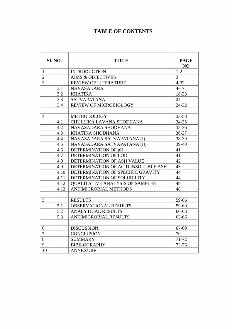
TABLE OF CONTENTS
SI. NO.
TITLE
PAGE
NO 1 INTRODUCTION 1-2 2 AIMS & OBJECTIVES 3 3 REVIEW OF LITERATURE 4-32 3.1 NAVASADARA 4-17 3.2 KHATIKA 18-23 3.3 SATVAPATANA 24 3.4 REVIEW OF MICROBIOLOGY 24-32 4 METHODOLOGY 33-58 4.1 CHULLIKA LAVANA SHODHANA 34-35 4.2 NAVASADARA SHODHANA 35-36 4.3 KHATIKA SHODHANA 36-37 4.4 NAVASADARA SATVAPATANA (I) 38-39 4.5 NAVASADARA SATVAPATANA (II) 39-40 4.6 DETERMINATION OF pH 41 4.7 DETERMINATION OF LOD 41 4.8 DETERMINATION OF ASH VALUE 42 4.9 DETERMINATION OF ACID INSOLUBLE ASH 43 4.10 DETERMINATION OF SPECIFIC GRAVITY 44 4.11 DETERMINATION OF SOLUBILITY 44 4.12 QUALITATIVE ANALYSIS OF SAMPLES 48 4.13 ANTIMICROBIAL METHODS 48 5 RESULTS 59-66 5.1 OBSERVATIONAL RESULTS 59-60 5.2 ANALYTICAL RESULTS 60-63 5.3 ANTIMICROBIAL RESULTS 63-66 6 DISCUSSION 67-69 7 CONCLUSION 70 8 SUMMARY 71-72 9 BIBILOGRAPHY 73-76 10 ANNEXURE
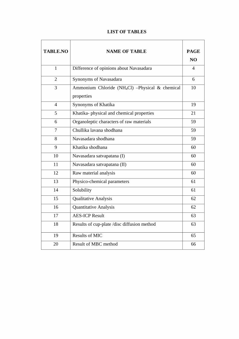
LIST OF TABLES
TABLE.NO
NAME OF TABLE
PAGE
NO
1 Difference of opinions about Navasadara 4
2 Synonyms of Navasadara 6
3 Ammonium Chloride (NH4Cl) –Physical & chemical
properties
10
4 Synonyms of Khatika 19
5 Khatika- physical and chemical properties 21
6 Organoleptic characters of raw materials 59
7 Chullika lavana shodhana 59
8 Navasadara shodhana 59
9 Khatika shodhana 60
10 Navasadara satvapatana (I) 60
11 Navasadara satvapatana (II) 60
12 Raw material analysis 60
13 Physico-chemical parameters 61
14 Solubility 61
15 Qualitative Analysis 62
16 Quantitative Analysis 62
17 AES-ICP Result 63
18 Results of cup-plate /disc diffusion method 63
19 Results of MIC 65
20 Result of MBC method 66
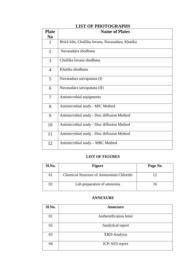
LIST OF PHOTOGRAPHS Plate No
Name of Plates
1 Brick kiln, Chullika lavana, Navasadara, Khatika
2 Navasadara shodhana
3 Chullika lavana shodhana
4 Khatika shodhana
5 Navasadara satvapatana (I)
6 Navasadara satvapatana (II)
7 Antimicrobial equipments
8 Antimicrobial study - MIC Method
9 Antimicrobial study - Disc diffusion Method
10 Antimicrobial study - Disc diffusion Method
11 Antimicrobial study - Disc diffusion Method
12 Antimicrobial study – MBC Method
LIST OF FIGURES
Sl.No. Figure
Page No
01 Chemical Structure of Ammonium Chloride
12
02 Lab preparation of ammonia
16
ANNEXURE
Sl.No. Annexure
01 Authentification letter
02 Analytical report
03 XRD-Analysis
04 ICP-AES report

Introduction
INTRODUCTION
Rasashastra is a new development in Ayurveda, as it is not mentioned in
traditional eight specialties i.e. Ashtanga Ayurveda. Rasashastra is a branch of
Ayurveda which deals with the various pharmaceutical processes such as shodhana,
marana, jarana, murchana, satwapatana etc. and description of metals, minerals,
animal products & poisonous herbal drugs, most precious things like gold, gems etc
& their therapeutic usage. These drugs are categorized or classified in different
groups like Maharasa, Uparasa, Sadharanarasa, Dhatu, Upadhatu, Ratna and so on.
Navasadara is one of the rasadravya included in Sadharana rasa1 group
of drug. Explanation of Navasadara is found in all most all Rasagranthas.
Rasendramangala the earliest book to mention about Navasadara and used as
dhatuvadartha, in parada jarana. But the amayika prayoga of Navasadara is explained
in the text Rasahridaya tantra in 8th century. Process of Navasadara satvapatana is
only explained in the text Rasatarangini2. Navasadara is used as a one of the jarana
drvya, i.e used in parada jarana and also used as lohadraavanaartha.
There is difference of opinions regarding Navasadara. Some of them explained
that it is one of the kshara and some are explained that it is a type of lavana3. To rule
out these differences of opinions proper identification by classical and modern
method, pharmaceutical, pharmacological study should be carried out.
While explaining the occurance of Navasadara it is told that the Chullika
lavana or ishtika lavana is the source for Navasadara i.e we can extract Navasadara
from this chullika lavana or ishtika lavana4.
“A Comparative Antimicrobial study of Navasadara Satva, by taking two different samples
of Navasadara”
1

Introduction
It is also extracted from Naramootra, Oustra mutra, oustra mala etc. In Punjab
it is extracted from mud5. As Navasadara is indicated as netrya, shweta kustahara,
deepana, kaphanissaraka, puppusashothahara,etc6. To assess its efficacy, its
antimicrobial study is carried out on gram positive and gram negative organisms.
. This study has been undertaken in three headings. A) Preparation of
Navasadara satva as per Rasatarangini, B) Comparative physico chemical analysis of
both raw samples as well as final product, and C) Comparative antimicrobial activity
of Navasadara satva using Disc Diffusion and MIC methods .
All the results obtained out of Analytical and Antimicrobial studies has been
tabulated and compared among them as per set of parameters.
Chullika lavana doesn’t yield Satva but the market sample given good amount
of Satva after subjecting both samples for satvapatana as per the reference
Rasatarangini. Antimicrobial studies revealed that both samples shown sensitive
against all four organisms except E-coli indicating good bacteriostatic action. Market
sample proven to be having good bactericidal action against E-coli and Staph
pneumoniae.
“A Comparative Antimicrobial study of Navasadara Satva, by taking two different samples
of Navasadara”
2
Aims and Objectives
AIMS AND OBJECTIVES
1. Preparation of Navasadara Satva by using both Chullika lavana and Navasadara
2. To screen Antimicrobial activity of Navasadara Satva.
3. Procurement of Chullika lavana from brick kiln.
4. Collection of Navasadara and Khatika.
5. Identification of Navasadara and Khatika according to grahya laxanas.
6. Shodhana of Chullika lavana, Navasadara, Khatika.
7. Physicochemical analysis of-
(a) Raw materials- Chullika lavana, Navasadara, Khatika
(b) Shodhita - Chullika lavana, Navasadara, Khatika
(c) Navasadara Satva
“A comparative Antimicrobial study of Navasadara Satva by taking two different samples Of Navasadara”
3

Review of Literature
REVIEW OF LITERATURE
3.1 NAVASADARA
Historical Review:
In vedakala and samhita kala there is no explanation about Navasadara. But
first it was mentioned in the text Rasendramangala in 7th century7. But it was used
only for lohavedartha. Later on during 8th century it was used for dehaveda by
Rasahrudaya tantrakara. Later on all rasa granthas have explained about this drug.
In Rasa Granthas:
• Among Rasa granthas, a reference regarding Navasadara is found in
Rasahridaya tantra8, grouped under lavana varga.
• Rasakamadhenu gives more detailed explanation regarding special varieties.
• Difference of openions were found regarding Navasadara, some authers
included under uparasa, some are included under sadharana rasa, lavana varga,
kshara varga, shown below.
Table No.1: Difference of openions about Navasadara
Sl.No. Reference Uparasa Sadharana
Rasa
Lavana
Varga
Kshara
Varga
01 Rasahridaya tantra - - + -
02 Rasarnava9 - - + -
03 Anandakanda10 + - + +
04 Rasendrachudamani - + + +
05 Rasendrasara
Samgraha11
- - + -
06 Rasaratna Samuchaya - + + +
“A comparative Antimicrobial study of Navasadara Satva by taking two different samples of Navasadara”
4
Review of Literature
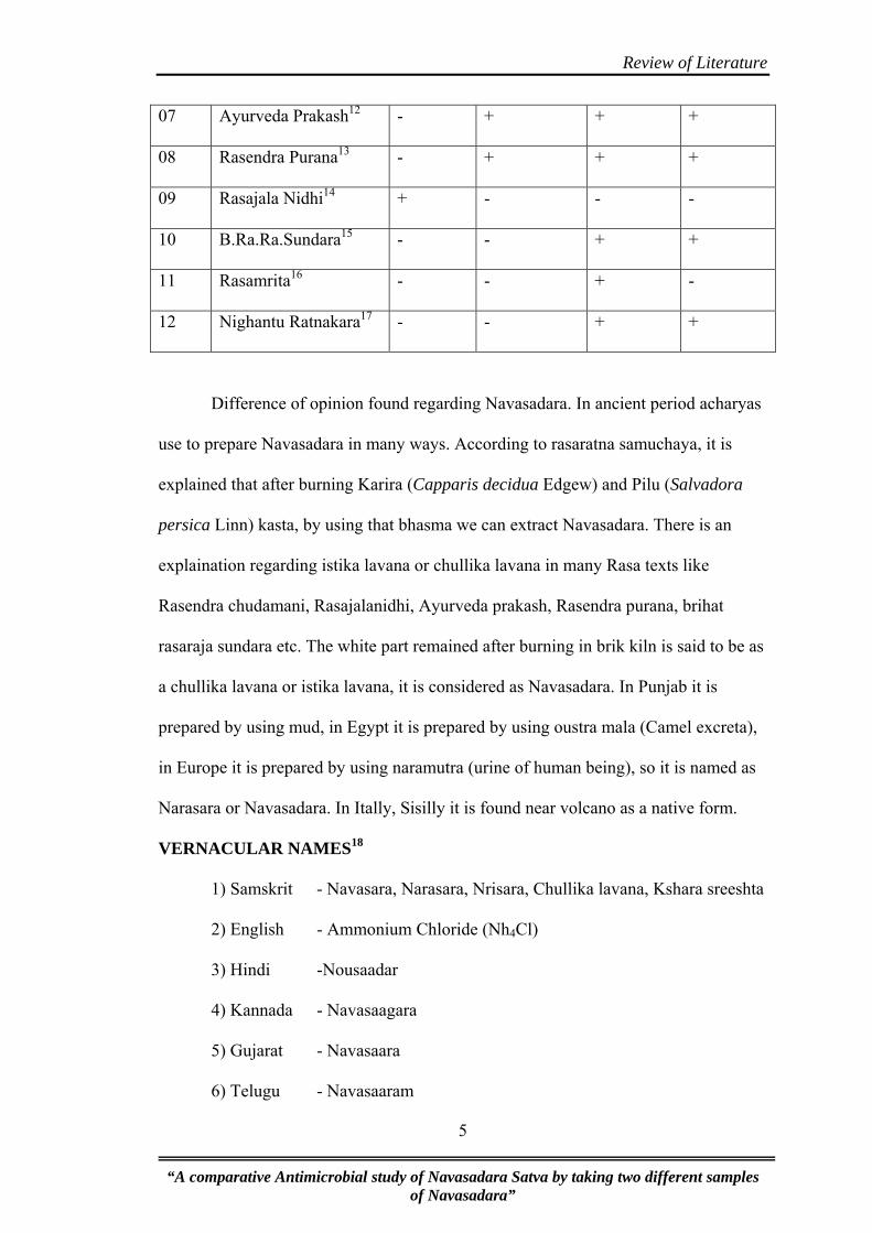
07 Ayurveda Prakash12 - + + +
08 Rasendra Purana13 - + + +
09 Rasajala Nidhi14 + - - -
10 B.Ra.Ra.Sundara15 - - + +
11 Rasamrita16 - - + -
12 Nighantu Ratnakara17 - - + +
Difference of opinion found regarding Navasadara. In ancient period acharyas
use to prepare Navasadara in many ways. According to rasaratna samuchaya, it is
explained that after burning Karira (Capparis decidua Edgew) and Pilu (Salvadora
persica Linn) kasta, by using that bhasma we can extract Navasadara. There is an
explaination regarding istika lavana or chullika lavana in many Rasa texts like
Rasendra chudamani, Rasajalanidhi, Ayurveda prakash, Rasendra purana, brihat
rasaraja sundara etc. The white part remained after burning in brik kiln is said to be as
a chullika lavana or istika lavana, it is considered as Navasadara. In Punjab it is
prepared by using mud, in Egypt it is prepared by using oustra mala (Camel excreta),
in Europe it is prepared by using naramutra (urine of human being), so it is named as
Narasara or Navasadara. In Itally, Sisilly it is found near volcano as a native form.
VERNACULAR NAMES18
1) Samskrit - Navasara, Narasara, Nrisara, Chullika lavana, Kshara sreeshta
2) English - Ammonium Chloride (Nh4Cl)
3) Hindi -Nousaadar
4) Kannada - Navasaagara
5) Gujarat - Navasaara
6) Telugu - Navasaaram
“A comparative Antimicrobial study of Navasadara Satva by taking two different samples of Navasadara”
5
Review of Literature
7) Tamil - Navachchaaram
8) Konkani - Navasagar
9) Punjabi - Nousadar
10) Pharsi - Noushadar
11) Malayalam - Navasaagaram
12) Latin - Ammonium chloride
13) Marathi -Navsaagar
14) Bengali -Nishaadal
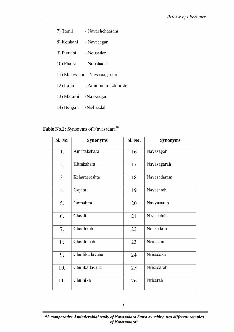
Table No.2: Synonyms of Navasadara19
Sl. No. Synonyms Sl. No. Synonyms
1. Amritakshara 16 Navasagah
2. Kittakshara 17 Navasagarah
3. Ksharasreshta 18 Navasadaram
4. Gojam 19 Navasarah
5. Gomalam 20 Navyasarah
6. Chooli 21 Nishaadala
7. Choolikah 22 Nousadara
8. Choolikaah 23 Nrirasara
9. Chullika lavana 24 Nrisadaka
10. Chulika lavana 25 Nrisadarah
11. Chulhika 26 Nrisarah
“A comparative Antimicrobial study of Navasadara Satva by taking two different samples of Navasadara”
6
Review of Literature
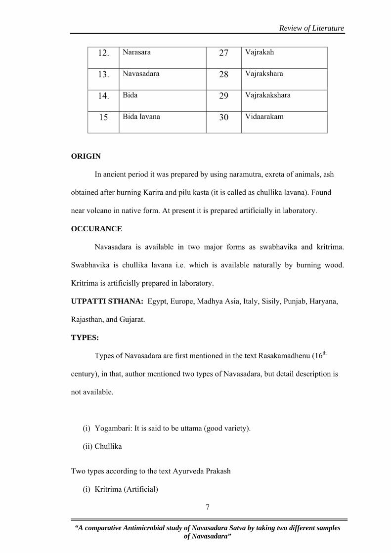
12. Narasara 27 Vajrakah
13. Navasadara 28 Vajrakshara
14. Bida 29 Vajrakakshara
15 Bida lavana 30 Vidaarakam
ORIGIN
In ancient period it was prepared by using naramutra, exreta of animals, ash
obtained after burning Karira and pilu kasta (it is called as chullika lavana). Found
near volcano in native form. At present it is prepared artificially in laboratory.
OCCURANCE
Navasadara is available in two major forms as swabhavika and kritrima.
Swabhavika is chullika lavana i.e. which is available naturally by burning wood.
Kritrima is artificislly prepared in laboratory.
UTPATTI STHANA: Egypt, Europe, Madhya Asia, Italy, Sisily, Punjab, Haryana,
Rajasthan, and Gujarat.
TYPES:
Types of Navasadara are first mentioned in the text Rasakamadhenu (16th
century), in that, author mentioned two types of Navasadara, but detail description is
not available.
(i) Yogambari: It is said to be uttama (good variety).
(ii) Chullika
Two types according to the text Ayurveda Prakash
(i) Kritrima (Artificial)
“A comparative Antimicrobial study of Navasadara Satva by taking two different samples of Navasadara”
7
Review of Literature
(ii) Akritrima (Natural)
Navasadara which is obtained by the mutra, purisha of manushya, pashu is called
as akritrima.
On the base of Praaptisthana – Two types
(i) Khanija
(ii) Vaanaspatika
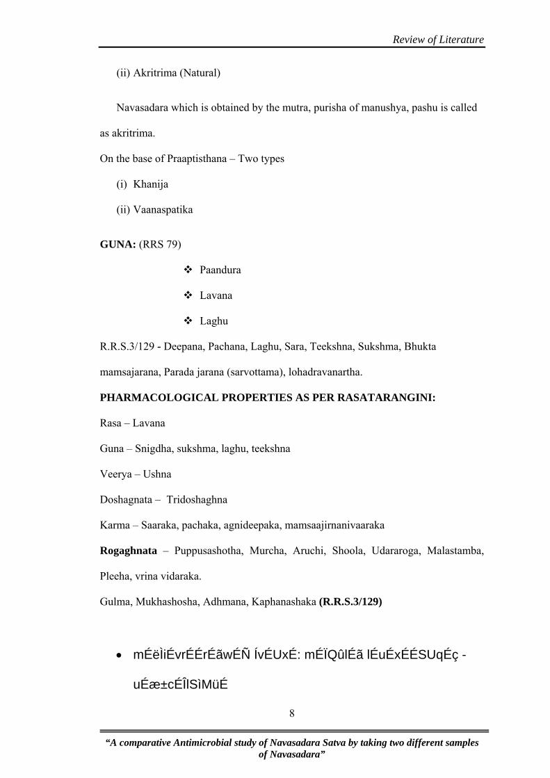
GUNA: (RRS 79)
Paandura
Lavana
Laghu
R.R.S.3/129 - Deepana, Pachana, Laghu, Sara, Teekshna, Sukshma, Bhukta
mamsajarana, Parada jarana (sarvottama), lohadravanartha.
PHARMACOLOGICAL PROPERTIES AS PER RASATARANGINI:
Rasa – Lavana
Guna – Snigdha, sukshma, laghu, teekshna
Veerya – Ushna
Doshagnata – Tridoshaghna
Karma – Saaraka, pachaka, agnideepaka, mamsaajirnanivaaraka
Rogaghnata – Puppusashotha, Murcha, Aruchi, Shoola, Udararoga, Malastamba,
Pleeha, vrina vidaraka.
Gulma, Mukhashosha, Adhmana, Kaphanashaka (R.R.S.3/129)
• mÉëÌiÉvrÉÉrÉãwÉÑ ÍvÉUxÉ: mÉÏQûlÉã lÉuÉxÉÉSUqÉç -
uÉæ±cÉÎlSìMüÉ
“A comparative Antimicrobial study of Navasadara Satva by taking two different samples of Navasadara”
8

Review of Literature
Navasadara is indicated for Pratisyaya and shira shoola by Vaidya chandrika
Method of extraction:
• SÕÌiÉ uÉæ± cÉÎlSìMüÉã£ãü cÉÔhÉïiÉÉãrÉÉprÉÉÇ
RûÉãsÉÉrÉl§ÉãhÉ mÉÉÍcÉiÉã YuÉÉjÉqÉãSã lÉuÉxÉÉSUqÉmrÉ§É |
-Vachaspatyam vol 5
Explanation regarding Navasadara in Vachaspatyam is, Navasadar churna is
dissolved in water and pachan is carried out in Dolayantra to get shuddha Navasadara.
NAVASADARA SHODHANA:
According to various Rasagranthas written by different rasavaidyas,
navasadara shodhana is mentioned in various methods.
lÉuÉxÉÉUliÉÑ xÉÍsÉsÉå ̧ÉaÉÑhÉå SìÉuÉrÉåΰwÉMçü |
uÉx§ÉmÉÔiÉqÉç iÉiÉ: M×üirÉÉ pÉÉeÉlÉå xjÉÉmÉrÉå¨ÉiÉ: ||
cÉÑÎssÉMüÉrÉÉqÉç ÌlÉkÉÉrÉÉjÉ mÉcÉå¨ÉÏuÉëÉÎalÉlÉÉ
pÉ×zÉqÉ |
eÉsÉÇ zÉÑwMÇü iÉÑsÉxrÉ¶É lÉ×xÉÉUÇ ÌuÉqÉsÉÇ WûUåiÉç ||
- RT14/3-4
1 part of Navasadara is dissolved completely in 3 parts of jala, and then it is
filtered. This filtrate is kept on intense heat. Heat is given until all water part get
evaporates. Then it is collected.
lÉuÉxÉÉUÉã pÉuÉãcNÒûkSÇ ¶ÉÔhÉïiÉÉãrÉæÌuÉïmÉÉÍcÉiÉ: |
RûÉãsÉÉrÉl§ÉãhÉ rɦÉãlÉ ÍpÉwÉÎapÉrÉÉãïaÉÍxÉkSrÉã || -
Rasendra sambhava
“A comparative Antimicrobial study of Navasadara Satva by taking two different samples of Navasadara”
9
Navasadara is made into churna mixed with jala and pachana is carried out in
Dolayantra to purify.

Review of Literature
According to Parada samhita –
Navasadara is taken in khalwa yantra, and bhavana is given with Jambeera
swarasa / Nimbu swarasa, then it is dried in sun light, after complete drying it is kept
in urdwa paatana yantra. Then collect the shuddha Navasadara from inner part of the
upper pot.
According to Rasaratna Samuchaya – Navasadara is dissolved in boiling
water, and then filtered through cloth, dried. Thus formed navasadara is shodhita.
NAVASADARA YOGAS:20
1) Gulmari rasa 10) Navasara churna yoga
2) Kshara parpati 11) Talakeshwara rasa,
3) Vrishchikdamshahara lepa. 12) Kshaya kesari rasa,
4) Pleehari vati 13) Amlapittantaka rasa
5) Talasindhura 14) Navasadara druti
6) Meghanada rasa 15) Lavanadya churna,
7) Netrasudha anjana 16) Tripura bhairava rasa
8) Jwarashara arka 17) Deepana churna
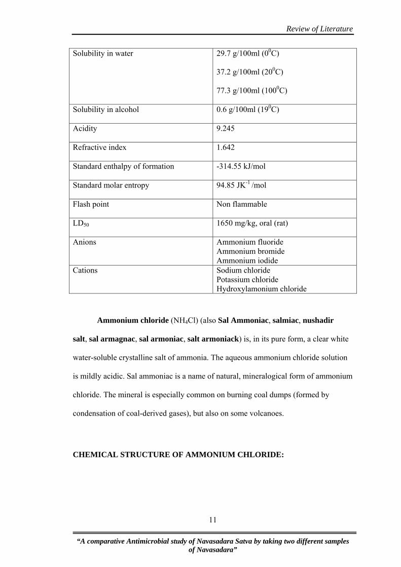
Table No. 3: Ammonium Chloride (NH4Cl) –Physical & chemical properties21
IUPAC Name Ammonium Chloride
Other Name Sal ammoniac
Molecular formula NH4Cl
Molar mass 53.49g/mol
Appearance White solid
Density 1.5274g/cm3
Melting Point 3380 C
“A comparative Antimicrobial study of Navasadara Satva by taking two different samples of Navasadara”
10
Review of Literature
Solubility in water 29.7 g/100ml (00C)
37.2 g/100ml (200C)
77.3 g/100ml (1000C)
Solubility in alcohol 0.6 g/100ml (190C)
Acidity 9.245
Refractive index 1.642
Standard enthalpy of formation -314.55 kJ/mol
Standard molar entropy 94.85 JK-1 /mol
Flash point Non flammable
LD50 1650 mg/kg, oral (rat)
Anions Ammonium fluoride Ammonium bromide Ammonium iodide
Cations Sodium chloride Potassium chloride Hydroxylamonium chloride
Ammonium chloride (NH4Cl) (also Sal Ammoniac, salmiac, nushadir
salt, sal armagnac, sal armoniac, salt armoniack) is, in its pure form, a clear white
water-soluble crystalline salt of ammonia. The aqueous ammonium chloride solution
is mildly acidic. Sal ammoniac is a name of natural, mineralogical form of ammonium
chloride. The mineral is especially common on burning coal dumps (formed by
condensation of coal-derived gases), but also on some volcanoes.
CHEMICAL STRUCTURE OF AMMONIUM CHLORIDE:
“A comparative Antimicrobial study of Navasadara Satva by taking two different samples of Navasadara”
11

Review of Literature
Fig.No 1. Chemical structure of ammonium chloride
HISTORY:
The Romans called the ammonium chloride deposits they collected from near
the Temple of Jupiter Amun (Greek �μμων Ammon) in ancient Libya 'sal
ammoniacus' (salt of Amun) because of proximity to the nearby temple. Salts of
ammonia have been known from very early times; thus the term Hammoniacus sal
appears in the writings of Pliny, although it is not known whether the term is identical
with the more modern sal-ammoniac.
In the form of sal-ammoniac, ammonia was known to the alchemists as
early as the 13th century, being mentioned by Albertus Magnus. It was also used by
dyers in the middle Ages in the form of fermented urine to alter the colour of
vegetable dyes. In the 15th century, Basilius Valentinus showed that ammonia could
be obtained by the action of alkalis on sal-ammoniac. The Haber process to produce
ammonia from the nitrogen in the air was developed by Fritz Haber and Carl Bosch in
1909 and patented in 1910. It was first used on an industrial scale by the Germans
during World War I.
“A comparative Antimicrobial study of Navasadara Satva by taking two different samples of Navasadara”
12
Review of Literature
Sal Ammoniac was named after it was observed in the Temple of Zeus-
Ammon in Egypt; its name means "salt of Ammon". It was the white crystalline
substance that remained on the ceiling and walls after camel dung was burned. The
modern name "ammonium" comes from Sal Ammoniac. There are a few stories of
Alexander the Great finding such crystals in the coal seams of Tajikistan. The
substance was known as nao sha in China, nao sadar in India, and nushadir in Persia
and Arabic countries.
Sources:
The substance occurs naturally in volcanic regions, forming on volcanic
rocks near fume-releasing vents. The crystals deposit directly from the gaseous state, and tend
to be short-lived, as they dissolve easily in water. It is a by-product of the Solvay process used
to produce sodium carbonate.
Ammonium chloride is prepared commercially by reacting ammonia (NH3) with hydrogen
chloride (HCl). As these chemicals are corrosive, this process has to be performed in vessels
lined with nonreactive materials (e.g. glass, enamel,lead, or PVC).
NH3 + HCl → NH4Cl
SYNTHESIS AND PRODUCTION:
Because of its many uses, ammonia is one of the most highly produced
inorganic chemicals. About 80% or more of the ammonia produced is used for
fertilizing agricultural crops.
Before the start of World War I, most ammonia was obtained by the dry
distillation of nitrogenous vegetable and animal waste products, including camel
dung, where it was distilled by the reduction of nitrous acid and nitrites with
“A comparative Antimicrobial study of Navasadara Satva by taking two different samples of Navasadara”
13

Review of Literature
hydrogen; in addition, it was produced by the distillation of coal, and also by the
decomposition of ammonium salts by alkaline hydroxides such as quicklime, the salt
most generally used being the chloride (sal-ammoniac) thus:
2 NH4Cl + 2 CaO → CaCl2 + Ca (OH) 2 + 2 NH3
Medical Properties and Physiological Action:
Ammonia gas is very alkaline, and an irritant to mucous surfaces. Inhaled, it
causes an overpowering sense of suffocation and spasm of the glottis, and when
prolonged, violent inflammation of the air-passages. Solution of ammonia when
swallowed causes destructive inflammation of the mucous membrane, extending to
the stomach. The long-continued use of ammonia interferes with digestion by
neutralizing the gastric juice, and by increased waste of tissue causes pallor,
Emaciation and feebleness. In the blood it injures the red blood globules, and thus
affects the nutrition of the body, being largely converted into urea. The preparations
of ammonia are stimulant expectorants.
Aqua ammonia is administered by inhalation in syncope and shock, and as a
counter-irritant; for which purpose ammonia liniment is also employed. The
incautious inhalation of ammonia may cause inflammation of the fauces and glottis,
but when cautiously employed sometimes gives relief to acute catarrh and hay
asthma. The diluted aqua ammonia will relieve the pain of stings of insects, and the
strong aqua ammonia is an antidote, when at once applied, to the bite of venomous
snakes, and of rabid animals. The aromatic spirits of ammonia is useful in acidity of
stomach, gaseous eructations and abdominal distensions; also in sick headache and
migraine; but the bromides are more effective in the latter affection. Ammonia salts
“A comparative Antimicrobial study of Navasadara Satva by taking two different samples of Navasadara”
14

Review of Literature
stimulate the liver and increase the secretions of the kidneys and intestinal mucous
glands and the action of the heart, hence are frequently used in adynamic states,
constipation, coated tongue and scanty urine.
Ammonia and Its Salts (NH3):
When pure, ammonia is a colourless gas, capable of being liquified; of very
pungent odour, the fumes producing an alkaline reaction; it forms salts with acids,
but always takes an atom of basic water, and hence by most chemists these salts are
regarded as containing an oxide of a hypothetical metal called ammonium (Nh4);
thus sal ammoniac may be regarded as a hydrochlorate of ammonia (Nh3, Hc1) or
chloride of ammonium (Nh4 Cl). Ammonia also forms direct combinations with
acids, as carbonic acid, not true salts; a compound of carbonic acid and ammonia
(Nh3, Co2) is perhaps present in the sesquicarbonate. Gaseous ammonia is
sometimes made use of therapeutically, evolved usually when thus employed, from
liquor ammonias, in which it is contained.
Laboratory Preparation of Ammonia
For use in the laboratory, ammonia is prepared by heating an ammonium salt
with a strong base. It can also be prepared by reacting a metal nitride with water. All
ammonium salts heated with alkali evolve ammonia. Ammonium nitrate is not used in
laboratory preparation, since it is explosive in nature and may decompose forming
nitrous oxide and water vapour.
“A comparative Antimicrobial study of Navasadara Satva by taking two different samples of Navasadara”
15

Review of Literature
Fig No.2 Ammonia is prepared in the laboratory by heating a mixture of solid
ammonium chloride and calcium hydroxide.
Reactants: Ammonium chloride and calcium hydroxide, Ratio 2:3
Procedure: Ammonium chloride and calcium hydroxide are ground together and
heated slowly in a round bottomed flask with its neck sloping downwards. The gas
is passed through a drying tower before collection.
Collection: Ammonia gas is lighter than air and hence collected by the downward
displacement of air. It is not collected over water since it is highly soluble in water.
“A comparative Antimicrobial study of Navasadara Satva by taking two different samples of Navasadara”
16

Review of Literature
DETECTION OF AMMONIA:
Ammonia and ammonium salts can be readily detected, in very minute traces, by the
addition of Nessler's solution, which gives a distinct yellow coloration in the
presence of the least trace of ammonia or ammonium salts.
Nessler's reagent: In analytical chemistry Nessler's reagent is a reagent used to detect
small concentrations of ammonia. A yellow coloration indicates the presence of
ammonia. At high concentrations, a brown precipitate may form.
Larger quantities can be detected by warming the salts with a caustic alkali or
with quicklime, when the characteristic smell of ammonia will be at once apparent.
The amount of ammonia in ammonium salts can be estimated quantitatively
by distillation of the salts with sodium or potassium hydroxide, the ammonia evolved
being absorbed in a known volume of standard sulfuric acid and the excess of acid
then determined volumetrically; or the ammonia may be absorbed in hydrochloric
acid and the ammonium chloride so formed precipitated as ammonium chlorplatinate,
(NH4)2PtCl6.
Test: Mixture + NaOH Boil and hold a wet litmus paper at the mouth of
the test tube.
Observation: Gas with ammonia smell having alkaline action on litmus paper
and white dense fumes with glass rod dipped in con HCl NH4 present
Toxicity:
It is toxic if swallowed, inhaled or absorbed through the skin. It presents
neurological hazard and may act as a carcinogen and be a reproductive hazard. It is
“A comparative Antimicrobial study of Navasadara Satva by taking two different samples of Navasadara”
17
Review of Literature
corrosive and causes burns. Always wear safety glasses and gloves and manipulate in
a well ventilated environment.
3.2 KHATIKA
Historical review –
In Vedas
There is no description about Khatika in Vedas.
In Samhitas
A little description is available in Samhitas.
In Rasagranthas
• Bhava Misra has included Khatika in Uparasa Varga.
• Yasodhara Bhatta in Rasa Prakasa sudhakara has mentioned the Khatika for
the preparation of Rasa karpura. He has also used khatika for the shodhana of
Swrna. He has used the word khatika to describe the white variety of sulphur.
• Vagbhatta in his text book Rasaratna samucchaya has also used the word
Khatika to describe the white variety of sulphur.
• In Ayurveda Prakash, Acharya Madhava.has included khatika in Uparasa
group. He also described the synonyms, varieties and therapeutic indications
of Khatika.
• Sri Sadananda Sharma in his text Rasa Tarangini has described the synonyms,
varieties and therapeutic indications of Khatika along with its purification
method.
“A comparative Antimicrobial study of Navasadara Satva by taking two different samples of Navasadara”
18
Review of Literature
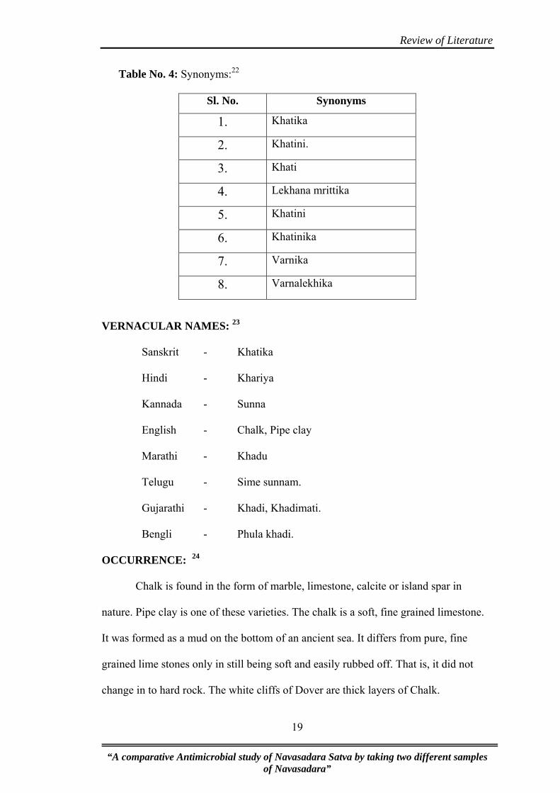
Table No. 4: Synonyms:22
Sl. No. Synonyms 1. Khatika
2. Khatini.
3. Khati
4. Lekhana mrittika
5. Khatini
6. Khatinika
7. Varnika
8. Varnalekhika
VERNACULAR NAMES: 23
Sanskrit - Khatika
Hindi - Khariya
Kannada - Sunna
English - Chalk, Pipe clay
Marathi - Khadu
Telugu - Sime sunnam.
Gujarathi - Khadi, Khadimati.
Bengli - Phula khadi.
OCCURRENCE: 24
Chalk is found in the form of marble, limestone, calcite or island spar in
nature. Pipe clay is one of these varieties. The chalk is a soft, fine grained limestone.
It was formed as a mud on the bottom of an ancient sea. It differs from pure, fine
grained lime stones only in still being soft and easily rubbed off. That is, it did not
change in to hard rock. The white cliffs of Dover are thick layers of Chalk.
“A comparative Antimicrobial study of Navasadara Satva by taking two different samples of Navasadara”
19
Review of Literature
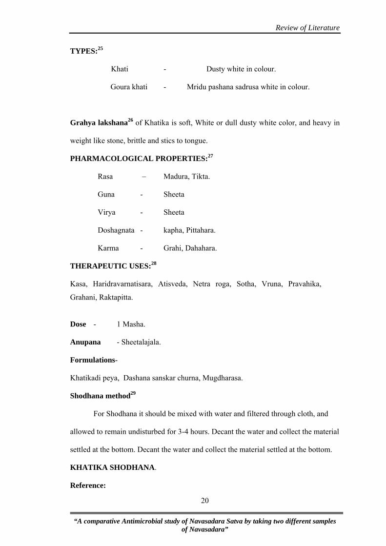
TYPES:25
Khati - Dusty white in colour.
Goura khati - Mridu pashana sadrusa white in colour.
Grahya lakshana26 of Khatika is soft, White or dull dusty white color, and heavy in
weight like stone, brittle and stics to tongue.
PHARMACOLOGICAL PROPERTIES:27
Rasa – Madura, Tikta.
Guna - Sheeta
Virya - Sheeta
Doshagnata - kapha, Pittahara.
Karma - Grahi, Dahahara.
THERAPEUTIC USES:28
Kasa, Haridravarnatisara, Atisveda, Netra roga, Sotha, Vruna, Pravahika,
Grahani, Raktapitta.
Dose - 1 Masha.
Anupana - Sheetalajala.
Formulations-
Khatikadi peya, Dashana sanskar churna, Mugdharasa.
Shodhana method29
For Shodhana it should be mixed with water and filtered through cloth, and
allowed to remain undisturbed for 3-4 hours. Decant the water and collect the material
settled at the bottom. Decant the water and collect the material settled at the bottom.
KHATIKA SHODHANA.
Reference:
“A comparative Antimicrobial study of Navasadara Satva by taking two different samples of Navasadara”
20

Review of Literature
ZÉOûÏcÉÔhÉïÇ vÉÑ®mÉɧÉã ÌlÉkÉÉrÉ ÌuÉqÉsÉã eÉsÉã |
mÉë¤ÉÉsÉrÉã̲kÉÉlÉalÉÉã ÌuÉvÉÑkrÉÌiÉ lÉ xÉÇvÉrÉ: || - R
T 11/210
Khatika is purified by nirmalikarna process by using water. 1 part of khatika
churna is dissolved in 4 parts of water kept for some time and filtered through cloth
and dried in sunlight and collected.
Table No.5: Khatika physical, chemical properties30:
Name Calcium carbonate
Chemical formula CaCO3
Appearance White solid
Formula weight 100.1 amu
Melting point Liquifies under high pressure at1612K (13390C)
Boiling point Decomposes at 1172 K (8990C)
Density 2.7 × 10³ kg/m3
Crystal structure Calcite, aragonite, or vaterite
Solubility 0.0013gm in 100gm water
Standard enthalpy formation liquid -1154 kJ/mol
Standard enthalpy formation solid -1207 kJ/mol
Standard molar enthalpy 93 J/(mol K)
CHALK:
Chalk is a soft, white, porous form of limestone composed of the mineral
calcium carbonate. It is relatively resistant to erosion and slumping compared to the
clays that it is usually associated with, and so forms tall steep cliffs where chalk
“A comparative Antimicrobial study of Navasadara Satva by taking two different samples of Navasadara”
21

Review of Literature
ridges meet the sea. Chalk hills, known as chalk downland, usually form where bands
of chalk reach the surface at an angle. Because chalk is porous, chalk downland
usually holds a large water table, providing a natural reservoir that releases water
slowly through dry seasons.
Chalk has been quarried from prehistory, providing building material and marl
for fields. In southeast England, deneholes are a notable example of ancient chalk pits.
Blackboard chalk is a substance used for drawing on rough surfaces, as it
readily crumbles leaving particles that stick loosely to these surfaces. Blackboard
chalk, often supplied in sticks about 5 cm long, is not actually made from the mineral
chalk but from gypsum (calcium sulfate). Similarly, the "chalk" used by tailors is
usually made from talc (magnesium silicate).
CHALK FORMATION:31
The Chalk Formations of Europe are thick deposits of chalk, a soft porous
white limestone, deposited in a marine environment during the upper Cretaceous
period. They appear most prominently in England. The formations are divided into
three parts: The Upper Chalk, the Middle Chalk, and the Lower Chalk. The famous
White cliffs of Dover, England are a good example of a Chalk Formation deposit.
Another good example displaying the sequence of the Chalk Formation are the
southern cliffs on the Isle of Wight, England and the quarries and motorway cutting at
Blue Bell Hill, Kent, England (which has been classified as a Site of Special Scientific
Interest).
“A comparative Antimicrobial study of Navasadara Satva by taking two different samples of Navasadara”
22

Review of Literature
• Chemistry: CaCO3, Calcium Carbonate
• Class: Carbonates
• Group: Calcite
• Uses: In cements and mortars, production of lime, limestone is used in the
steel industry; glass industry, ornamental stone, chemical and optical uses and
as mineral specimens.
Calcite, which gets its name from "chalix" the Greek word for lime, is a
most amazing and yet, most common mineral. It is one of the most common minerals
on the face of the Earth, comprising about 4% by weight of the Earth's crust and is
formed in many different geological environments. Calcite can form rocks of
considerable mass and constitutes a significant part of all three major rock
classification types. It forms oolitic, fossiliferous and massive limestones in
sedimentary environments and even serves as the cements for many sandstones and
shales. Limestone becomes marble from the heat and pressure of metamorphic events.
Calcite is even a major component in the igneous rock called carbonatite and forms
the major portion of many hydrothermal veins. Some of these rock types are
composed of better than 99% calcite.
The crystals of calcite can form literally a thousand different shapes by
combining the basic forms of the positive rhombohedron, negative rhombohedron,
steeply, moderately and slightly inclined rhombohedrons, various scalahedrons, prism
and pinacoid to name a few of the more common forms. There are more than 300
crystal forms identified in calcite and these forms can combine to produce the
thousand different crystal variations. Calcite also produces many twin varieties that
are favorites among twin collectors. There are also phantoms, included crystals, color
“A comparative Antimicrobial study of Navasadara Satva by taking two different samples of Navasadara”
23

Review of Literature
varieties, pseudomorphs and unique associations. There simply is no end to the
varieties of calcite.
3.3 SATVAPATANA:32
The term satvapatana is composed of two words, satva and patana. Satva is an
essence of drug, wher as patana means to extract.
Shloka:
¤ÉÉUÉqsÉ mÉëÉuÉMæürÉÑï£üÇ kqÉÉiÉÉqÉÉÇ MüU
MüÉã¹Mãü |
rɦÉiÉÉã ÌlÉaÉïiÉ: xÉÉU: xÉiuÉÍqÉirÉÍpÉSÏrÉiÉã || - Rasadarpana
The drugs of mineral origin when mixed with alkali (kshara), acid (amla) and
fusing materials (dravaka gana) chakrikas prepared and subjected to intense heat in a
furnace, it will liberates the metallic portion of the material is called satva.
History of Satvapatana:
In Vedic literature metals like gold, silver, iron etc are mentioned. Mention of
these metals which are not available in Free State in nature means people of Vedic era
were having knowledge of satvapatana. Acharya Nagarjuna in 7th century AD
described the process in details in his book Rasendramangala. The research workers
of Rasashastra have developed newer techniques decided the fuel and various signs
which indicated the melting of mineral substances and separation of metallic contents.
Also various types of fuels, equipments and furnces are mentioned in these ancient
texts. Further the literature of Rasashastra describes the form appearance, colour, and
etc. characters of satva bhasmeekarana process and uses of satva.
3.4 REVIEW OF MICROBIOLOGY: 33
Medical microbiology is the study of microbes that infect humans, the
diseases, which they cause, their diagnosis, prevention and treatment. As microbes are
“A comparative Antimicrobial study of Navasadara Satva by taking two different samples of Navasadara”
24

Review of Literature
invisible to the unaided eye, definitive knowledge about them had to await the
development of microscopes.
Microbiology a branch of science, dealing with microorganisms. These
microorganisms could be pathogenic or nonpathogenic with varied applications.
Some of them play an important role in maintaining the ecological balance
on earth. Some of them have commercial applications as in synthesis of chemical
products and food, some others are pathogenic that is disease causing.
The group of microbes includes.
1. Bacteria.
2. Fungi.
3. Protozoa.
4. Microscopic algae.
It also includes the viruses the non cellular entities sometimes regarded as
being at the border between life and non life.
ESCHERICHIA COLI:
Classification:-
Domain : Bacteria
Phylum : Proteo Bacteria
Class : Gamma Proteo Bacteria
Order : Entero Bacteriales
Family : Entero Bacteriaceae
Genus : Escherichia
Species : Escherichia Coli
Escherichia Coli:
• The proteins group.
“A comparative Antimicrobial study of Navasadara Satva by taking two different samples of Navasadara”
25

Review of Literature
• Is the most common cause of GIT, UTI, and gram-negative.
• It is main cause of infantile gastroenteritis, traveller’s diarrhoea and neonatal
meningitis.
Bacteriology:
• E. coli: Belongs to the germs Escherichia of the Coliform group.
• It is gram-negative motile bacillus which grows on ordinary media.
• It ferments lactose, a property that distinguishes it from the two major
intestinal pathogens, Shigella and Salmonella.
• It produces pink colonies on MacConkey agar and yellow colonies on agar
because of lactose fermentation.
• It gives a positively methyl red test and a negative vages proskaur reaction and
citrate test.
Pathogenicity:
Although a normal gut commensal, many strain of E. coli have powerful
toxins and other enteropathgenic mechanism which can be responsible for
diarrhea and other symptoms.
STAPHYLOCOCCUS:
Scientific classification:-
Kingdom – Monora
Phylum – Firmicutes
Class – Bacilli
Order – Bacillales
Family – Staphylococcacea
Genus – Staphylococcus
Species – S. aureus
“A comparative Antimicrobial study of Navasadara Satva by taking two different samples of Navasadara”
26

Review of Literature
GRAM - POSITIVE COCCI:-
Identification of gram positive cocci is an important part of the work the
clinical microbiologist performs. The most common gram-positive cocci infections
include staphylococcus and streptococcus.
STAPHYLOCOCCUS
The genus staphylococcus comprises approximately 19 species, of which there
all important pathogens for humans – S. aureus, S. epidermidis and sprophyticus.
Staphylococci generally appear in stains as grape – like clusters but they can
also appear singly or in pairs. They are approximately 1 mm in diameter.
Staphylococcus is normal flora on the skin and mucous membrane but can
cause infection under certain circumstances.
S. aureus has been recognized historically as a virulent human pathogen. Its
capacity to cause human disease has not been diminished by the introduction of
antibiotics.
For Eg. Such staphylococci can infect prosthetic or indwelling devices (Eg –
artificial body parts and catheters). S.aureus is recovered from various infections,
including skin lesions, basal wound infections, pneumonia and others. Organisms
from these sites on invade the blood and spread throughout the body organisms.
S. aureus can cause such problems as food poisoning, toxic shock syndrome, gastro
enteritis, pneumonia, endocarditis, osteomyelitis and ear and eye infections.
STAPHYLOCOCCUS AUREUS:
Morphology: They are spherical cocci, approximately in grape like clusters. They
may also be found singly, in pairs and in short chain of three or four cell especially
when examined from liquid culture. They are non-motile, non-sporing. They stain
readily with aniline dyes and are uniformly gram positive.
“A comparative Antimicrobial study of Navasadara Satva by taking two different samples of Navasadara”
27

Review of Literature
Culture characteristic:
They grow readily on ordinary media within a temperature range of 10-42OC.
the optimum temperature being 37OC and pH 7.4 – 7.6. They are aerobes and
facultative anaerobes.
On nutrient agar, after incubation for 24 hrs. The colonies are large (2-4 mm
diameter), circular, convex, smooth, shiny, opaque and easily emulsifiable. Most
strains produce golden yellow pigment, though some may be while orange or yellow.
On nutrient agar slope, their contluent growth presents a characteristic “oil
paint” appearance.
For determining S. aureus a test called coagulase test is performed S. aureus
produce coagulase enzymes that is able to clot rabbit plasma, differentiating this
staphylococcus species from the rest of the clinically significant staphylococci.
PNEUMOCOCCUS:
Pneumococcus, a gram positive lanceolate diplococcus, formerly classified as
Str.pneumoniae, has been reclassified as Stre.pneumoneae because of its genetic
relatedness to Streptococcus. Pneumococcus differs from other Streptococci chiefly in
its morphology, bile solubility, optachin sensitivity and possession of a specific
polysaccharide capsule. Pneumococci are normally inhabitants of the human upper
respiratory tract. They are the single most prevalent bacterial agent in Pneumonia and
in otitis media in children. They can also cause sinusitis, bronchitis, bacteremia,
meningitis and other infections. Pneumococci were first noticed in 1881 by Pasteur
and Steinberg independently.
Morphology:
“A comparative Antimicrobial study of Navasadara Satva by taking two different samples of Navasadara”
28

Review of Literature
Pneumococci are typically small 1μ m, slightly elongated cocci, with one end
broad or rounded and other end pointed, presenting a flame shaped or lanceolate
appearance. They occur in pairs, with the broad ends in apposition, the long axis of
the coccus parallel to the line joining the two cocci in a pair. They are capsulated, the
capsule enclosing each pair. The capsules are best seen in material taken directly from
exudates and may be lost on repeated cultivation.
In culture the typical morphology may not be apparent and the cocci are more
rounded, tending to occur in short chains. They are non-motile and non-sporing. They
are readily stained with aniline dyes and are Gram+ve.
Cultural characteristics:
Pneumococci have complex growth requirements and grow only in enriched
media. They are aerobes and facultative anaerobes. The optimum temperature being
370C [range 250-420C] and pH7.8 [range 6.5-8.3] growth is improved by 5-10% CO2.
On blood agar, after incubation for 18 hrs, the colonies are small [0.5-1mm], dome
shaped and glistening with an area of green discoloration around them resembling
colonies of str.viridans.
Under anaerobic conditions, colonies on blood agar are surrounded by a zone of beta
hemolysis due to oxygen liable hemolysin O. In liquid media such as glucose broth,
growth occurs as uniform turbidity. The cocci readily undergo autolysis in cultures
due to the activity of intracellular enzymes. Autolysis is enhanced by bile salts,
sodium lauryl sulphate and other surface active agents. Heat cultures do not undergo
autolysis.
“A comparative Antimicrobial study of Navasadara Satva by taking two different samples of Navasadara”
29
Review of Literature
KLEBSILLA PNEUMONIAE
Klebsiella pneumoniae is a Gram-negative, non-motile, encapsulated, lactose
fermenting, facultative anaerobic, rod shaped bacterium found in the normal flora of
the mouth, skin, and intestines.They are short, plump, straight rods, about 1-2× 0.5-
0.8 mm in size. The capsule is often prominent and can be made out even in Gram
stained smears as holoes around the basilli.
It naturally occurs in the soil and about 30% of strains can fix nitrogen in
anaerobic condition.
HISTORY:
The Danish scientist Hans Christian Gram (1853–1938), developed the
technique now known as Gram staining in 1884 to discriminate between K.
pneumoniae and Streptococcus pneumoniae.
Klebssadsadiella was named after the German bacteriologist Edwin Klebs (1834–
1913). Community-acquired pneumonia caused by Klebsiella pneumoniae may be
called Friedländer's Pneumonia, after Carl Friedländer.
Types:
Their classification has undergone various modifications. They have been
classdsified into three species based on biiochemiocal reactions and into over 80
serotypes based on capsular antigens.
Species are,
i. Klebsiella pneumoniae
ii. Klebsiella ozaenae.
iii. Klebsiellarhinoscleromatis.
“A comparative Antimicrobial study of Navasadara Satva by taking two different samples of Navasadara”
30
Review of Literature
Biochemical Reactions:
It ferments sugars (glucose, lactose, sucrose, and mannitol) with the
production of acid and abundant gas. It is indole and MR negative and VP and citrate
positive. Biochemically variant strains are common. It forms Urease. It is second most
populous member of the aerobic bacterial flora of the human intestine. It has become
a very important cause of nosocomial infections.
Pathogenesis:
K. pneumoniae can cause bacterial pneumonia, typically due to aspiration by
alcoholics, though it is more commonly implicated in hospital-acquired urinary tract
and wound infections, particularly in immunocompromised individuals. Klebsiella
ranks second to E. coli for urinary tract infections in older persons. It is also an
opportunistic pathogen for patients with chronic pulmonary disease, enteric
pathogenicity, nasal mucosa atrophy, and rhinoscleroma. Feces are the most
significant source of patient infection, followed by contact with contaminated
instruments.
Members of the Klebsiella genus typically express 2 types of antigens on their
cell surface. The first, O antigen is a lipopolysaccharide of which 9 varieties exist.
The second is K antigen, a capsular polysaccharide with more than 80 varieties. Both
contribute to pathogenicity and form the basis for subtyping.
Klebsiella pneumoniae is a serious disease with high case fatality.It occurs in
middle aged or older persons who have medical problems such as alcoholism, chronic
bronchopulmanary disease or diabetes mellitus.
“A comparative Antimicrobial study of Navasadara Satva by taking two different samples of Navasadara”
31

Review of Literature
RESEARCH PROFILE:
• Baldaniya K R,Navasadara nirman ki vidiyon ka proyogika adhyayana evam
stara nirdharana,1979,IPGTR Jamnagar
• Nagendrappa, Management of Kamala with Hareetaki katuki and
Navasadara,1994,GAMC Mysore
“A comparative Antimicrobial study of Navasadara Satva by taking two different samples of Navasadara”
32

Methodology
“A comparative Antimicrobial study of Navasadara Satva by taking two different Samples of Navasadara” 33
METHODOLOGY
MATERIALS:
Source of collection of raw material;
The different Samples of chullika lavana were collected from brick
kiln. Three samples were collected from three different brik kilns,
from Koppal, Desur, and Kolhapur and named as sample 1, 2, 3
respectively.
Grahya Navasadara and Khatika were procured from the market,
got authentified by the experts in the subject.
Source of Data:
Literary data is collected from K.L.E.’s B. M. K. Ayurved
Mahavidyalaya Library, K.L.E.’s College of Pharmacy Library,
from well known national journals, and form internet sources.
Navasadara satva was prepared as per the reference, in P.G
Department of KLE’s BMK Ayurvedia Mahavidyalaya,
Belgaum.
Cultures of pathogens, Staphylococcus aureus, E.coli,
S.pneumoniae, K.pneumoniae, are obtained from Maratha
mandal’s Dental College, Microbiology Department in
association with the Microbiologist.
Chullika lavana collection:34
As collection method is not available in any texts of Rasashastra. As
per the reference a material obtained after burning Karira and Pilu kastas in
Methodology
“A comparative Antimicrobial study of Navasadara Satva by taking two different Samples of Navasadara” 34
brick kiln is said to be a chullika lavana.. It is available at the bottom of the
brick kiln. Three samples were collected from different places, Koppal, Desur,
and from Kolhapur. Percentage of ammonium chloride in all three samples is
assessed.
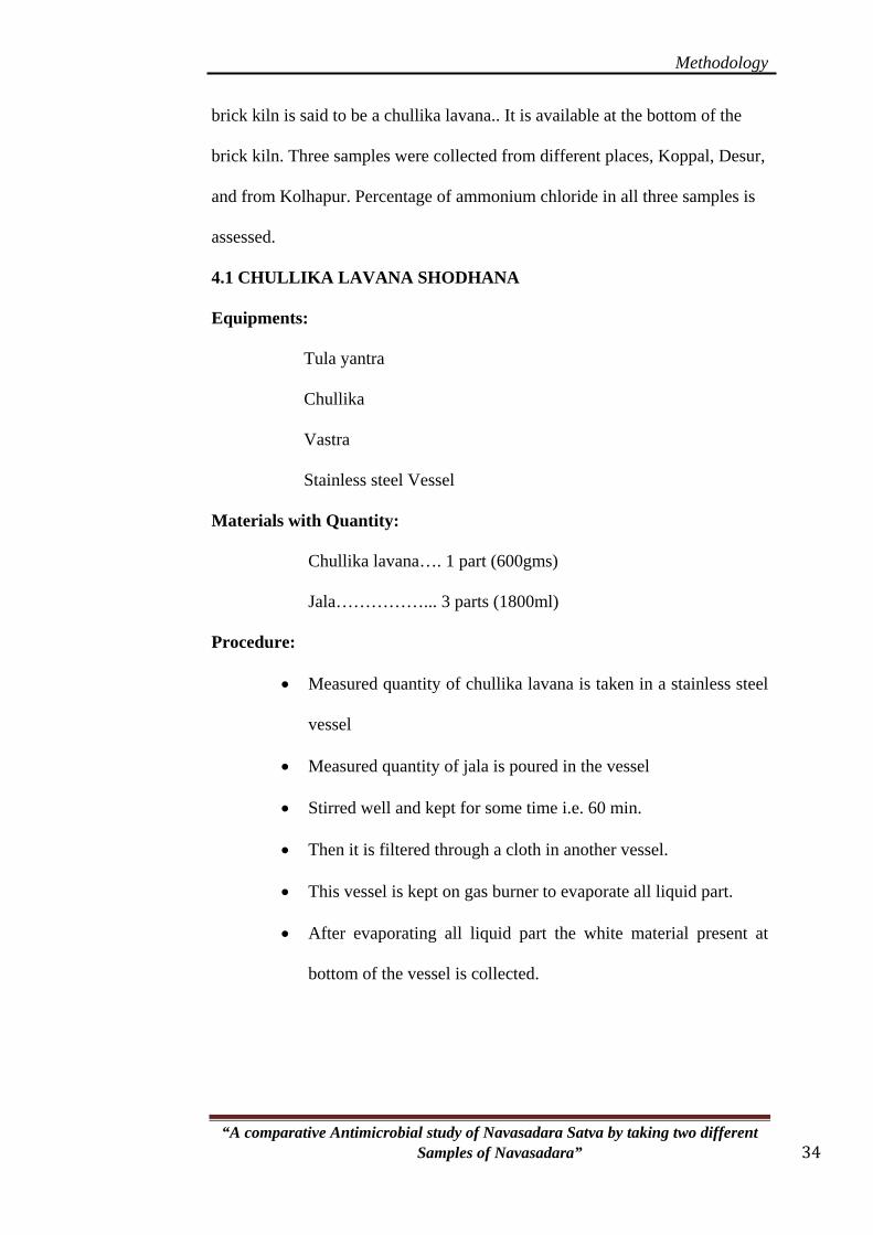
4.1 CHULLIKA LAVANA SHODHANA
Equipments:
Tula yantra
Chullika
Vastra
Stainless steel Vessel
Materials with Quantity:
Chullika lavana…. 1 part (600gms)
Jala……………... 3 parts (1800ml)
Procedure:
• Measured quantity of chullika lavana is taken in a stainless steel
vessel
• Measured quantity of jala is poured in the vessel
• Stirred well and kept for some time i.e. 60 min.
• Then it is filtered through a cloth in another vessel.
• This vessel is kept on gas burner to evaporate all liquid part.
• After evaporating all liquid part the white material present at
bottom of the vessel is collected.

Methodology
“A comparative Antimicrobial study of Navasadara Satva by taking two different Samples of Navasadara” 35
Observation:
• It is observed that the chullika lavana is not easily dissolved in
water, so it is stirred well and kept for 60 min.
• Room Temperature—300C
• After adding water no changes were observed in temperature.
• Total quantity taken- 600gms
• Qty obtained- 92 gms
• Loss – 508 gms
4.2) NAVASADARA SHODHANA:35
Equipments: Tula yantra
Chullika
Vastra
Stainless steel Vessel
Materials with quantity:
Navasadara…. 1 part (400gms)
Jala……………... 3 parts (1200ml)
Procedure:
• Measured quantity of Navasadara is taken in a stainless steel
vessel
• Measured quantity of jala is poured in the vessel
• Stirred well and kept for some time i.e. 20 min.
• It will dissolve completely.
• Then it is filtered through a cloth in another vessel.
• This vessel is kept on gas burner to evaporate all liquid part.
Methodology
“A comparative Antimicrobial study of Navasadara Satva by taking two different Samples of Navasadara” 36
• After evaporating all liquid part the white material present at
bottom of the vessel is collected.
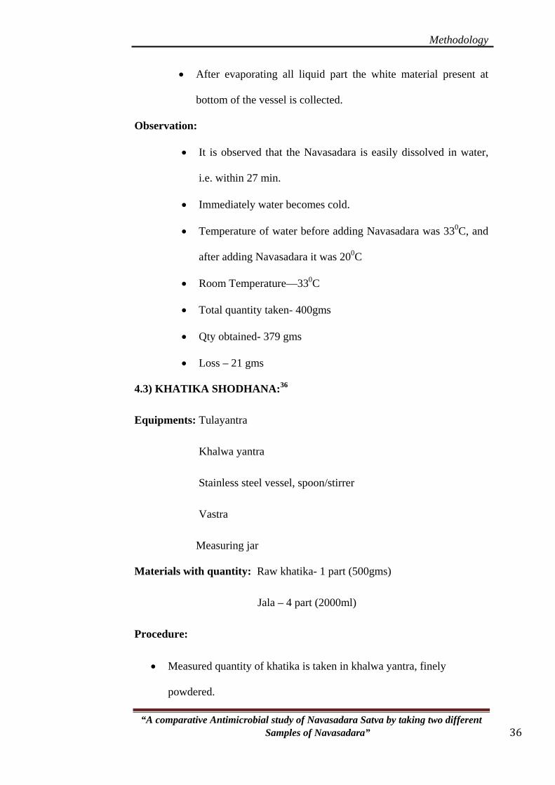
Observation:
• It is observed that the Navasadara is easily dissolved in water,
i.e. within 27 min.
• Immediately water becomes cold.
• Temperature of water before adding Navasadara was 330C, and
after adding Navasadara it was 200C
• Room Temperature—330C
• Total quantity taken- 400gms
• Qty obtained- 379 gms
• Loss – 21 gms
4.3) KHATIKA SHODHANA:36
Equipments: Tulayantra
Khalwa yantra
Stainless steel vessel, spoon/stirrer
Vastra
Measuring jar
Materials with quantity: Raw khatika- 1 part (500gms)
Jala – 4 part (2000ml)
Procedure:
• Measured quantity of khatika is taken in khalwa yantra, finely
powdered.

Methodology
“A comparative Antimicrobial study of Navasadara Satva by taking two different Samples of Navasadara” 37
• It is transferred in a vessel containing 4 parts of water.
• Stirred well and filtered through a filter cloth
• Kept undisturbed for 6 hrs
• Then upper water portion is removed
• Khatika present at the bottom of the vessel is dried and collected.
Precaution:
• To observe the separation glass vessel should be used.
Observation:
• Before shodhana khatika is grey in color.
• After mixing water, it easily mixes, and the mixture turns in to
dull grey suspension form.
• After filtering through the cloth, the foreign materials, mud
particles seen on the filtered cloth.
• The filtrate is clean and has the consistency thicker than water.
• For drying, it takes about 2 days.
• Dried shodhita Khatika looks brighter then raw Khatika.
• Room Temperature—330C
• Total quantity taken- 500gms
• Qty obtained- 472 gms
• Loss – 28 gms
Methodology
“A comparative Antimicrobial study of Navasadara Satva by taking two different Samples of Navasadara” 38
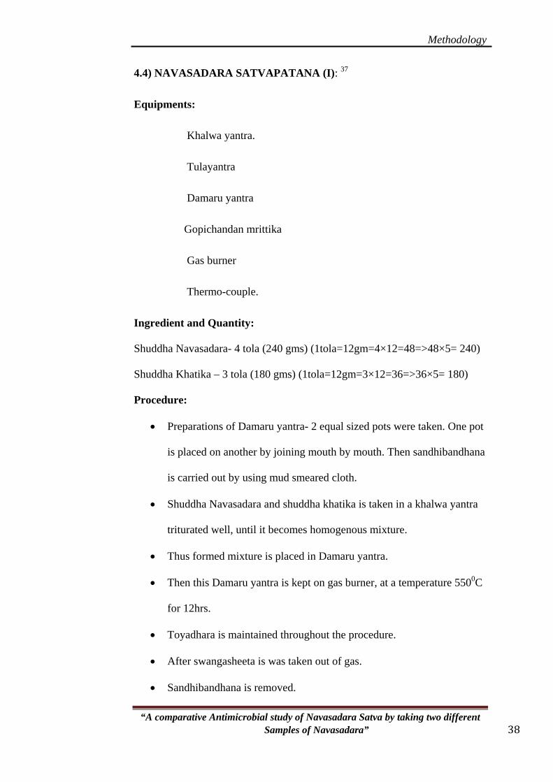
4.4) NAVASADARA SATVAPATANA (I): 37
Equipments:
Khalwa yantra.
Tulayantra
Damaru yantra
Gopichandan mrittika
Gas burner
Thermo-couple.
Ingredient and Quantity:
Shuddha Navasadara- 4 tola (240 gms) (1tola=12gm=4×12=48=>48×5= 240)
Shuddha Khatika – 3 tola (180 gms) (1tola=12gm=3×12=36=>36×5= 180)
Procedure:
• Preparations of Damaru yantra- 2 equal sized pots were taken. One pot
is placed on another by joining mouth by mouth. Then sandhibandhana
is carried out by using mud smeared cloth.
• Shuddha Navasadara and shuddha khatika is taken in a khalwa yantra
triturated well, until it becomes homogenous mixture.
• Thus formed mixture is placed in Damaru yantra.
• Then this Damaru yantra is kept on gas burner, at a temperature 5500C
for 12hrs.
• Toyadhara is maintained throughout the procedure.
• After swangasheeta is was taken out of gas.
• Sandhibandhana is removed.
Methodology
“A comparative Antimicrobial study of Navasadara Satva by taking two different Samples of Navasadara” 39
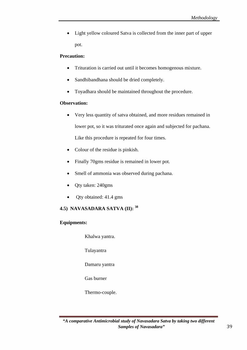
• Light yellow coloured Satva is collected from the inner part of upper
pot.
Precaution:
• Trituration is carried out until it becomes homogenous mixture.
• Sandhibandhana should be dried completely.
• Toyadhara should be maintained throughout the procedure.
Observation:
• Very less quantity of satva obtained, and more residues remained in
lower pot, so it was triturated once again and subjected for pachana.
Like this procedure is repeated for four times.
• Colour of the residue is pinkish.
• Finally 70gms residue is remained in lower pot.
• Smell of ammonia was observed during pachana.
• Qty taken: 240gms
• Qty obtained: 41.4 gms
4.5) NAVASADARA SATVA (II): 38
Equipments:
Khalwa yantra.
Tulayantra
Damaru yantra
Gas burner
Thermo-couple.

Methodology
“A comparative Antimicrobial study of Navasadara Satva by taking two different Samples of Navasadara” 40
Ingredient and Quantity:
Shuddha Navasadara- 4 tola(48 gms) (1tola=12gm=4×12=48)
Shuddha Khatika – 3 tola (36 gms) (1tola=12gm=3×12=36)
Procedure:
• Shuddha chullika lavana and shuddha khatika is taken in a khalwa
yantra triturated well, until it becomes homogenous mixture.
• Thus formed mixture is placed in Damaru yantra.
• Then this Damaru yantra is kept on gas burner, at a temperature 5500C
for 6hrs.
• Toyadhara is maintained throughout the procedure.
• After swangasheeta is was taken out of gas.
• Sandhibandhana is removed.
• Satva was not found.
Precaution:
• Trituration is carried out until it becomes homogenous mixture.
• Sandhibandhana should be dried completely.
• Toyadhara should be maintained throughout the procedure.
Observation:
• Satva not obtained.
• Grey coloured material is found at the bottom pot.
• No smell was observed during pachana.
ANALYTICAL STUDY:
For analytical study samples were labeled as shown below,
Sample A – Raw chullika lavana
Sample B – Shodhita chullika lavana

Methodology
“A comparative Antimicrobial study of Navasadara Satva by taking two different Samples of Navasadara” 41
Sample C – Raw Navasadara
Sample D – Shodhita Navasadara
Sample E- Raw Khatika
Sample F- Shodhita khatika
Sample G-Navasadara satvapatana (I)
Sample H-Residue
Sample I-Navasadara satvapatana (II)
PHYSICO CHEMICAL ANALYSIS:39
4.6) Determination of pH value:
Apparatus: pH meter, Glass beaker.
Procedure:
1% of sample solution (1gm sample powder + 100ml of distilled
water) was prepared, Shaken well and homogenized just before taking pH
reading and then the tip of electrode was dipped in such a way that the glass
bulb of the electrode is fully immersed in solution and recorded. In the same
manner pH of all samples were taken out.
4.7) Determination of loss on drying:
Apparatus: Oven, Crucible, and Chemical balance.
Procedure:
1) First evaporating dish is weighed & preconditioned.
2) 2 Gms of sample is taken in pre-weighed, preconditioned
evaporating dish.
3) Then it is placed in hot air oven at 1100 C for half an hour.
4) Difference in the weights of sample before & after placing
in hot air oven is the Loss on drying at 1100C.
Methodology
Calculation:
Wt of the sample – A
Wt of the sample + evaporating dish – B
Wt of evaporating dish + sample after LOD – C
Wt of residue = B-C = D
% Moisture content = D x 100
A
4.8) Determination of Ash value:
Apparatus: Crucible, oven, desiccators, & chemical balance.
Procedure:
1) Crucible is to be conditioned in the oven at 1050 C for one
hour & cooled in Desiccators.
2) Then that crucible is weighed accurately in a chemical
balance.
3) 2 gm of air-dried & powdered sample is taken in conditioned
Crucible.
4) Then it is kept in furnace for incineration at 4500C for 1 hr.
5) % of the total ash content is calculated with reference to the
original weight taken of Air- dried drug for the analysis.
Calculation:
Wt of sample – A
Wt of crucible – B
Wt of crucible after incineration – C
“A comparative Antimicrobial study of Navasadara Satva by taking two different Samples of Navasadara” 42
Wt of ash = C – B = D
Methodology
% of Ash value = D × 100
A
4.9) Determination of Acid insoluble ash:
Apparatus: Crucible, Muffle furnace, desiccators, & chemical balance,
beaker.
“A comparative Antimicrobial study of Navasadara Satva by taking two different Samples of Navasadara” 43
Procedure:
• After completion of ash value, the ash present in that crucible is
taken for AIA, crucible is washed with 25ml of dilute
Hydrochloric acid, and whole content of that crucible is
transferred into a 100 ml beaker.
• Beaker is kept in water bath for 10 min.
• Then it is filtered through an ash less filter paper.
• Then that filter paper is kept in crucible.
• Crucible is kept in furnace at 4500C for 1 hr.
• Cooled in desiccators, weighed and calculated
Calculation:
Wt of sample = A
Wt of crucible = B
Wt crucible + filter paper after filtering, incinerating = C
AIA = C – B = D
% of AIA = D × 100
A
Methodology
4.10) Specific gravity:40
It is done for 3 samples, raw Navasadara, shodhita Navasadara, and
Navasadara satva (I).
Apparatus: Picnometer, chemical balance.
Procedure: Empty Specific Gravity bottle (picnometer) was weighed and the
bottle filled with distilled water and again weighed, the same bottle was then
filled with 1% (100ml of water + 1gm sample) of sample and weighed. All
these 3 weights were noted; the Specific Gravity of sample was calculated.
Calculation:
Wt of empty picnometer = A
Wt of picnometer + water = B
Wt of picnometer + sample = C
Wt of water = B – A = D
Wt of sample = C – A = E
Specific gravity = E × 1
D
4.11) Solubility:41
Apparatus: Beaker /test tubes, spatula
Procedure: Solubility test of Navasadara satva (I) was subjected with the
following solvents.
Distilled water
Petroleum ether
Alcohol
Chloroform
“A comparative Antimicrobial study of Navasadara Satva by taking two different Samples of Navasadara” 44

Methodology
“A comparative Antimicrobial study of Navasadara Satva by taking two different Samples of Navasadara” 45
A pinch of sample was taken in a dry test tube with 1ml of solvent and
shaken for 1min. then observed for solubility, non-solubility and sparingly
solubility.
Determination of Elements by ICP-AES Method:
Sample preparation:
0.2500 g of sample was weighed into a Teflon bomb. 8 ml of Milli-Q water,
7 ml of HNO3 and 1 ml of H2O2 was added. The mixture was microwaved for
20 mins in microwave digestor (Model: Milestone Start D). The samples were
then diluted to 50ml with Milli-Q water.Further dilutions may be done if
required to attain the working range of the instrument.
Make of the instrument; Jobin Vyon Horiba
Model: Ultima 2
Procedure:
Normal speed of pump : 20 (rates/min)
Plasma gas flow rate : PL1 (l/min)
Sheath flow rate : G1 (1/min)
Auxiliary flow rate : 0.0 (1/min)
Sheath stabilization time : 15.0 (min)
Nebulisation flow rate : 0.02 (1/min)
Nebulisation pressure : 1.0(bar)
XRD ANALYSIS:42
Definition:
“X-Ray are diffracted because crystalline solids are constructed from a
regularly simple assemblage of components (atoms or atomic groupings)

Methodology
“A comparative Antimicrobial study of Navasadara Satva by taking two different Samples of Navasadara” 46
repeated at regular intervals in three dimensions, and X – Ray wavelengths are
the same order of magnitude as the spacing of atom centers.”
From Morphological view-point, a crystal may be defined as the
regular polyhedral form, bounded by smooth surfaces, which is assumed by a
chemical compound under the influences of it’s inter atomic forces when
passing from the gaseous or liquid state to solid state.
The geometric shapes of crystals reflect an internal symmetry of atoms
and molecules arranged in a regular and repeated pattern in space, which
distinguish crystalline solids from amorphous.
An important aspect of solid state is the ability of compounds to
crystallize in a variety of symmetrical arrangements of their molecules in
space i.e. polymorphic forms. Which are often quite different from each other
in physical characters (Habits, Melting point, solubility) although chemically
identical.
The pharmaceutical relevance of polymorphisms lies in the fact that
the differences in physico-chemical properties between forms may influence
manufacturing processes and importantly, dissolution rate and therefore
bioavailability.
The X – Ray diffraction methods are necessary to distinguish between
different crystallographic arrangements.
The condition for diffraction of a beam of X – Ray from crystal is
given by Bragg’s equation:
2 d Sin θ = n λ
Where,
λ = Wavelength of X – Ray beam
Methodology
“A comparative Antimicrobial study of Navasadara Satva by taking two different Samples of Navasadara” 47
θ = Angle between incidents
X-Ray and crystal lattice plan
d = Distance between lattice plane’s
The Bragg’s equation is fundamental relation underlying all X-Ray
Diffraction measurements.
Only for angles of incidence such as Sin θ = n λ / 2 d will W – Rays be
reflected, and at all other angles destructive interference occurs.
The specimens can be identified by reference libraries such as Powder
Date File or the Joint Committee on Powder Diffraction Standards (JCPDS),
which contains the diffraction patterns of some tens of thousands of
substances in numerical form.
Advantages:
• The particular advantage of diffraction analysis is that it discloses the
presence of a substance as that actually exists in the sample, and not in
terms of its constituent chemical elements. i.e. this analysis shows both
the elemental form and compound form of the element as it is present
in the sample.
• Helps to identify and characterize the raw materials of mineral/metal
origin.
• It is also used to fix up the particular characteristics pattern (finger
print) of prepared bhasma.

Methodology
“A comparative Antimicrobial study of Navasadara Satva by taking two different Samples of Navasadara” 48
4.12) Qualitative analysis of samples:43
Calcium:
Dissolve 0.8gm of sample in 20 ml con HCl => boil the solution =>
filter => collect the filtrate and test.
Test: Filtrate + ammonia+ potassium ferocynide => yellow ppt => calcium
present
Sodium / Potassium / Chlorides:
Dissolve 2 gm of sample in 20 ml conc. HCl => boil the solution
=>filter through filter paper => collect the filterate and test.
Test: Filtrate + Potassium Pyroantimonate => Milkyness ppt => Sodium
present.
Filtrate + Sodium cobalt nitrate => Yellow ppt => Potassium present.
Filtrate + Silver nitrate solution => White ppt => Chlorides present.
Ammonia:
Sample + NaOH => boil and hold a wet litmus paper at the mouth of
the test tube.
Observation:
Gas with ammonia smell having alkaline action on litmus paper (red
turns to blue) and white dense fumes with glass rod dipped in concentrated
HCl.
4.13) Antimicrobial study:44
Aim of the study
In the present study, Disc diffusion / cup plate method and Broth
dilution both tests are performed to determine the level of bacteriostatic
and bactericidal concentration.

Methodology
“A comparative Antimicrobial study of Navasadara Satva by taking two different Samples of Navasadara” 49
4 samples were selected for antimicrobial study they are,
shodhita chullika lavana, shodhita Navasadara, Navasadara satva (I) and
Navasadara satva (II), they are labeled as sample 1, 2, 3, 4 respectively.
Methods:
The antimicrobial study was designed by following two methods as;
1. Agar disc diffusion method
2. Broth dilution method.
1) Agar disc diffusion method
This is one of the methods officials in Indian Pharmacopeia. Where
the Antimicrobial powder dissolved in saline water is diffused from the
cup through an agar layer in petri-dish to an extent such that the growth
of added microorganisms is restricted entirely in circular area or zone
around the cavity containing the solution of an antibiotic substance.
The Antimicrobial activity is expressed as zone diameter in
millimeters, which is measured with a divider.
Samples were screened for Antimicrobial activity against micro-
organisms and the activity was compared with appropriate standard
allopathic drugs.
Antimicrobial activity:
The Antimicrobial activity of a drug is generally expressed as its
inhibiting effect towards the growth of bacterium in nutrient broth or
nutrient agar.
For this study following conditions are required:
o The substance must be contact with the test organism.
Methodology
“A comparative Antimicrobial study of Navasadara Satva by taking two different Samples of Navasadara” 50
o Condition must be favorable for the growth of
microorganisms in the absence of Antimicrobial substances.
o There must be a means of estimating the amount of growth
and thereby, percentage of growth of inhibition.
o The activity of powder substance should be observed and
determined by the growth response of microorganisms.
Materials used:
o BHI Agar media
o Stock solution
o Inoculated Microorganisms solution
o Sterile micro-pipettes
o Petri dishes
o Sterile (metal)borer
o Cotton swab
o Incubator
o Spirit lamp
o Micropipettes
Standards used in the study:
o Ciprofloxacin 2mg/ml
Preparation of stock solution:
1. 500 mg of all 4 samples are taken and dissolved in 1 ml of
saline water to get a concentration of 500mg/ml.
2. From prepared stock solution main test solutions are
prepared by taking different four concentrations i.e.

Methodology
“A comparative Antimicrobial study of Navasadara Satva by taking two different Samples of Navasadara” 51
1:2,1:5,1:10, & 1:20 and tested for its antibacterial activity,
totally four concentrations were tested.
Preparation of test solution:
The test solution here prepared using saline water to get the
following concentrations
1. 1:2 -------- 125 mg/ml
2. 1:5 ------- 62.5 mg/ml
3. 1:10 ------- 13mg/ml
4. 1:20 ------- 6mg/ml
Preparation of inoculums:
Mueller - Hinton agar medium (Himedia labs) of the following
composition was used for preparation of slants.
Peptone ---- 5.0 g
Beef extract ---- 1.5 g
Sodium chloride ---- 5.0 g
Agar ---- 15.0 g
Yeast extract ---- 15.0 g
Distilled water to make ----- 1000 ml
• About 28 gm of prepared medium was taken in 1000 ml of
distilled water and boiled to dissolve completely. After that
streaked with micro organisms.
• The slants were incubated at 37OC + 1OC for 24 hrs.
• These 24 hrs cultures were used for preparation of inoculums.
Methodology
“A comparative Antimicrobial study of Navasadara Satva by taking two different Samples of Navasadara” 52
• The suspension of micro organisms was prepared in 10 ml of
sterile water and 0.5 ml of this suspense was added for 10 ml of
the agar medium.
Culture Medium:
Different types of media have been used according to the types of
organisms.
In the present study, BHI (Brain heart infusion) agar media employed
possessing the following composition (Ready made medium).
Agar ---- 15.0 g
Calf brain infusion from ---- 200.00
Beef heart infusion from ---- 250.00
Proteose peptone ---- 10.00
Dextrose ---- 2.00
Sodium chloride ---- 5.00
Disodium phosphate ---- 2.50
About 52.0 gm of above readymade medium was dissolved in
distilled water (1000ml) by gentle heating. Heat to boiling to dissolve
the medium completely. Sterilize by autoclaving at 15lbs pressure
(1210C) for 15mins. Mix well before pouring.
Sterilization:
Sterilization of the medium, tubes for slants, borer etc. was done
by autoclaving at 15 ibs / square inch for 20 mins. The glass wares like
syringes, petridishes, pipettes, empty test tubes, were sterilized by dry
heat in an oven at a temperature of 160OC for 1 hrs.
Methodology
“A comparative Antimicrobial study of Navasadara Satva by taking two different Samples of Navasadara” 53
Preparation of Agar plates:
The sterilized medium was cooled at 40OC and 0.5 ml of
inoculums per 100 ml of medium was added to the conical flask.
This was shaken gently to avoid the formation of air bubbles and then
transferred into petri-dishes. So as to obtain 6mm thickness of medium.
The medium in the plate was allowed to solidify at room temperature.
Thus prepared agar used for three organisms except S.pneumoniae
Readymade Chocolate agar was used for S.pneumoniae.
Experimental Procedure:
• The sterile borer was used to prepare five cups of 8 mm diameter
in the medium of each petri-dish.
• Accurately measured 50 mL solutions of each concentration of
solutions of dissolved powder (prepared standard drug) were
added to the cups with the help of micropipette.
• All the plates were kept at room temperature for effecting
diffusion of drug dissolved and standards.
• Later, they were incubated at 37+1OC for 24 hrs.
• The presence of definite zones around the cup of any size
indicated antibacterial activity.
• The control was run simultaneously to assess the activity of
saline water which was used as the vehicle for drug powder.
• The diameter of the zone of inhibition was measured and
recorded.

Methodology
“A comparative Antimicrobial study of Navasadara Satva by taking two different Samples of Navasadara” 54
• The zones of inhibition for the antibacterial activities of samples
were calculated by measuring the inhibitory effect towards the
growth of bacteria around nutrient agar cup.
1) Broth dilution test Method:
The Broth Dilution method is a simple procedure for testing a
small number of isolates, even single isolate. It has the added advantage
that the same tubes can be taken for MBC tests also:
Materials:
o B H I Broth media
o Overnight broth culture of test and control organisms
(Inoculated microorganisms)
o Samples (required antibiotic in powder form).
o Required solvent- sterile Distilled Water.
o Sterile capped 7.5 x 1.3 cm tubes / small screw-capped
bottles,
o Incubator
o Cotton swab
o Sterile graduated Micropipettes-5, 10, 50, 200, 500mL size.
o A suitable rack to hold 40 tubes in four rows i.e. 10 tubes in
each row.
Preparation of Broth media:
Brain Heart Infusion media was used for the preparation of Broth
solution.

Methodology
“A comparative Antimicrobial study of Navasadara Satva by taking two different Samples of Navasadara” 55
Calf brain infusion from ---- 200.00
Beef heart infusion from ---- 250.00
Proteose peptone ---- 10.00
Dextrose ---- 2.00
Sodium chloride ---- 5.00
Disodium phosphate ---- 2.50
Suspend 37.0gms in 1000ml distilled water dispense in to bottles
or tubes as desired. Sterile by autoclaving at 15lbs pressure (1210C).
Preparation of Stock solution:
• Stock solution is prepared by using saline water.
• The stock solution is prepared in the concentration 1gm/ml. The
volume of the solution used during the procedure is 0.2mL, so
the concentration of the drug finally stands as 1mg/ml.
Experimental procedure:
• To start with, glass test tubes plugged with cotton all are
autoclaved.
• Ten Borosilicate glass test tubes are taken in four rows each, one
corresponding for the serial dilution w.r.t one organism.
• Test tubes are serially numbered from 1-10.
• The first test tube placed in the row in rack with the name of the
organism used in the study.
• 200 mL of the (BHI) Brain heart infusion broth is initially taken
in all the test tubes of eight rows.
• To the first tube of all the four rows, 200mL of the stock solution
is added.

Methodology
“A comparative Antimicrobial study of Navasadara Satva by taking two different Samples of Navasadara” 56
• It is pipette in and out to ensure uniform mixing.
• 200 mL of the first test tube is then pipette to the second tube and
the process is repeated till the 10th tube.
• From the 10th tube 200 mL is pipette out and discarded.
• Every time when 200 mL of solution is introduced in the test
tube it is gently pipette in and out within the test tube to ensure
proper mixing.
• During every withdrawal of 200 mL of the solution and
introduction to another tube, a new pipette is made use of.
• In the above said method, serial dilutions are made similarly with
10 test tubes arranged in sixteen rows corresponding for four
microorganisms of four drug samples.
• The concentration in the test tube now stands as 250mg (tube-1),
125mg(tube-2),62.5mg(tube-3),31.5mg(tube-4),16mg(tube-5),
8mg (tube-6), 4mg(tube-7), 2mg(tube-8), 1mg(tube-9), 0.5mg
(tube 10)
• Now adjust the bacterial count 1.58*108 organisms per ml by
comparing with McFarland’s standards. The suspension of the
organisms, , Staphylococcus aureus, E.coli, K.pneumoniae,
S.pneumoniae in the ( BHI) Brain heart infusion Broth is then
inoculated to all the test tubes of a corresponding four rows ,in
the concentration of 200 MicroLitres.
• All tubes are inoculated at 350C for 24hrs without shaking or
agitation.
Methodology
“A comparative Antimicrobial study of Navasadara Satva by taking two different Samples of Navasadara” 57
• After 24hrs of incubation the test tubes are read for MIC is
defined as the concentration of the first tube in the series to show
visible traces of growth of bacteria, which is evident, by turbidity
or cloudiness of the solution in the test tube.
• Turbidity indicates that bacterial growth has not been inhibited
by the concentration of the preparation contained in the medium.
• The main advantage of the ‘Broth dilution’ method for the MIC
determination lies in the fact that it can readily be converted to
determine the MBC as well.
Reading of result:
• MIC is expressed as the lowest dilution, which inhibited growth judged
by lack of turbidity in the tube. Because very faint turbidity may be
given by the inoculums itself, the inoculated tube kept in the
refrigerator overnight may be used as the standard for the
determination of complete inhibition.
Minimum Bactericidal Concentrations (MBC):
• The MBC values also known as MLC (Minimal lethal
concentration) is the estimation of bacterial activity and is
defined as the lowest concentration of the preparation of the
sample to kill 99.9% of the initial inoculums after incubation for
24hrs under a standardized set of conditions. By observing MIC
result 3rd sample (Navasadara Satva I) was taken for MBC. Here
in the present study MBC was obtained by platting out the
contents on an agar medium to count the growth.

Methodology
“A comparative Antimicrobial study of Navasadara Satva by taking two different Samples of Navasadara” 58
Method
• Dilutions and inoculations are prepared in the same manner as
described for the determination of MIC or the dilutions prepared
for MIC can be taken directly for MBC.
• Next day readings are taken, after visually clear tubes 0.1ml from
each of four tubes were taken i.e. 2 sensitive and 2 turbid &
spread across the surface of dried agar plates with the help of
sterile bent glass rods. The plates were inoculated overnight at
350C for 24hrs.
• Next day plates are observed for growth of colonies and readings
are noted. The number of colonies that grown on each MBC
subculture plates after overnight incubation was counted.
• The concentration in which petriplate showed either no growth or
reduction in 99.9%, was considered the MBC of specific
antimicrobial agent.
Reading of result
• These subcultures may show similar number of colonies-
indicating bacteriostasis only.
• A reduced number of colonies-indicating a partial or slow
bactericidal activity.
• No growth- if the whole inoculum has been killed.
• The highest dilution showing at least 99% inhibition is taken as
MBC.
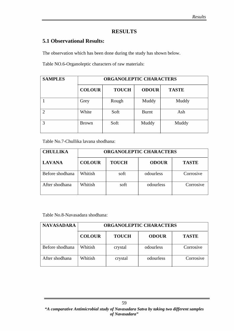
Results
RESULTS 5.1 Observational Results: The observation which has been done during the study has shown below.
Table NO.6-Organoleptic characters of raw materials:
SAMPLES ORGANOLEPTIC CHARACTERS
COLOUR TOUCH ODOUR TASTE
1 Grey Rough Muddy Muddy
2 White Soft Burnt Ash
3 Brown Soft Muddy Muddy
“A comparative Antimicrobial study of Navasadara Satva by taking two different samples
of Navasadara”
59
Table No.7-Chullika lavana shodhana:
CHULLIKA
LAVANA
ORGANOLEPTIC CHARACTERS
COLOUR TOUCH ODOUR TASTE
Before shodhana Whitish soft odourless Corrosive
After shodhana Whitish soft odourless Corrosive
Table No.8-Navasadara shodhana:
NAVASADARA ORGANOLEPTIC CHARACTERS
COLOUR TOUCH ODOUR TASTE
Before shodhana Whitish crystal odourless Corrosive
After shodhana Whitish crystal odourless Corrosive

Results
Table No.9- Khatika shodhana:
KHATIKA ORGANOLEPTIC CHARACTERS
COLOUR TOUCH ODOUR TASTE
Before shodhana Dull Grey soft Characteristic Tasteless
After shodhana Bright Grey soft Characteristic Tasteless
“A comparative Antimicrobial study of Navasadara Satva by taking two different samples
of Navasadara”
60
Table No.10-Navasadara satvapatana (I):
NAVASADARA
SATVA (I)
ORGANOLEPTIC CHARACTERS
COLOUR TOUCH ODOUR TASTE
Yellow soft characteristic Alkaline
odour of ammonia
Table No.11-Navasadara satvapatana (II):
NAVASADARA
SATVA(2)
ORGANOLEPTIC CHARACTERS
COLOUR TOUCH ODOUR TASTE
Grey soft Odourless Tasteless
5.2 ANALYTICAL RESULTS: Table No.12-Raw material analysis: Sr. No. Parameters Sample 1 Sample 2 Sample 3
1 % of Ammonia 2.34% 0.82% 0.64%
2 % of Chloride 1.36% 14.68% 1.90%
3 % of Calcium 2.20% 40.67% 1.48%

Results
“A comparative Antimicrobial study of Navasadara Satva by taking two different samples
of Navasadara”
61
4 % of Magnesium 0.48% 2.68% 1.80%
5 % of Potassium 0.66% 0.38% 0.26%
6 % of Iron 1.49% 0.76% 2.94%
7 % of Silica 90.16% 9.88% 61.94%
9 % of NH4Cl 2.01% 2.32% 1.054%
Table No.13-Physico-chemical parameters: SAMPLE pH LOD ASH
VALUE AIA
SPECIFIC GRAVITY
A 11.50 1.84%w/w 84.47% 17.56% NA
B 12.43 7.11%w/w 81.47% 11.38% NA
C 7.8 13.23%w/w 0.046% NIL 1.0045
D 8.0 15.86%w/w 87.83% NIL 1.0046
E 13.0 2.63%w/w 80.15% 65.62% NA
F 9.0 4.15%w/w 77.98% 22.24% NA
G 10.1 14%w/w 0.099% NIL 1.0046
Table no. 14- Solubility: 01 Petroleum ether + Navasadara Satva(I) Insoluble
02 Alcohol + Navasadara Satva(I) Sparingly soluble
03 Chloroform + Navasadara Satva(I) Insoluble
04 Water + Navasadara Satva(I) Soluble
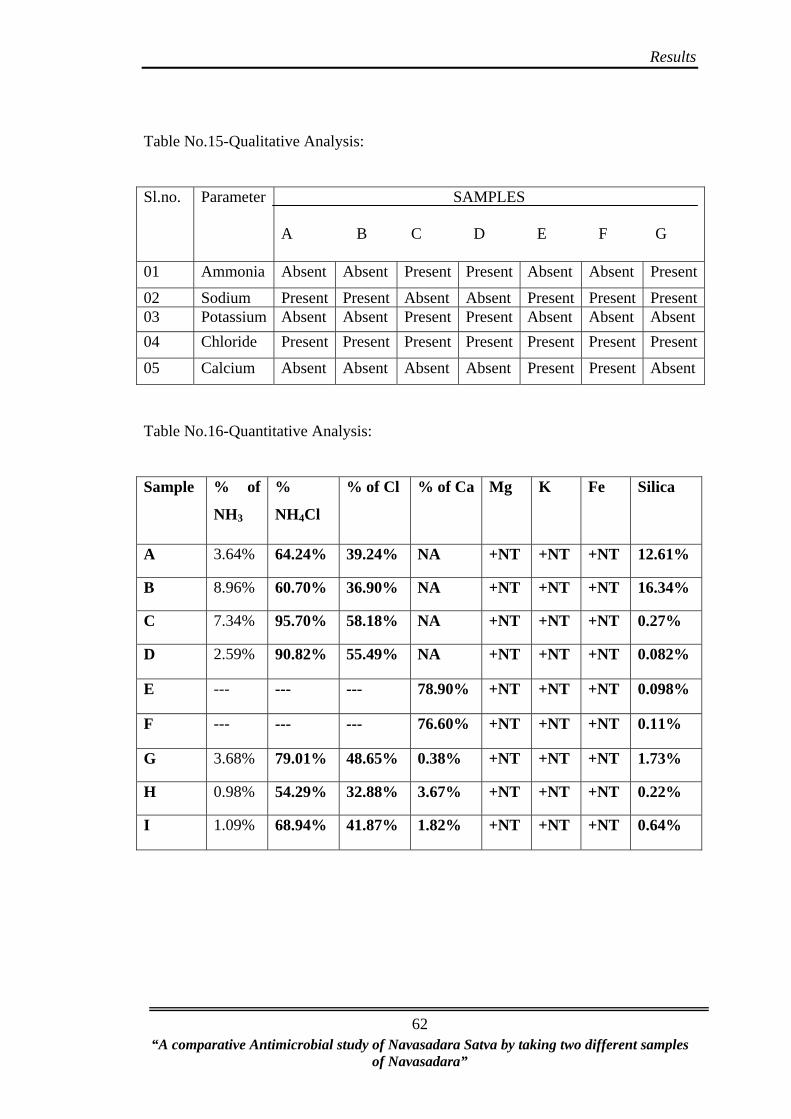
Results
Table No.15-Qualitative Analysis:
Sl.no. Parameter SAMPLES
A B C D E F G
01 Ammonia Absent Absent Present Present Absent Absent Present
02 Sodium Present Present Absent Absent Present Present Present03 Potassium Absent Absent Present Present Absent Absent Absent 04 Chloride Present Present Present Present Present Present Present
05 Calcium Absent Absent Absent Absent Present Present Absent
Table No.16-Quantitative Analysis:
Sample % of
NH3
%
NH4Cl
% of Cl % of Ca Mg K Fe Silica
A 3.64% 64.24% 39.24% NA +NT +NT +NT 12.61%
B 8.96% 60.70% 36.90% NA +NT +NT +NT 16.34%
C 7.34% 95.70% 58.18% NA +NT +NT +NT 0.27%
D 2.59% 90.82% 55.49% NA +NT +NT +NT 0.082%
E --- --- --- 78.90% +NT +NT +NT 0.098%
F --- --- --- 76.60% +NT +NT +NT 0.11%
G 3.68% 79.01% 48.65% 0.38% +NT +NT +NT 1.73%
H 0.98% 54.29% 32.88% 3.67% +NT +NT +NT 0.22%
I 1.09% 68.94% 41.87% 1.82% +NT +NT +NT 0.64%
“A comparative Antimicrobial study of Navasadara Satva by taking two different samples
of Navasadara”
62
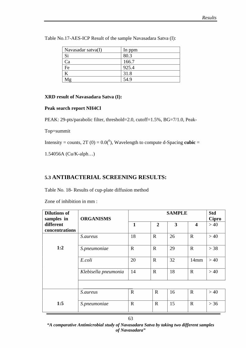
Results
Table No.17-AES-ICP Result of the sample Navasadara Satva (I):
Navasadar satva(I) In ppm Si 80.3 Ca 166.7 Fe 925.4 K 31.8 Mg 54.9
XRD result of Navasadara Satva (I):
Peak search report NH4Cl
PEAK: 29-pts/parabolic filter, threshold=2.0, cutoff=1.5%, BG=7/1.0, Peak-
Top=summit
Intensity = counts, 2T (0) = 0.0(0), Wavelength to compute d-Spacing cubic =
1.54056A (Cu/K-alph…)
5.3 ANTIBACTERIAL SCREENING RESULTS:
Table No. 18- Results of cup-plate diffusion method
Zone of inhibition in mm :
SAMPLE
Std Cipro
Dilutions of samples in different concentrations
ORGANISMS
1 2 3 4 > 40
S.aureus
18 R 26 R > 40
S.pneumoniae
R R 29 R > 38
E.coli
20 R 32 14mm > 40
Klebisella pneumonia
14 R 18 R > 40
1:2
S.aureus
R R 16 R > 40
1:5 S.pneumoniae
R R 15 R > 36
“A comparative Antimicrobial study of Navasadara Satva by taking two different samples
of Navasadara”
63
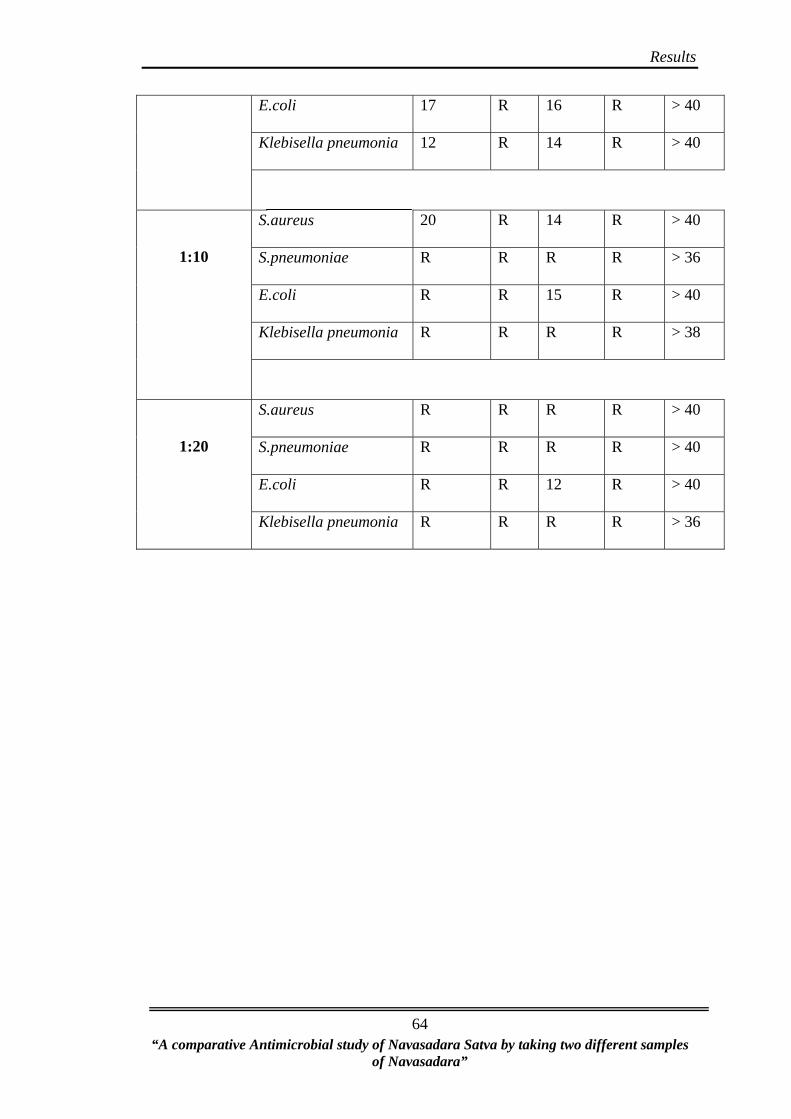
Results
E.coli
17 R 16 R > 40
Klebisella pneumonia
12 R 14 R > 40
S.aureus
20 R 14 R > 40
“A comparative Antimicrobial study of Navasadara Satva by taking two different samples
of Navasadara”
64
S.pneumoniae
R R R R > 36
E.coli
R R 15 R > 40
Klebisella pneumonia
R R R R > 38
1:10
S.aureus
R R R R > 40
S.pneumoniae
R R R R > 40
E.coli
R R 12 R > 40
1:20
Klebisella pneumonia
R R R R > 36

Results
Table No.19-Results of Macrodilution broth susceptibility method:
Sample Micro
Organisms
250
mg/ml
125
mg/ml
62.5
mg/ml
31.25
mg/ml
16
mg/ml
8
mg/ml
4
mg/ml
2
mg/ml
1
mg/ml
0.5
mg/
ml
S.aureus S S S S R R R R R R
S.pneumoniae
S S S S S R R R R R
E.coli R R R R R R R R R R
1
Klebisella pneumonia
S S S S R R R R R R
S.aureus S S S S R R R R R R
S.pneumoniae
S S R R R R R R R R
E.coli R R R R R R R R R R
2
Klebisella pneumonia
S S R R R R R R R R
S.aureus S S S S S S R R R R
S.pneumoniae
S S S S S R R R R R
E.coli S S S S R R R R R R
3
Klebisella pneumonia
S S S S S R R R R R
S.aureus S S R R R R R R R R
S.pneumoniae
S R R R R R R R R R
E.coli R
“A comparative Antimicrobial study of Navasadara Satva by taking two different samples
of Navasadara”
65
R R R R R R R R R
4
Klebisella pneumonia
S R R R R R R R R R
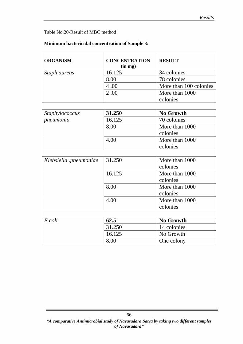
Results
“A comparative Antimicrobial study of Navasadara Satva by taking two different samples
of Navasadara”
66
Table No.20-Result of MBC method
Minimum bactericidal concentration of Sample 3:
ORGANISM
CONCENTRATION (in mg)
RESULT
16.125 34 colonies 8.00 78 colonies 4 .00 More than 100 colonies
Staph aureus
2 .00 More than 1000 colonies
31.250 No Growth 16.125 70 colonies 8.00 More than 1000
colonies
Staphylococcus pneumonia
4.00 More than 1000 colonies
31.250 More than 1000
colonies 16.125 More than 1000
colonies 8.00 More than 1000
colonies
Klebsiella .pneumoniae
4.00 More than 1000 colonies
62.5 No Growth 31.250 14 colonies 16.125 No Growth
E coli
8.00 One colony
Conclusion
CONCLUSION
• Chullika lavana couldn’t yield satva.
• Even though chullika lavana is a source of Navasadara, it showed less % of
ammonium chloride and also it is less effective when compared to market
sample of Navasadara.
• Shodhita chullika lavana, shodhita navasadara and satva (II) showed
bacteriostatic activity against all organisms except E coli.
• Navasadara Satva (I) showed bacteriostatic activity on all four organisms.
• Navasadara Satva (I) prepared by using market sample of Navasadara) is said
to be a effective drug against the organisms S.pneumoniae and E coli, at a
concentration of 31.250mg and 62.5mg respectively, at this stage it exhibited
bactericidal effect.
• Artificially prepared Ammonium Chloride can be used in place of Navasadara.
• Ammonium chloride prepared artificially has satisfied properties mentioned in
classics.
• Compared to shodhita Navasadara, Navasadara Satva is more effective.
• Preparation of Navasadara satva is less laborious and also cost effective.
70 “A comparative Antimicrobial study of Navasadara Satva by taking two different samples of
Navasadara”

Discussion
“A comparative Antimicrobial study of Navasadara Satva by taking two different Samples of Navasadara”
67
DISCUSSION
Whole work can be discussed under following headings
Discussion on raw material collection
Discussion on Preparation
Discussion on Analytical part
Discussion on Antimicrobial part
• As per the classics Navasadara is extracted from chullika lavana that to
particular plants should be used as a fuel, i.e Karira (Capparis decidua
Edgew), and Pilu (Salvadora persica Linn), and now a day these drugs are not
used as fuel in brick kiln, so less quantity of Navasadara is found.
• The white part which is remained after burning brick in kiln is also said to be
ishtika lavana or chullika lavana or Navasadara. Now a day in brick kiln
nimba kasta is used as a fuel, and in some kilns coal is used, so required
amount of Navasadara is not extracted from chullika lavana.
• All three samples of chullika lavana differ in colour, it might be because of the
things used as a fuel in that brick kiln.
• Pattern of agni is not mentioned for satvapatana, only for Navasadara
shodhana teevragni is mentioned. More amount of agni is required for
satvapatana i.e. 12 hrs pachana has been done to get Satva. It is fixed after
doing pilot study.
• Chullika lavana couldn’t yield Satva; it might be because of the very less
percentage of ammonium chloride in it.
• Compared to Navasadara, pH of chullika lavana is high i.e. more alkaline
when compared to Navasadara.
Discussion
“A comparative Antimicrobial study of Navasadara Satva by taking two different Samples of Navasadara”
68
• LOD is maximum in raw Navasadara, shodhita Navasadara and Navasadara
Satva because of more amount of moisture content in it.
• Acid insoluble ash of raw Navasadara, shunavasadara and Satva is nil because
it will not sustain 4500 C temperatures i.e. it evaporated.
• No significant difference observed in specific gravity of the samples C,D&G.
• % of ammonia in Navasadara Satva (I) is 3.68%, in Navasadara Satva (II) it
was 1.09%, and in residue it was 0.98%.
• After extraction of Satva also some amount of ammonia is found in residue.
• ICP-AES results of Navasadara Satva (I) reveals presence of elements like Si,
Ca, Fe, K, Mg in ppm level of in the in ppm level.
• XRD showed highest peak of NH4Cl, it showed presence of ammonium
chloride in the sample Navasadara Satva (I), and it cubic in structure.
• For antimicrobial study 4 samples were taken to assess the activity, they are
shodhita chullika lavana, shodhita Navasadara, Navasadara satva (I) and
Navasadara satva (II) and they are labeled as sample 1, 2, 3, 4 respectively.
• Antimicrobial study revealed,
Minimum inhibition concentration (MIC):
Sample 1- Shodita chullika lavana
• E-coli showed resistance.
• Sensitivity at a concentration of 31.250mg/ml onwards for k pneumoniae &
s. aureus.
• S.pneumoniae showed sensitivity at a concentration of 16.125mg/ml onwards.
Sample 2- Shodhita Navasadara
• E-coli showed resistance.

Discussion
“A comparative Antimicrobial study of Navasadara Satva by taking two different Samples of Navasadara”
69
• S aureus showed sensitivity at 31.250mg/ml
• S.pneumoniae & K pneumoniae showed sensitivity at 125mg/ml onwards
Sample 3-Navasadara Satva (I)
• E coli showed sensitivity at 31.125mg/ml
• S aureus showed sensitivity at 8mg/ml onwards
• S pneumoniae & k pneumoniae showed sensitivity at 16.125mg/ml
Sample 4-Navasadara Satva (II)
• E coli showed resistance S aureus sensitive at 125mg/ml S pneumoniae & k
pneumoniae sensitive at 250mg/ml onwards.
• Sample 1, 2 showed sensitivity in all organisms except E-coli, it might be
because of its high pH level i.e. alkaline property.
• Among all 4 samples, 3rd sample showed good bacteriostatic effect, so it was
selected for MBC to evaluate its bactericidal effect.
• MBC- sample 3 exhibited bactericidal effect for S.pneumoniae at a
concentration of 31.250mg and for E coli it was 62.5 mg, means at this level
of concentration no growth was found.
• Disc diffusion - at 1:2 & 1:20 concentration 3rd sample showed maximum
zone of inhibition against E-coli. At 1:10 & 1:5 concentration sample 1
showed maximum zone of inhibition against S.aureus and E coli respectively.
• Hence disc diffusion method was adopted by comparing zone of inhibition
between test drugs against control drug i.e. ciprofloxacin. It was found that
drug has shown antimicrobial result against S.pneumoniae and E-coli
compared to other organisms.
Summary
“A comparative Antimicrobial study of Navasadara Satva by taking two different samples of Navasadara”
72
SUMMARY
Whole work is summarized as follows-
Navasadara satvapatana is only mentioned in the text Rasatarangini. It is
indicated in Puppusashotha, Shweat kutsa, Amlapitta, Murcha, Kaphanissaraka etc. so
this study has been conducted to screen antimicrobial activity of the Navasadara
satva. It is said that we can extract Navasadara from chullika lavana. So here an effort
has been taken to extract Navasadara from chullika lavana. The present study
conducted to assess the physico-chemical features of chullika lavana, Navasadara and
Navasadara Satva and its anti microbial activity on two gram positive and two gram
negative organisms.
For the study raw materials like Navasadara and khatika were collected from
market and chullika lavana is collected from three different brick kiln, i.e. koppal,
Desur, and from Kolhapur. After analysis, on the base of % of Ammonium chloride
one sample of chullika lavana i.e. Desur sample is selected for study.
Chullika lavana, Navasadara and Khatika are subjected for shodhana process
by nirmalikarana. Navasadara Satva is prepared by doing pachana in damaru yantra.
Navasadara satvapatana is carried out as per the reference Rasatarangini. Got yellow
coloured Satva from the market sample Navasadara, whereas from chullika lavana
Satva was not found. It might be because of the less % of Ammonium chloride in it.
The physico–chemical analysis of Chullika lavana, Navasadara and khatika
before and after shodhana and Satva has been carried out to evaluate their qualitative
and quantitative assessment. Final product contains maximum quantity of ammonium
chloride i.e. 79.01%, remaining are trace elements such as, Calcium, Magnesium,
Silica.
Summary
“A comparative Antimicrobial study of Navasadara Satva by taking two different samples of Navasadara”
72
For Antimicrobial study four samples were selected, they are Shodhita
chullika lavana, Shodhita Navasadara, Navasadara Satva (I), and Navasadara Satva
(II), labeled as sample 1, 2, 3, 4 respectively.
Antimicrobial study was carried on four different strains of Micro-
organisms by Agar disc diffusion method compared with standard drug with the four,
and Minimal inhibitory concentration method for the more accuracy of the study.
After the study it is revealed that the Navasadara Satva (I) shown good
sensitivity on micro-organisms. All four samples showed bacteriostatic effect on all
organisms except E-coli. Whereas 3rd sample showed bacteriostatic effect on all
organisms. So the 3rd sample is selected for MBC to assess its bactericidal effect. 3rd
sample is said to be a good drug against the organisms S.pneumoniae and E coli, at a
concentration of 31.250mg and 62.5mg respectively, at this stage it exhibited
bactericidal effect.
Bibliography
BIBLIOGRAPHY
1) Tripathi Indradev ‘Rasaratna Samucchaya’ – Choukambha Samskrit Bhavan,
Varanasi 2st Edition – 2003, 3rd chp shloka 126, Pp 37.
2) Sharma Sadanand ‘Rasa Tarangini’ – Motilal Banarasidas 1994, Reprint, chap
14, Pp No.329.
3) Tripathi Indradeva ‘Rasarnavama athava Rasatantram’ – Choukambha
Samskrit Series Office, Varanasi,chap-5,shloka no 32,pp no 61
4) Shastri Ambikadatta ‘Rasaratna Samucchaya’ – Choukambha Amarabharati
Prakashana ,Varanasi 9st Edn – 1995, shloka: 134, Pp No.81
5) Mishra siddhinandan, Rasendrachudamani, chaukhambha orientalia, Varanasi,
1st edn, 1984, pp.193
6) Sharma Sadanand ‘Rasa Tarangini’ – Motilal Banarasidas 1994, Reprint, chap
14,shloka;5-7, Pp No.326
7) Sharma kaviraj harishankar,AcharyaNagarjuna,Rasendra mangala,Choukamba
orientalia,chap-3,pp no 48.
8) Bhagaavt pada Govinda, chaturbhuja mishra, ‘Rasa hridaya tantra’ Krishna
gopal ayurveda bhavan, Ajmir,2nd Edn 2002, pp no.128
9) Tripathi Indradeva ‘Rasarnavama athava Rasatantram’ – Choukambha
Samskrit Series Office, Varanasi, chap-5,shloka no 32,pp no 61.
10) Mishra Sidhinanadana ,Anandakanda, Chaukhambha publication,Varanasi,1st
Edn 2008,chapter 23/7-10th shloka, Pp 683.
11) Satpute.Ashok ‘Rasendra Sarasangraha’ – Choukambha Amarbharathi
Prakashan, Varanasi, 1st Edition – 2003 Pp 51.
73
“A comparative Antimicrobial study of Navasadara Satva by taking two different samples of Navasadara”
Bibliography
12) Mishra Gulraj Sharma, Ayurveda Prakasha, Chaukhambha publication,
Varanasi, 1st Edn. Reprint 1999 Pp-339
13) Vaidya Ramprasad, Rasendra Purana, Mumbai, Shri Krishnadas Lakshmi
Venkateshwar Publishers, M H, 1848 edition. Pp 385.
14) Mukherji Bhudeb Rasajala Nidhi Vol-2’ – Choukambha Publishers, Varanasi,
3rd Edition, Pp 214.
15) Choube Dattaram ‘Brihad Rasaraja Sundara’ – Choukhamba orientalia,
Varanasi 2007, Pp 183.
16) Joshi Damodar ‘Rasamritam’ – Choukambha Samskrit Bhavan, Varanasi, 1st
Edition – 1998, Pp 131.
17) Shastry Vasudev ,Laxman, Krishnaji Vittal, Nighantu ratnakara, Vol I,
Nirnaya sagar press, Bombay, 1936, pp no.510
18) Nadakarni K M, Nadakarni.A. Indian Materia medica Vol II, Bombay popular
prakashana, 3rd Edn, 1976 Pp M.11.
19) Mishra Sidhinanadana, Ayurvediya Rasashastra, Chaukhamba publication,
Varanasi, 6th Edn, 1996, Pp 475.
20) Vaidya pandit Hariprapannaji Rasayoga sagara, Vol I and II, Choukhamba
Krishnadas Academy, Varanasi reprint 2004.
21) www.wikipedia.com
22) Mishra Gulraj Sharma, Ayurveda Prakasha, Chaukhambha publication,
Varanasi, 1st Edn. 1999 Pp-324.
23) Joshi Damodar ‘Rasamritam’ – Choukambha Samskrit Bhavan, Varanasi, 1st
Edition, 1998, Pp 119.
74
“A comparative Antimicrobial study of Navasadara Satva by taking two different samples of Navasadara”
24) Dole Vilas, ‘A Text book of Rasashastra’, Paranjape prakash Ist edn Delhi,
Choukhamba Sanskrit pratistan; Edn - 2004, Pp380.

Bibliography
25) Mishra B shankarananda vaishya editor, Bhavamishra’s Bhavaprakash
choukhambha samskrit samsthan, Varanasi, Bhava Prakash Pp.621
26) Joshi Damodar ‘Rasamritam’ – Choukambha Samskrit Bhavan, Varanasi, 1st
Edition, 1998, Pp.119
27) Joshi Damodar ‘Rasamritam’ – Choukambha Samskrit Bhavan, Varanasi, 1st
Edition, 1998, Pp.120.
28) Sharma Sadanand ‘Rasatarangini’ – Motilal Banasidas, New-Delhi, Reprint
1994, Pp. 278.
29) Joshi Damodar ‘Rasamritam’ – Choukambha Samskrit Bhavan, Varanasi, 1st
Edition, 1998, Pp 119.
30) www.wikipedia.com
31) www.chemistrydaily.com
32) Dadupantha Swamy Bhajandas Rasadarpana part I, Natha pustaka bhandara,
Haryana, 3rd edn, Pp 179.
33) Satish Gupte, The short textbook of Medical Microbiology, Jaypee
publication, 8th Edn, 2002, Pp 11, 66, 157,166,173.
34) Shastry Ambikadatta Rasaratna Samuchaya, – Choukambha Amarabharati
Prakashana ,Varanasi 9th Edn, -1995, Shloka 134, Pp 81.
35) Sharma Sadanand ‘Rasa Tarangini’ – Motilal Banarasidas 1994, Reprint,
chap 14,shloka;3-4 Pp No 326.
36) Sharma Sadanand ‘Rasa Tarangini’ – Motilal Banarasidas 1994, Reprint, chap
11,shloka;210, Pp No 279.
37) Sharma Sadanand ‘Rasa Tarangini’ – Motilal Banarasidas 1994, Reprint, chap
14,shloka;20-21, Pp No -329.
75
“A comparative Antimicrobial study of Navasadara Satva by taking two different samples of Navasadara”

Bibliography
38) Sharma Sadanand Rasa Tarangini-Motilal Banarasidas 1994, Reprint, Chp
14, shloka 20-21, pp no.329.
39) Pharmacopeial standards for Ayurvedic Formulations, Central Council For
research in Ayurveda and Siddha, Revised Edn 1987, pp 522.
40) Pharamacopoeia of India, Determination of pH values, Spacific Gravity,
Pharmacopoeia of India. Pub: Contorller of Publications, Delhi. 3rd Edn:1985 :
2: A 124, A 137.
41) C.K.Kokate and et. Al., Physicochemical Investigations Moisture content,
Solubility, extractions. 5th chapter: Analytical Pharmacognosy, Publication:
Nirali Prakashan Pune.1st Edn: 1990:122-124:111,112,120).
42) Rutley’s elements of minerology27th edn, revised by c.d. gribble, CBS
publishers & distributors, Delhi, Pp-117
43) Vogels a textbook of qualititative chemicals analysis J Mendham, R C Denney
J d Barnes, M J K Thomas 6th edn 2003 pear sons education Singapore.
44) Jacquelyn. G. Black. Microbiology Principles and Explorations, John Wiley
& sons, 5th edn, 2002, Pp, 76-77.
76
“A comparative Antimicrobial study of Navasadara Satva by taking two different samples of Navasadara”
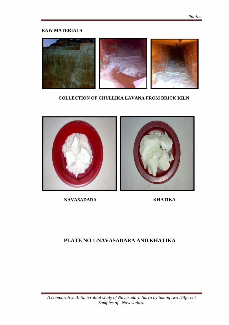
Photos
RAW MATERIALS
COLLECTION OF CHULLIKA LAVANA FROM BRICK KILN
KHATIKA NAVASADARA
PLATE NO 1:NAVASADARA AND KHATIKA
A comparative Antimicrobial study of Navasadara Satva by taking two Different Samples of Navasadara
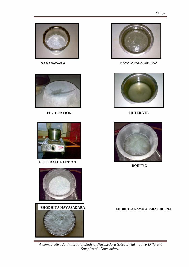
Photos
NAVASADARA CHURNA NAVASADARA
FILTERATE
FILTERATION
FILTERATE KEPT ONBOILING
SHODHITA NAVASADARA SHODHITA NAVASADARA CHURNA
A comparative Antimicrobial study of Navasadara Satva by taking two Different Samples of Navasadara

Photos
NAVASADARA SHODHANA PLATE NO 2:
CHULLIKA LAVANA CHULLIKA LAVANA + JALA
RESIDUE KEPT ON AGNI
A comparative Antimicrobial study of Navasadara Satva by taking two Different Samples of Navasadara
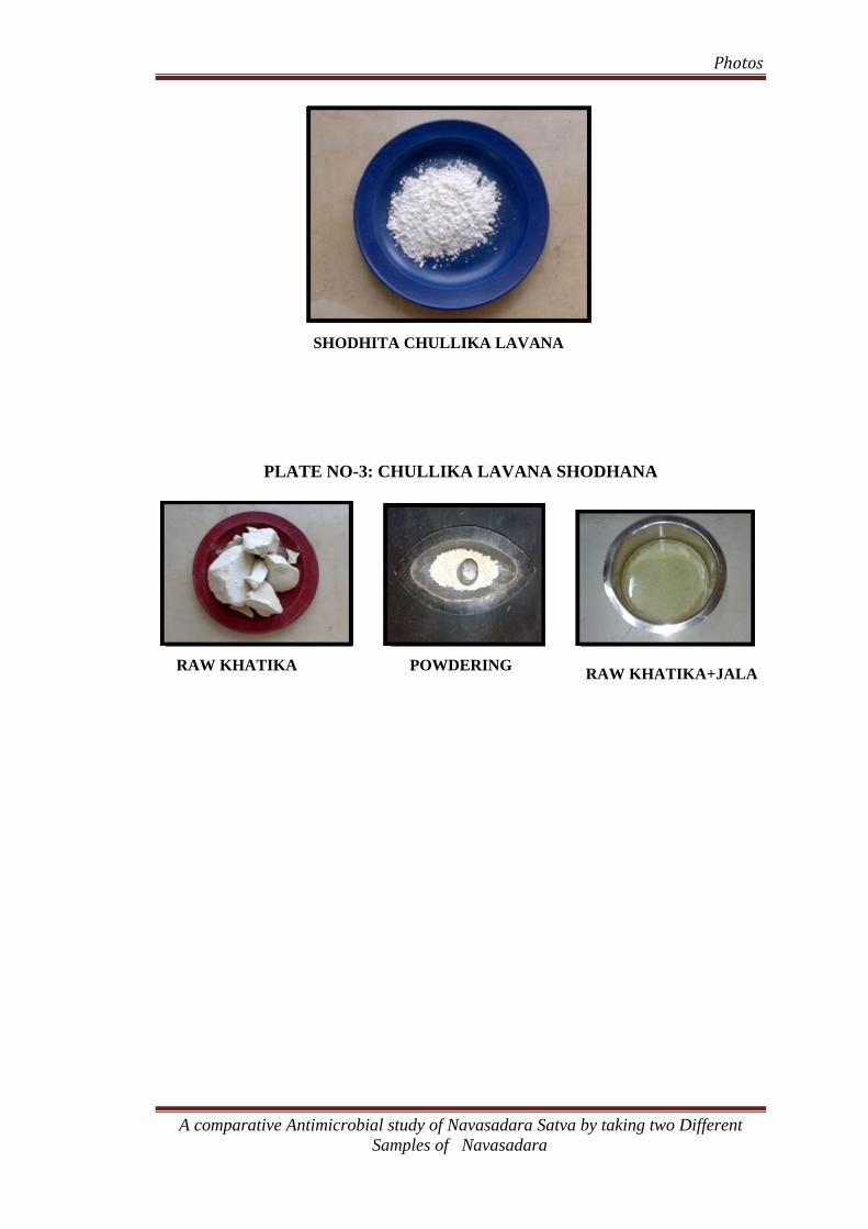
Photos
SHODHITA CHULLIKA LAVANA
PLATE NO-3: CHULLIKA LAVANA SHODHANA
RAW KHATIKA POWDERING RAW KHATIKA+JALA
A comparative Antimicrobial study of Navasadara Satva by taking two Different Samples of Navasadara
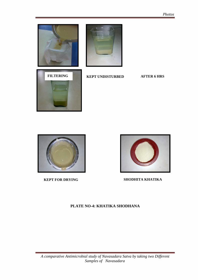
Photos
FILTERING AFTER 6 HRSKEPT UNDISTURBED
SHODHITA KHATIKA KEPT FOR DRYING
PLATE NO-4: KHATIKA SHODHANA
A comparative Antimicrobial study of Navasadara Satva by taking two Different Samples of Navasadara
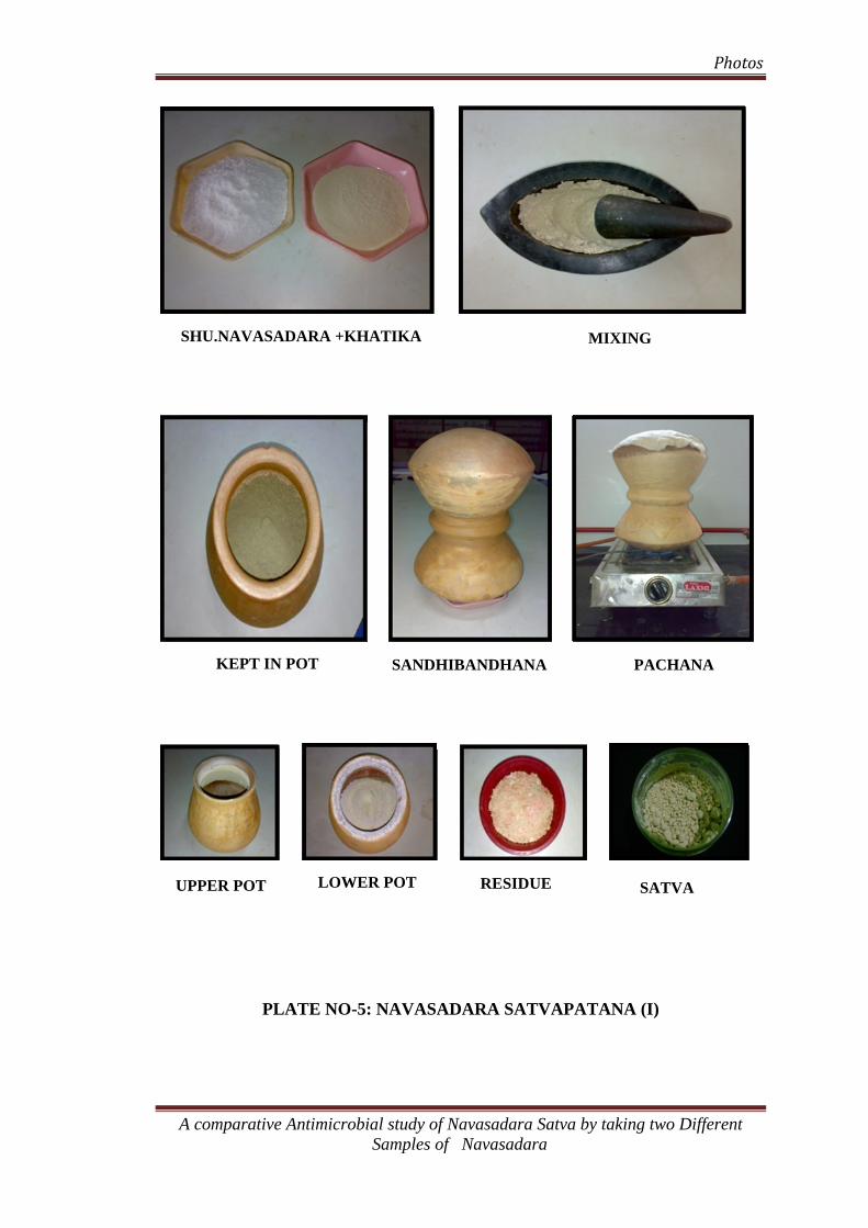
Photos
SHU.NAVASADARA +KHATIKA MIXING
KEPT IN POT SANDHIBANDHANA PACHANA
LOWER POT RESIDUE UPPER POT SATVA
PLATE NO-5: NAVASADARA SATVAPATANA (I)
A comparative Antimicrobial study of Navasadara Satva by taking two Different Samples of Navasadara

Photos
SHU.CHULLIKA LAVANA & KHATIKA MIXING
A comparative Antimicrobial study of Navasadara Satva by taking two Different Samples of Navasadara
KEPT IN POT PACHANA

Photos
SATVA (II)
PLATE NO-6: NAVASADARA SATVA (II)
MICROPIPETTE BHI BROTH
A comparative Antimicrobial study of Navasadara Satva by taking two Different Samples of Navasadara
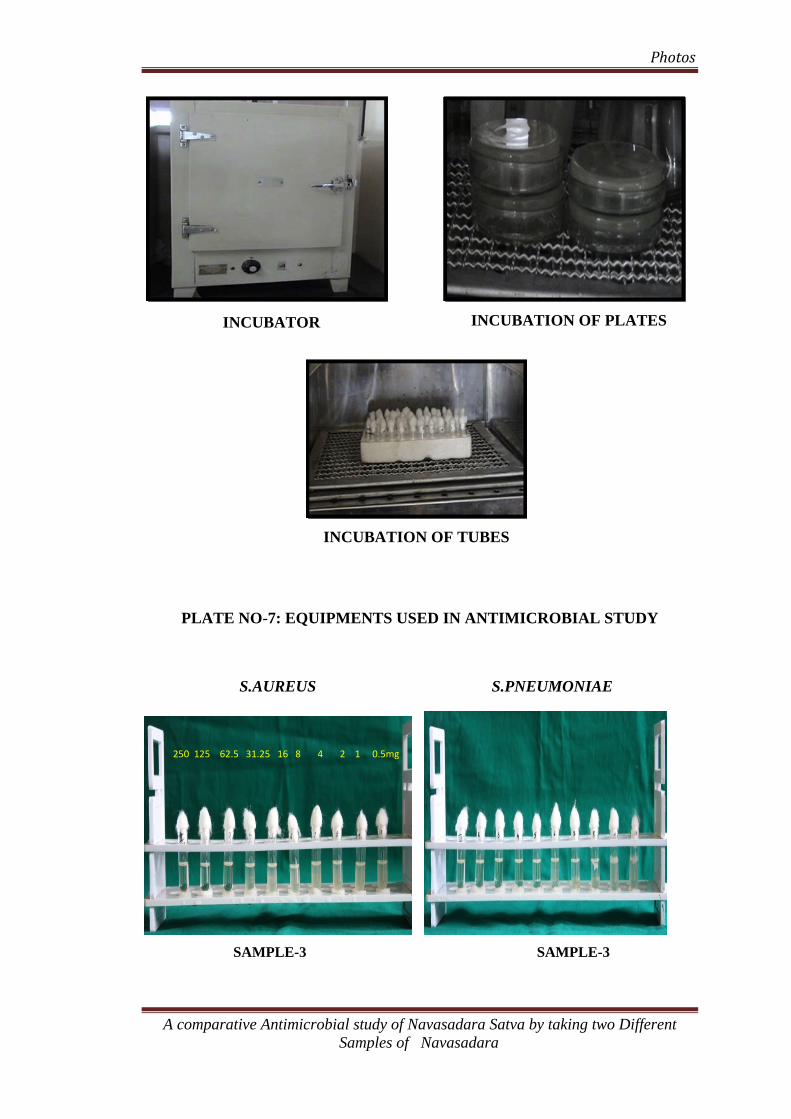
Photos
INCUBATION OF PLATES INCUBATOR
INCUBATION OF TUBES
PLATE NO-7: EQUIPMENTS USED IN ANTIMICROBIAL STUDY
S.AUREUS S.PNEUMONIAE
250 125 62.5 31.25 16 8 4 2 1 0.5mg
SAMPLE-3 SAMPLE-3
A comparative Antimicrobial study of Navasadara Satva by taking two Different Samples of Navasadara
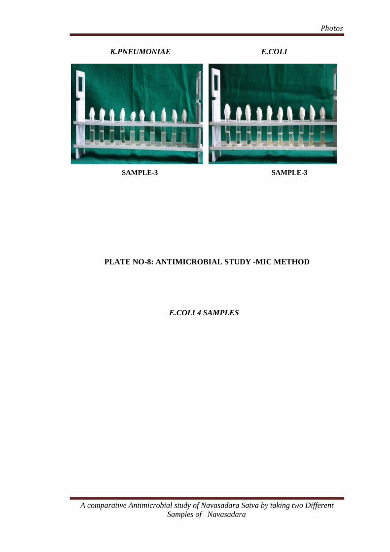
Photos
K.PNEUMONIAE E.COLI
SAMPLE-3 SAMPLE-3
PLATE NO-8: ANTIMICROBIAL STUDY -MIC METHOD
E.COLI 4 SAMPLES
A comparative Antimicrobial study of Navasadara Satva by taking two Different Samples of Navasadara

Photos
32mm
Zone of Inhibition
S.AUREUS 4 SAMPLES:
PLATE NO-9: ANTIMICROBIAL STUDY- DISC DIFFUSION METHOD
K.PNEUMONIAE 4 SAMPLES:
A comparative Antimicrobial study of Navasadara Satva by taking two Different Samples of Navasadara
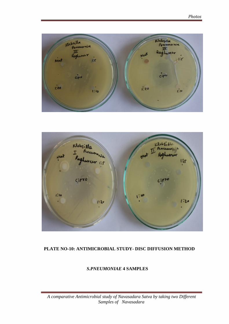
Photos
PLATE NO-10: ANTIMICROBIAL STUDY- DISC DIFFUSION METHOD
S.PNEUMONIAE 4 SAMPLES
A comparative Antimicrobial study of Navasadara Satva by taking two Different Samples of Navasadara
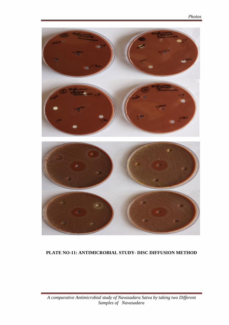
Photos
PLATE NO-11: ANTIMICROBIAL STUDY- DISC DIFFUSION METHOD
A comparative Antimicrobial study of Navasadara Satva by taking two Different Samples of Navasadara
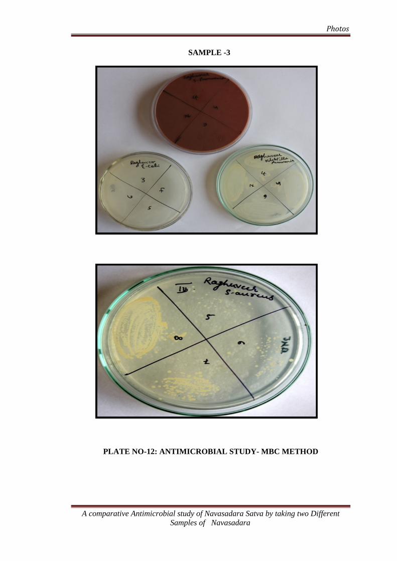
Photos
SAMPLE -3
PLATE NO-12: ANTIMICROBIAL STUDY- MBC METHOD
A comparative Antimicrobial study of Navasadara Satva by taking two Different Samples of Navasadara