a fully integrated microfluidic genetic analysis system … · 2006-12-15 · a fully integrated...
TRANSCRIPT

A fully integrated microfluidic genetic analysis systemwith sample-in–answer-out capabilityChristopher J. Easley*, James M. Karlinsey*, Joan M. Bienvenue*, Lindsay A. Legendre*, Michael G. Roper*,Sanford H. Feldman†, Molly A. Hughes‡, Erik L. Hewlett‡, Tod J. Merkel§, Jerome P. Ferrance*, and James P. Landers*¶�
*Department of Chemistry, University of Virginia, Charlottesville, VA 22904; Departments of †Comparative Medicine, ‡Infectious Disease, and ¶Pathology,University of Virginia Health System, Charlottesville, VA 22908; and §Center for Biologics Evaluation and Research, U.S. Food and Drug Administration,Bethesda, MD 28092
Edited by Robert H. Austin, Princeton University, Princeton, NJ, and approved October 16, 2006 (received for review June 5, 2006)
We describe a microfluidic genetic analysis system that representsa previously undescribed integrated microfluidic device capable ofaccepting whole blood as a crude biological sample with theendpoint generation of a genetic profile. Upon loading the sample,the glass microfluidic genetic analysis system device carries outon-chip DNA purification and PCR-based amplification, followed byseparation and detection in a manner that allows for microlitersamples to be screened for infectious pathogens with sample-in–answer-out results in <30 min. A single syringe pump deliverssample/reagents to the chip for nucleic acid purification from abiological sample. Elastomeric membrane valving isolates eachdistinct functional region of the device and, together with resistiveflow, directs purified DNA and PCR reagents from the extractiondomain into a 550-nl chamber for rapid target sequence PCRamplification. Repeated pressure-based injections of nanoliter ali-quots of amplicon (along with the DNA sizing standard) allowelectrophoretic separation and detection to provide DNA fragmentsize information. The presence of Bacillus anthracis (anthrax) in 750nl of whole blood from living asymptomatic infected mice and ofBordetella pertussis in 1 �l of nasal aspirate from a patientsuspected of having whooping cough are confirmed by the result-ant genetic profile.
full integration � micro total analysis system � microdevice � pumping �valving
The next revolution in personalized medicine, forensic science,and biowarfare defense will be impelled by analysis systems
that provide a quantum leap in terms of functionality, time toresult, and cost effectiveness. These systems need to meet severalrequirements, including a design conducive with low-cost man-ufacturing, turn-key operation with fast analysis times, and theability to manipulate small volumes from crude samples. Oneexample is the micrototal analysis system (�-TAS) describedconceptually more than a decade ago by Manz et al. (1).Prophetically, they stated that, ‘‘. . . the detector or sensor in aTAS does not need high selectivity, because the sample pre-treatment serves to eliminate most of the interfering chemicalcompounds.’’ There are multiple examples in the literature ofsteps taken toward the advancement of integrated microfluidicgenetic analysis (MGA) systems (refs. 2–4; also see ref. 5 for acomprehensive review); however, after a decade and a half, nobona fide microfluidic device has been presented that is capableof nanoliter flow control and integration of an electrophoreticseparation with comprehensive sample pretreatment (DNApurification and PCR amplification).
The MGA system described in this report brings togethermany advances in microfluidics over the last decade, exploitingdifferential channel f low resistances (6), elastomeric valves (7,8), laminar flow (9), and electrophoretic mobility within thedevice, in concert with external f luid flow control from a syringepump for sample and reagent delivery. Nucleic acid purificationthrough solid-phase extraction (SPE), followed by target se-quence amplification by PCR and microchip electrophoretic
(ME) amplicon separation and detection is completed in �30min. This represents a previously undescribed integrated mi-crofluidic system that can accept biological samples as crude aswhole blood, extract high-purity nucleic acids, and generate aPCR-targeted amplicon that can be characterized to provide agenotypic readout.
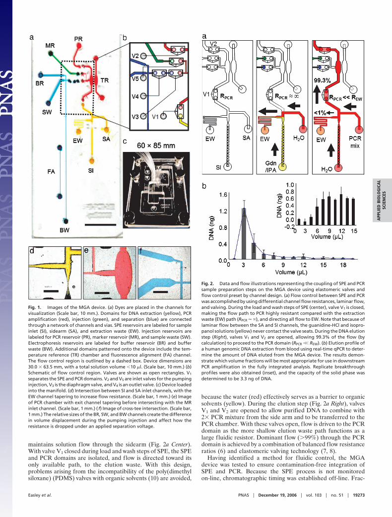
ResultsMicrodevice Design. The MGA system has a microchannel archi-tecture with three distinct functional domains, two for samplepreparation (SPE and PCR) and one for analysis (ME) (Fig. 1).A total of five elastomeric normally closed valves (8) direct f lowfrom a single syringe pump and localize the chemistries andreaction conditions that exist (Fig. 1b). The reagents used forDNA extraction in the SPE domain were isolated from the PCRchamber (valve V1), because these are known PCR inhibitors.The PCR domain, gated from the ME domain by two valves (V3and V4), must be passivated to avoid protein fouling anddeactivation of the Taq polymerase. Valves V3 and V4 functionto gate the two domains and/or pump amplicon from the PCRchamber, whereas the DNA standard from the marker reservoiris injected with valves V2 and V5, respectively. The sample ismobilized across the analysis channel for injection, after whichthe components are separated and detected by laser-inducedfluorescence.
Flow Control and Method Development. The major challenge as-sociated with integrating sample treatment steps into a microflu-idic format is the incompatibility of SPE reagents (guanidine andisoproponal) with the PCR process. Fluidic isolation of the SPEand PCR domains was accomplished by combining severalmethods used in microfluidic flow control. Fig. 2 illustrates howdifferential channel f low resistances, elastomeric valves, andlaminar flow are used to isolate SPE solvents from the otherdomains without compromising DNA extraction. The SPE do-main consists of a sample inlet reservoir, a silica extraction bed,a patterned weir, a sidearm for solution loading, and an extrac-tion waste arm (Fig. 2a Left). Lysed sample and 80% isopropanol(yellow) are sequentially delivered through the sample inlet andthe replaceable silica bed, while distilled deionized water (red)
Author contributions: C.J.E., J.M.K., J.M.B., and L.A.L. contributed equally to this work;C.J.E., J.M.K., J.M.B., L.A.L., J.P.F., and J.P.L. designed research; C.J.E., J.M.K., J.M.B., andL.A.L. performed research; S.H.F., M.A.H., E.L.H., and T.J.M. contributed new reagents/analytic tools; C.J.E., J.M.K., J.M.B., L.A.L., M.G.R., and J.P.L. analyzed data; and C.J.E.,J.M.K., J.M.B., L.A.L., M.G.R., M.A.H., and J.P.L. wrote the paper.
The authors declare no conflict of interest.
This article is a PNAS direct submission.
Abbreviations: �-TAS, micrototal analysis system; MGA, microfluidic genetic analysis; SPE,solid-phase extraction; ME, microchip electrophoretic; qPCR, quantitative PCR; PDMS,poly(dimethyl siloxane).
�To whom correspondence should be addressed. E-mail: [email protected].
This article contains supporting information (SI) online at www.pnas.org/cgi/content/full/0604663103/DC1.
© 2006 by The National Academy of Sciences of the USA
19272–19277 � PNAS � December 19, 2006 � vol. 103 � no. 51 www.pnas.org�cgi�doi�10.1073�pnas.0604663103
maintains solution flow through the sidearm (Fig. 2a Center).With valve V1 closed during load and wash steps of SPE, the SPEand PCR domains are isolated, and flow is directed toward itsonly available path, to the elution waste. With this design,problems arising from the incompatibility of the poly(dimethylsiloxane) (PDMS) valves with organic solvents (10) are avoided,
because the water (red) effectively serves as a barrier to organicsolvents (yellow). During the elution step (Fig. 2a Right), valvesV1 and V2 are opened to allow purified DNA to combine with2� PCR mixture from the side arm and to be transferred to thePCR chamber. With these valves open, f low is driven to the PCRdomain as the more shallow elution waste path functions as alarge fluidic resistor. Dominant flow (�99%) through the PCRdomain is achieved by a combination of balanced flow resistanceratios (6) and elastomeric valving technology (7, 8).
Having identified a method for fluidic control, the MGAdevice was tested to ensure contamination-free integration ofSPE and PCR. Because the SPE process is not monitoredon-line, chromatographic timing was established off-line. Frac-
Fig. 1. Images of the MGA device. (a) Dyes are placed in the channels forvisualization (Scale bar, 10 mm.). Domains for DNA extraction (yellow), PCRamplification (red), injection (green), and separation (blue) are connectedthrough a network of channels and vias. SPE reservoirs are labeled for sampleinlet (SI), sidearm (SA), and extraction waste (EW). Injection reservoirs arelabeled for PCR reservoir (PR), marker reservoir (MR), and sample waste (SW).Electrophoresis reservoirs are labeled for buffer reservoir (BR) and bufferwaste (BW). Additional domains patterned onto the device include the tem-perature reference (TR) chamber and fluorescence alignment (FA) channel.The flow control region is outlined by a dashed box. Device dimensions are30.0 � 63.5 mm, with a total solution volume �10 �l. (Scale bar, 10 mm.) (b)Schematic of flow control region. Valves are shown as open rectangles. V1
separates the SPE and PCR domains. V2 and V5 are inlet valves for the pumpinginjection, V3 is the diaphragm valve, and V4 is an outlet valve. (c) Device loadedinto the manifold. (d) Intersection between SI and SA inlet channels, with theEW channel tapering to increase flow resistance. (Scale bar, 1 mm.) (e) Imageof PCR chamber with exit channel tapering before intersecting with the MRinlet channel. (Scale bar, 1 mm.) ( f) Image of cross-tee intersection. (Scale bar,1 mm.) The relative sizes of the BR, SW, and BW channels create the differencein volume displacement during the pumping injection and affect how theresistance is dropped under an applied separation voltage.
Fig. 2. Data and flow illustrations representing the coupling of SPE and PCRsample preparation steps on the MGA device using elastomeric valves andflow control preset by channel design. (a) Flow control between SPE and PCRwas accomplished by using differential channel flow resistances, laminar flow,and valving. During the load and wash steps of SPE (center), valve V1 is closed,making the flow path to PCR highly resistant compared with the extractionwaste (EW) path (RPCR � �), and directing all flow to EW. Note that because oflaminar flow between the SA and SI channels, the guanidine-HCl and isopro-panol solutions (yellow) never contact the valve seats. During the DNA elutionstep (Right), valves V1 and V2 are opened, allowing 99.3% of the flow (bycalculation) to proceed to the PCR domain (RPCR �� REW). (b) Elution profile ofa human genomic DNA extraction from blood using real-time qPCR to deter-mine the amount of DNA eluted from the MGA device. The results demon-strate which volume fractions will be most appropriate for use in downstreamPCR amplification in the fully integrated analysis. Replicate breakthroughprofiles were also obtained (inset), and the capacity of the solid phase wasdetermined to be 3.3 ng of DNA.
Easley et al. PNAS � December 19, 2006 � vol. 103 � no. 51 � 19273
APP
LIED
BIO
LOG
ICA
LSC
IEN
CES
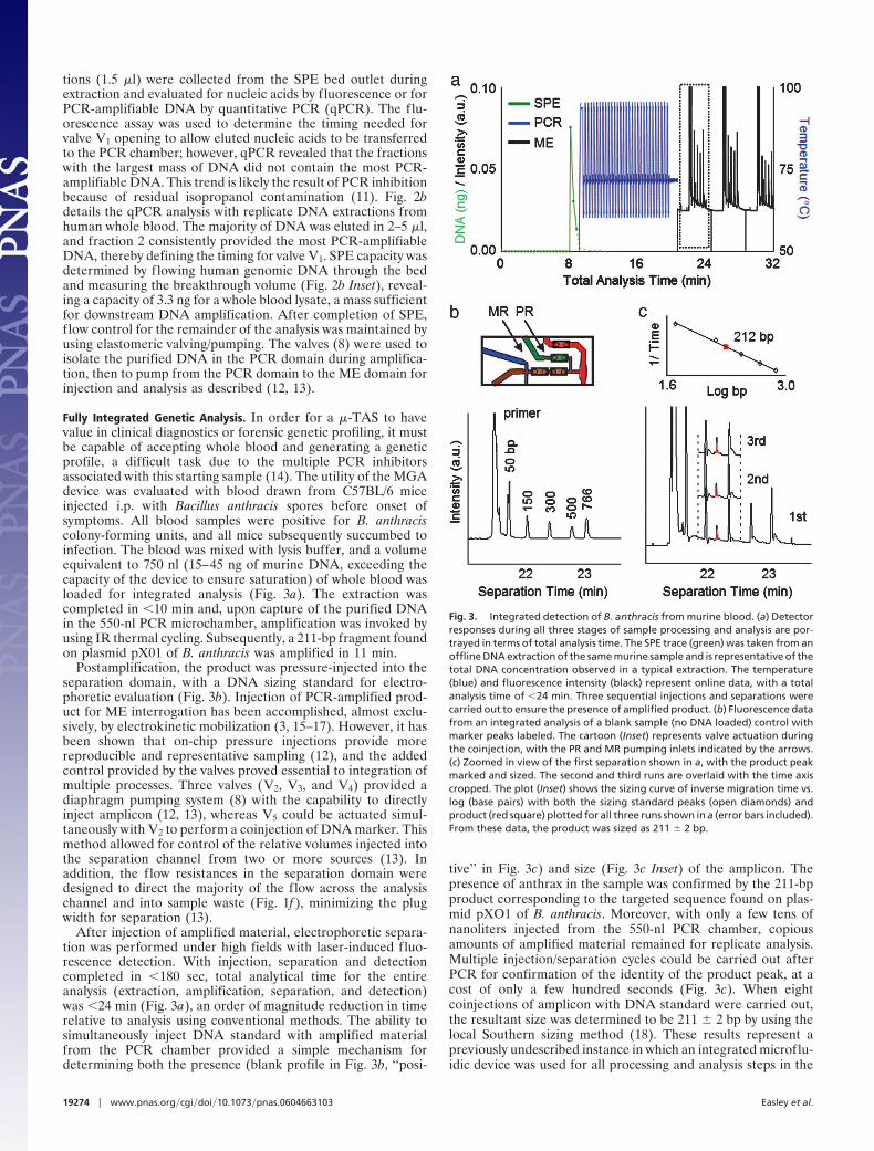
tions (1.5 �l) were collected from the SPE bed outlet duringextraction and evaluated for nucleic acids by fluorescence or forPCR-amplifiable DNA by quantitative PCR (qPCR). The flu-orescence assay was used to determine the timing needed forvalve V1 opening to allow eluted nucleic acids to be transferredto the PCR chamber; however, qPCR revealed that the fractionswith the largest mass of DNA did not contain the most PCR-amplifiable DNA. This trend is likely the result of PCR inhibitionbecause of residual isopropanol contamination (11). Fig. 2bdetails the qPCR analysis with replicate DNA extractions fromhuman whole blood. The majority of DNA was eluted in 2–5 �l,and fraction 2 consistently provided the most PCR-amplifiableDNA, thereby defining the timing for valve V1. SPE capacity wasdetermined by flowing human genomic DNA through the bedand measuring the breakthrough volume (Fig. 2b Inset), reveal-ing a capacity of 3.3 ng for a whole blood lysate, a mass sufficientfor downstream DNA amplification. After completion of SPE,flow control for the remainder of the analysis was maintained byusing elastomeric valving/pumping. The valves (8) were used toisolate the purified DNA in the PCR domain during amplifica-tion, then to pump from the PCR domain to the ME domain forinjection and analysis as described (12, 13).
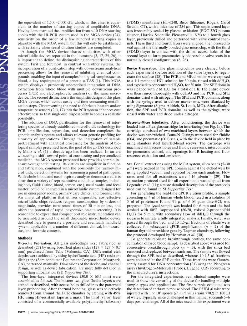
Fully Integrated Genetic Analysis. In order for a �-TAS to havevalue in clinical diagnostics or forensic genetic profiling, it mustbe capable of accepting whole blood and generating a geneticprofile, a difficult task due to the multiple PCR inhibitorsassociated with this starting sample (14). The utility of the MGAdevice was evaluated with blood drawn from C57BL/6 miceinjected i.p. with Bacillus anthracis spores before onset ofsymptoms. All blood samples were positive for B. anthraciscolony-forming units, and all mice subsequently succumbed toinfection. The blood was mixed with lysis buffer, and a volumeequivalent to 750 nl (15–45 ng of murine DNA, exceeding thecapacity of the device to ensure saturation) of whole blood wasloaded for integrated analysis (Fig. 3a). The extraction wascompleted in �10 min and, upon capture of the purified DNAin the 550-nl PCR microchamber, amplification was invoked byusing IR thermal cycling. Subsequently, a 211-bp fragment foundon plasmid pX01 of B. anthracis was amplified in 11 min.
Postamplification, the product was pressure-injected into theseparation domain, with a DNA sizing standard for electro-phoretic evaluation (Fig. 3b). Injection of PCR-amplified prod-uct for ME interrogation has been accomplished, almost exclu-sively, by electrokinetic mobilization (3, 15–17). However, it hasbeen shown that on-chip pressure injections provide morereproducible and representative sampling (12), and the addedcontrol provided by the valves proved essential to integration ofmultiple processes. Three valves (V2, V3, and V4) provided adiaphragm pumping system (8) with the capability to directlyinject amplicon (12, 13), whereas V5 could be actuated simul-taneously with V2 to perform a coinjection of DNA marker. Thismethod allowed for control of the relative volumes injected intothe separation channel from two or more sources (13). Inaddition, the flow resistances in the separation domain weredesigned to direct the majority of the flow across the analysischannel and into sample waste (Fig. 1f ), minimizing the plugwidth for separation (13).
After injection of amplified material, electrophoretic separa-tion was performed under high fields with laser-induced fluo-rescence detection. With injection, separation and detectioncompleted in �180 sec, total analytical time for the entireanalysis (extraction, amplification, separation, and detection)was �24 min (Fig. 3a), an order of magnitude reduction in timerelative to analysis using conventional methods. The ability tosimultaneously inject DNA standard with amplified materialfrom the PCR chamber provided a simple mechanism fordetermining both the presence (blank profile in Fig. 3b, ‘‘posi-
tive’’ in Fig. 3c) and size (Fig. 3c Inset) of the amplicon. Thepresence of anthrax in the sample was confirmed by the 211-bpproduct corresponding to the targeted sequence found on plas-mid pXO1 of B. anthracis. Moreover, with only a few tens ofnanoliters injected from the 550-nl PCR chamber, copiousamounts of amplified material remained for replicate analysis.Multiple injection/separation cycles could be carried out afterPCR for confirmation of the identity of the product peak, at acost of only a few hundred seconds (Fig. 3c). When eightcoinjections of amplicon with DNA standard were carried out,the resultant size was determined to be 211 � 2 bp by using thelocal Southern sizing method (18). These results represent apreviously undescribed instance in which an integrated microflu-idic device was used for all processing and analysis steps in the
Fig. 3. Integrated detection of B. anthracis from murine blood. (a) Detectorresponses during all three stages of sample processing and analysis are por-trayed in terms of total analysis time. The SPE trace (green) was taken from anoffline DNA extraction of the same murine sample and is representative of thetotal DNA concentration observed in a typical extraction. The temperature(blue) and fluorescence intensity (black) represent online data, with a totalanalysis time of �24 min. Three sequential injections and separations werecarried out to ensure the presence of amplified product. (b) Fluorescence datafrom an integrated analysis of a blank sample (no DNA loaded) control withmarker peaks labeled. The cartoon (Inset) represents valve actuation duringthe coinjection, with the PR and MR pumping inlets indicated by the arrows.(c) Zoomed in view of the first separation shown in a, with the product peakmarked and sized. The second and third runs are overlaid with the time axiscropped. The plot (Inset) shows the sizing curve of inverse migration time vs.log (base pairs) with both the sizing standard peaks (open diamonds) andproduct (red square) plotted for all three runs shown in a (error bars included).From these data, the product was sized as 211 � 2 bp.
19274 � www.pnas.org�cgi�doi�10.1073�pnas.0604663103 Easley et al.

direct analysis of a blood sample to genetically verify thepresence of a pathogen in �25 min. Because the early detectionof anthrax is critical to the survival of the host by earlyrecognition and administration of antibiotics with postexposurevaccination, the MGA system and its integrated methods providea microfluidic path to improving biodefense surveillance mea-sures.
To demonstrate the broader utility of the MGA system, adifferent sample and nucleic acid target was evaluated. A nasalaspirate was obtained from a human patient symptomatic ofwhooping cough, a respiratory infection caused by the Gram-negative bacterium Bordetella pertussis, which can be isolatedfrom the mouth, nose, and throat (19, 20). This infection ischaracterized by severe spasms of coughing that can last severalweeks or months and, although not particularly threatening tothose beyond their first year, it can lead to serious complicationsor fatality in infants (19, 20). Using the same method describedabove, a volume equivalent to 1 �l of nasal aspirate was preparedin lysis buffer and loaded into the MGA device, with DNA
purification carried out as described earlier. The presence of B.pertussis can be confirmed by an amplification of a 181-bpfragment of the IS481 repeated insertion sequence, and afterPCR amplification of this target, the amplicon was injected intothe separation channel for electrophoretic separation (Fig. 4a).Again, coinjection of a DNA sizing standard was used to aid inthe sizing of amplified product for comparison with the expected181-bp fragment, confirmed by off-chip sequencing of the re-sultant amplicon. With a total analysis time of 24 min, the MGAsystem could provide physicians with a method to rapidly screenfor B. pertussis respiratory infection in patients during earlyinfection/exposure or for screening during outbreaks. This tech-nological advance is timely, because �25,000 B. pertussis caseswere reported in 2004, a 12-fold increase since 1980 (19). Therapid turnaround time not only provides a dramatic improve-ment over conventional culturing methods for diagnosis [requir-ing a minimum of 24–48 h (20)] but also presents the possibilityof point-of-care testing, a rapidly growing concept applicable toclinical diagnostics, forensics, environmental testing, food safetytesting, and biothreat sensing in the field for armed forces.
DiscussionThe advantages of the MGA system are obvious: rapid turn-around time, decreased reagent consumption per test, decreasedoperator variability (human error factor), and improved opera-tor safety. The comparisons in Fig. 4 b and c showcase thecapabilities of a MGA system with respect to reduction of volumeanalysis time. Fig. 4c compares the turnaround time of the MGAsystem for detecting B. pertussis from a sample, relative toconventional molecular-, serologic-, and culture-based methods.The 24-min turnaround time compares favorably with �2 h foranalysis using conventional methods, a minimum of 24 h forPCR-based analysis in a clinical microbiological testing lab , and�48 h for serology and/or culturing of the organism (20). Fig. 4cInset highlights the comparison of the MGA system with con-ventional methods for extraction (green), amplification (blue),and detection (black), assuming standard laboratory instrumen-tation used by the same operators, with no lost time betweenprocesses, and does not take into account ‘‘batching-related’’delays. Although not insignificant, the 5-fold reduction in anal-ysis time is outweighed by the potential for automation of theintegrated analysis, which will further decrease technician labortime and isolate the operator from the analysis. Finally, Fig. 4bhighlights the value of a microfluidic system with respect toreduced consumption of reagents for DNA extraction andamplification. Microfluidic devices are expected to inherentlyscale reduction to the analytical system and, consistent with theother elegant microfluidic developments from various (2–4)groups, the MGA system allows for submicroliter PCR. Thisreduced size not only enhances amplification speed but alsoprovides a 50-fold reduction in PCR volume. Consuming lessTaq polymerase, the most costly reagent in this molecularanalysis, yields the potential to dramatically decrease the cost pertest. Concordantly, the �25-fold reduction in volume of reagentsused for DNA extraction reduces the hazardous waste that mustbe disposed of. The microfluidic nature of the MGA system [likemicrodevices (2–4)] distinguishes it from larger-volume com-mercial systems (21–23) that do not reap the benefits of submi-croliter fluid manipulation.
Although a comprehensive evaluation of device sensitivity forthese two diverse sample matrices is ongoing, the proof-of-principle experimentation accomplished with anthrax-infectedmurine blood suggests the following regarding detection levelswith the MGA system. The average day 2 serum level of anthraxin immunoprecipitation-challenged mice was determined to be2.5 (�1) � 106 cfu/ml. Of the total blood drawn, only 0.75 �l ofblood was loaded onto the silica bed (purposefully overloaded toensure saturation of the phase for the purification), representing
Fig. 4. Fully integrated microchip detection of B. pertussis from a humannasal aspirate in only 24 min. One microliter of human nasal aspirate wasextracted, PCR was performed on the purified DNA, and products werepressure-injected and electrophoresed. (a) The ME trace was plotted alone toshow the separation of the coinjected DNA sizing standard (peak sizes labeledin number of base pairs) with the PCR amplicon for product verification. Theamplicon (red) migrates between the expected size standards, and sequencinganalysis was used to further verify the product (see SI Supporting Text). (b)Volumes for SPE (green) and PCR (blue) are compared for MGA and Conv.,showing a significant reduction for both processes. (c) Total analysis times forcrude biological samples of the MGA device (from a), conventional analysisperformed in the research lab (Conv.) and a clinical lab (Clin.), and analysis byserology/cell culture. Analysis times for MGA and Conv. are shown in (Inset),with SPE (green), PCR (blue), and ME (black) denoted.
Easley et al. PNAS � December 19, 2006 � vol. 103 � no. 51 � 19275
APP
LIED
BIO
LOG
ICA
LSC
IEN
CES
the equivalent of 1,500–2,000 cfu, which, in this case, is equiv-alent to the number of starting copies of amplifiable DNA.Having demonstrated the amplification from �10 DNA startingcopies with the IR-PCR system used in the MGA device (24),sensitivity on the order of a few hundred starting copies isplausible with the MGA system, but this will only be establishedwith certainty when serial dilution studies are completed.
Although the MGA device shares similarities with othermicrofluidic devices reported in the literature (3, 17, 25, 26), itis important to define the distinguishing characteristics of thissystem. First and foremost, in contrast with other systems, theincorporation of a purification step with downstream analyticalprocessing allows for the removal of inhibiting chemical com-pounds, enabling the input of complex biological samples such asblood, a key requirement of a genetic �-TAS (1). This MGAsystem displays a previously undescribed integration of DNAextraction from whole blood with multiple downstream pro-cesses (PCR and electrophoretic analysis) on the same micro-device. The second distinction is the simplistic design of this glassMGA device, which avoids costly and time-consuming metalli-zation steps. Circumventing the need to fabricate heaters and/ortemperature sensors (2, 3, 17) into the PCR system enhances costeffectiveness so that single-use disposability becomes a realisticpossibility.
The addition of DNA purification for the removal of inter-fering species to already established microfluidic technology forPCR amplification, separation, and detection completes thegenetic analysis system and allows relevant genetic profiling fora variety of applications. Through the integration of samplepretreatment with analytical processing for the analysis of bio-logical samples presented here, the goal of the �-TAS describedby Manz et al. (1) a decade ago has been realized. In an erawitnessing a shift toward point-of-care testing and personalizedmedicine, the MGA system presented here provides sample-in–answer-out genetic testing. Its virtues are simplicity in functionand fabrication, combined with the possibility for turnkey mi-crofluidic detection systems for screening a panel of pathogens.With whole-blood and nasal-aspirate analyses demonstrated, it isclear that a variety of representative candidate samples, includ-ing body fluids (urine, blood, semen, etc.), nasal swabs, and fecalmatter, could be analyzed in a microfluidic system designed foruse in emergency rooms, primary care clinics, and forensic labs.An analytical platform that utilizes disposable, cost-effectivemicrofluidic chips reduces reagent consumption by orders ofmagnitude, provides turnaround times of 30 min or less, andoffers the potential of rapid inexpensive on-site screening. It isreasonable to expect that compact portable instrumentation canbe assembled around the small disposable microfluidic devicedescribed here to generate a portable and eventually handheldsystem, applicable in a number of different clinical, biohazard-ous, and forensic contexts.
MethodsMicrochip Fabrication. All glass microchips were fabricated asdescribed (27) by using borofloat glass slides (127 � 127 � 0.7mm) purchased from Telic (Valencia, CA). Differential etchdepths were achieved by using hydrofluonic acid (HF) resistantdicing tape (Semiconductor Equipment Corporation, Moorpark,CA), patterned manually. Dimensions of the device and channeldesign, as well as device fabrication, are more fully detailed insupporting information (SI) Supporting Text.
The four-layer integrated devices (30.0 � 63.5 mm) wereassembled as follows. The bottom two glass fluidic layers wereetched as described, with access holes drilled into the patternedlayer prebonding. After thermal bonding, glass was selectivelyremoved from around the PCR chamber by etching with 49%HF, using HF-resistant tape as a mask. The third (valve) layerconsisted of a commercially available poly(dimethyl siloxane)
(PDMS) membrane (HT-6240, Bisco Silicones, Rogers, CarolStream, CT), with a thickness of 254 �m. This unpatterned layerwas irreversibly sealed by plasma oxidation (PDC-32G plasmacleaner, Harrick Scientific, Pleasantville, NY) to a fourth glasslayer, previously drilled and patterned with valve control chan-nels. These third and fourth layers were aligned, then pressed toseal against the thermally bonded glass microchip, with the third(PDMS) layer in contact with the drilled access holes of thesecond layer to form pneumatically addressable valve seats in anormally closed configuration (8, 26).
Device Preparation. The glass microchips were cleaned beforeeach experiment (before addition of the valve layer), to regen-erate the surface (28). The PCR and ME domains were exposedto a 1:1 methanol:HCl solution for 30 min, rinsed with ddH2O,and exposed to concentrated H2SO4 for 30 min. The SPE domainwas cleaned with 2 M HCl for a total of 1 h. The entire devicewas then rinsed thoroughly with ddH2O and the PCR and SPEdomains dried with nitrogen. The SPE and PCR domains, alongwith the syringe used to deliver master mix, were silanized byusing Sigmacote (Sigma-Aldrich, St. Louis, MO). After silaniza-tion, the SPE and PCR domains, as well as the syringe, wererinsed with water and dried under nitrogen.
Macro-to-Micro Interfacing. After conditioning, the device wasloaded into a Plexiglas cartridge for interfacing (see Fig. 1c). Thecartridge consisted of two machined layers between which thedevice was sandwiched. Buna-N O-rings were used for fluidic(004) and pneumatic (001) seals, with the device held in place byusing stainless steel knurled-head screws. The cartridge wasmachined with access holes and fluidic reservoirs, interconnectsfor pneumatic control, and openings for IR heating and fluo-rescence excitation and emission.
SPE. For all extractions using the MGA system, silica beads (5–30�m) were packed in the SPE domain against the etched weir byusing applied vacuum and replaced before each analysis. Flowrates used for all extractions were 4.16 �l�min�1 (29). Theextraction protocol used for all experiments was adapted fromLegendre et al. (11); a more detailed description of the protocolsused can be found in SI Supporting Text.
For generating the real-time qPCR elution profile, a sampleconsisting of 4 �l of human whole blood, lysed in a solution of5 �l of proteinase K and 91 �l of 6 M guanidine�HCl, wasprepared. The lysed sample was loaded for 6 min and the bedwashed with 80% isopropanol (80/20, vol/vol 2-propanol/ddH2O) for 5 min, with secondary flow of ddH2O through thesidearm to imitate a fully integrated analysis. Finally, water waspassed through the bed, and 13 1.5-�l fractions of eluate werecollected for subsequent qPCR amplification (n � 2) of thehuman thyroid peroxidase gene by Taqman chemistry, followingthe protocol developed by Horsman et al. (30).
To generate replicate breakthrough profiles, the same con-centration of lysed blood sample as described above was used forconsecutive breakthrough plots (n � 3), with the silica bedremoved and replaced between each run. The sample was flowedthrough the SPE bed as described, whereas 10 1.5-�l fractionswere collected at the SPE outlet. These fractions were fluores-cently assayed for DNA concentration (31) using the Picogreenassay (Invitrogen–Molecular Probes, Eugene, OR) according tothe manufacturer’s instructions.
For the integrated experiments, real clinical samples wereused to show the versatility of the device for handling multiplesample types and applications. The first sample evaluated wasthe detection of anthrax in mouse blood. The C57BL/6 mice wereinjected with 1 � 109 spores (B. anthracis strain 7702) in 100 �lof water. Typically, mice challenged in this manner succumb 5–6days post-challenge. All of the mice used in this experiment were
19276 � www.pnas.org�cgi�doi�10.1073�pnas.0604663103 Easley et al.
positive for cfus in the blood, liver, and spleen by day 2postchallenge and were asymptomatic when sampled on day 2.The clinical patient sample was a discarded clinical sample fromUniversity of Virginia Medical Laboratories, and all patientidentification information was removed from the sample beforeit was obtained. The sample consists of a nasopharyngeal washthat was tested and diagnosed as a strong positive for B. pertussis.Samples were prepared by mixing the appropriate volume ofsample (8 �l of nasal aspirate or 6 �l of murine whole blood) with5 �l of Proteinase K, diluted to 100 �l of total volume with 6 MGuHCl and vortexed for 30 sec to mix thoroughly. Humansamples were used directly in the analysis, whereas mouse bloodsamples were first boiled for 10 min. The sample was loaded for3 min, followed by a 5-min rinse with 80% isopropanol. Apreconditioning rinse step was used in which PCR master mixwas flowed through R2 and PCR domain with valves V1 and V2open for 2 min to condition the valve between SPE and PCR.DNA was eluted with water, with the valves closed until theappropriate time as previously determined, followed by subse-quent opening and closing of the valves to allow PCR mastermixture and eluting DNA to be trapped within the PCR chamberfor thermal cycling.
PCR Amplification. For fully integrated analysis, the PCR mastermixture was made with the following final concentrations: 20mM Tris/100 mM KCl (pH 8.3)/6 mM MgCl2/0.8 �M of eachprimer/0.4 mM dNTP/0.5 units/�l Taq polymerase. The thermalcycling protocols used were 95°C for 30 sec (initial denatur-ation), then 30 cycles of 95°C for 2 sec, 62°C/55°C for 3 sec (forB. anthracis/B. pertussis, respectively), and 72°C for 5 sec, fol-lowed by a single final extension for 1 min at 72°C after the 30cycles were completed. The primers for B. pertussis amplificationwere adapted from Loeffelholz et al. (32). The primers used inthe B. anthracis amplification were 5-CAAATCAGCTC-GAAAGTTAGGA (for) and 5-CAGTAACTGTTCAGAAG-GTACATCTGA (rev) for the amplification of a 211-bp frag-ment of the virulence B gene on pXO1 and were designedin-house (33). The noncontact thermal cycling PCR system (see
SI Supporting Text) was constructed in-house, as described (34).Amplicon from the analysis of B. pertussis was removed from thedevice postanalysis and sequenced at the Biomolecular ResearchFacility at the University of Virginia.
Microchip Electrophoresis. Glass microchips were cleaned as de-scribed above. The separation channels were not dried post-cleaning. During PCR, the separation domain was filled with 1.0M HNO3. After PCR, the separation channels were rinsed withddH2O and filled with the sieving matrix, 3.5% HPC in 80/40 mMMes/Tris (35) with 1.0 �M YOPRO DNA intercalating dye(Invitrogen–Molecular Probes). The pressure injection and valv-ing instrumentation (see SI Supporting Text, for more detaileddescriptions of both) were used as described (13). After pressureinjection, separation was achieved by applying voltage using adual polarity high-voltage power supply built in-house using twoSpellman high-voltage sources (Hauppauge, NY). For B. an-thracis analysis, �200 V was applied to the buffer reservoir and1,050 V to the buffer waste. For the B. pertussis analysis, �150and 790 V were applied. An argon ion laser (Model LS200,Dynamic Laser, Salt Lake City, UT) was used for excitation witha conventional confocal detection setup (�16 objective, 1-mmpinhole). Emission was collected with a PMT (Hamamatsu,Bridgewater, CT) through a 515-nm bandpass filter (OmegaOptical, Brattleboro, VT). The instrument and data acquisitionwere controlled through a LabVIEW application written in-house.
We gratefully acknowledge the pioneering developments of Drs. A.Huhmer, B. C. Giordano, and Q. Wu, who laid the foundation for thiswork. We also thank the Mathies Laboratory at the University ofCalifornia, Berkeley, CA, for sharing their valving innovations early on.Funding was provided by the National Institutes of Health/NationalHuman Genome Research Institute through Grants R01 HG001832 andR01 HG002613 (to J.P.L.). Funding for C.J.E. was provided by theAmerican Chemical Society, Division of Analytical Chemistry, and EliLilly and Co. M.A.H., E.L.H., and T.J.M. acknowledge funding from theNational Institutes of Health National Institute of Allergy and InfectiousDieseases Mid-Atlantic Regional Center of Excellence for Biodefense(Grant U54 AI057168).
1. Manz A, Graber N, Widmer HM (1990) Sens Act B 1:244–248.2. Lagally ET, Scherer JR, Blazej RG, Toriello NM, Diep BA, Ramchandani M,
Sensabaugh GF, Riley LW, Mathies RA (2004) Anal Chem 76:3162–3170.3. Pal R, Yang M, Lin R, Johnson BN, Srivastava N, Razzacki SZ, Chomistek KJ,
Heldsinger DC, Haque RM, Ugaz VM, et al. (2005) Lab Chip 5:1024–1032.4. Liu J, Enzelberger M, Quake S (2002) Electrophoresis 23:1531–1536.5. Auroux PA, Koc Y, deMello A, Manz A, Day PJ (2004) Lab Chip 4:534–546.6. Attiya S, Jemere AB, Tang T, Fitzpatrick G, Seiler K, Chiem N, Harrison DJ
(2001) Electrophoresis 22:318–327.7. Unger MA, Chou, H-P, Thorsen T, Scherer A, Quake SR (2000) Science
288:113–116.8. Grover WH, Skelley AM, Liu CN, Lagally ET, Mathies RA (2003) Sens Act B
89:315–323.9. Kenis PJ, Ismagilov RF, Whitesides GM (1999) Science 285:83–85.
10. Lee JN, Park C, Whitesides GM (2003) Anal Chem 75:6544–6554.11. Legendre LA, Bienvenue JM, Roper MG, Ferrance JP, Landers JP (2006) Anal
Chem 78:1444–1451.12. Karlinsey JM, Monahan J, Marchiarullo DJ, Ferrance JP, Landers JP (2005)
Anal Chem 77:3637–3643.13. Easley CJ, Karlinsey JM, Landers JP (2006) Lab Chip 6:601–610.14. Al-Soud WA, Radstrom P (2001) J Clin Microbiol 39:485–493.15. Lagally ET, Medintz I, Mathies RA (2001) Anal Chem 73:565–570.16. Waters LC, Jacobson SC, Kroutchinina N, Khandurina J, Foote RS, Ramsey
JM (1998) Anal Chem 70:158–162.17. Burns MA, Johnson BN, Brahmasandra SN, Handique K, Webster JR,
Krishnan M, Sammarco TS, Man PM, Jones D, Heldsinger D, et al. (1998)Science 282:484–487.
18. Elder JK, Southern EM (1983) Anal Biochem 128:227–231.19. Hewlett EL, Edwards KM (2005) N Engl J Med 352:1215–1222.20. Mattoo S, Cherry JD (2005) Clin Microbiol Rev 18:326–382.
21. Pourahmadi F, Taylor M, Kovacs G, Lloyd K, Sakai S, Schafer T, Helton B,Western L, Zaner S, Ching J, et al. (2000) Clin Chem 46:1511–1513.
22. Liu RH, Yang J, Lenigk R, Bonanno J, Grodzinski P (2004) Anal Chem76:1824–1831.
23. Anderson RC, Su X, Bogdan GJ, Fenton J (2000) Nucleic Acids Res 28:E60.24. Legendre LAC, WC, Piper, K, Ferrance, JP, Patel, R, Landers JP (2006) in
Proceedings of the �TAS 2006 Conference (Tokyo, Japan).25. Blazej RG, Kumaresan P, Mathies RA (2006) Proc Natl Acad Sci USA
103:7240–7245.26. Skelley AM, Scherer JR, Aubrey AD, Grover WH, Ivester RHC, Ehrenfreund
P, Grunthaner FJ, Bada JL, Mathies RA (2005) Proc Natl Acad Soc USA102:1041–1046.
27. Manz A, Fettinger JC, Verpoorte E, Ludi H, Widmer HM, Harrison DJ (1991)Trends Anal Chem 10:144–149.
28. Cras JJ, Rowe-Taitt CA, Nivens DA, Ligler FS (1999) Biosens Bioelectron14:683–688.
29. Bienvenue JM, Duncalf N, Marchiarullo D, Ferrance JP, Landers JP (2006) JForens Sci 51:266–273.
30. Horsman KM, Hickey JA, Cotton RW, Landers JP, Maddox LO (2006) J ForensSci 51:758–765.
31. Ahn SJ, Costa J, Emanuel JR (1996) Nucleic Acids Res 24:2623–2625.32. Loeffelholz MJ, Thompson CJ, Long KS, Gilchrist MJ (1999) J Clin Microbiol
37:2872–2876.33. Breadmore MC, Wolfe KA, Arcibal IG, Leung WK, Dickson D, Giordano BC,
Power ME, Ferrance JP, Feldman SH, Norris PM, Landers JP (2003) AnalChem 75:1880–1886.
34. Easley CJ, Legendre A, Roper MG, Wavering T, Ferrance JP, Landers JP(2005) Anal Chem 77:1038–1045.
35. Sanders JC, Breadmore MC, Kwok YC, Horsman KM, Landers JP (2003) AnalChem 75:986–994.
Easley et al. PNAS � December 19, 2006 � vol. 103 � no. 51 � 19277
APP
LIED
BIO
LOG
ICA
LSC
IEN
CES