a morphological study of the brain of solea senegalensis ... morphological study of the brain... ·...
TRANSCRIPT
Histol Histopathol (2000) 15: 355-364
001: 10.14670/HH-15.355
http://www.hh.um.es
Histology and Histopathology
Cellular and Molecular Biology
A morphological study of the brain of Solea senegalensis. I. The telencephalon F.J. Rodriguez-Gomez1, C. Sarasquete2 and J.A. Munoz-Cueto1
1 Department of Animal Biology, Plant Biology and Ecology, Faculty of Marine Sciences, University of Cadiz, Poligono Rro San Pedro,
Puerto Real, Cadiz, Spain and 21nstitute of Marine Sciences of Andalucfa (CSIC), Poligono Rro San Pedro, Puerto Real, Cadiz,
Spain
Summary. In this paper we present an anatomical description of the telencephalon of Solea senegaLensis based on cresyl violet and haematoxilin-eosin-stained serial transverse sections. This work was conducted as a basis for the precise localization of neuroendocrine territories in the brain of a species with growing interest in marine aquaculture. The external asymmetric morphology of Senegalese sole is correlated with the asymmetry of the forebrain . The right olfactory nerve and bulb are larger than the contralateral ones and this asymmetry is also extended to the cerebral hemispheres. The olfactory bulb comprises an outer olfactory nerve fiber layer, a glomerular layer, an external cellular layer, a secondary olfactory fiber layer and an internal cellular layer. The telencephalic hemispheres can be divided in area dorsalis and area ventralis, consisting of eleven and eight cell masses, respectively. The area dorsalis comprises five subareas : a pars medialis (Dm), subdivided into four nuclei termed Dm 1 to Dm4; a pars dorsalis (Dd); a pars lateral is (Dl), which consists of dorsal (DId) , ventral (Dlv) and posterior (Dip) subdivisions; a pars centralis (Dc); and more caudally, a pars posterioris (Dp), which is very prominent in this species. A nucleus taenia (NT) was observed in the transitional region between area dorsalis and area ventralis. The area ventralis consists of pars dorsalis (Vd), pars ventralis (Vv), pars supracommissuralis (Vs), pars postcommissuralis (Vp), pars lateralis (VI), pars centralis (Vc), pars intermedia (Vi) and nucleus entopeduncularis (E). A peri ventricular organ, that we have termed lateral septal organ (LSO), was observed in the ventral telencephalon, medial to Vv.
Key words: Telencephalon, Anatomy, Sole, Pleuronectiformes, Teleost
Offprint requests to: Dr. Jose Antonio Munoz-Cueto, Department of Animal Biology, Plant Biology and Ecology, Faculty of Marine Sciences, University of Cadiz, Poligono Rio San Pedro, 11510, Puerto Real,
Cadiz, Spain. Fax: + 34-956-016019. e-mail: [email protected].
Introduction
As other vertebrates, ray-finned fishes posses paired olfactory bulbs and cerebral hemispheres in the rostral forebrain (Northcutt and Davis, 1983; Braford, 1995). However, the telencephalic hemispheres of gnathostomes exhibit a remarkable degree of variation as a result of differences during development. In ray-finned fishes, the cerebral hemispheres result from an eversion of the dorsal half of the lateral walls of the forebrain while in all other gnathostomes they are originated by inversion and evagination processes (Nieuwenhuys, 1963; Northcutt, 1995). Within actinopterygians, there is also a considerable variation in the anatomical telencephalic organization and the amplitude of this variation increases with the evolutionary distance separating groups and species (Northcutt and Braford, 1980; Northcutt and Davis, 1983).
Pleuronectiformes are characteristic asymmetric animals during adult life, although they are externally symmetric until metamorphosis (Amaoka, 1971; Policansky and Sieswerda, 1979; Policansky, 1982). In many flatfishes, the left eye migrates to the right side, which faces upward, its left side facing the bottom (Policansky, 1982). Metamorphosis of flatfishes also implies modifications of the mouth, opercular orifices, fins and pigmentation (Parker, 1903; Chabanaud, 1948; Policansky and Sieswerda, 1979). In winter flounder, the olfactory organ, nerve and bulb are larger in the right side, the right telencephalon also being slightly larger than the left one (Prasada Rao and Finger, 1984). A telencephalic asymmetry is also observed during the metamorphic process in turbot, but in this case the left hemisphere is larger (Brinon et aI., 1993).
The Senegalese sale, Solea senegaLensis, belongs to the order of the flatfishes, Pleuronectiformes. It is a species adapted to temperate waters and commonly exploited in extensive aquaculture in the south of Europe (Drake et aI., 1984; Dinis, 1992) and the north of Africa (Fehri-Bedoui, 1997). In the latter years, some studies tried to understand some biological and pathological processes in Senegalese sale (Rodriguez, 1984; Gutierrez et aI., 1985; Sarasquete et aI., 1993a,b;
Telencephalic anatomy of Senegalese sole
Mourente and Vázquez, 1996; Sarasquete et al., 1996), as well as the organization of pituitary secretory cells (Rendón et al., 1997) and the molecular characteristic of pituitary hormones (Pendón et al., 1994a,b). However, no studies aiming at identifying the brain regions potentially implicated in neuroendocrine regulation of pituitary functions and at tracing the neurona1 systems participating in this regulation have been carried out, in part, because precise neuroanatomical tools for this species are not available. Thus, as a first approach in this direction, we present in this paper a precise anatomical description of the telencephalon of Senegalese sole.
Materials and rnethods
Adult s~ecimens. females and males. of Senegalese sole, SoIeaLsenegalensis, with a mean lehgth of 5 cm, were purchased from a local fishery (C.I.C.E.M. El Toruno, Puerto de Santa María, Cadiz, Spain) and kept in the laboratory in running seawater. Specimens (n=17) were anaesthetized with 2-phenoxiethanol (Sigma, St Louis, MO, USA) and perfused via the aortic bulb with 0.6% saline solution followed by Bouin fixative (4% paraformaldehyde in 0.1M phosphate buffer, pH 7.4, 0.2% picric acid). Brains were carefully removed and postfixed in the same fixative for 12 h. After fixation, tissues were washed in distilled water and embedded in paraffin. Corona1 (transverse) and sagittal serial sections (10 pm) were cut on a rotary microtome, mounted on gelatin-coated glass slides and deparaffinized through xylene-ethanol-water. For coronal sectioning, brains were oriented in order to obtain sections perpendicular to the mid sagittal and horizontal planes. The cyto- architectonic organization of the telencephalon was determined in brain sections stained with 1% cresyl violet or haematoxylin-eosin (Gabe, 1968), which were
analyzed on an Olympus photomicroscope and photographed using panchromatic Agfapan APX 25 Films (AGFA). Size, shape, density, staining and pattern of distribution of perikarya, as well as the spatial discontinuity of cell masses were used as major criteria to identify different cell groups on both sagittal and coronal sections. For the description, we subdivided the cells into three categories: small (5-15 pm); medium (15-25 pm); and large (26-40 pm) cells. The boundaries of cell masses and fiber tracts were drawn on photographs of the brain sections and copied on transparent paper. Serial drawings were digitized by using an HP 4L scanner (Hewlett Packard) and processed on an IBM compatible personal computer with the help of the Aldus Photostyler 2.0 program. The section through the anterior commissure was chosen as transverse zero point. Distantes from the zero point are expressed in p m and drawings anterior or posterior to the zero point are indicated as - or +, respectively.
Results
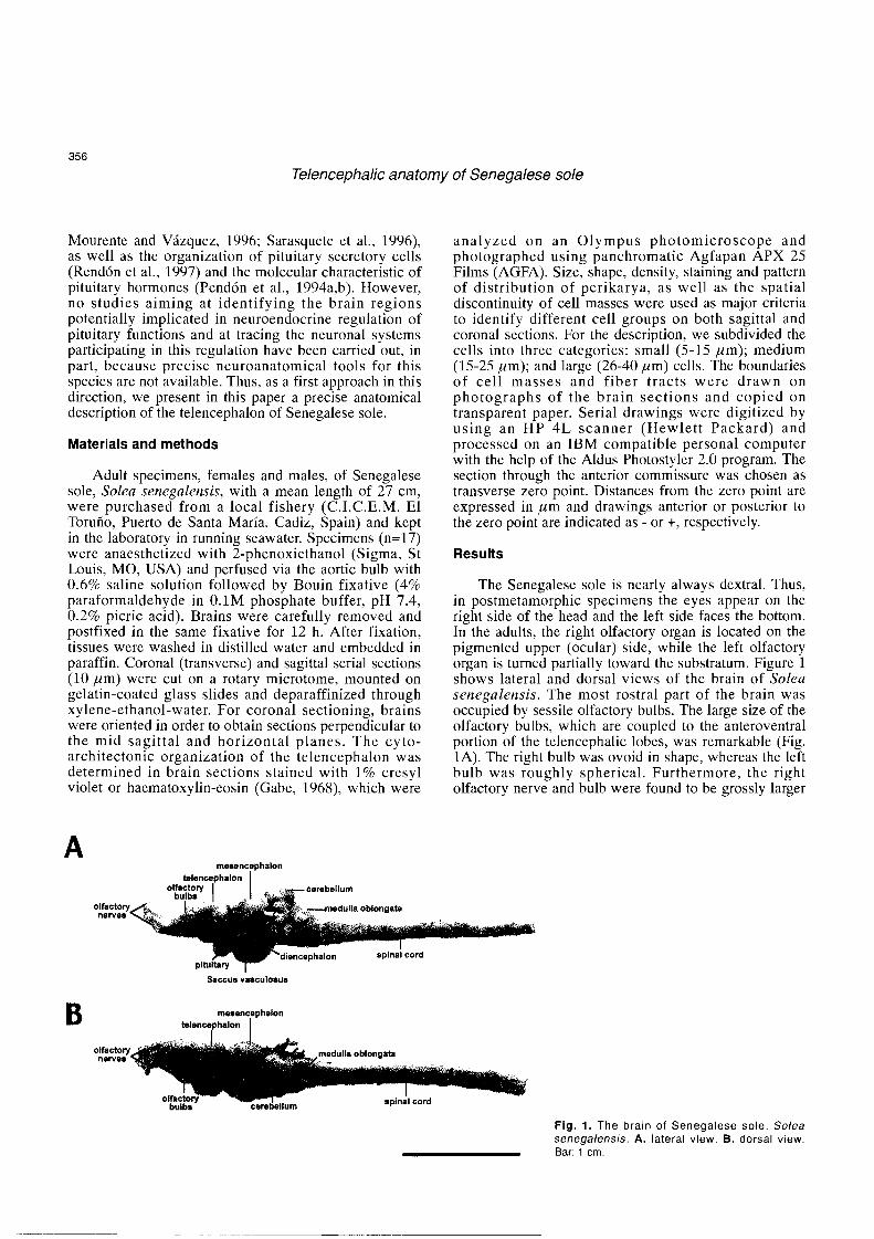
The Senegalese sole is nearly always dextral. Thus, in postmetamorphic specimens the eyes appear on the right side of the head and the left side faces the bottom. In the adults, the right olfactory organ is located on the pigmented upper (ocular) side, while the left olfactory organ is turned partially toward the substratum. Figure 1 shows lateral and dorsal views of the brain of Solea senegalensis. The most rostral part of the brain was occupied by sessile olfactory bulbs. The large size of the olfactory bulbs, which are coupled to the anteroventral portion of the telencephalic lobes, was remarkable (Fig. 1A). The right bulb was ovoid in shape, whereas the left bulb was roughly spherical. Furthermore, the right olfactory nerve and bulb were found to be grossly larger
melrencephalon
olfactory newes
~ i e ~ a l o n splnal cord piiuiiary
saccus vasculosus
mo8oncephalon telencephslon
I I
lum spinal cord
Fig. 1. The brain of Senegalese sole, Solea senegalensis. A. lateral view. B. dorsal view. Bar: 1 cm.
Telencephalic anatomy of Senegalese sole
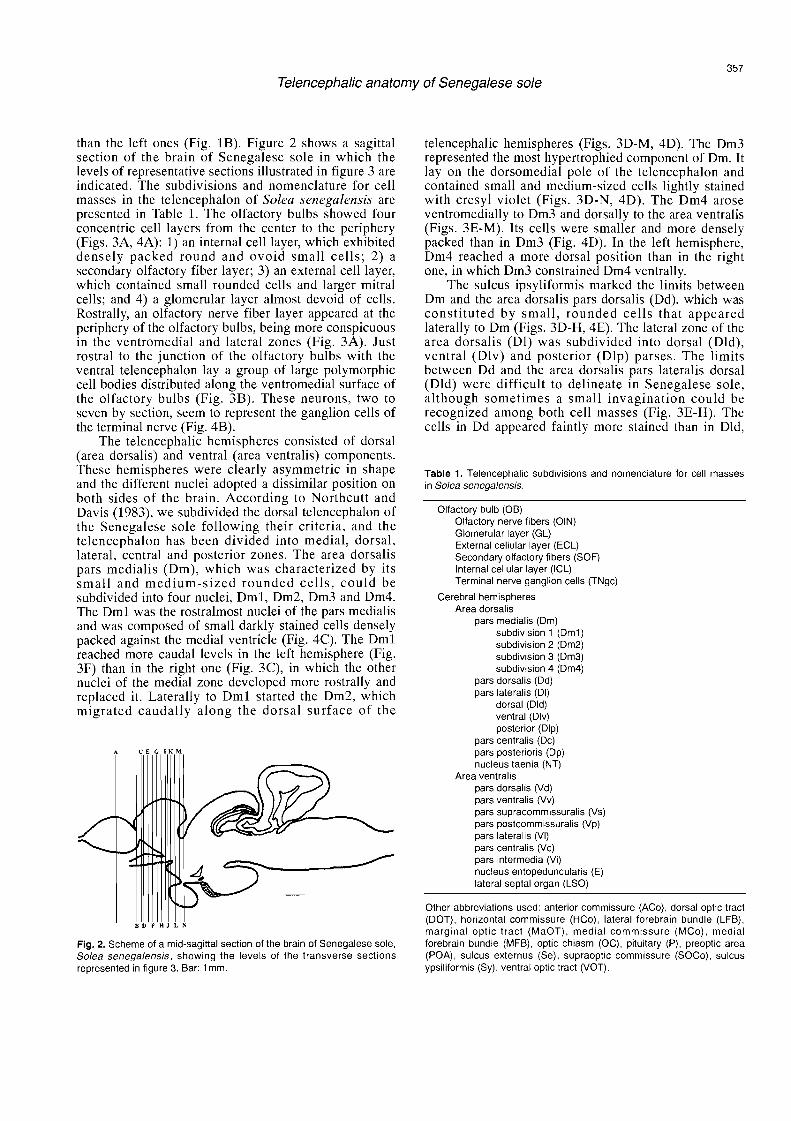
than the left ones (Fig. 1B). Figure 2 shows a sagittal section of the brain of Senegalese sole in which the levels of representative sections illustrated in figure 3 are indicated. The subdivisions and nomenclature for cell masses in the telencephalon of Solea senegalensis are presented in Table 1. The olfactory bulbs showed four concentric cell layers from the center to the periphery (Figs. 3A, 4A): 1) an interna1 cell layer, which exhibited densely packed round and ovoid small cells; 2) a secondary olfactory fiber layer; 3) an externa1 cell layer, which contained small rounded cells and larger mitra1 cells; and 4) a glomerular layer almost devoid of cells. Rostrally, an olfactory nerve fiber layer appeared at the periphery of the olfactory bulbs, being more conspicuous in the ventromedial and lateral zones (Fig. 3A). Just rostral to the junction of the olfactory bulbs with the ventral telencephalon lay a group of large polymorphic cell bodies distributed along the ventromedial surface of the olfactory bulbs (Fig. 3B). These neurons, two to seven by section, seem to represent the ganglion cells of the terminal nerve (Fig. 4B).
The telencephalic hemispheres consisted of dorsal (area dorsalis) and ventral (area ventralis) components. These hemispheres were clearly asymmetric in shape and the different nuclei adopted a dissimilar position on both sides of the brain. According to Northcutt and Davis (1983), we subdivided the dorsal telencephalon of the Senegalese sole following their criteria, and the telencephalon has been divided into medial, dorsal, lateral, central and posterior zones. The area dorsalis pars medialis (Dm), which was characterized by its small and medium-sized rounded cells, could be subdivided into four nuclei, Dml, Dm2, Dm3 and Dm4. The Dml was the rostralmost nuclei of the pars medialis and was composed of small darkly stained cells densely packed against the media1 ventricle (Fig. 4C). The Dml reached more caudal levels in the left hemisphere (Fig. 3F) than in the right one (Fig. 3C), in which the other nuclei of the media1 zone developed more rostrally and replaced it. Laterally to Dml started the Dm2, which migrated caudally along the dorsal surface of the
Fig. 2. Scherne of a rnid-sagittal section of the brain of Senegalese sole, Solea senegalensis, showing the levels of the transverse sections represented in figure 3. Bar: 1 rnrn.
telencephalic hemispheres (Figs. 3D-M, 4D). The Dm3 represented the most hypertrophied component of Dm. It 1ay on the dorsomedial pole of the telencephalon and contained small and medium-sized cells lightly stained with cresyl violet (Figs. 3D-N, 4D). The Dm4 arose ventromedially to Dm3 and dorsally to the area ventralis (Figs. 3E-M). Its cells were smaller and more densely packed than in Dm3 (Fig. 4D). In the left hemisphere, Dm4 reached a more dorsal position than in the right one, in which Dm3 constrained Dm4 ventrally.
The sulcus ipsyliformis marked the limits between Dm and the area dorsalis pars dorsalis (Dd), which was constituted by small, rounded cells that appeared laterally to Dm (Figs. 3D-H, 4E). The lateral zone of the area dorsalis (Dl) was subdivided into dorsal (Dld), ventral (Ulv) and posterior (Dlp) parses. The limits between Dd and the area dorsalis pars lateralis dorsal (Dld) were difficult to delineate in Senegalese sole, although sometimes a small invagination could be recognized among both cell masses (Fig. 3E-H). The cells in Dd appeared faintly more stained than in Dld,
Table 1. Telencephalic subdivisions and nornenclature for cell rnasses in Solea senegalensis.
Olfactory bulb (OB) Olfactory nerve fibers (OIN) Glornerular layer (GL) External cellular layer (ECL) Secondary olfactory fibers (SOF) Interna1 cellular layer (ICL) Terminal nerve ganglion cells (TNgc)
Cerebral hernispheres Area dorsalis
pars rnedialis (Drn) subdivision 1 (Dml) subdivision 2 (Dm2) subdivision 3 (Drn3) subdivision 4 (Dm4)
pars dorsalis (Dd) pars lateralis (DI)
dorsal (Dld) ventral (Dlv) posterior (Dlp)
pars centralis (Dc) pars posterioris (Dp) nucleus taenia (NT)
Area ventralis pars dorsalis (Vd) pars ventralis (Vv) pars supracornrnissuralis (Vs) pars postcornmissuralis (Vp) pars lateralis (VI) pars centralis (Vc) pars intermedia (Vi) nucleus entopeduncularis (E) lateral septal organ (LSO)
Other abbreviations used: anterior cornrnissure (ACo), dorsal optic tract (DOT), horizontal cornmissure (HCo), lateral forebrain bundle (LFB), marginal optic tract (MaOT), rnedial cornmissure (MCo), medial forebrain bundle (MFB), optic chiasm (OC), pituitary (P), preoptic area (POA), sulcus externus (Se), supraoptic cornmissure (SOCO), sulcus ypsiliformis (Sy), ventral optic tract (VOT).
Telencephalic anatomy of Senegalese sole
ECL
o. SOF' , : , : ICL ,,, ;; m1 .. ..
'.'.. :,.. . . .. ... ICL*ECL .....
T N a C TNgc
- 200 prn
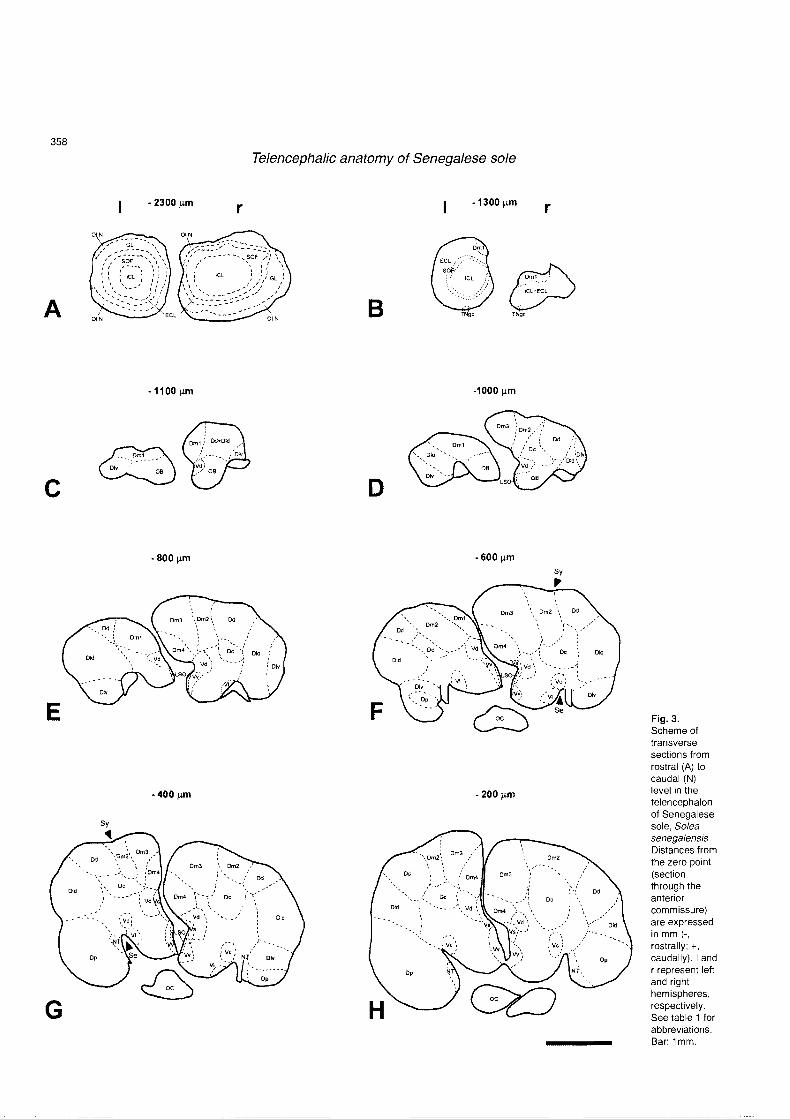
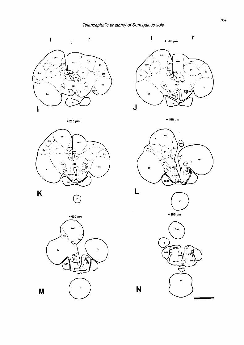
Fig. 3. Scheme of transverse sections from rostral (A) to caudal (N) level in the telencephalon of Senegalese sole, Solea senegalensis Distances from the zero point (section through the anterior commissure) are expressed in mm (-, rostrally; t, caudally). I and r represent leit and right hemispheres, respectively. See table 1 for abbreviations. Bar: 1 mm.
Telencephalic anatomy of Senegalese sole
Telencephalic anatomy of Senegalese sole
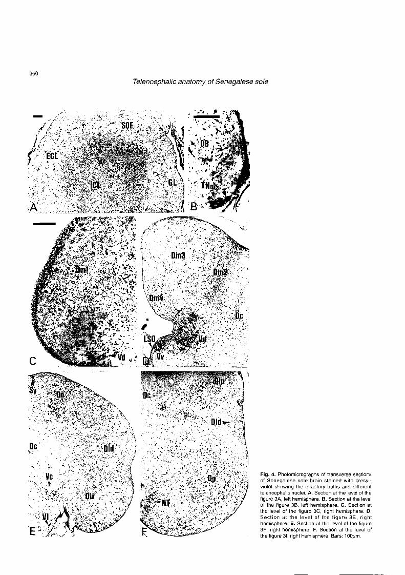
Fig. 4. Photomicrographs of transverse sections of Senegalese sole brain stained with cresyl- violet showing the olfactory bulbs and different telencephalic nuclei. A. Section at the level of the figure 3A, left hemisphere. B. Section at the level of the figure 30, leít hemisphere. C. Section at the level of the figure 3C, right hemisphere. D. Section at the level of the figure 3E, right hemisphere. E. Section at the level of the figure 3F, right hemisphere. F. Section at the level of the figure 31, right hemisphere. Bars: 100pm.

Telencephalic anatomy of Senegalese sole
. . . . ,. . ,. . . 1 . ' : 0 ' : .. . .. . m : . - - '. s.; . . . . especially in the central zone, and the neuropil between
. ' . . , . * . J . ~ - . .. . .. cell clusters was more evident in Dld than in Dd (Fig. 4E). The cells in Dlv were smaller than in Dld and exhibited an organization in tight columns oriented perpendicularly to the ependymal surface (Fig. 4E). At the caudal end of Dld and dorsal to it, the pars lateralis posterior of the area dorsalis (Dlp) arose (Fig. 31-L). The
. . Dlp cells, which were slightly larger and more intensely stained than those of Dld, appeared organized in clusters in the lateral surface of the telencephalon (Fig. 4F). The area dorsalis pars centralis (Dc) contained intensely stained medium-sized cells dispersed in the center of the telencephalon (Figs. 4D-F, 5A, B). In spite of the fact
. . that this nucleus was composed of heterogeneous cell aggregations, we grouped these cells in a single Dc because of the dif f icul ty in observing a c lear discontinuity between cell groups. Lateral to the sulcus
s. ?. externus lay the nucleus taenia (NT). This nucleus, which adopted a character is t ic t r iangular shape,
. , , ., %., ,,, .-. . . -... .. . .. . ..,.,. . ..,. exhibited darkly stained cells arranged in clusters (Fig. . . . ., ..f.. ,..#?.. . / - f : ; T , : ? . * * . . ?,e. . , ' .
.L.:, ,. , , . , , . , - - : 4F). The caudal telencephalon was occupied ventrally by . . . .te . - ' :-;, í t ..* - .. Dm3:-?!;?.>.:.. u.. ,, ,. . - the pars posterioris of the area dorsalis (Dp). This
-, !.* * , : : : . . , . . ,. , :,q '.A,.. . . . a!
. ., . .., __.* . . . , . ..: : / .. -.'. "..;' .. (-,*.:.' %: nucleus was hypertrophied in Senegalese sole (Figs. 3F- . . . . '
r ' , i ; , : . , , - ' .;%. . .. , , ''.:..*, .. : .. .. N). It contained medium-sized cells although intensely . , .' ' . ,:;:- ..m. ' . . . -.:., , ,;-% , '., . . . . .. pkt. stained larger cells could also be observed adopting a .fJ ,.;.;'.~ .
: ~.Zt.c; t*.: . . ? . , ,XI> laminar arrangement in parallel to the ependymal surface . .- cc .*: h.+.. . :7; - ; . ..
, .I , . , . (Fig. 4F). . , . . . . . . . , . -
. . . . The ventral telencephalon could be divided into - . , . . . .DE , , ' eight cells masses: pars dorsalis (Vd), pars ventralis
. . (Vv) , pars supracommissural is (Vs) , pars post- . . , . .. . .
, , commissuralis (Vp), pars lateralis (Vl), pars centralis
. . (Vc), pars intermedia (Vi) and nucleus entopeduncularis , ,. (E). The Vd represented the most rostral cell mass of the . . .. . . area ventralis and appeared in the midline, dorsal to the
. . , cauda l ol factory bulbs (Fig. 3C-E). T h e Vd was .., . > . . . ' . composed of small and medium-sized intensely stained
: . cells which appeared organized in discrete groups in the . -. , - . .
' , Ycc'.. neuropil (Figs. 4D, 5A). Slightly caudal to the onsct of :B .. . : . ' ".. . . . -., . , , . . - Vd and ventrally to it, started Vv, that extended from the caudal end of the olfactory bulbs to the anterior commissure (Fig. 3E-H). The cells in Vv were smaller, less intensely stained and more densely packed than in Vd (Figs. 4D, 5A). As Vv developed, Vd was displaced dorsally, reaching a more dorsal position in the left hemisphere than in the right one (Fig. 3F-J). A structure which resembled the lateral septal organ (LSO) described in other vertebrales could be recognized in the ventral telencephalon of Senegalese sole (Fig. 3D-G). This organ was composed of columnar densely packed, small and darkly stained cells which appeared in a periventricular position, media1 to Vv (Figs. 4D. 5A). Further caudal ly , Vd migrated laterally, left its periventricular position (Fig. 5A) and its place was
. .. : -
7 ' Fig. 5. Photomicrographs of transverse sections of Senegalese sole
, . .,e-.) . - + brain stained with cresyl-violet showing different telencephalic nuclei A.
B .- Section at the level of the figure 3G B. Sections at the level of the figure a . 3J C. Section at the level of the figure 3L Bar IOOpm
Telencephalic anatomy of Senegalese sole
progressively occupied by the pars supracommissuralis (Vs) and more caudally by the pars postcommissuralis (Vp). The Vs was constituted by small, rounded cells lightly stained with cresyl violet (Figs. 3F-K. 5A.B). The cells of Vp were slightly larger and more darkly stained than those of Vs (Figs. 31-M, 5B). Two migrated cell masses were observed in the lateral zone of the area ventralis, associated to the sulcus externus: the pars lateralis (Vl) and the pars centralis (Vc). The cells of these nuclei were surrounded by nerve fibers of the olfactory tracts and the lateral forebrain bundles (LFB). The V1 contained a few small, round and fusiform cells loosely scattered along the media1 portion of the sulcus externus (Figs. 3F,G, 4E, 5A). The Vc represented a more conspicuous nucleus that contained slightly larger and more intensely stained cells than those of V1 (Figs. 3F-J , 4E , 5 A ) . T h e V c ended where the an te r io r commissure a rose , jus t at the ros t ra l pole of the diencephalic preoptic area (Figs. 31,J). Finally, two nuclei were observed at postcommissural level, in the caudal zone of the area ventralis; the pars intermedia (Vi) and the nucleus entopeduncularis (E). The Vi contained small, darkly stained neurons, lying dorsally to the LFB and medially to the Dp (Figs. 3K,L, 5C). The nucleus entopeduncularis (E), which was small in size in Senegalese sole, was composed of very small granular cells denselly packed in the media1 zone of the LFB (Figs. 31-M, 5C).
Discussion
The telencephalon of Senegalese sole exhibits a clear asymmetry, which seems to be the consequence of complex metamorphic processes, implying the migration of the eyes towards the zenithal face. Thus, in this species the right olfactory nerve and bulb are larger in size than the left ones. A similar asymmetry has been descr ibed in the forebrain of the winter f lounder Pseudopleuronectes americanus (Prasada Rao and Finger, 1984). In winter flounder, there is also an asymmetry in the projections of the olfactory bulbs, the dorsomedial and ventromedial olfactory tracts issued by the right bulb being larger than the corresponding tracts arising from the left bulb (Prasada Rao and Finger. 1984). Also in this species, the olfactory fiber bundle that crosses the midline from the right telencephalon to the contralateral side via the anterior commissure is th icker than the bundle that en te r s in to the right telencephalon from the left side (Prasada Rao and Finger, 1984). Further studies should be directed to e lucidate if the re a re a l so di f ferences in cen t ra l projections and decussations of olfactory fibers from the right and left hemispheres in Senegalese sole.
In Senega lese so le , s o m e large ce l l s c a n be recognized ventromedially at the transitional zone between the olfactory bulb and the telencephalon. Unpubl ished data indicate that these neurons a re immunoreactive for salmon GnRH, suggesting that this group of cells constitute the terminal nerve ganglion. In
winter flounder, these neurons have been described as a source of afferents to the olfactory bulbs (Prasada Rao and Finger, 1984). The large size of the olfactory bulbs in Senegalese sole is also remarkable when compared to the cerebral hemispheres. This fact could be explained because this species is largely dependent on smell sense, which compensates the poorly developed visual system (unpublished observations).
The telencephalic asymmetry of the forebrain is also extended to the cerebral hemispheres of Senegalese sole. in which the disposition of nuclei varies considerably among the right and left hemispheres. This asymmetry in shape is also evident in other flatfishes and is reflected in differences in the size of the hemispheres. In winter flounder, the right telencephalon is about 8% larger than the left (Prasada Rao and Finger. 1984), while in turbot the left hemisphere is almost 21% larger than the right (Briñon et al., 1993). The apparent symmetry of the brain in the Senegalese sole begins at the diencephalon and extends further caudally (unpublished observations), while olfactory bulbs and telencephalic hemispheres are asymmetrical regions.
The area dorsalis and area ventralis have been generally homologized to the pallium and subpallium of other vertebrates (Nieuwenhuys, 1963; Braford, 1995; Northcutt, 1995). In the present study, it was found that the organization of the telencephalic hemispheres of the Senegalese sole resembled that described in other percomorph fishes like the flatfish Pseudopleuronectes americanus (Prasada Rao and Finger, 1984) or the perciforms Lepomis cyarrellus (Northcutt and Davis: 1983), Bettu splendcrrs (Marino-Neto and Sabbatini, 1988) and Dicentrarckus labrax (J.M. Cerdá-Reverter, S . Zanuy and J . A . Mufioz-Cueto, unpubl ished obse rva t ions ) . However , some dif ferences in t e lencepha l i c cy toarch i t ec tu re be tween Solea sctzegulensis and the flatfish Pseudopleuronectes atncricanus could be observed, especially in the area dorsalis.
In Senegalese sole four subdivisions could be clearly recognized in Dm while in the winter founder Dm was considered as a single component (Prasada Rao and Finger, 1984). A single Dm zone was also contemplated in lctalurus punctatus (Bass, 1981a,b), but different subdivisions have been reported in the Dm of other teleosts. Thus, two subdivisions were observed in Sebasticus marmoratus (Murakami et al., 1983) and Barbus meridionalis (Díez et al., 1987) and three subdivisions were described in Betta splendens (Marino- Neto and Sabbatini, 1988). As in Senegalese sole, four nuclei have been observed in Dm of Salmo (Northcutt and Bradford, 1980), green sunfish (Northcutt and Davis, 1983), blind cave fish Astyanax hubbsi (Riedel. 1997) and gilthead seabream (Muñoz-Cueto et al., in press).
The Dd of Senegalese sole adopts a similar position to that of Pseudopleuronectes americanus (Prasada Rao and Finger, 1984) and other teleosts (Northcutt and Davis, 1983). However, in winter flounder Dd reaches