a new lantibiotic from bacillus subtilis strain mma7 isolated from the marine sponge haliclona
TRANSCRIPT

Mar. Drugs 2013, 11, 1878-1898; doi:10.3390/md11061878
Marine Drugs ISSN 1660-3397
www.mdpi.com/journal/marinedrugs
Article
Subtilomycin: A New Lantibiotic from Bacillus subtilis Strain
MMA7 Isolated from the Marine Sponge Haliclona simulans
Robert W. Phelan 1,2
, Matthieu Barret 1,2
, Paul D. Cotter 3,4
, Paula M. O’Connor 3,4
, Rui Chen 5,
John P. Morrissey 1,6
, Alan D. W. Dobson 1,6
, Fergal O’Gara 1,2,6,
* and Teresa M. Barbosa 1,7,
*
1 Department of Microbiology, University College Cork, Cork, Ireland;
E-Mails: [email protected] (R.W.P.); [email protected] (M.B.);
[email protected] (J.P.M.); [email protected] (A.D.W.D.) 2
Biomerit Research Centre, Department of Microbiology, University College Cork, Cork, Ireland 3
Teagasc Food Research Centre, Teagasc, Moorepark, Fermoy, Co. Cork, Ireland;
E-Mails: [email protected] (P.D.C.); [email protected] (P.M.O.) 4
Alimentary Pharmabiotic Centre, University College Cork, Cork, Ireland 5
Department of Molecular and Human Genetics, Baylor College of Medicine, Houston, TX 77030,
USA; E-Mail: [email protected] 6
Marine Biotechnology Centre, Environmental Research Institute, University College Cork,
Cork, Ireland 7
School of Pharmacy, University College Cork, Cork, Ireland
* Authors to whom correspondence should be addressed; E-Mails: [email protected] (T.M.B.);
[email protected] (F.O.); Tel.: +353-21-4901792 (T.M.B.); +353-21-4902646 (F.O.);
Fax: +353-21-4901656 (T.M.B.); +353-21-4275934 (F.O.).
Received: 25 April 2013; in revised form: 13 May 2013 / Accepted: 15 May 2013 /
Published: 3 June 2013
Abstract: Bacteriocins are attracting increased attention as an alternative to classic
antibiotics in the fight against infectious disease and multidrug resistant pathogens.
Bacillus subtilis strain MMA7 isolated from the marine sponge Haliclona simulans
displays a broad spectrum antimicrobial activity, which includes Gram-positive and
Gram-negative pathogens, as well as several pathogenic Candida species. This activity is
in part associated with a newly identified lantibiotic, herein named as subtilomycin. The
proposed biosynthetic cluster is composed of six genes, including protein-coding genes for
LanB-like dehydratase and LanC-like cyclase modification enzymes, characteristic of the
class I lantibiotics. The subtilomycin biosynthetic cluster in B. subtilis strain MMA7 is
found in place of the sporulation killing factor (skf) operon, reported in many B. subtilis
isolates and involved in a bacterial cannibalistic behaviour intended to delay sporulation.
OPEN ACCESS

Mar. Drugs 2013, 11 1879
The presence of the subtilomycin biosynthetic cluster appears to be widespread amongst
B. subtilis strains isolated from different shallow and deep water marine sponges.
Subtilomycin possesses several desirable industrial and pharmaceutical physicochemical
properties, including activity over a wide pH range, thermal resistance and water solubility.
Additionally, the production of the lantibiotic subtilomycin could be a desirable property
should B. subtilis strain MMA7 be employed as a probiotic in aquaculture applications.
Keywords: antimicrobial; subtilomycin; lantibiotic; marine sponge; Bacillus subtilis
1. Introduction
The quest for novel antimicrobial compounds from the marine environment has been driven by a
widespread resistance to antibiotics and emerging multi drug resistant pathogens. The large majority of
the antimicrobials currently used in clinical therapy are, or are derived from, natural products of
microbial origin [1]. While antimicrobials have been traditionally isolated from soil-dwelling
microorganisms, the number of bioactive metabolites isolated from marine organisms, and in particular
from marine sponges, has increased dramatically in recent years [2–4].
Marine sponges (Porifera) are simple multicellular, sessile filter feeding invertebrates, which have
been in existence for 700–800 million years [4]. They house a dense and diverse microbial population,
which is believed to contribute to their unmatched prolific production of bioactive metabolites [3].
These bioactives are thought to play an important protective role against pathogens which might enter
the sponge habitat. Bacillus species appear to be part of the microbiota of many marine sponges [5–8],
although their diversity, properties and specific ecological contribution still remain poorly
understood [9].
Bacillus are renowned for their ability to produce a wide variety of bioactive compounds [10,11].
The production of antimicrobials coupled with the development of environmentally highly resistant
endospores, facilitates their survival in competitive ecological niches such as that which they are likely
to encounter within sponges. B. subtilis is the best characterised member of the genus. Although
initially described as a soil dwelling bacterium, numerous reports suggest that this species can be
isolated from, and can grow, sporulate and germinate in, many different ecological niches [9,12–17].
B. subtilis isolates produce a vast array of chemically different antimicrobial peptides, which include
non-ribosomally synthesised peptides, such as polymyxins and the lipopetide surfactin, gene encoded
bacteriocins, such as subtilosin and, within this latter group, lantibiotics, such as subtilin [10].
Lantibiotics are ribosomally synthesised post-translationally modified antimicrobial peptides
typically produced by Gram-positive bacteria, although the array of producer organisms is currently
expanding to other groups of bacteria [18]. Lantipeptides, i.e.; the name used to describe lantibiotics
and related peptides which lack antimicrobial activity, are currently classified into four classes
(I to IV) according to the biosynthetic pathway involved in their production. While dedicated
dehydratase (LanB) and cyclase enzymes (LanC) are involved in the modification of class I
lantibiotics, for the other three classes this is mediated by a single multifunctional enzyme [18,19].

Mar. Drugs 2013, 11 1880
The bio-conversion from a linear lantibiotic precursor, with an N-terminal leader peptide and a
C-terminal core peptide, to the mature active molecule involves different post-translational
modification events of the C-terminal portion, such as the enzymatic conversion of serine and
threonine residues into the dehydrated amino acids dehydroalanine (Dha) and dehydroamino-2-butyric
acid (Dhb), respectively. When one of these modified amino acids interacts with an intrapeptide
cysteine, a thioether bond is formed generating meso-lanthionine (Lan) or (2S,3S,6R)-3-methyll
anthionine (MeLan) [20]. Besides these, other less common modifications have also been described [19].
The post-translational modifications appear to influence not only the structure of the peptides, but also
their activity and stability against protease degradation and heat denaturation [18,21,22]. For class I
lantibiotics, the mature peptide is then translocated through the membrane generally by a
transmembrane ATP-binding cassette (ABC) transporter (LanT). This can happen before or after
cleavage of the leader peptide from the modified core peptide, which is frequently mediated by a
dedicated serine protease LanP [19]. Dedicated immunity proteins (LanI) and/or specialised ABC
transporter proteins (LanFE(G)) protect the host from the action of its own lantibiotic. All of the genes
involved in the processing, transport, immunity and regulation are organised in genetic clusters, which
are frequently composed of multiple transcriptional units. While the mode of action of most
lantibiotics is thought to involve inhibition of peptidoglycan biosynthesis through their interaction with
specific molecular targets, such as the cell wall precursor lipid II, many will additionally cause pore
formation in the cytoplasmic membrane and ultimately, result in cell death [19].
Bacillus lantibiotics, despite their broad spectrum of antimicrobial activity, have attracted limited
attention regarding their potential applicability in food, agricultural and clinical fields.
Endospore-forming bacteria, particularly Bacillus, are known to play an important role in the
preparation of Asian fermented foods [23,24] and are currently being used as spore probiotic
preparations in human therapy, animal production and aquaculture [14,25,26]. In this regard, the
production of antimicrobial compounds, such as lantibiotics, is regarded as a highly desirable property
if candidate strains are to be used as probiotics and/or starter cultures.
Despite several reports describing the isolation of bioactive Bacillus strains from marine
sponges [27–29], very few have successfully identified the associated antimicrobial compounds. In this
study we identify and characterise a novel class I lantibiotic, here named subtilomycin, produced by
B. subtilis MMA7 isolated from the marine sponge Haliclona simulans.
2. Results
2.1. Antimicrobial Activity of B. subtilis Strain MMA7
Strain B. subtilis MMA7 isolated from the marine sponge H. simulans, was previously shown to
display a strong antimicrobial activity against different indicator bacteria [9]. The spectrum of activity
of this isolate has been extended in the current study to include marine associated Gram-negative
bacteria, such as Aeromonas hydrophila, Vibrio anguillarum and Alteromonas sp., and nosocomial
pathogens, such as VRE, VISA, hVISA and MRSA (Tables 1 and S1). Strong inhibitory activity was
also seen towards multiple Candida species, such as C. albicans, C. dubliniensis, C. lusitaniae and
C. parapsilosis.

Mar. Drugs 2013, 11 1881
Table 1. Antimicrobial activity of B. subtilis strain MMA7 and derived bioactive peptide #.
Indicators MMA7 culture Peptide
B. cereus +++ ++
B. megaterium ++++ ++
L. monocytogenes ++++ ++
L. innocua ++++ ++
C. sporogenes +++++ +++
C. perfringens ++++ - *
C. difficile ++ - *
E. faecium ++ - *
S. aureus ++ +
MRSA ++ +
hVISA + +
VRE + - *
L. lactis HP ++++ +
A. hydrophila ++++ + *
V. anguillarum + + *
Alteromonas sp. +++ + *
P. aeruginosa - + *
C. albicans ++++ - *
C. dubliniensis ++ - *
C. lusitaniae ++ - *
C. parapsilosis + - * #
Producer B. subtilis strain MMA7 was grown on Marine agar (MA) for the deferred antagonism assays.
Antimicrobial activity of purified peptide (~10 μM) derived from the MMA7 ∆sbo-albF::cat mutant strain
was tested by a spot on lawn assay. Testing was repeated at least three times and representative results are
shown; +, clear halo of growth inhibition; multiple +, increased activity as assessed visually by the increased
diameter of the inhibition zone; -, no inhibition; * Results detected with 100 fold more peptide than that used
for the susceptible Gram-positive indicators. No activity was detected on deferred antagonism assays against
Escherichia coli, Enterobacter aerogenes, Salmonella typhimurium, Burkholderia cenocepacia,
Stenotrophomonas maltophilia, Klebsiella pneumoniae, Serratia marcescens, Morganella morganii and
Candida glabrata.
Antimicrobial activity by B. subtilis strains is commonly associated with the production of
bacteriocins. In order to test for the presence of genes encoding the B. subtilis bacteriocins subtilosin,
sublancin or subtilin in strain MMA7, PCR primers sets were designed to amplify sequences
corresponding to the associated biosynthetic gene clusters (Table S2). While no amplification was
observed with primers targeting sublancin and subtilin, a PCR product was obtained with
subtilosin-specific primers. However, no marked difference in antimicrobial activity was observed
between a deletion insertion mutant of the subtilosin biosynthetic cluster (∆sbo-albF::cat) of
B. subtilis MMA7 and the wild type strain (Figure 1A). This suggested the likely production of other
antimicrobial compound(s) by the strain. Although all the work hereafter was carried out on the mutant
background, for simplicity it will be referred to as strain MMA7.
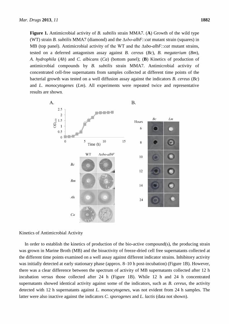
Mar. Drugs 2013, 11 1882
Figure 1. Antimicrobial activity of B. subtilis strain MMA7. (A) Growth of the wild type
(WT) strain B. subtilis MMA7 (diamond) and the ∆sbo-albF::cat mutant strain (squares) in
MB (top panel). Antimicrobial activity of the WT and the ∆sbo-albF::cat mutant strains,
tested on a deferred antagonism assay against B. cereus (Bc), B. megaterium (Bm),
A. hydrophila (Ah) and C. albicans (Ca) (bottom panel); (B) Kinetics of production of
antimicrobial compounds by B. subtilis strain MMA7. Antimicrobial activity of
concentrated cell-free supernatants from samples collected at different time points of the
bacterial growth was tested on a well diffusion assay against the indicators B. cereus (Bc)
and L. monocytogenes (Lm). All experiments were repeated twice and representative
results are shown.
Kinetics of Antimicrobial Activity
In order to establish the kinetics of production of the bio-active compound(s), the producing strain
was grown in Marine Broth (MB) and the bioactivity of freeze-dried cell free supernatants collected at
the different time points examined on a well assay against different indicator strains. Inhibitory activity
was initially detected at early stationary phase (approx. 8–10 h post-incubation) (Figure 1B). However,
there was a clear difference between the spectrum of activity of MB supernatants collected after 12 h
incubation versus those collected after 24 h (Figure 1B). While 12 h and 24 h concentrated
supernatants showed identical activity against some of the indicators, such as B. cereus, the activity
detected with 12 h supernatants against L. monocytogenes, was not evident from 24 h samples. The
latter were also inactive against the indicators C. sporogenes and L. lactis (data not shown).
Mar. Drugs 2013, 11 1883
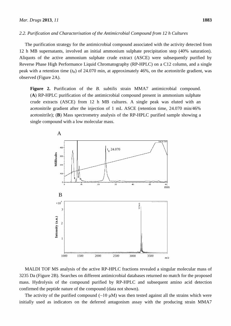
2.2. Purification and Characterisation of the Antimicrobial Compound from 12 h Cultures
The purification strategy for the antimicrobial compound associated with the activity detected from
12 h MB supernatants, involved an initial ammonium sulphate precipitation step (40% saturation).
Aliquots of the active ammonium sulphate crude extract (ASCE) were subsequently purified by
Reverse Phase High Performance Liquid Chromatography (RP-HPLC) on a C12 column, and a single
peak with a retention time (tR) of 24.070 min, at approximately 46%, on the acetonitrile gradient, was
observed (Figure 2A).
Figure 2. Purification of the B. subtilis strain MMA7 antimicrobial compound.
(A) RP-HPLC purification of the antimicrobial compound present in ammonium sulphate
crude extracts (ASCE) from 12 h MB cultures. A single peak was eluted with an
acetonitrile gradient after the injection of 1 mL ASCE (retention time, 24.070 min/46%
acetonitrile); (B) Mass spectrometry analysis of the RP-HPLC purified sample showing a
single compound with a low molecular mass.
MALDI TOF MS analysis of the active RP-HPLC fractions revealed a singular molecular mass of
3235 Da (Figure 2B). Searches on different antimicrobial databases returned no match for the proposed
mass. Hydrolysis of the compound purified by RP-HPLC and subsequent amino acid detection
confirmed the peptide nature of the compound (data not shown).
The activity of the purified compound (~10 μM) was then tested against all the strains which were
initially used as indicators on the deferred antagonism assay with the producing strain MMA7
×104
1
2
1000 1500 2000 2500 3000 3500 m/z
B
3
Inte
nsi
ty (
a.u
.)
3234.6
min
tR 24.070
Mil
livo
lts
A
Mar. Drugs 2013, 11 1884
(Table 1). While inhibitory activity was readily detected against most of the Gram-positive indicators,
the strong activity seen on the deferred antagonism assay against V. anguillarum, A. hydrophila and
Alteromonas sp., although clear, was only detected with 100 fold more compound than that required to
inhibit the Gram-positive indicators.
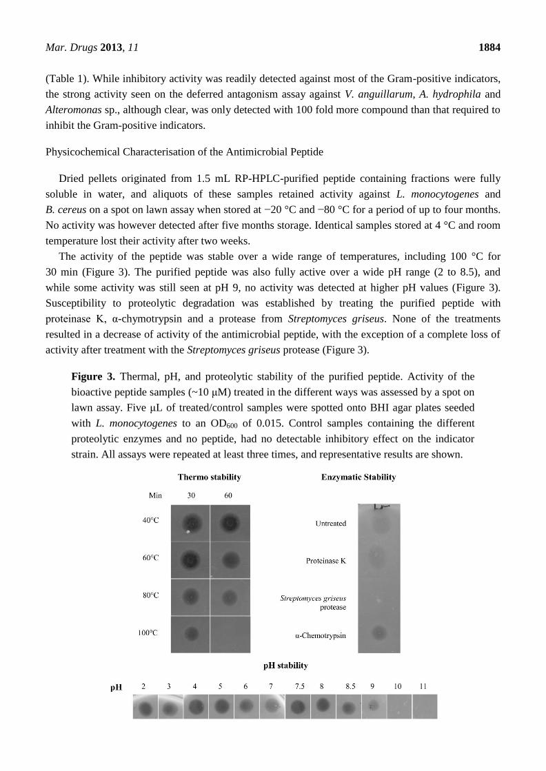
Physicochemical Characterisation of the Antimicrobial Peptide
Dried pellets originated from 1.5 mL RP-HPLC-purified peptide containing fractions were fully
soluble in water, and aliquots of these samples retained activity against L. monocytogenes and
B. cereus on a spot on lawn assay when stored at −20 °C and −80 °C for a period of up to four months.
No activity was however detected after five months storage. Identical samples stored at 4 °C and room
temperature lost their activity after two weeks.
The activity of the peptide was stable over a wide range of temperatures, including 100 °C for
30 min (Figure 3). The purified peptide was also fully active over a wide pH range (2 to 8.5), and
while some activity was still seen at pH 9, no activity was detected at higher pH values (Figure 3).
Susceptibility to proteolytic degradation was established by treating the purified peptide with
proteinase K, α-chymotrypsin and a protease from Streptomyces griseus. None of the treatments
resulted in a decrease of activity of the antimicrobial peptide, with the exception of a complete loss of
activity after treatment with the Streptomyces griseus protease (Figure 3).
Figure 3. Thermal, pH, and proteolytic stability of the purified peptide. Activity of the
bioactive peptide samples (~10 μM) treated in the different ways was assessed by a spot on
lawn assay. Five μL of treated/control samples were spotted onto BHI agar plates seeded
with L. monocytogenes to an OD600 of 0.015. Control samples containing the different
proteolytic enzymes and no peptide, had no detectable inhibitory effect on the indicator
strain. All assays were repeated at least three times, and representative results are shown.

Mar. Drugs 2013, 11 1885
2.3. Genetic and Structural Characterisation of the Antimicrobial Peptide
2.3.1. Primary Amino Acid Sequence of the Antimicrobial Peptide
MS and MS/MS analysis showed that the intact mass of the peptide has a number of oxidised forms
(Figure S1), typical of the presence of thioethers, indicating that the compound was potentially
a lantibiotic.
Initial structural analysis of the peptide proved problematic given that Edman degradation was not
possible as the N-terminal end of the peptide was blocked. Trypsin digestion and alkylation of the
peptide followed by MALDI MS and MS/MS analysis however provided a tentative primary amino
acid sequence of the N-terminal part of the peptide; and established that the peptide contained at least
six or seven unusual amino acids which are common in lantibiotic peptides, such as Dhb, Dha
and lanthionine.
The presumptive amino acid sequence was then used in subsequent BLASTp and TBLASTn
searches against the draft genome sequence of B. subtilis MMA7. Following theses searches,
homology was detected with a small protein-coding gene. This gene is located in the vicinity of
protein-coding genes showing similarities to genes encoding lantibiotic modification enzymes. This
allowed for the aforementioned experiments to be repeated, refined and the N-terminal sequence of the
peptide to be confirmed as T-W-A-T-I-G-K-T-I-V-Q-S-V-K-K. We have here named this novel
lantibiotic as subtilomycin.
2.3.2. Genetic Organisation and Location of the Subtilomycin Biosynthetic Gene Cluster
As previously noted, a single match was found between the primary amino acid sequence of the
N-terminal part of subtilomycin and the 5’end of a small gene (subA, 171 nt), located immediately
upstream from ORFs that encode for proteins which have been shown to be involved in the processing
of class I lantibiotics, such as LanB-like dehydratase and LanC-like cyclase modification enzymes
(SubB and SubC, respectively, Figure 4). These three genes are clustered with two other ORFs that
encode a putative extracellular serine protease (subP) and an ABC transporter (subT), respectively.
These are likely to be involved in the processing and transport of the lantibiotic. The final ORF within
the cluster (subI) has no homology to any other sequence in the database and could encode an
immunity protein (Table S3). The ORFs flanking these sequences show high homology (greater than
95%) with the genome of B. subtilis 168, including a putative two-component response regulator,
ybdJ, and sensor histidine kinase, ybdK, genes located downstream of the cluster (Figure 4). In silico
analysis of the biosynthetic cluster identified two putative promoter sequences, located upstream of
subA and subT, respectively.
Further sequence analysis of the flanking regions of the putative subtilomycin biosynthetic gene
cluster allowed us to establish that the cluster is inserted in the same genomic locus as the sporulation
killing factor (skf) operon in B. subtilis subsp. subtilis strain 168 [30] (Figure 4). The nucleotide
sequence encompassing the putative subtilomycin biosynthetic cluster and neighbouring genes has
been deposited in GenBank under accession number JX912247.

Mar. Drugs 2013, 11 1886
Figure 4. Structural organisation of the putative subtilomycin biosynthetic cluster and
flanking regions: subA, subtilomycin structural gene; subP, serine protease; subB,
lanthionine dehydratase; subC, lanthionine synthetase; subT, ABC transporter. The
function of subI cannot be predicted from its sequence, although its genetic location and
lack of homologues is consistent with a possible involvement in immunity. Predicted
promoters are indicated by arrows. A comparison of the genomic location of the
subtilomycin biosynthetic cluster in strain MMA7 with that of the skf operon in strain 168
is provided.
2.3.3. Further Structural Characterisation of Subtilomycin
The presence of an N-terminal 2-oxo butyrate residue was established by MS/MS analysis of the
peptide. Such residues arise as a consequence of the fact that N-terminal Dhb residues are not stable
and undergo spontaneous oxidative deamination [31]. The deaminated Dhb (Dhb*) weighs 85 Da
instead of 83 Da, resulting in a mass gain of 2 Da.
The mass difference from the alanine at position 3 to the N-terminal of subtilomycin equals
271.3 Da, which corresponds to the sum of 186.07 Da for the tryptophan residue with 85.03 Da for the
deaminated Dhb. The C-terminal of the peptide was not suitable for de novo sequencing, but data from
the analysis of tryptic digests suggested that this region of the peptide could possibly contain up to
five ring structures, three Lan and two MeLan, since five cysteines, three Dha and two Dhb are present
in this section of the peptide.
Therefore the most likely sequence for the lantibiotic is Dhb*-W-A-Dhb-I-G-K-Dhb-I-V-Q-Dha-V-
K-K-C-R-Dhb-F-Dhb-C-G-C-Dha-L-G-Dha-C-Dha-N-C-N, where the underlined amino acids have
been experimentally confirmed by mass spectrometric analysis and the subsequent sequence predicted
from the genomic sequence of strain MMA7. There was no mass change following DTT reductions
suggesting that no disulphide bonds were present in the peptide. In summary, the 32 amino acid long
lantibiotic contains nine modified aminoacids, five of which are likely to be involved in the formation

Mar. Drugs 2013, 11 1887
of thioether bridges. The theoretical mass of the proposed sequence is 3234.5, while the observed mass
was 3234.6 (m/z = 3235.6).
Search of the bactibase and the antimicrobial peptide databases highlighted some similarities with
previously identified lantibiotics. The closest homology was with paenibacillin produced by
Paenibacillus polymyxa OSY-DF (54%) [32,33], followed by epilancin 15X produced by
Staphylococcus epidermidis 15X154 (43%) (Figure 5A,B). Although the structure of purified
paenibacillin has been elucidated, the genetics of the associated biosynthetic cluster have not been
published. As a result comparative analysis of the paenibacillin and subtilomycin pre-peptides
(Figure 5B) and respective gene clusters was not possible.
Figure 5. Comparison of the amino acid sequence between subtilomycin and nisin
pro-petides and their respective closest homologue (A). Comparison of the amino acid
sequence of the N-terminal leader peptide and the C-terminal core peptide of subtilomycin
and different class I lantibiotics (B). Sequences obtained from Uniprot were aligned with
ClustalX (Multiple Sequence Alignment version 2.0.11, [34]) and sequence analysis
processed with GeneDoc (Multiple sequence Alignment Editor & Shading Utility, Version
2.7.000). Conserved amino acids are boxed in black and gaps are indicated by hyphen. A
vertical arrow indicates the first amino acid of the pro-peptide *, indicates the conserved
motif of class I leader peptides documented to be important for efficient production.
Proposed conformational structure of the mature lantibiotic subtilomycin (C). Top,
unmodified propeptide. Bottom, mature peptide, where Ser and Thr residues which are
posttranslationally dehydrated to Dha and Dhb, or involved in the formation of Lan and
MeLan, respectively, with cysteine residues, are shaded in grey. The location of the
thioether bridges was estimated from the amino acid sequence and by comparison with that
of paenibacillin [32]. The presence of the N-terminal 2-oxobutyrate residue is also indicated.
Mar. Drugs 2013, 11 1888
Based on the primary amino acid sequence of subtilomycin, we have here proposed a structure for
subtilomycin (Figure 5C), which reflects that experimentally established for paenibacillin [32].
However further analysis will be required to definitively establish the proposed structure.
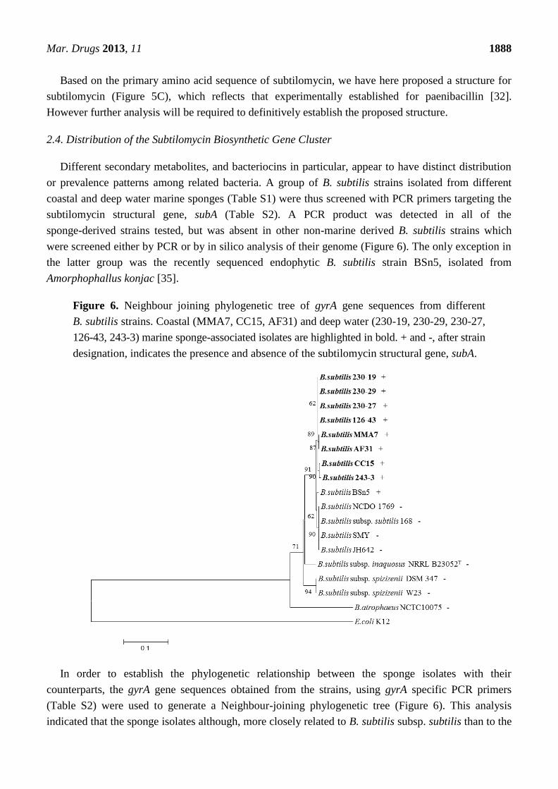
2.4. Distribution of the Subtilomycin Biosynthetic Gene Cluster
Different secondary metabolites, and bacteriocins in particular, appear to have distinct distribution
or prevalence patterns among related bacteria. A group of B. subtilis strains isolated from different
coastal and deep water marine sponges (Table S1) were thus screened with PCR primers targeting the
subtilomycin structural gene, subA (Table S2). A PCR product was detected in all of the
sponge-derived strains tested, but was absent in other non-marine derived B. subtilis strains which
were screened either by PCR or by in silico analysis of their genome (Figure 6). The only exception in
the latter group was the recently sequenced endophytic B. subtilis strain BSn5, isolated from
Amorphophallus konjac [35].
Figure 6. Neighbour joining phylogenetic tree of gyrA gene sequences from different
B. subtilis strains. Coastal (MMA7, CC15, AF31) and deep water (230-19, 230-29, 230-27,
126-43, 243-3) marine sponge-associated isolates are highlighted in bold. + and -, after strain
designation, indicates the presence and absence of the subtilomycin structural gene, subA.
In order to establish the phylogenetic relationship between the sponge isolates with their
counterparts, the gyrA gene sequences obtained from the strains, using gyrA specific PCR primers
(Table S2) were used to generate a Neighbour-joining phylogenetic tree (Figure 6). This analysis
indicated that the sponge isolates although, more closely related to B. subtilis subsp. subtilis than to the

Mar. Drugs 2013, 11 1889
other subspecies, appear to form subgroups which cluster together, and are more closely related to each
other than to other, non-sponge, isolates.
3. Discussion
During our search for new antimicrobial compounds produced by marine sponge associated
bacteria, B. subtilis strain MMA7 isolated from H. simulans demonstrated broad range antimicrobial
activity towards important clinical and aquaculture pathogens. Interestingly, activity against a number
of indicator strains, such as L. monocytogenes and C. sporogenes, was detected from 12 to 14 h
cultures but not from 24 h cultures. This activity was here shown to be associated with a novel
lantibiotic, subtilomycin. Lantibiotics are typically produced by specific strains and in general target
Gram-positive organism, although examples are emerging of those that have an extended spectrum of
activity that includes Gram-negative bacteria [18,36]. Subtilomycin shows antimicrobial activity
against a broad spectrum of Gram-positive and some Gram-negative bacteria, although the
susceptibility of the latter group appears to be significantly less than the former. It also possesses
several physicochemical properties which make it interesting for use in the food and/or pharmaceutical
industry, including solubility in water and stability under different conditions such as physiologic pH.
Based on MS, MS/MS and genome sequence analysis the subtilomycin precursor undergoes
extensive posttranslational modification. Three Dhb residues are present at positions T1, T4 and T8.
However, similar to Pep5 and Lacticin 3147A2, the N-terminal Dhb is modified to 2-oxo
butyrate [31,37]. The post translational modification of the N-terminal residues in lantibiotics is
believed to provide protection from aminopeptidases [18]. The remaining threonine residues, T18 and
T20, are involved in the formation of β-methyllanthionines. Additionally, one Dha residue, and three
lanthionines result from serine residues at positions 12, 24, 27 and 29, respectively. The closest
homologue to subtilomycin is paenibacillin, a 30 amino acid long lantibiotic (m/z = 2984.61) isolated
from the sporeformer P. polymyxa OSY-DF [32]. The two molecules seem to be particularly similar at
the C-terminal region with a similar ring pattern structure, a feature which is also seen between nisin
and its closest relative subtilin [38]. This could be associated with the similarities found within the
physicochemical properties described for both subtilomycin and paenibacillin [32,33]. However, in
contrast paenibacillin has an N-terminal acetylation rather than 2-oxo butyrate. Also, although
subtilomycin could be detected from 12 h MB cultures but not from 24 h fermentates, paenibacillin
could be equally detected from both time points [32], which may indicate differences in the regulation
of production.
The proposed subtilomycin biosynthetic gene cluster appears to have several of the sequence
features associated with class I lantibiotics [39], including the presence of LanP, LanB and LanC-like
encoding sequences immediately downstream from the structural gene subA. The conserved four
amino acid residue box (FNLD) of class I leader peptides known to be important for efficient
production [18] is also present in the leader peptide of subtilomycin supporting its grouping with class
I lantibiotics. Contrary to the other aforementioned elements involved in the synthesis of lantibiotics,
there is very little homology between the peptides involved in providing immunity to the different
lantibiotics [19]. With this in mind, we propose that SubI, which has no homologues on the database,
could be fulfilling this role. If this is confirmed, similarly to Pep5 and lactocin S, host resistance to

Mar. Drugs 2013, 11 1890
subtilomycin, would be provided by a single dedicated immunity protein, dispensing the additional
ABC transport proteins. These questions will be the focus of further investigations.
The subtilomycin biosynthetic cluster in B. subtilis strain MMA7 is found in place of the skf
operon, which is present in different B. subtilis isolates, such as strain 168. This encodes for
production, export and immunity towards the sporulation killing factor (SkfA). This peptide mediates a
well characterised bacterial cannibalistic behaviour intended to delay sporulation, by which producing
sporulating sibling cells feed on non-producing non-sporulating cells by mediating their lysis [30]. It
will be interesting to establish if the lack of the skf operon in strain MMA7 results in any change in the
temporal entry to sporulation. Of note, the rhizocticin biosynthetic gene cluster (rhi) of B. subtilis
ATCC6633 was also inserted nearby, and in place of, the skf operon [40]. This may indicate that in the
B. subtilis genome this may be a hotspot for the location of genetic elements associated with the
production of secondary metabolites. It will be interesting to survey different natural B. subtilis isolates
at this site to see the prevalence and identity of these elements. Importantly, the GC content of the
proposed subtilomycin biosynthetic gene cluster (29.5%) is considerably different to the overall GC
content of the genome of strain MMA7 (44%), suggesting a potential exogenous acquisition of
this cluster.
The structural gene coding for subtilomycin has been found in several other B. subtilis strains
isolated from different shallow and deep sea marine sponges, but appears to be absent from many
B. subtilis isolates from other environments, with the exception of strain BSn5, an endophytic
B. subtilis from Amorphophallus konjac [35]. Interestingly, a closer phylogenetic relationship based on
analysis of gyrA sequences, exists between the marine isolates, than with the other strains. This closer
phylogenetic relationship occurs despite clear morphological and phenotypic differences between the
marine sponge isolates (data not shown). Apart from its industrial and pharmaceutical potential the
biological role of subtilomycin remains unknown, although its presence in all the marine sponge
B. subtilis isolates may indicate a potential niche-related role associated with providing the strain with
some competitive advantage within the stringent environment found within marine sponges.
Nevertheless, its presence in the endophytic BSn5 isolate may indicate a more complex role in the
symbiotic relationship with the host.
4. Experimental Section
4.1. Bacterial Strains, Media and Growth Conditions
Bacterial strains used in this study are listed in Table S1. B. subtilis strain MMA7, was isolated
from the marine sponge H. simulans collected during scuba diving in Gurraig Sound Kilkieran Bay,
Galway, on the west coast of Ireland [9]. Strains AF31 and CC15 were isolated from the shallow water
sponges Amphilectus fucorum and Cliona celata, respectively, both collected from the Lough Hyne
Marine Reserve, Ireland, by Scuba diving. Strains BD230-19, BD230-27, BD230-29, BD243-3 and
BD126-43 were isolated from the deep-water sponges BD230, BD126 and BD243, collected from the
Rockall Ridge and Porcupine Bank, 300 nautical miles North West of Galway harbour, at a depth of
2900, 2129 and 1300 m, respectively. Unless stated otherwise, marine sponge B. subtilis isolates were
routinely grown and maintained aerobically on Difco™ Marine agar (MA) and broth (MB)
Mar. Drugs 2013, 11 1891
(Difco 2216), at 30 °C. Other strains used in this study, including indicator strains, were grown in the
media specified in Table S1, aerobically at 37 °C, with the exception of Aeromonas hydrophila,
Vibrio anguillarum, Carnobacterium malteromaticum and all the Candida strains that were incubated
at 30 °C. Clostridium perfringens and Clostridium sporogenes were grown anaerobically in an
anaerobic jar at 37 °C. E. coli DH5α used for cloning experiments was grown in Luria-Bertani (LB).
For B. subtilis strains chloramphenicol was used at 5 μg·mL−1
.
4.2. Antimicrobial Activity Screening Assays
Antimicrobial activity of producing strains against the indicator strains was initially assessed with a
deferred antagonism assay as described by Phelan et al. [9]. Subsequent testing of freeze dried crude
extracts and purified compounds was carried out with a well and/or a spot on lawn assays. Essentially,
5 μL of samples were spotted onto 20 mL agar plates seeded with the indicator strain to a final OD600
of 0.015, or 30 μL aliquots were applied to a 5 mm well made on identical plates. Controls of freeze
dried MB or solvent were tested in parallel.
4.3. DNA Extraction, PCR Amplification, DNA Sequencing and Phylogenetic Analysis
Genomic DNA was extracted from 5 mL overnight MB cultures by a modification of the guanidine
thiocyanate method described by Pitcher et al. [41]. Plasmid DNA was purified from 5 mL overnight
E. coli LB cultures with the QIAprep spin Miniprep Kit (Qiagen, GmbH, Hilden, Germany).
All oligonucleotide primers used in this study are listed in Table S2. Unless stated otherwise PCR
reactions (50 μL) contained 1× BioTaq PCR buffer (Bioline, London, UK), 1.5 mM MgCl2, 0.2 mM
dNTPs, 0.5 μM of each primer, 2.5 U BioTaq DNA polymerase (Bioline), and 3 μL bacterial lysates or
50 ng of genomic DNA as template. With the exception of the annealing temperature (Table S2), and
the extension time, the cycling parameters for the amplification of the different target sequences
remained constant. An initial denaturation at 94 °C for 4 min, was followed by 30 cycles of 94 °C for
30 s, X °C for 30 s, and 72 °C for X min, with a final extension of 10 min at 72 °C. When required,
PCR products were purified using the QIAquick PCR purification kit (Qiagen GmbH, Hilden,
Germany). Routine DNA sequencing was performed by GATC-Biotech, AG (Konstanz, Germany).
Partial gyrA gene sequences of the marine sponge B. subtilis isolates were deposited in GenBank
with the following accession numbers: CC15, JX977124; AF31, JX977125; 243-3, JX977126; 230-19,
JX977127; 230-29, JX977128; 126-43, JX977129; 230-27, JX977131; MMA7, JX977130. For
phylogenetic analysis, 620 nt of the gyrA genes (nt 232 to 852 in the E. coli gyrA gene, accession
number AP012306.1) were aligned using Clustal X2 [34]. Neighbour-joining phylogenetic trees were
constructed using MEGA 4 [42] and bootstrap tests were performed once with 1000 replicates.
4.4. Construction of Strain MMA7 ∆Sbo-albF::Cat Mutant
Methods for the preparation and transformation of E. coli DH5α and B. subtilis competent cells
were as described previously [43,44]. To create a sbo-albF insertion-deletion mutant, DNA fragments
containing 284 nt of the upstream and coding region of the structural gene, sbo (nt 136 to 420,
accession number AJ430547) and 284 nt internal nucleotides of the albF gene (nt 5684 to 5968,

Mar. Drugs 2013, 11 1892
accession number AJ430547), were PCR amplified from the genomic DNA of strain MMA7 with the
primer pair SboUp-F/Sbo-R and AlbF-F/AlbF-R, respectively (Table S2). Digested amplified
fragments were sequentially inserted between the HindIII-BamHI sites and EcoRI-XhoI sites flanking
the chloramphenicol resistance cassette in the pMK3 derivative integrational vector, pMS38 [45]. The
resulting construct was named pRP1. The mutant strain, MMA7Δsbo-albF::cat, ChlR, in which the
sbo-albF region (nucleotides 420 to 5684, accession number AJ430547) was replaced by a
chloramphenicol cassette by a double-crossover event, was generated by the transformation of strain
MMA7 with ScaI linearised pRP1, with subsequent selection for chloramphenicol resistance (ChlR).
Disruption of the cluster in the mutants was confirmed by PCR with a combination of primers specific
for the chloramphenicol cassette, and primers flanking or internal to, the deleted region. Specifically,
primer pairs ywiB-F & albA-R2, ywiB-F & cat255-R, albD-F & alb-FDown-R, cat958-D &
albFDown-R (Table S2).
4.5. Kinetics of Antimicrobial Production
MB (100 mL) were inoculated with overnight MB cultures of the producing strain (1:100) and
grown at 30 °C, 150 rpm shaking on an orbital incubator. The optical density (600 nm) was read
hourly until 14 h incubation and then at 24 h incubation and 5 mL aliquots removed at each time point
and centrifuged at 3200 rpm, 15 min, 20 °C. Supernatants were filter sterilised (33 mm Cellulose
Acetate, 0.45 μm syringe filters, Anachem Ltd., Luton, UK), and freeze dried (Labconco Freezone6,
Labconco Kansas City, MO, USA) for 16 h. Freeze dried material was resuspended in 500 μL of
sterile HPLC grade water and tested for activity in a well diffusion assay against different
indicator strains.
4.6. Purification and Characterisation of the Antimicrobial Compound
Approximately 1 L of B. subtilis MMA7 12 h MB cultures were centrifuged at 3200 rpm, for
15 min at 20 °C and the supernatant filter sterilized using a Nalgene® vacuum filtration system
(0.45 μm, Sigma-Aldrich®, Munich, Germany). Ammonium sulphate was added intermittently to the
cell free supernatant to 40% saturation, and gently stirred overnight at 4 °C. The resulting precipitate
was collected by centrifugation of 100 mL aliquots at 12,500 rpm, for 90 min, at 4 °C and resuspended
in total volume of 2 mL of sterile HPLC grade water (Sigma-Aldrich, CHROMASOLV®
Plus,
Munich, Germany) (1/500 of original volume). Subsequent purification by RP-HPLC was achieved by
applying 1 mL of the ammonium sulphate crude extract onto a Phenomenex® C12 column (Jupiter 4 μ
proteo 90 Å, 250 × 10.0, 4 μm). Separation was carried out with a gradient of acetonitrile (33% to
65%) containing 0.1% TFA from 5 to 50 min, at a flow rate of 2.1 mL·min−1
and separation monitored
at 214 nm. Fractions (1.5 mL) were evaporated to dryness on a speedvac (Savant DNA 120, Speedvac,
Thermo Scientific, London, UK), and the resulting pellets resuspended in sterile HPLC grade water.
The aforementioned purification process routinely yielded 0.4–0.5 mg of compound per litre of a
12 h culture.
The purity and molecular weight of the compound in the active fraction was initially estimated on
an AXIMA-TOF2 MALDI Mass spectrometer (Shimadzu Biotech, Manchester, UK). Further analysis
of the nature and structure of the compound was carried out by Alphalyse A/S (Odense, Denmark).

Mar. Drugs 2013, 11 1893
This involved determination of the amino acid composition, MALDI MS and MALDI MS/MS
analysis. 1.77 μg of RP-HPLC purified sample resuspended in 0.1% TFA was used for amino acid
analysis. The acid hydrolysis was performed for 20 h at 110 °C, in 6 N HCl, 0.1% phenol,
0.1% thioglycolic acid. The hydrolysis took place under reduced pressure in an atmosphere of argon.
Identification and quantification of the amino acids took place on a BioChrom 30 amino acid analyzer
using ion exchange chromatography, post-column derivatization with ninhydrin and detection at two
wavelengths, 570 nm and 440 nm. A known amount of the non-natural amino acid norleucine (Nle)
was added as an internal control standard.
For MALDI MS and MS/MS of intact peptide, approx. 5 pmol peptide was spotted onto a steel
target with alpha-cyano-4-hydroxycinnamic acid (CHCA) matrix and was analyzed on a Bruker
Autoflex Speed instrument. For MALDI MS and MS/MS analysis of trypsin digests, the peptide was
digested with trypsin at 37 °C overnight and the resulting peptides were concentrated on a C18 ziptip
from Millipore and eluted with CHCA matrix in 50% acetonitrile/0.1% TFA. Subsequent analysis
involved alkylation of the peptide with β-mercaptoethanol in a basic solution. Alkylation increased the
peptide mass by 78 Da for each alkylated amino acid. Samples were taken out at different times to
follow the alkylation process. When samples were removed they were acidified with TFA and purified
on a C18 ziptip from Millipore before being eluted with CHCA matrix in 50% acetonitrile/0.1% TFA
and analysed by MALDI MS and MS/MS.
Reduction of the peptide with DTT was used to ascertain the potential presence of disulfide bonds,
as the peptide mass would be increased by 2 Da when a disulfide bond is broken due to the addition of
two hydrogen atoms.
4.7. Heat, pH and Proteolytic Treatment of the Antimicrobial Peptide
To establish the thermal stability of the purified peptide, aliquots (~10 μM) were exposed to 40, 60,
80 and 100 °C for 30 and 60 min respectively before being tested for bioactivity on a spot on lawn
assay. Untreated samples were used as controls. In a similar way, samples were checked for activity
following storage for different time periods at room temperature, 4 °C, −20 °C and −80 °C.
The activity of the purified peptide at different pH values was established by resuspending the
peptide in 50 μL of 100 mM Tris buffer (~10 μM), with pH values between 2 and 11. Samples were
incubated for 1 h at room temperature before being tested for antimicrobial activity against
L. monocytogenes on spot on lawn assays. The different buffers were also individually tested as
negative controls.
Susceptibility to proteolytic enzymes was tested by incubating the purified peptide resuspended in
water (~10 μM) with proteinase K, α-chymotrypsin and a protease mixture from Streptomyces griseus
(Sigma Aldrich® 81748), to a final concentration of 1 mg·mL
−1, at 37 °C for 1 h. Samples were then
heated to 100 °C for 2 min to inactivate the proteolytic enzymes and bioactivity was tested with a spot
on lawn assay.

Mar. Drugs 2013, 11 1894
4.8. Genome Sequencing of B. subtilis MMA7 and Sequence Analysis of the Subtilomycin Biosynthetic
Gene Cluster
The genome of isolate MMA7 was sequenced by Genospec, Inc., (Houston, TX, USA) using
Illumina Sequencing technology on a Illumina High Seq 2000 (Illumina Inc., California, CA, USA).
The sequencing data, which provided 100X coverage of the genome, was assembled into 48 contigs,
spanning approximately 4.2 Mb. A total of 4277 open reading frames (ORFs) were determined and
annotated using the RAST Server (Rapid Annotation using Subsystem Technology) and the Basic
Local Alignment Search Tool (BLAST) programme at NCBI [46]. Further details on the analysis of
the genomic sequence of strain MMA7 will be published elsewhere.
BPROM and the Berkeley Drosophila Genome Project (BDGP) [47,48], promoter prediction
algorithms were used to identify putative promoters within the subtilomycin gene cluster. Only
promoters identified by both algorithms were given further consideration.
For comparison of the amino acid sequence of lantibiotic precursors sequences obtained from
Uniprot, were aligned with ClustalX (Multiple Sequence Alignment version 2.0.11, [34]) and sequence
analysis processed with GeneDoc (Multiple sequence Alignment Editor & Shading Utility,
Version 2.7.000).
5. Conclusions
This study describes the identification of a novel class I lantibiotic, subtilomycin, produced by a
marine sponge B. subtilis isolate. Lantibiotics are an important class of antimicrobial peptides with
potential commercial application. Of relevance, subtilomycin possesses several physicochemical
properties which make it interesting for use in the food and/or pharmaceutical industry.
Despite the abundance and broad range of mechanism of antimicrobial compounds produced by the
marine sponge microbiota, to the best of our knowledge, subtilomycin is the first lantibiotic to be
purified and identified from a marine sponge associated bacteria. This emphasises the importance of
pursuing the search for novel antimicrobial compounds within this still untapped environment. It
additionally reinforces the importance of the Bacillus genus as a prolific resource of new
bioactive compounds.
Acknowledgments
This research was supported in parts by grants awarded to FOG by the Science Foundation of
Ireland (08/RFP/GEN1295, 08/RFP/GEN1319, SFI09/RFP/BMT2350); the Department of
Agriculture, Fisheries and Food (RSF grants 06-321 and 06-377; FIRM grants 06RDC459 06RDC506
and 08RDC629); the European Commission (MicroB3-287589-OCEAN2012, MACUMBA-CP-TP
311975; PharmaSea-CP-TP 312184), the Marine Institute Beaufort award (C & CRA 2007/082) and
the HRB (RP/2006/271, RP/2007/290, HRA/2009/146). We thank Lorraine Draper for assistance with
the RP-HPLC.
Conflict of Interest
The authors declare no conflict of interest.

Mar. Drugs 2013, 11 1895
References
1. Pelaez, F. The historical delivery of antibiotics from microbial natural products-can history
repeat? Biochem. Pharmacol. 2006, 71, 981–990.
2. Taylor, M.W.; Radax, R.; Steger, D.; Wagner, M. Sponge-associated microorganisms: evolution,
ecology, and biotechnological potential. Microbiol. Mol. Biol. Rev. 2007, 71, 295–347.
3. Kennedy, J.; Baker, P.; Piper, C.; Cotter, P.D.; Walsh, M.; Mooij, M.J.; Bourke, M.B.;
Rea, M.C.; O’Connor, P.M.; Ross, R.P. Isolation and analysis of bacteria with antimicrobial
activities from the marine sponge Haliclona simulans collected from Irish waters. Mar.
Biotechnol. (NY) 2009, 11, 384–396.
4. Thomas, T.R.; Kavlekar, D.P.; LokaBharathi, P.A. Marine drugs from sponge-microbe
association—A review. Mar. Drugs 2010, 8, 1417–1468.
5. Muscholl-Silberhorn, A.; Thiel, V.; Imhoff, J.F. Abundance and bioactivity of cultured
sponge-associated bacteria from the Mediterranean sea. Microb. Ecol. 2008, 55, 94–106.
6. Zhu, P.; Li, Q.; Wang, G. Unique microbial signatures of the alien Hawaiian marine sponge
Suberites zeteki. Microb. Ecol. 2008, 55, 406–414.
7. Lee, O.O.; Wong, Y.H.; Qian, P.Y. Inter- and intraspecific variations of bacterial communities
associated with marine sponges from san juan island, washington. Appl. Environ. Microbiol.
2009, 75, 3513–3521.
8. Menezes, C.B.; Bonugli-Santos, R.C.; Miqueletto, P.B.; Passarini, M.R.; Silva, C.H.;
Justo, M.R.; Leal, R.R.; Fantinatti-Garboggini, F.; Oliveira, V.M.; Berlinck, R.G.; Sette, L.D.
Microbial diversity associated with algae, ascidians and sponges from the north coast of Sao
Paulo state, Brazil. Microbiol. Res. 2009, 165, 466–482.
9. Phelan, R.W.; O’Halloran, J.A.; Kennedy, J.; Morrissey, J.P.; Dobson, A.D.; O’Gara, F.;
Barbosa, T.M. Diversity and bioactive potential of endospore-forming bacteria cultured from the
marine sponge Haliclona simulans. J. Appl. Microbiol. 2012, 112, 65–78.
10. Stein, T. Bacillus subtilis antibiotics: Structures, syntheses and specific functions. Mol. Microbiol.
2005, 56, 845–857.
11. Westers, L.; Westers, H.; Quax, W.J. Bacillus subtilis as cell factory for pharmaceutical proteins:
A biotechnological approach to optimize the host organism. Biochim. Biophys. Acta 2004, 1694,
299–310.
12. Alcaraz, L.D.; Moreno-Hagelsieb, G.; Eguiarte, L.E.; Souza, V.; Herrera-Estrella, L.;
Olmedo-Alvarez, G. Understanding the evolutionary relationships and major traits of
Bacillus through comparative genomics. BMC Genomics 2010, 11, 332.
13. Barbosa, T.M.; Serra, C.R.; Henriques, A.O. Gut Sporeformers. In Bacterial Spore Formers:
Probiotics and Emerging Applications; Ricca, E., Henriques, A.O., Cutting, S.M., Eds.; Horizon
Bioscience: Norfolk, UK, 2004; pp. 183–191.
14. Barbosa, T.M.; Serra, C.R.; La Ragione, R.M.; Woodward, M.J.; Henriques, A.O. Screening for
Bacillus isolates in the broiler gastrointestinal tract. Appl. Environ. Microbiol. 2005, 71, 968–978.
15. Tam, N.K.; Uyen, N.Q.; Hong, H.A.; Le Duc, H.; Hoa, T.T.; Serra, C.R.; Henriques, A.O.;
Cutting, S.M. The intestinal life cycle of Bacillus subtilis and close relatives. J. Bacteriol. 2006,
188, 2692–2700.
Mar. Drugs 2013, 11 1896
16. Hong, H.A.; Khaneja, R.; Tam, N.M.; Cazzato, A.; Tan, S.; Urdaci, M.; Brisson, A.;
Gasbarrini, A.; Barnes, I.; Cutting, S.M. Bacillus subtilis isolated from the human gastrointestinal
tract. Res. Microbiol. 2009, 160, 134–143.
17. Hong, H.A.; To, E.; Fakhry, S.; Baccigalupi, L.; Ricca, E.; Cutting, S.M. Defining the natural
habitat of Bacillus spore-formers. Res. Microbiol. 2009, 160, 375–379.
18. Knerr, P.J.; van der Donk, W.A. Discovery, biosynthesis, and engineering of lantipeptides.
Annu. Rev. Biochem. 2012, 81, 479–505.
19. Willey, J.M.; van der Donk, W.A. Lantibiotics: Peptides of diverse structure and function.
Annu. Rev. Microbiol. 2007, 61, 477–501.
20. Cotter, P.D.; Hill, C.; Ross, R.P. Bacterial lantibiotics: Strategies to improve therapeutic potential.
Curr. Protein Pept. Sci. 2005, 6, 61–75.
21. Brotz, H.; Sahl, H.G. New insights into the mechanism of action of lantibiotics—Diverse
biological effects by binding to the same molecular target. J. Antimicrob. Chemother. 2000, 46,
1–6.
22. Suda, S.; Westerbeek, A.; O’Connor, P.M.; Ross, R.P.; Hill, C.; Cotter, P.D. Effect of
bioengineering lacticin 3147 lanthionine bridges on specific activity and resistance to heat and
proteases. Chem. Biol. 2010, 17, 1151–1160.
23. Kim, T.W.; Kim, Y.H.; Kim, S.E.; Lee, J.H.; Park, C.S.; Kim, H.Y. Identification and distribution
of Bacillus species in doenjang by whole-cell protein patterns and 16S rRNA gene sequence
analysis. J. Microbiol. Biotechnol. 2010, 20, 1210–1214.
24. Kim, T.W.; Lee, J.H.; Park, M.H.; Kim, H.Y. Analysis of bacterial and fungal communities in
Japanese- and Chinese-fermented soybean pastes using nested PCR-DGGE. Curr. Microbiol.
2010, 60, 315–320.
25. Hong, H.A.; Le Duc, H.; Cutting, S.M. The use of bacterial spore formers as probiotics. FEMS
Microbiol. Rev. 2005, 29, 813–835.
26. Lalloo, R.; Maharajh, D.; Gorgens, J.; Gardiner, N.; Gorgens, J.F. High-density spore production
of a B. cereus aquaculture biological agent by nutrient supplementation. Appl. Microbiol.
Biotechnol. 2009, 83, 59–66.
27. Anand, T.P.; Bhat, A.W.; Shouche, Y.S.; Roy, U.; Siddharth, J.; Sarma, S.P. Antimicrobial
activity of marine bacteria associated with sponges from the waters off the coast of South East
India. Microbiol. Res. 2006, 161, 252–262.
28. Hentschel, U.; Schmid, M.; Wagner, M.; Fieseler, L.; Gernert, C.; Hacker, J. Isolation and
phylogenetic analysis of bacteria with antimicrobial activities from the Mediterranean sponges
Aplysina aerophoba and Aplysina cavernicola. FEMS Microbiol. Ecol. 2001, 35, 305–312.
29. Pabel, C.T.; Vater, J.; Wilde, C.; Franke, P.; Hofemeister, J.; Adler, B.; Bringmann, G.;
Hacker, J.; Hentschel, U. Antimicrobial activities and matrix-assisted laser desorption/ionization
mass spectrometry of Bacillus isolates from the marine sponge Aplysina aerophoba.
Mar. Biotechnol. (NY) 2003, 5, 424–434.
30. Gonzalez-Pastor, J.E.; Hobbs, E.C.; Losick, R. Cannibalism by sporulating bacteria. Science
2003, 301, 510–513.
31. Sahl, H.G.; Jack, R.W.; Bierbaum, G. Biosynthesis and biological activities of lantibiotics with
unique post-translational modifications. Eur. J. Biochem. 1995, 230, 827–853.
Mar. Drugs 2013, 11 1897
32. He, Z.; Yuan, C.; Zhang, L.; Yousef, A.E. N-Terminal acetylation in paenibacillin, a novel
lantibiotic. FEBS Lett. 2008, 582, 2787–2792.
33. He, Z.; Kisla, D.; Zhang, L.; Yuan, C.; Green-Church, K.B.; Yousef, A.E. Isolation and
identification of a Paenibacillus polymyxa strain that coproduces a novel lantibiotic and
polymyxin. Appl. Environ. Microbiol. 2007, 73, 168–178.
34. Deng, Y.; Zhu, Y.; Wang, P.; Zhu, L.; Zheng, J.; Li, R.; Ruan, L.; Peng, D.; Sun, M. Complete
genome sequence of Bacillus subtilis BSn5, an endophytic bacterium of Amorphophallus konjac
with antimicrobial activity for the plant pathogen Erwinia carotovora subsp. carotovora.
J. Bacteriol. 2011, 193, 2070–2071.
35. Foulston, L.C.; Bibb, M.J. Microbisporicin gene cluster reveals unusual features of lantibiotic
biosynthesis in actinomycetes. Proc. Natl. Acad. Sci. USA 2010, 107, 13461–13466.
36. Xie, L.; van der Donk, W.A. Post-translational modifications during lantibiotic biosynthesis.
Curr. Opin. Chem. Biol. 2004, 8, 498–507.
37. Kleerebezem, M. Quorum sensing control of lantibiotic production; nisin and subtilin autoregulate
their own biosynthesis. Peptides 2004, 25, 1405–1414.
38. Field, D.; Hill, C.; Cotter, P.D.; Ross, R.P. The dawning of a 'Golden era' in lantibiotic
bioengineering. Mol. Microbiol. 2010, 78, 1077–1087.
39. Borisova, S.A.; Circello, B.T.; Zhang, J.K.; van der Donk, W.A.; Metcalf, W.W. Biosynthesis of
rhizocticins, antifungal phosphonate oligopeptides produced by Bacillus subtilis ATCC6633.
Chem. Biol. 2010, 17, 28–37.
40. Pitcher, D.G.; Saunders, N.A.; Owen, R.J. Rapid extraction of bacterial genomic DNA with
guanidium thiocyanate. Lett. Appl. Microbiol. 1989, 8, 151–156.
41. Larkin, M.A.; Blackshields, G.; Brown, N.P.; Chenna, R.; McGettigan, P.A.; McWilliam, H.;
Valentin, F.; Wallace, I.M.; Wilm, A.; Lopez, R. Clustal W and Clustal X version 2.0.
Bioinformatics 2007, 23, 2947–2948.
42. Tamura, K.; Dudley, J.; Nei, M.; Kumar, S. MEGA4: Molecular evolutionary genetics analysis
(MEGA) software version 4.0. Mol. Biol. Evol. 2007, 24, 1596–1599.
43. Sambrook, J.; Fritsch, E.F.; Maniatis, T. Molecular Cloning: A Laboratory Manual; Cold Spring
Harbor Laboratory Press: Woodbury, NY, USA, 1989.
44. Cutting, S.M.; Vander Horn, P.B. Genetic analysis. In Molecular Biology Methods for Bacillus;
Harwood, C.R., Cutting, S.M., Eds.; John Wiley and Sons: Chichester, UK, 1990; pp. 53–54.
45. Zilhao, R.; Serrano, M.; Isticato, R.; Ricca, E.; Moran, C.P.; Henriques, A.O. Interactions among
CotB, CotG, and CotH during assembly of the Bacillus subtilis spore coat. J. Bacteriol. 2004,
186, 1110–1119.
46. Altschul, S.F.; Gish, W.; Miller, W.; Myers, E.W.; Lipman, D.J. Basic local alignment search
tool. J. Mol. Biol. 1990, 215, 403–410.
47. Tomancak, P.; Beaton, A.; Weiszmann, R.; Kwan, E.; Shu, S.; Lewis, S.E.; Richards, S.;
Ashburner, M.; Hartenstein, V.; Celniker, S.E.; Rubin, G.M. Systematic determination of patterns
of gene expression during Drosophila embryogenesis. Genome Biol. 2002, 3, 1–14.

Mar. Drugs 2013, 11 1898
48. Tomancak, P.; Berman, B.P.; Beaton, A.; Weiszmann, R.; Kwan, E.; Hartenstein, V.;
Celniker, S.E.; Rubin, G.M. Global analysis of patterns of gene expression during
Drosophila embryogenesis. Genome Biol. 2007, 8, doi:10.1186/gb-2007-8-7-r145.
© 2013 by the authors; licensee MDPI, Basel, Switzerland. This article is an open access article
distributed under the terms and conditions of the Creative Commons Attribution license
(http://creativecommons.org/licenses/by/3.0/).