a new technique to improve the mechanical and biological performance of ultra high molecular weight...
TRANSCRIPT
Available online at www.sciencedirect.com
www.elsevier.com/locate/jmbbm
j o u r n a l o f t h e m e c h a n i c a l b e h a v i o r o f b i o m e d i c a l m a t e r i a l s 3 2 ( 2 0 1 4 ) 1 9 8 – 2 0 9
1751-6161/$ - see frohttp://dx.doi.org/10.
Abbreviations: (CN
High density polyet
carbon nanotubes;
(UHMWPE), Ultra hnCorresponding autnnCorresponding a
Tel.: þ20 22 2615 290E-mail addresses:
Research Paper
A new technique to improve the mechanicaland biological performance of ultra high molecularweight polyethylene using a nylon coating
Dariush Firouzi a, Aya Youssef b, Momen Amer b, Rami Sroujic,Asma Amlehb,c,nn, Daniel A. Foucher d, Habiba Bougheraraa,n
aDepartment of Mechanical and Industrial Engineering, Ryerson University, 350 Victoria Street, Toronto,Canada M5B 2K3bBiotechnology Program, American University in Cairo, AUC Avenue, P.O. Box 74, New Cairo 11835, EgyptcDepartment of Biology, American University in Cairo, EgyptdDepartment of Chemistry and Biology, Ryerson University, Toronto, Canada
a r t i c l e i n f o
Article history:
Received 16 August 2013
Received in revised form
30 December 2013
Accepted 6 January 2014
Available online 15 January 2014
Keywords:
Nylon coating
UHMWPE fiber
MTT Assay
Mechanical properties
Osteolysis
Cell viability
nt matter & 2014 Elsevie1016/j.jmbbm.2014.01.001
Ts), Carbon nanotubes;
hylene; (HA), Hydroxya
(PBS), Phosphate buffere
igh molecular weight pohor. Tel.: þ1 416 9795 000uthor at: [email protected] (
a b s t r a c t
A new patent pending technique is proposed in this study to improve the mechanical and
biological performance of ultra high molecular weight polyethylene (UHMWPE), i.e., to
uniformly coat nylon onto the UHMWPE fiber (Firouzi et al., 2012). Mechanical tests were
performed on neat and new nylon coated UHMWPE fibers to examine the tensile strength
and creep resistance of the samples at different temperatures. Cytotoxicity and osteolysis
induced by wear debris of the materials were investigated using (MTT) assay, and RT-PCR
for tumor necrosis factor alpha (TNFα) and interleukin 6 (IL-6) osteolysis markers. Mechan-
ical test results showed substantial improvement in maximum creep time, maximum
breaking force, and toughness values of Nylon 6,6 and Nylon 6,12 coated UHMWPE fibers
between average 15% and 60% at 25, 50, and 70 1C. Furthermore, cytotoxicity studies have
demonstrated significant improvement in cell viability using the nylon coated UHMWPE
over the neat one (72.4% vs 54.8%) for 48 h and (80.7 vs 5%) for 72 h (Po0.01). Osteolysis test
results have shown that the expression levels of TNFα and IL-6 markers induced by the neat
UHMWPE fiber were significantly higher than those induced by the Nylon 6,6 coated
UHMWPE (2.5 fold increase for TNFα at 48 h, and three fold increase for IL-6 at 72 h
(Po0.01)). This study suggests that UHMWPE coated with nylon could be used as a novel
material in clinical applications with lower cytotoxicity, less wear debris-induced osteolysis,
and superior mechanical properties compared to neat UHMWPE.
& 2014 Elsevier Ltd. All rights reserved.
r Ltd. All rights reserved.
(DMEM), Dulbecco0s Modified Eagle0s Medium; (FBS), Fetal Bovine Serum; (HDPE),
patite; (IL-6), Interleukin 6; (MTT), Methylthiazol Tetrazolium; (MWCNTs), Multi wall
d saline; (THA), Total hip arthroplasty; (TNFα), Tumor necrosis factor alpha;
lyethylenex7092; fax: þ1 416 9795265.Program, American University in Cairo, AUC Avenue, P.O. Box 74, New Cairo 11835, Egypt.
A. Amleh), [email protected] (H. Bougherara).
j o u r n a l o f t h e m e c h a n i c a l b e h a v i o r o f b i o m e d i c a l m a t e r i a l s 3 2 ( 2 0 1 4 ) 1 9 8 – 2 0 9 199
1. IntroductionUltra high molecular weight polyethylene (UHMWPE) hasbeen extensively used as a successful load bearing materialin orthopedic applications due in part to its superior proper-ties of biocompatibility, lightweight, good wear resistance,chemical stability, and lubricity which fit the biological andmechanical requirements for such applications. However, theproperties of UHMWPE strongly depend on the synthesis andprocessing conditions which may alter the overall perfor-mance of the implanted device (Kurtz, 2009). Despite therelative high abrasion resistance of UHMWPE, wear andthe induction of cytotoxicity due to leachable eluates fromthe material could limit the long term performance of totalhip and knee replacement prostheses. The most commonreasons for implant failure are osteolysis (a painful inflam-matory reaction) due to wear debris and stress shieldinginduced by high-stiffness materials (Bougherara et al., 2007,2010). Implants should have high strength and low stiffnessto allow the underlying bone to carry a considerable amountof load, while having high wear resistance to prevent osteo-lysis at the joint (articulating components) and bone-implantinterface (Bartel et al., 2006). Several studies have evaluatedthe wear resistance of this polymer and identified it as theprimary cause for osteolysis and the subsequent asepticloosening of the implant. In order to minimize the wear ofUHMWPE, several solutions were introduced such as irradia-tion cross linking and composite technology. On the otherhand, little improvement in wear resistance of UHMWPE wasobserved when reinforcement fillers such as glass or aramidfibers were used, however substantial improvement wasachieved by extensive gamma radiation crosslinking andthermal processing (Kurtz, 2009). Deng and Shalaby (1997,1998) demonstrated that the longitudinal tensile propertiesand creep resistance of continuous UHMWPE-fiber (Spectra1000)/resin-(GUR 405 UHMWPE) homocomposites wereimproved compared with the control samples, although wearresistance was not improved and transverse mechanicalproperties were drastically degraded.
Xue et al. (2006) reported the improvement in wearresistance of high density polyethylene (HDPE)/UHMWPEcomposite reinforced with pre-treated multiwalled carbonnanotubes (MWCNTs). Chang et al. (2000) reported that thewear resistance of UHMWPE-fabric/resin-(GUR 4150HP)homocomposites were similar or worse than the controlsample because of poor fiber–matrix adhesion. Xie et al.(2003) demonstrated that the wear resistance of UHMWPEfilled with micron-sized quartz particles was improved usingan organosiloxane coupling agent which increased the adhe-sion between the particles and the matrix. Poor adhesion offiber-UHMWPE was also cited as a primary problem sourcewhich in turn caused inadequate consolidation and shortterm clinical failures in carbon fiber reinforced UHMWPEcomposites. Hofste et al. (1998) showed that pre-treatments(i.e., plasma irradiation and chemical etching with chromicacid) of aramid fiber could improve the fiber-UHMWPE inter-face and enhanced the mechanical (i.e., elastic modulus, yieldstress, and stress at break) and tribological properties.Because of inconsistency of results and difficulties in manu-facturing processes, self-reinforced UHMWPE composites
have not been commercialized for orthopedic bearing appli-cations. Cross linking of UHMWPE by gamma irradiationcould enhance the wettability and wear resistance ofUHMWPE, although it is associated with some importantsetbacks i.e., degradation of other mechanical properties(e.g., toughness) during the shelf ageing period and increas-ing the vulnerability of the polymer to oxidation due togeneration of free radicals. Martínez-Morlanes et al. (2011)and Sreekanth et al. (2012) reported that the addition ofcarbon nanotubes (CNTs) to UHMWPE matrix was effectiveto compensate the negative effects of gamma irradiation aftershelf aging period. In addition, Jia et al. (2005) introducedCNTs as a cytotoxic substance with characteristic features ofnecrosis and degeneration, although Haniu et al. (2012)reported that CNTs could also be biocompatible. Kang andNho (2001) showed that the wear rate of UHMWPE wasdecreased after gamma irradiation. They proved that recrys-tallization of UHMWPE could be helpful to enhance polymercross linking after gamma irradiation. In results, the tensilestrength of irradiated recrystallized sample was continuouslyincreased with the radiation dose. On the other hand, theelongation at break of the sample was reduced by increasingthe radiation dose which was interpreted to be the result ofincreasing in crosslinking structure of polymer chains.In contrast, Lewis and Carroll (2001) showed that gammairradiated GUR 1050 UHMWPE samples provided less tensileand compressive creep performance compared with thecontrols. Viscoelastic UHMWPE is inherently weak in creepand fatigue resistance when compared to the metal stem andcortical bone. Long term stability of body implants is depen-dent on creep characteristics of the components as well aswear resistance which maintains device dimensions. Mejiaand Brierley (1995) showed that creep of UHMWPE implantsunder continuous stress (i.e., body weight) is important inorthopedic applications. Jacobs et al. (2000) showed that HDPEreinforced with UHMWPE fiber composites has lower creepbut higher wear rate than UHMWPE which make themunsuitable for use as a tribal component for a total kneejoint replacement. Penmetsa et al. (2006) suggested that thecreep rate of polyethylene acetabular liner has an importanteffect on early penetration and wear rate due to smallerhead-liner contact areas after total hip arthroplasty (THA),while it was found to have much less influence on the criticalfactor of long term volumetric wear rate. This finding wasalso confirmed by the study conducted by Hernigou et al.(2008) showing that creep has a significant influence on thepenetration rate of the polyethylene femoral condyleimplant. The rate of creep was found to dramatically increaseif the implanted device was thin, misaligned, or if the patientwas overweight. Pal et al. (2005) reported that a bioactivesprayed hydroxyapatite (HA) coating over alumina ceramicparticulate reinforced UHMWPE composite could facilitatebiological fixation between the prosthesis and the hardtissue. The integrity and long term stability of the compositewere also claimed to be improved. Silva et al. (2010) coatedtitanium and titanium/HA on UHMWPE to stabilize bearingsurfaces of the arthroplasty devices and to improve osteo-blastic interfaces with bone.
Driven by the great interest in improving both themechanical and biological performance of UHMWPE and

j o u r n a l o f t h e m e c h a n i c a l b e h a v i o r o f b i o m e d i c a l m a t e r i a l s 3 2 ( 2 0 1 4 ) 1 9 8 – 2 0 9200
ultimately extending the lifespan of orthopedic implants, thispaper introduces a new strategy to increase the creep resis-tance and tensile strength of UHMWPE and decrease itscytotoxic and osteolysis effects using nylon coating.
Fig. 1 – Tensile and creep test setup.
2. Materials and methods
Nylon 6,6 (C12H26N2O2, density of 1.14 g/ml at 25 1C) and 6,12(C18H34O2N2, density of 1.3 g/ml at 25 1C) pellets were pro-vided by Sigma-Aldrich to coat UHMWPE fiber. Anhydrousmethyl (MeOH) and ethyl (EtOH) alcohol and ASC gradeanhydrous calcium chloride, CaCl2 (VWR, Canada) were usedto dissolve both types of nylon pellets. UHMWPE fiber (1200denier, consisting of 360 filaments each with the diameter of20 μm) was supplied by Montaki Canada Inc.
2.1. Nylon coating preparation
Nylon 6,6 and Nylon 6,12 pellets were dissolved in 99%anhydrous methyl or ethyl alcohol respectively in the presenceof anhydrous CaCl2. Different weight fractions of the constitu-ents in the reflux system were examined to explore theoptimum one which was found out to be 1:5:21 (nylon:CaCl2:alcohol). The alcohol solution containing CaCl2 was heated toreflux for several hours in order to dissolve all the nylon pellet.In a similar experiment, Benhui (1994) showed that intermole-cular interaction/hydrogen bonds between the nylon moleculeswere severed when dissolved in heated CaCl2/MeOH solvent,i.e. Ca2þ ions were replaced to the extensive hydrogen bondingof the amide protons of one nylon polymer chain with thecarbonyl oxygens of another (or folded polymer). A spooledsample of neat UHMWPE fiber was placed in line on theprototype automatic vertically-made coating machine. TheUHMWPE fiber was drawn using a DC motor operating at60 RPM with a traveling speed of 0.1 m/s through a holdingtank (65 cm�5 cm�2 cm) containing the solution of nylon forapproximately 6 s. The wet coated fiber was then directed intotwo cone shaped dies consequently (with 1mm and 0.7mmhole diameter, respectively) to remove excess material and tocreate a uniform coating. The coated fiber was then drawnupward and downward several times to give enough time todry the fiber with the help of multiple fans. The fiber was drawnto a distilled deionized water tank to remove CaCl2 from thenylon coating. Eventually, the coated fiber was spooled onto asecond (and final) spool. The thickness and weight of UHMWPEfiber were measured before and after the coating. Thicknessmeasurements were done on neat and coated fiber using aRenishaw Ramascope 2000 confocal laser scanning microscope.
2.2. Tensile and creep tests
Tensile and creep tests were performed in a hot chamber of aSFM United Tensile Testing Machine. Special Tensile FineYarn Grips (1 KN, TestResources) were used to perform thetest with no fiber slippage (Fig. 1). Tensile tests were per-formed on each single fiber sample at 25 1C, 50 1C, and 70 1Cwith the gauge length of 250 mm and cross speed of 2.5 mm/saccording to the ASTM D2256. The whole length of the fiberand two grips were completely inside the chamber and the
holes at top and bottom of the chamber were blocked. Testswere repeated several times to obtain reproducible resultsand to ensure that breakage occurs within the gauge lengthand not close or within the grips. Breaking force, elongation,and toughness of each single fiber were then reported. Creeptests were performed with the same setup at a fixed load of190 N at 25 1C, 50 1C, and 120 N at 70 1C with the initial crossspeed of 10 mm/min for neat and nylon coated fibers. Strainvs time curves for each sample at different temperatureswere produced according to ASTM D2990. Prior to all tensileand creep test at high temperature, the samples were held for5 min to reach the thermal equilibrium condition.
2.3. SEM
Scanning electron microscopy (SEM) studies were carried outusing a JEOL 6380LV scanning electron microscope to analyzethe morphological characteristics of nylon coated UHMWPEfiber. The samples were coated with gold/palladium to pre-vent charge build-up by the electron absorption that couldresult in poor image.
2.4. Cytotoxicity of neat and Nylon 6,6 coatedUHMWPE fiber
2.4.1. Preparation of neat and Nylon 6,6 coatedUHMWPE extractsNylon 6,6 and Nylon 6,12 have similar chemical composition,differing in only the length of the alkyl spacer chain structure

j o u r n a l o f t h e m e c h a n i c a l b e h a v i o r o f b i o m e d i c a l m a t e r i a l s 3 2 ( 2 0 1 4 ) 1 9 8 – 2 0 9 201
of the acid component (6 carbons vs 12) of the polyamide. Forthis study, cytotoxicity was only evaluated for the Nylon 6,6.Extracts from both the neat and Nylon 6,6 coated UHMWPEwere prepared according to the ISO standards (10993-5: 1999)related to in vitro cytotoxicity tests and as reported by Wanget al. (2001). Both samples were cut into 2.5 cm length piecesand sterilized in 97% ethanol for 5 min then washed twicewith phosphate buffered saline (PBS) and finally left to air dryfor 15 min. Sterilized material was immersed in Dulbecco0sModified Eagle0s Medium (DMEM) at a ratio 2.5 cm/3 ml andincubated for 72 h at 37 1C and 5% CO2 at constant agitation of200 RPM using an orbital shaker. The media were harvestedand filtered then kept at �80 1C until used.
2.4.2. Cell culture for L929 fibroblasts and U-937 humanmacrophage cell linesTo investigate the cytotoxicity of our materials, we used theL929 fibroblast cell-line because this type of cell allows for thestudy of certain common cellular functions such as mem-brane state, viability, proliferation, attachment and cellularproperties which are factors related to the basal cytocompat-ibility of the biomaterial. In addition, this type of cell is indirect contact with the orthopedic implants during clinicaluse (Trentani et al., 2002). L929 cell line (ATCC) was grown inDMEM (Lonza, USA) that was supplemented with 10% fetalbovine serum (FBS) (Lonza, USA) and 5% penicillin/strepto-mycin (Lonza, USA). The cells were seeded at 3�104 cells/wellin 96-well plate and left for 24 h at 37 1C and 5% CO2 in orderto adhere and attach. The culture medium was replaced bythe prepared conditioned media for both the neat and Nylon6,6 coated UHMWPE after supplementing it with 10% FBS and5% penicillin/streptomycin. Two controls were used; the firstone was the L929 cells treated with fresh complete mediavoid of the test material extract, the second was the cellstreated with shaken media that has not been in contact withthe test material yet processed under the same conditionsused for preparing the conditioned media. The aim of thesecond control is to test if the agitation process alone affectedcell survival or not. Treated and untreated cells were incu-bated at 37 1C and 5% CO2 humidified atmosphere for thedurations of 24, 48, and 72 h.
To investigate osteolysis induced by wear debris, we usedU-937 human macrophages. These cells were cultured inRoswell Park Memorial Institute (RPMI) 1640 Medium supple-mented with 10% FBS, and 5% penicillin/streptomycin in astandard cell culture environment (37 1C, 5% CO2 in airatmosphere). Cells were incubated in T-25 flasks with 10 mlconditioned media also formulated by the addition of 10%fetal bovine serum, and 5% penicillin/streptomycin at a celldensity of 100,000 cells/ml. Cells were harvested from 2 mlof cell suspension culture after 24, 48 and 72 h for RNAextraction.
2.4.3. Cytotoxicity assay by MTTThe cytotoxicity of the test material was assessed in vitroon L929 cells using the Methylthiazol Tetrazolium (MTT)assay (Olivier et al., 2003). MTT assay is based on the abilityof living cells to convert a water-soluble yellow dye, 3-(4,5-dimethylthiazole-2-yl)-2,5-diphenyl tetrazolium bromide intoviolet formazan crystals by the action of mitochondrial
dehydrogenase enzymes present in living cells. Therefore,the intensity of the violet color formed indicates the viability.The conditioned media of cells were discarded after 24, 48,and 72 h and replenished with 120 μL MTT solution composedof 100 μL complete media plus 20 μL MTT (5 mg/ml). Cellswere incubated in the MTT solution for 4 h at 37 1C and 5%CO2. The MTT solution was then removed and the formazancrystals were solubilized with 100 μL DMSO and the absor-bance was measured at 544 nm using a microplate readerFLUOstar OPTIMA (BMG LabTech, Germany). Cytotoxicity wascalculated as the percentage of control cell viability where thecontrol cells were considered to be of 100% survival.
2.5. Investigation of osteolysis by RT-PCR
1 μg of the extracted RNA was used as a template forsynthesizing cDNA using RevertAid™ First Strand cDNASynthesis Kit (Fermentas) in accordance to the manufacturerprotocol. PCR was then performed to amplify tumor necrosisfactor alpha (TNFα) and interleukin 6 (IL-6) genes as osteolysismarkers from the U-937 human macrophages and the house-keeping gene β-actin as endogenous loading control. The PCRconditions were the same except for the annealing tempera-tures. Initial denaturation: 94 1C for 3 min, followed by35 cycles of denaturation at 94 1C for 30 s, annealing for 30 sand extension: 72 1C for 45 s, then a final extension for 10 minwere used.
2.6. Data analysis and statistical comparison
PCR gel electrophoresis results were analyzed using ImageJSoftware (National Institute of Health, U.S.A.) and all datawere normalized to the internal control β-actin. The levels ofexpression of tested genes were calculated in cell cultured inconditioned media with the neat and coated biomaterialrelative to their expression in the media conditioned withoutany biomaterial. The results were compared using one-wayanalysis of variance (ANOVA) followed by Tukey–Kramermultiple comparisons test using GraphPad Prism 5 software.For the MTT assay, raw data, which was corrected withrespect to the blank readings, was used as input data forthe statistical analysis program R© version 2.14.1 (2011-12-22).These results were compared using ANOVA, and Po0.05 wereconsidered significant.
3. Results
3.1. Nylon coating
After immersion of the UHMWPE in the nylon solution, thecoated fiber was subjected to an inline water rinse to removethe CaCl2 salt. The nylon coating immediately collapsedaround the UHMWPE fiber forming a tight mechanicallyinterlocked composite fiber. Measurements of the neat andnylon coated UHMWPE fibers were found to have essentiallythe same thickness (diameter¼0.62570.025 mm). This waslikely a result of the shrinkage of the nylon coating onto theUHMWPE fiber upon drying. The weight increase of fibersafter nylon coating was measured to be 2075%. From Fig. 2,

j o u r n a l o f t h e m e c h a n i c a l b e h a v i o r o f b i o m e d i c a l m a t e r i a l s 3 2 ( 2 0 1 4 ) 1 9 8 – 2 0 9202
it was shown that the final nylon coated product wasrelatively uniform and stable on the UHMWPE fiber. SEMmicrographs presented in Fig. 3 show pictures of nyloncoated UHMWPE fiber at different magnifications from thecross section. The images confirm that the nylon (6,6 or 6,12)is well coated on the interior and exterior filaments.
Fig. 4 – Tensile test results of neat and nylon coatedUHMWPE fibers at 25 1C.
3.2. Tensile and creep test results
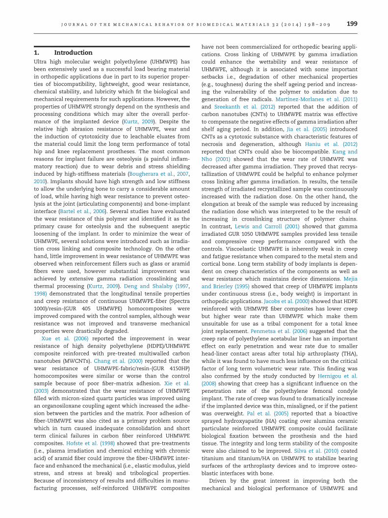
Tensile test results of neat and nylon coated UHMWPE fibersat three different temperatures were presented in Figs. 4–6.In all the cases it was shown that nylon coated UHMWPEfibers provided superior tensile properties compared to theneat UHMWPE fiber. Nylon 6,6 and Nylon 6,12 coated fibersshowed 17% and 16% improvement (respectively) in max-imum breaking force at 25 1C. Improvements of 14% and 6%for Nylon 6,6 coated fiber and 15% and 2% for Nylon 6,12coated fiber at 50 1C and 70 1C (respectively) were alsorecorded (Table 1). It was also observed that the elongationof nylon coated fiber in tension was comparable with the neatone which showed the flexibility and ductile nature of thenylon coating onto the fibers. Toughness values of neat andnylon coated UHMWPE fibers were compared from tensiletest results by measuring the areas under the stress–straincurves and presented in Table 1. From the calculated
Fig. 2 – A close view at the nylon coated UHMWPE fiber(25� ).
Fig. 3 – Cross section area of nylon coated
toughness results, it can be observed that nylon coatedUHMWPE fibers showed higher values compared to the neatUHMWPE fiber (average 32%). The increase in toughnessvalues is more significant at higher temperatures (averageincrease of 54% and 46% at 50 1C and 70 1C, respectively)
UHMWPE fiber (a) 140� and (b) 700� .
Fig. 5 – Tensile test results of neat and nylon coatedUHMWPE fibers at 50 1C.
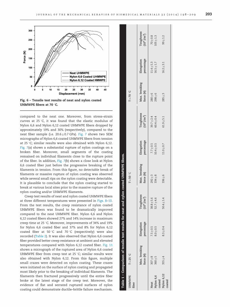
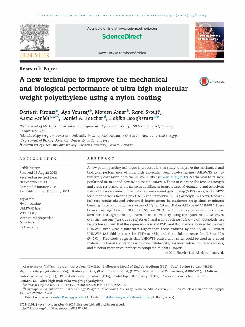
Fig. 6 – Tensile test results of neat and nylon coatedUHMWPE fibers at 70 1C.
Tab
le1–Com
parison
oftensile
test
resu
ltsfornea
tan
dnylon
coated
UHMW
PEfibe
rs.
UHMW
PEfiber
T¼25
1CT¼50
1CT¼70
1C
Max
.break
ing
force(N
)Elon
gation
perce
ntage
Tou
ghnes
s(106
J/m
3)
Max
.break
ing
force(N
)Elon
gation
perce
ntage
Tou
ghnes
s(106
J/m
3)
Max
.break
ing
force(N
)Elon
gation
perce
ntage
Tou
ghnes
s(106
J/m
3)
Nea
t30
778
7.57
0.5
38.875.6
2927
37.77
0.5
39.772.4
2807
611
.475.3
7070.8
Nylon
6,6
coated
3607
58.07
0.5
52.774.6
3347
810
.470.5
60.470.4
2987
616
.571.7
1057
1.3
Nylon
6,12
coated
3557
48.37
0.4
50.471.4
3367
611
.070.7
61.975.1
2857
316
.273.1
9971.2
j o u r n a l o f t h e m e c h a n i c a l b e h a v i o r o f b i o m e d i c a l m a t e r i a l s 3 2 ( 2 0 1 4 ) 1 9 8 – 2 0 9 203
compared to the neat one. Moreover, from stress–straincurves at 25 1C, it was found that the elastic modulus ofNylon 6,6 and Nylon 6,12 coated UHMWPE fibers dropped byapproximately 19% and 30% (respectively), compared to theneat fiber sample (i.e. 20.670.7 GPa). Fig. 7 shows two SEMmicrographs of Nylon 6,6 coated UHMWPE fibers from tensionat 25 1C; similar results were also obtained with Nylon 6,12.Fig. 7(a) shows a substantial rupture of nylon coatings on abroken fiber. Moreover, small segments of the coatingremained on individual filaments close to the rupture pointof the fiber. In addition, Fig. 7(b) shows a close look at Nylon6,6 coated fiber just before the progressive breaking of thefilaments in tension. From this figure, no detectable break offilaments or massive rupture of nylon coating was observedwhile several small rips on the nylon coating were detectable.It is plausible to conclude that the nylon coating started tobreak at various local sites prior to the massive rupture of thenylon coating and/or UHMWPE filaments.
Creep test results of neat and nylon coated UHMWPE fibersat three different temperatures were presented in Figs. 8–10.From the test results, the creep resistance of nylon coatedUHMWPE fibers was found to be dramatically improvedcompared to the neat UHMWPE fiber. Nylon 6,6 and Nylon6,12 coated fibers showed 27% and 14% increase in maximumcreep time at 25 1C. Moreover, improvements of 34% and 19%for Nylon 6,6 coated fiber and 37% and 8% for Nylon 6,12coated fiber at 50 1C and 70 1C (respectively) were alsorecorded (Table 2). It was also observed that Nylon 6,6 coatedfiber provided better creep resistance at ambient and elevatedtemperatures compared with Nylon 6,12 coated fiber. Fig. 11shows a micrograph of the ruptured area of Nylon 6,6 coatedUHMWPE fiber from creep test at 25 1C; similar results werealso obtained with Nylon 6,12. From this figure, multiplesmall crazes were detected on nylon coating. These crazeswere initiated on the surface of nylon coating and propagatedmost likely prior to the breaking of individual filaments. Thefilaments then fractured progressively until the entire fiberbroke at the latest stage of the creep test. Moreover, theevidence of flat and serrated ruptured surfaces of nyloncoating could demonstrate ductile–brittle failure mechanism.
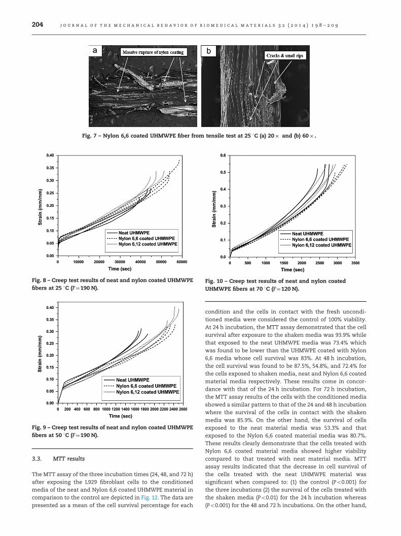
Fig. 7 – Nylon 6,6 coated UHMWPE fiber from tensile test at 25 1C (a) 20� and (b) 60� .
Fig. 8 – Creep test results of neat and nylon coated UHMWPEfibers at 25 1C (F¼190 N).
Fig. 9 – Creep test results of neat and nylon coated UHMWPEfibers at 50 1C (F¼190 N).
Fig. 10 – Creep test results of neat and nylon coatedUHMWPE fibers at 70 1C (F¼120 N).
j o u r n a l o f t h e m e c h a n i c a l b e h a v i o r o f b i o m e d i c a l m a t e r i a l s 3 2 ( 2 0 1 4 ) 1 9 8 – 2 0 9204
3.3. MTT results
The MTT assay of the three incubation times (24, 48, and 72 h)after exposing the L929 fibroblast cells to the conditionedmedia of the neat and Nylon 6,6 coated UHMWPE material incomparison to the control are depicted in Fig. 12. The data arepresented as a mean of the cell survival percentage for each
condition and the cells in contact with the fresh uncondi-tioned media were considered the control of 100% viability.At 24 h incubation, the MTT assay demonstrated that the cellsurvival after exposure to the shaken media was 93.9% whilethat exposed to the neat UHMWPE media was 73.4% whichwas found to be lower than the UHMWPE coated with Nylon6,6 media whose cell survival was 83%. At 48 h incubation,the cell survival was found to be 87.5%, 54.8%, and 72.4% forthe cells exposed to shaken media, neat and Nylon 6,6 coatedmaterial media respectively. These results come in concor-dance with that of the 24 h incubation. For 72 h incubation,the MTT assay results of the cells with the conditioned mediashowed a similar pattern to that of the 24 and 48 h incubationwhere the survival of the cells in contact with the shakenmedia was 85.9%. On the other hand, the survival of cellsexposed to the neat material media was 53.3% and thatexposed to the Nylon 6,6 coated material media was 80.7%.These results clearly demonstrate that the cells treated withNylon 6,6 coated material media showed higher viabilitycompared to that treated with neat material media. MTTassay results indicated that the decrease in cell survival ofthe cells treated with the neat UHMWPE material wassignificant when compared to: (1) the control (Po0.001) forthe three incubations (2) the survival of the cells treated withthe shaken media (Po0.01) for the 24 h incubation whereas(Po0.001) for the 48 and 72 h incubations. On the other hand,

Table 2 – Comparison of creep test results for neat and nylon coated UHMWPE fibers.
UHMWPE fiber T¼25 1C T¼50 1C T¼70 1CMax. creep time (s) Max. creep time (s) Max. creep time (s)
Neat 4196375% 175176% 262376%Nylon 6,6 coated 5534475% 234074% 313476%Nylon 6,12 coated 4774777% 239775% 283575%
Fig. 11 – Nylon 6,6 coated UHMWPE fiber from creep test at25 1C (15� ).
Fig. 12 – Effect of neat and Nylon 6,6 coated UHMWPEextracts on the viability of L929 fibroblast cells at 24, 48, and72 h as % of negative control (nPo0.05, nnPo0.01, andnnnPo0.001).
j o u r n a l o f t h e m e c h a n i c a l b e h a v i o r o f b i o m e d i c a l m a t e r i a l s 3 2 ( 2 0 1 4 ) 1 9 8 – 2 0 9 205
the decrease in cell survival of the cells treated with thenylon coated UHMWPE material was significant compared to:(1) the control (Po0.01) for the 24 h incubation, (Po0.001)for the 48 h incubation, and (Po0.05) for the 72 h incubation(2) the survival of the cells treated with the shaken media(Po0.01) for the 48 h incubation only. Thus the cell deathinduced by both materials in relation to the control demon-strate that the neat material induced significant cell deathcompared to the control with the same P-value (Po0.001) inthe three incubations. The nylon coated material inducedsignificant cell death compared to the control with differentP-values in the three incubations. These results indicatethat the neat UHMWPE material caused a more significant
decrease in cell survival than the nylon coated UHMWPEmaterial in relation to the control. Moreover, there was nosignificant difference in cell survival of the control comparedto the cells treated with the shaken media without thematerial except at the 48 h incubation (Po0.01). This indi-cates that the process of conditioning the media throughshaking it at 200 RPM for 72 h at 37 1C and 5% CO2 had nosignificant effect on the cell death and that any decrease inthe cell survival was due to the material tested and not theshaking process itself. Finally, the decrease in cell survival ofthe cells treated with the neat material was significant to thatof the cells treated with the nylon coated material (Po0.001)for the 48 h incubation and (Po0.01) for the 72 h incubation.This shows that the neat material induced more significantcell death compared to the nylon coated material. Accord-ingly, the Nylon 6,6 coated UHMWPE material is less cytotoxicand that the nylon improved the UHMWPE material biocom-patibility. Moreover, L929 fibroblast cell cultures0 viabilitysubjected to three conditions is depicted in Fig. 13. From thisfigure, it is evident that the cell density in the tissue cultureplate upon exposure to media conditioned with neat nylon isless than that observed in media conditioned with nylon 6,6coated UHMWPE. This effect of conditioned media on the rateof cell division was observed throughout the three incubationtimes.
3.4. Osteolysis results
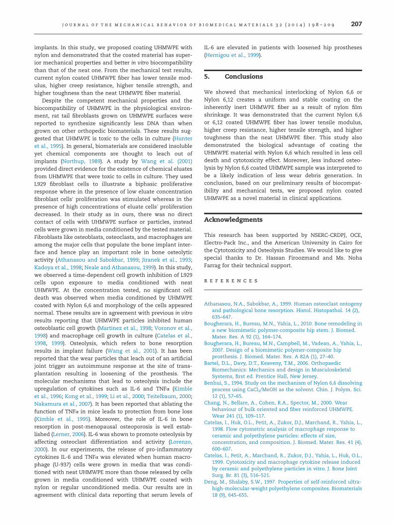
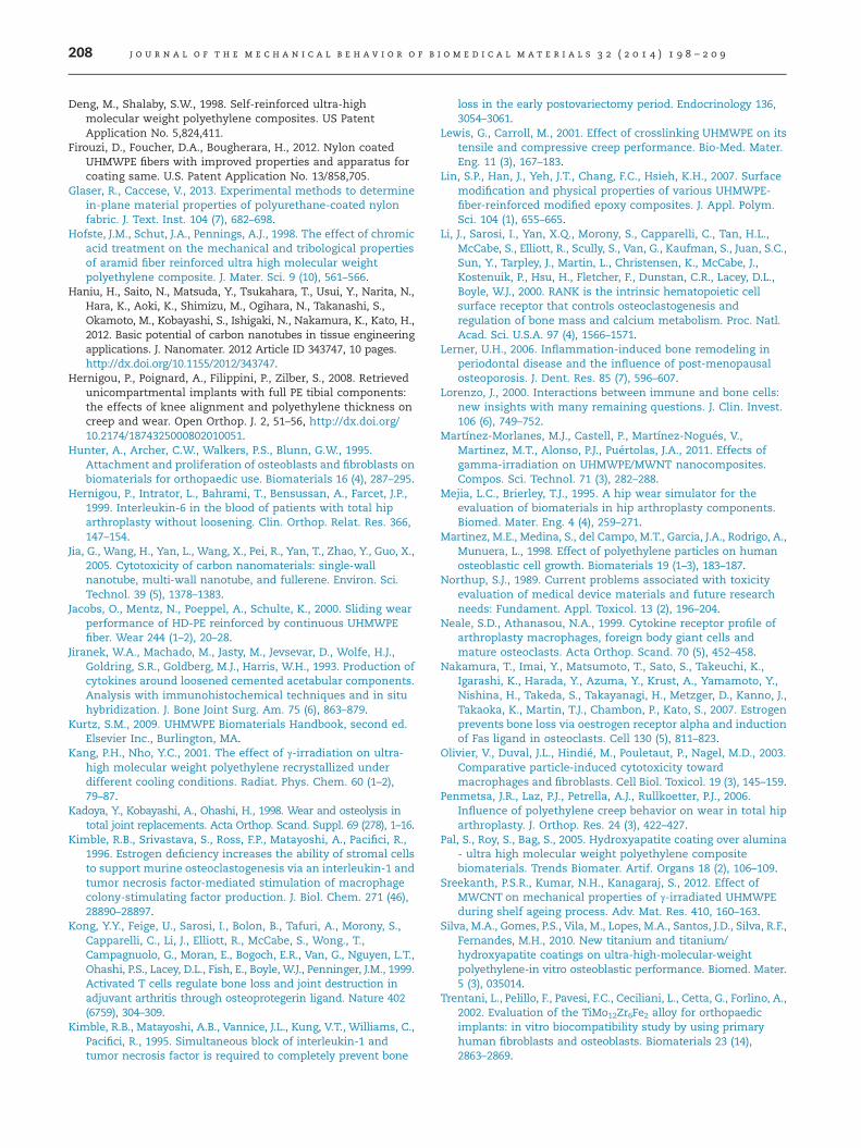
The osteolysis results of the three incubation times (24, 48,and 72 h) after exposing the U-97 human macrophage cells tothe conditioned media of the neat or Nylon 6,6 coatedUHMWPE material in comparison to the control are depictedin Figs. 14 and 15. The expression level of TNFα in theunconditioned media was nearly double its level in the mediaconditioned with both materials after 24 h (Po0.01), whileafter 48 h, the expression level from the media conditionedwith the neat material was about 2.5 fold more than thatfrom the nylon coated material (Po0.01) whose expressionwas even less than that from the unconditioned media(Po0.05). After 72 h, no significant difference in TNFα expres-sion level was observed in all three conditions. For IL-6, nosignificant difference in expression level was observed exceptafter 72 h where macrophages cultured in the unconditionedmedia and in media conditioned with the neat materialshowed about three fold expression level more than thosecultured in the media conditioned with the nylon coatedmaterial. The difference in expression was shown to bestatistically significant with Po0.01. The coated material alsoshowed a statistically significant lower level of expressionfrom the control with Po0.05.
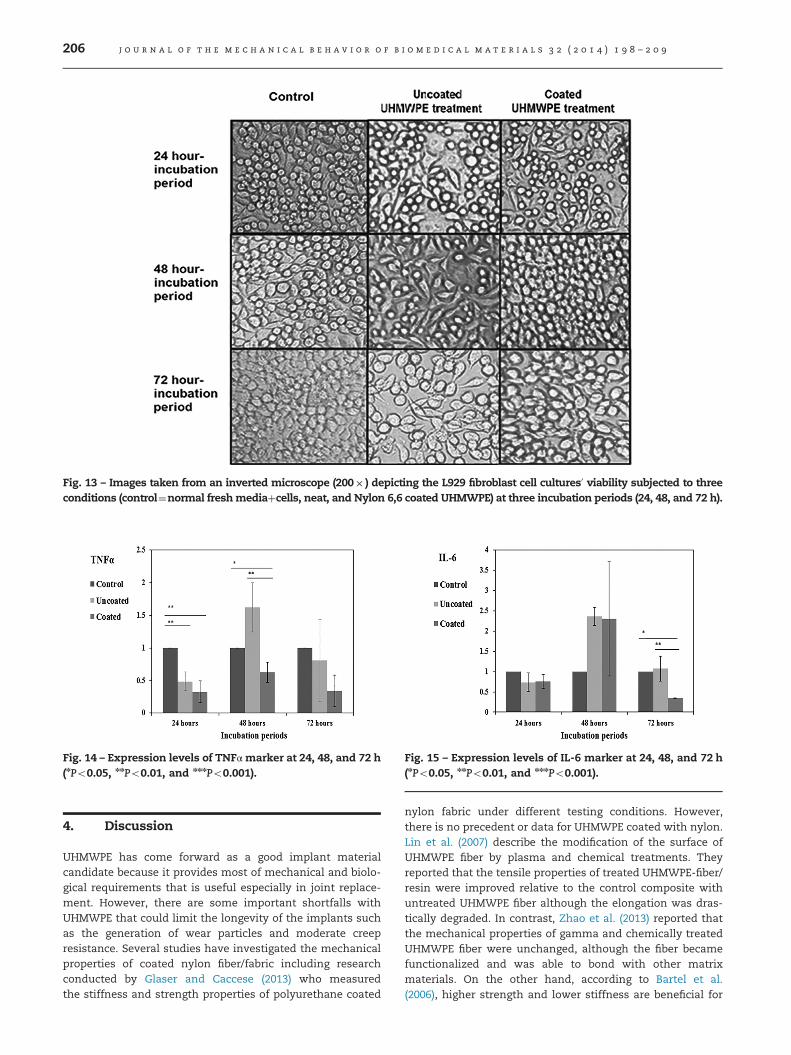
Fig. 13 – Images taken from an inverted microscope (200� ) depicting the L929 fibroblast cell cultures0 viability subjected to threeconditions (control¼normal freshmediaþcells, neat, and Nylon 6,6 coated UHMWPE) at three incubation periods (24, 48, and 72 h).
Fig. 14 – Expression levels of TNFα marker at 24, 48, and 72 h(nPo0.05, nnPo0.01, and nnnPo0.001).
Fig. 15 – Expression levels of IL-6 marker at 24, 48, and 72 h(nPo0.05, nnPo0.01, and nnnPo0.001).
j o u r n a l o f t h e m e c h a n i c a l b e h a v i o r o f b i o m e d i c a l m a t e r i a l s 3 2 ( 2 0 1 4 ) 1 9 8 – 2 0 9206
4. Discussion
UHMWPE has come forward as a good implant materialcandidate because it provides most of mechanical and biolo-gical requirements that is useful especially in joint replace-ment. However, there are some important shortfalls withUHMWPE that could limit the longevity of the implants suchas the generation of wear particles and moderate creepresistance. Several studies have investigated the mechanicalproperties of coated nylon fiber/fabric including researchconducted by Glaser and Caccese (2013) who measuredthe stiffness and strength properties of polyurethane coated
nylon fabric under different testing conditions. However,there is no precedent or data for UHMWPE coated with nylon.Lin et al. (2007) describe the modification of the surface ofUHMWPE fiber by plasma and chemical treatments. Theyreported that the tensile properties of treated UHMWPE-fiber/resin were improved relative to the control composite withuntreated UHMWPE fiber although the elongation was dras-tically degraded. In contrast, Zhao et al. (2013) reported thatthe mechanical properties of gamma and chemically treatedUHMWPE fiber were unchanged, although the fiber becamefunctionalized and was able to bond with other matrixmaterials. On the other hand, according to Bartel et al.(2006), higher strength and lower stiffness are beneficial for
j o u r n a l o f t h e m e c h a n i c a l b e h a v i o r o f b i o m e d i c a l m a t e r i a l s 3 2 ( 2 0 1 4 ) 1 9 8 – 2 0 9 207
implants. In this study, we proposed coating UHMWPE withnylon and demonstrated that the coated material has super-ior mechanical properties and better in vitro biocompatibilitythan that of the neat one. From the mechanical test results,current nylon coated UHMWPE fiber has lower tensile mod-ulus, higher creep resistance, higher tensile strength, andhigher toughness than the neat UHMWPE fiber material.
Despite the competent mechanical properties and thebiocompatibility of UHMWPE in the physiological environ-ment, rat tail fibroblasts grown on UHMWPE surfaces werereported to synthesize significantly less DNA than whengrown on other orthopedic biomaterials. These results sug-gested that UHMWPE is toxic to the cells in culture (Hunteret al., 1995). In general, biomaterials are considered insolubleyet chemical components are thought to leach out ofimplants (Northup, 1989). A study by Wang et al. (2001)provided direct evidence for the existence of chemical eluatesfrom UHMWPE that were toxic to cells in culture. They usedL929 fibroblast cells to illustrate a biphasic proliferativeresponse where in the presence of low eluate concentrationfibroblast cells0 proliferation was stimulated whereas in thepresence of high concentrations of eluate cells0 proliferationdecreased. In their study as in ours, there was no directcontact of cells with UHMWPE surface or particles, insteadcells were grown in media conditioned by the tested material.Fibroblasts like osteoblasts, osteoclasts, and macrophages areamong the major cells that populate the bone implant inter-face and hence play an important role in bone osteolyticactivity (Athanasou and Sabokbar, 1999; Jiranek et al., 1993;Kadoya et al., 1998; Neale and Athanasou, 1999). In this study,we observed a time-dependent cell growth inhibition of L929cells upon exposure to media conditioned with neatUHMWPE. At the concentration tested, no significant celldeath was observed when media conditioned by UHMWPEcoated with Nylon 6,6 and morphology of the cells appearednormal. These results are in agreement with previous in vitroresults reporting that UHMWPE particles inhibited humanosteoblastic cell growth (Martinez et al., 1998; Voronov et al.,1998) and macrophage cell growth in culture (Catelas et al.,1998, 1999). Osteolysis, which refers to bone resorptionresults in implant failure (Wang et al., 2001). It has beenreported that the wear particles that leach out of an artificialjoint trigger an autoimmune response at the site of trans-plantation resulting in loosening of the prosthesis. Themolecular mechanisms that lead to osteolysis include theupregulation of cytokines such as IL-6 and TNFα (Kimbleet al., 1996; Kong et al., 1999; Li et al., 2000; Teitelbaum, 2000;Nakamura et al., 2007). It has been reported that ablating thefunction of TNFα in mice leads to protection from bone loss(Kimble et al., 1995). Moreover, the role of IL-6 in boneresorption in post-menopausal osteoporosis is well estab-lished (Lerner, 2006). IL-6 was shown to promote osteolysis byaffecting osteoclast differentiation and activity (Lorenzo,2000). In our experiments, the release of pro-inflammatorycytokines IL-6 and TNFα was elevated when human macro-phage (U-937) cells were grown in media that was condi-tioned with neat UHMWPE more than those released by cellsgrown in media conditioned with UHMWPE coated withnylon or regular unconditioned media. Our results are inagreement with clinical data reporting that serum levels of
IL-6 are elevated in patients with loosened hip prostheses(Hernigou et al., 1999).
5. Conclusions
We showed that mechanical interlocking of Nylon 6,6 orNylon 6,12 creates a uniform and stable coating on theinherently inert UHMWPE fiber as a result of nylon filmshrinkage. It was demonstrated that the current Nylon 6,6or 6,12 coated UHMWPE fiber has lower tensile modulus,higher creep resistance, higher tensile strength, and highertoughness than the neat UHMWPE fiber. This study alsodemonstrated the biological advantage of coating theUHMWPE material with Nylon 6,6 which resulted in less celldeath and cytotoxicity effect. Moreover, less induced osteo-lysis by Nylon 6,6 coated UHMWPE sample was interpreted tobe a likely indication of less wear debris generation. Inconclusion, based on our preliminary results of biocompat-ibility and mechanical tests, we proposed nylon coatedUHMWPE as a novel material in clinical applications.
Acknowledgments
This research has been supported by NSERC-CRDPJ, OCE,Electro-Pack Inc., and the American University in Cairo forthe Cytotoxicity and Osteolysis Studies. We would like to givespecial thanks to Dr. Hassan Firoozmand and Ms. NohaFarrag for their technical support.
r e f e r e n c e s
Athanasou, N.A., Sabokbar, A., 1999. Human osteoclast ontogenyand pathological bone resorption. Histol. Histopathol. 14 (2),635–647.
Bougherara, H., Bureau, M.N., Yahia, L., 2010. Bone remodeling ina new biomimetic polymer-composite hip stem. J. Biomed.Mater. Res. A 92 (1), 164–174.
Bougherara, H., Bureau, M.N., Campbell, M., Vadean, A., Yahia, L.,2007. Design of a biomimetic polymer-composite hipprosthesis. J. Biomed. Mater. Res. A 82A (1), 27–40.
Bartel, D.L., Davy, D.T., Keaveny, T.M., 2006. OrthopaedicBiomechanics: Mechanics and design in MusculoskeletalSystems, first ed. Prentice Hall, New Jersey.
Benhui, S., 1994. Study on the mechanism of Nylon 6,6 dissolvingprocess using CaCl2/MeOH as the solvent. Chin. J. Polym. Sci.12 (1), 57–65.
Chang, N., Bellare, A., Cohen, R.A., Spector, M., 2000. Wearbehaviour of bulk oriented and fiber reinforced UHMWPE.Wear 241 (1), 109–117.
Catelas, I., Huk, O.L., Petit, A., Zukor, D.J., Marchand, R., Yahia, L.,1998. Flow cytometric analysis of macrophage response toceramic and polyethylene particles: effects of size,concentration, and composition. J. Biomed. Mater. Res. 41 (4),600–607.
Catelas, I., Petit, A., Marchand, R., Zukor, D.J., Yahia, L., Huk, O.L.,1999. Cytotoxicity and macrophage cytokine release inducedby ceramic and polyethylene particles in vitro. J. Bone JointSurg. Br. 81 (3), 516–521.
Deng, M., Shalaby, S.W., 1997. Properties of self-reinforced ultra-high-molecular-weight polyethylene composites. Biomaterials18 (9), 645–655.
j o u r n a l o f t h e m e c h a n i c a l b e h a v i o r o f b i o m e d i c a l m a t e r i a l s 3 2 ( 2 0 1 4 ) 1 9 8 – 2 0 9208
Deng, M., Shalaby, S.W., 1998. Self-reinforced ultra-highmolecular weight polyethylene composites. US PatentApplication No. 5,824,411.
Firouzi, D., Foucher, D.A., Bougherara, H., 2012. Nylon coatedUHMWPE fibers with improved properties and apparatus forcoating same. U.S. Patent Application No. 13/858,705.
Glaser, R., Caccese, V., 2013. Experimental methods to determinein-plane material properties of polyurethane-coated nylonfabric. J. Text. Inst. 104 (7), 682–698.
Hofste, J.M., Schut, J.A., Pennings, A.J., 1998. The effect of chromicacid treatment on the mechanical and tribological propertiesof aramid fiber reinforced ultra high molecular weightpolyethylene composite. J. Mater. Sci. 9 (10), 561–566.
Haniu, H., Saito, N., Matsuda, Y., Tsukahara, T., Usui, Y., Narita, N.,Hara, K., Aoki, K., Shimizu, M., Ogihara, N., Takanashi, S.,Okamoto, M., Kobayashi, S., Ishigaki, N., Nakamura, K., Kato, H.,2012. Basic potential of carbon nanotubes in tissue engineeringapplications. J. Nanomater. 2012 Article ID 343747, 10 pages.http://dx.doi.org/10.1155/2012/343747.
Hernigou, P., Poignard, A., Filippini, P., Zilber, S., 2008. Retrievedunicompartmental implants with full PE tibial components:the effects of knee alignment and polyethylene thickness oncreep and wear. Open Orthop. J. 2, 51–56, http://dx.doi.org/10.2174/1874325000802010051.
Hunter, A., Archer, C.W., Walkers, P.S., Blunn, G.W., 1995.Attachment and proliferation of osteoblasts and fibroblasts onbiomaterials for orthopaedic use. Biomaterials 16 (4), 287–295.
Hernigou, P., Intrator, L., Bahrami, T., Bensussan, A., Farcet, J.P.,1999. Interleukin-6 in the blood of patients with total hiparthroplasty without loosening. Clin. Orthop. Relat. Res. 366,147–154.
Jia, G., Wang, H., Yan, L., Wang, X., Pei, R., Yan, T., Zhao, Y., Guo, X.,2005. Cytotoxicity of carbon nanomaterials: single-wallnanotube, multi-wall nanotube, and fullerene. Environ. Sci.Technol. 39 (5), 1378–1383.
Jacobs, O., Mentz, N., Poeppel, A., Schulte, K., 2000. Sliding wearperformance of HD-PE reinforced by continuous UHMWPEfiber. Wear 244 (1–2), 20–28.
Jiranek, W.A., Machado, M., Jasty, M., Jevsevar, D., Wolfe, H.J.,Goldring, S.R., Goldberg, M.J., Harris, W.H., 1993. Production ofcytokines around loosened cemented acetabular components.Analysis with immunohistochemical techniques and in situhybridization. J. Bone Joint Surg. Am. 75 (6), 863–879.
Kurtz, S.M., 2009. UHMWPE Biomaterials Handbook, second ed.Elsevier Inc., Burlington, MA.
Kang, P.H., Nho, Y.C., 2001. The effect of γ-irradiation on ultra-high molecular weight polyethylene recrystallized underdifferent cooling conditions. Radiat. Phys. Chem. 60 (1–2),79–87.
Kadoya, Y., Kobayashi, A., Ohashi, H., 1998. Wear and osteolysis intotal joint replacements. Acta Orthop. Scand. Suppl. 69 (278), 1–16.
Kimble, R.B., Srivastava, S., Ross, F.P., Matayoshi, A., Pacifici, R.,1996. Estrogen deficiency increases the ability of stromal cellsto support murine osteoclastogenesis via an interleukin-1 andtumor necrosis factor-mediated stimulation of macrophagecolony-stimulating factor production. J. Biol. Chem. 271 (46),28890–28897.
Kong, Y.Y., Feige, U., Sarosi, I., Bolon, B., Tafuri, A., Morony, S.,Capparelli, C., Li, J., Elliott, R., McCabe, S., Wong., T.,Campagnuolo, G., Moran, E., Bogoch, E.R., Van, G., Nguyen, L.T.,Ohashi, P.S., Lacey, D.L., Fish, E., Boyle, W.J., Penninger, J.M., 1999.Activated T cells regulate bone loss and joint destruction inadjuvant arthritis through osteoprotegerin ligand. Nature 402(6759), 304–309.
Kimble, R.B., Matayoshi, A.B., Vannice, J.L., Kung, V.T., Williams, C.,Pacifici, R., 1995. Simultaneous block of interleukin-1 andtumor necrosis factor is required to completely prevent bone
loss in the early postovariectomy period. Endocrinology 136,3054–3061.
Lewis, G., Carroll, M., 2001. Effect of crosslinking UHMWPE on itstensile and compressive creep performance. Bio-Med. Mater.Eng. 11 (3), 167–183.
Lin, S.P., Han, J., Yeh, J.T., Chang, F.C., Hsieh, K.H., 2007. Surfacemodification and physical properties of various UHMWPE-fiber-reinforced modified epoxy composites. J. Appl. Polym.Sci. 104 (1), 655–665.
Li, J., Sarosi, I., Yan, X.Q., Morony, S., Capparelli, C., Tan, H.L.,McCabe, S., Elliott, R., Scully, S., Van, G., Kaufman, S., Juan, S.C.,Sun, Y., Tarpley, J., Martin, L., Christensen, K., McCabe, J.,Kostenuik, P., Hsu, H., Fletcher, F., Dunstan, C.R., Lacey, D.L.,Boyle, W.J., 2000. RANK is the intrinsic hematopoietic cellsurface receptor that controls osteoclastogenesis andregulation of bone mass and calcium metabolism. Proc. Natl.Acad. Sci. U.S.A. 97 (4), 1566–1571.
Lerner, U.H., 2006. Inflammation-induced bone remodeling inperiodontal disease and the influence of post-menopausalosteoporosis. J. Dent. Res. 85 (7), 596–607.
Lorenzo, J., 2000. Interactions between immune and bone cells:new insights with many remaining questions. J. Clin. Invest.106 (6), 749–752.
Martınez-Morlanes, M.J., Castell, P., Martınez-Nogues, V.,Martinez, M.T., Alonso, P.J., Puertolas, J.A., 2011. Effects ofgamma-irradiation on UHMWPE/MWNT nanocomposites.Compos. Sci. Technol. 71 (3), 282–288.
Mejia, L.C., Brierley, T.J., 1995. A hip wear simulator for theevaluation of biomaterials in hip arthroplasty components.Biomed. Mater. Eng. 4 (4), 259–271.
Martinez, M.E., Medina, S., del Campo, M.T., Garcia, J.A., Rodrigo, A.,Munuera, L., 1998. Effect of polyethylene particles on humanosteoblastic cell growth. Biomaterials 19 (1–3), 183–187.
Northup, S.J., 1989. Current problems associated with toxicityevaluation of medical device materials and future researchneeds: Fundament. Appl. Toxicol. 13 (2), 196–204.
Neale, S.D., Athanasou, N.A., 1999. Cytokine receptor profile ofarthroplasty macrophages, foreign body giant cells andmature osteoclasts. Acta Orthop. Scand. 70 (5), 452–458.
Nakamura, T., Imai, Y., Matsumoto, T., Sato, S., Takeuchi, K.,Igarashi, K., Harada, Y., Azuma, Y., Krust, A., Yamamoto, Y.,Nishina, H., Takeda, S., Takayanagi, H., Metzger, D., Kanno, J.,Takaoka, K., Martin, T.J., Chambon, P., Kato, S., 2007. Estrogenprevents bone loss via oestrogen receptor alpha and inductionof Fas ligand in osteoclasts. Cell 130 (5), 811–823.
Olivier, V., Duval, J.L., Hindie, M., Pouletaut, P., Nagel, M.D., 2003.Comparative particle-induced cytotoxicity towardmacrophages and fibroblasts. Cell Biol. Toxicol. 19 (3), 145–159.
Penmetsa, J.R., Laz, P.J., Petrella, A.J., Rullkoetter, P.J., 2006.Influence of polyethylene creep behavior on wear in total hiparthroplasty. J. Orthop. Res. 24 (3), 422–427.
Pal, S., Roy, S., Bag, S., 2005. Hydroxyapatite coating over alumina- ultra high molecular weight polyethylene compositebiomaterials. Trends Biomater. Artif. Organs 18 (2), 106–109.
Sreekanth, P.S.R., Kumar, N.H., Kanagaraj, S., 2012. Effect ofMWCNT on mechanical properties of γ-irradiated UHMWPEduring shelf ageing process. Adv. Mat. Res. 410, 160–163.
Silva, M.A., Gomes, P.S., Vila, M., Lopes, M.A., Santos, J.D., Silva, R.F.,Fernandes, M.H., 2010. New titanium and titanium/hydroxyapatite coatings on ultra-high-molecular-weightpolyethylene-in vitro osteoblastic performance. Biomed. Mater.5 (3), 035014.
Trentani, L., Pelillo, F., Pavesi, F.C., Ceciliani, L., Cetta, G., Forlino, A.,2002. Evaluation of the TiMo12Zr6Fe2 alloy for orthopaedicimplants: in vitro biocompatibility study by using primaryhuman fibroblasts and osteoblasts. Biomaterials 23 (14),2863–2869.

j o u r n a l o f t h e m e c h a n i c a l b e h a v i o r o f b i o m e d i c a l m a t e r i a l s 3 2 ( 2 0 1 4 ) 1 9 8 – 2 0 9 209
Teitelbaum, S.L., 2000. Osteoclasts, integrins, and osteoporosis.J. Bone Miner. Metab. 18 (6), 344–349.
Voronov, I., Santerre, J.P., Hinek, A., Callahan, J.W., Sandhu, J.,Boynton, E.L., 1998. Macrophage phagocytosis of polyethyleneparticulate in vitro. J. Biomed. Mater. Res. 39 (1), 40–51.
Wang, K.Y., Horne, J.G., Devane, P.A., Wilson, T., Miller, J.H., 2001.Chemical eluates from ultra-high molecular weightpolyethylene and fibroblast proliferation. J. Orthop. Surg.(Hong Kong) 9 (1), 25–33.
Xue, Y., Wu, W., Jacobs, O., Schadel, B., 2006. Tribologicalbehaviour of UHMWPE/HDPE blends reinforced with
multi-wall carbon nanotubes. Polym. Test. 25 (2),221–229.
Xie, X.L., Tang, C.Y., Chan, K.Y., Wu, X.C., Tsui, C.P., Cheung, C.Y.,2003. Wear performance of ultrahigh molecular weightpolyethylene/quartz composites. Biomaterials 24 (11),1889–1896.
Zhao, Y., Han, G., Hou, J., Wang, C., Wan, J., Ma, J., Sun, L., 2013.Surface modification of UHMWPE fiber by low temperatureplasma treatment and grafting using acrylic acid. Mater. Sci.Eng. 29 (6), 62–65.