aapm report no. 21 specification of
TRANSCRIPT
7/28/2019 Aapm Report No. 21 Specification Of
http://slidepdf.com/reader/full/aapm-report-no-21-specification-of 1/24
AAPM REPORT NO. 21
SPECIFICATION OF
BRACHYTHERAPY
SOURCE STRENGTH
Published for the American Association of Physicists in Medicine
by the American institute of Physics

7/28/2019 Aapm Report No. 21 Specification Of
http://slidepdf.com/reader/full/aapm-report-no-21-specification-of 2/24
AAPM REPORT NO. 21
SPECIFICATION OF
BRACHYTHERAPY
SOURCE STRENGTH
REPORT OF AAPMTask Group No. 32*
MembersRavinder Nath, Yale University, Chairman
Lowell Anderson, Memorial Sloan-Kettering Institute
Douglas Jones, Northwest Medical Physics Center
Clifton Ling, University of California at San Francisco
Robert Loevinger, National Bureau of Standards
Jeffrey Williamson, University of Arizona
William Hanson, Radiological Physics Center Consultant
*The Task Group is part of the AAPM Radiation Therapy Committee,
Faiz M. Khan, Chairman.
June 1987
Published for the
American Association of Physicists in Medicineby the American Institute of Physics

7/28/2019 Aapm Report No. 21 Specification Of
http://slidepdf.com/reader/full/aapm-report-no-21-specification-of 3/24
Further copies of this report may be obtained from
Executive Officer
American Association of Physicists in Medicine
335 E. 45 Street
New York, NY 10017
Library of Congress Catalog Card Number: 87-72124International Standard Book Number: 0-88318-545-8
International Standard Serial Number: 0271-7344
Copyright © 1987 by the American Associationof Physicists in Medicine
All rights reserved. No part of this publication may bereproduced, stored in a retrieval system, or transmittedin any form or by any means (electronic, mechanical, photocopying, recording, or otherwise) without the
prior written permission of the publisher.
Published by the American Institute of Physics, Inc.,
335 East 45 Street, New York, New York 10017
Printed in the United States of America

7/28/2019 Aapm Report No. 21 Specification Of
http://slidepdf.com/reader/full/aapm-report-no-21-specification-of 4/24
Table of Contents
I .
I I .
I I I .
I V.
V.
VI .
V I I .
I ntroduction . . . . . . . . . . . . . . . . . . . . . . . . . . . . . . . . . . . . . . . . . . . . . . . . 3
U.S. Standards for Brachytherapy Sources . . . . . . . . . . . . . . . . . . . . 4
Source Specification Methods Currently in Use . . . . . . . . . . . . . . . 6
A. Activity of the Radionuclide . . . . . . . . . . . . . . . . . . . . . . . . . . . . 6
B. Exposure Rate at a Reference Point in Free Space . . . . . . . . 9
C. Equivalent Mass and Apparent Activity . . . . . . . . . . . . . . . . . . . 9
D. A ir-K erma Rate at a Reference Point in F ree Space...... 10
Discussion . . . . . . . . . . . . . . . . . . . . . . . . . . . . . . . . . . . . . . . . . . . . . . . . . 11
Recommendations . . . . . . . . . . . . . . . . . . . . . . . . . . . . . . . . . . . . . . . . . . . . 13
Recommended Quantities and Their Relationships............. 14
Practical Considerations . . . . . . . . . . . . . . . . . . . . . . . . . . . . . . . . . . . 17
Appendix: Information Recommended for Manufacturer’s
Certificate of Calibration . . . . . . . . . . . . ................ 19
References . . . . . . . . . . . . . . . . . . . . . . . . . . . . . . . . . . . . . . . . . . . . . . . . . . . . . . . . . 20
7/28/2019 Aapm Report No. 21 Specification Of
http://slidepdf.com/reader/full/aapm-report-no-21-specification-of 5/24
Acknowledgments
We would like to thank the members of the Radiation Therapy
Committee and the Science Counci l f or a careful review of this
document. Many valuable comments from Montague Cohen, David
K ubiatowicz and J erome M el i are al so grateful ly acknowledged.
Finally, we would like to thank Ms. Deanna J acobs for preparing this
manuscript.
7/28/2019 Aapm Report No. 21 Specification Of
http://slidepdf.com/reader/full/aapm-report-no-21-specification-of 6/24
3
I . Introduct ion
I t is common practice to express source strength of brachytherapysources in terms of activity or equivalent mass of radium. The
output of a brachytherapy source, in terms of exposure rate in free
space, can then be obtained by multiplying the source strength by the
appropriate exposure-rate constant. I n 1974, the National Council of
Radiation Protection and Measurements (NCRP) pointed out several
shortcomings of these methods, the most important being that they do
not specify strength in terms of directly measured quantities.1
The
NCRP recommended that brachytherapy sources should be specified in
terms of exposure rate at 1 m in free air with corrections for air
attenuation. A similar suggestion was made by Dutreix and Wambersie2
in 1975. Despite the sound rationale of this proposal there has been
little progress towards the use of this specification method in the
clinical community. Recent adoption of SI units, and replacement of
exposure by air kerma, has reopened this discussion. Gibb and
Massey3, Aird and Day4, J ayaraman, L anzl and Agarwa15, comite
F rancais Measure des Rayonnements I onisants (CF MRI )6, L oevinger 7,
Williamson and Morin14, and British Committee on Radiation Units and
Measurements (BCRD)8 have contributed to the discussion in recent
publications. The BCRU and CFMRI have recommended specifying
brachytherapy source strength in terms of air-kerma rate, with a unit
of µGy m2h-1
.
I n the present paper , recommendations are set forth that, in
many r es p ect s , ar e s i mi l a r t o t h os e c i t ed a bov e. These
recommendations have been approved by the Radiation Therapy Committee
and the Science Council of the American Association of Physicists in
Medicine (AAPM) and thus represent the official policy of the AAPM
regarding thi s matter. This report considers the needs of the
clinical community and includes a discussion of the ramifications of
these recommendations.

7/28/2019 Aapm Report No. 21 Specification Of
http://slidepdf.com/reader/full/aapm-report-no-21-specification-of 7/24
4
I I . U.S. Standards for Brachytherapy Sources
I n the United States, the National Bureau of Standards (NBS)
provides standards for radiation dosimetry including the calibration
of brachytherapy sources. The NBS standards have been compared to
other nat i onal st andards and to the international standards
maintai ned by the I nternati onal B ureau of Weights and M easures
(Bureau International des Poid et Mesures, BI PM) Located in Pari s.
The BIPM has the responsibil ity of comparing BIPM primary standards
with those of the national standards laboratori es. I n the United
States, the convention has been adopted that instruments used for
calibration of radiation therapy beams shall have calibrations
d i rect ly t raceable to NBS.9,10
For that purpose, “d i rect ly
traceable” is defined to mean that the instrument has been calibrated
either at NBS or at one of the AAPM-accredited dosimetry calibration
laboratories (ADCLs).
F or brachytherapy sources, establishing suitable traceability
to NBS standards is more complicated than for instruments used for
calibration of therapy beams, due to the wide range of hal f-l i ves
encountered, and due also to the large number of sources used in some
cli nical procedures. As a result, two levels of traceabil ity to NBS
are defined and traceability by statistical inference is recognized
as an existing practice.
Direct traceability is established when the source is calibratedat NBS or at a suitably accredited brachytherapy calibration
laboratory.
Secondary traceability is established when the source is
calibrated by comparison with a source of the same kind, the
c a l i b r a t i o n o f w h i c h i s d i r e c t l y t r a c e a b l e t o N B S . For
long-half-life sources (such as137Cs), the comparison should be made
by the method of substitution, using either an external ionizationchamber or a well-type ionization chamber. For short -hal f - l i fe
sources (such as192
I r or 125 I ) the comparison should be made using a
well-type ionization chamber (or equivalent detector) that has been
calibrated using a directly traceable source; and the constancy of
the well-type chamber should be assured by use of a suitable
long-half-life source.

7/28/2019 Aapm Report No. 21 Specification Of
http://slidepdf.com/reader/full/aapm-report-no-21-specification-of 8/24
5
Secondary traceability by statistical inference is established
when a source is part of a group of sources, of which a suitablerandom sample has been calibrated by comparison with a directly
traceable source. The comparison should be carr ied out in the same
manner as for secondary traceabil ity. I n thi s case the foll owing
minimum information must be supplied: number of sources in the
batch, number of sources in the sample, mean and standard deviation
of the sample and range of values in the sample.
Brachytherapy sources other than radium are calibrated at NBS interms of exposure rate; the NBS standards of exposure are spherical
graphite ionization chambers and free-air chambers.137
Cs sources
are calibrated in terms of exposure rate at about one meter in air
using a work ing standard source (of the same ki nd) that has
previously been calibrated by using the spherical graphite ionization
chambers. The source to be cali brated is compared to the work ing
standard using a large-volume spherical aluminum chamber (2.811l i t e r ) .
Brachytherapy sources of 192
I r are ca l ibrated in a di f ferent12manner because of their lower exposure rate and shorter half-life .
A composite source containing about 50 seeds was prepared, and expo-
sure rate from this composite source was measured in open-air
scatter-free geometry at 1 m, in the same manner as for 1 3 7c s
sources, using the spherical graphite ionization chambers. Each192
I r source was then measured individually in a well -type ionization
chamber to establish the calibration of this chamber in terms of ex-
posure rate at 1 m. Thi s well-type ionization chamber now serves
as the working standard for calibration of 192
I r sources. Calibration
of other well-type ionization chambers can be obtained by comparison
with the NBS well-type ionization chamber using an192
I r source.
Brachytherapy sources of 125
I were cali brated i n t er ms of
exposure rate in free space at 1 m using a free-air ionization
chamber13. The routine calibration procedure is essential ly the same
as for192
I r seeds except for the use of a free-air chamber as the
primary standard,
At the time of writing, NBS does not offer calibration standards
for 252Cf and 1 9 8 Au, or for other potential brachytherapy sources.

7/28/2019 Aapm Report No. 21 Specification Of
http://slidepdf.com/reader/full/aapm-report-no-21-specification-of 9/24
I I I . . Source Specification Methods Currently in Use
Determination of the dose distribution from a brachytherapy
source in tissue requires a knowledge of source strength and a number
of conversion factors. I n t h i s sect i on , currently used methods
of source specification and the calculation of exposure rate in free
space are described. Not treated is the calculation of dose rate in
tissue, which may proceed from that of exposure rate in free spacebut is preferably based on dose measurements in phantom around a
source of known strength.
A. Activity of the Radionuclide
The source st rengt h can be speci fi ed in terms of the act ivi ty
of the radionuclide in the source. For an activity A, the exposure
rate at a point of interest in free space may be given by the
expression .X(r, θ ) = A ( Γ δ) x G(r, θ ) α (r, θ) (1)
The lengths and angles ar e shown in F ig. 1. L i s the act ive
l eng th o f the l i ne sou rce , t is the thickness of encapsulation
m a t e r i a l , µ i s the e f fec t i ve absorp t ion coe f f i c ien t of t h e
encapsulation material and ( Γ δ) X is the exposure-rate constant for a
bare point source of the radionuclide. G(r, θ) is a geometry factor
and a(r,0) is a dimensionless quantity that accounts for absorption
and scatter in the source material and encapsulation.
6

7/28/2019 Aapm Report No. 21 Specification Of
http://slidepdf.com/reader/full/aapm-report-no-21-specification-of 10/24
Fig. 1. Schematic drawing of the geometry of a line source
calculation.
7

7/28/2019 Aapm Report No. 21 Specification Of
http://slidepdf.com/reader/full/aapm-report-no-21-specification-of 11/24
8
The concept of ef fect i ve at tenuat i on coef f i ci ent for these
calculations has been a subject of many investigations, including
recent studies by Williamson and Morin14
and Casse l l1 5. I t should be
noted that, f or pol yen er get i c radionuclides e.g. 2 2 6Ra, the
effective attenuation coefficient depends on absorber thickness, due
to selective absorption of the lower energy gamma rays. I n the case
of 226Ra, measured values of µ are available and may be used 16,17
However, when such values are not available, calculated values using
mass energy-absorption coefficients have to be used14 .
F or a point source with spherical encapsulation, α is a constant
that depends upon photon energy and encapsulation thickness. For
heavily fi ltered sources with non-spherical encapsulation, the
effect of anisotropy may be large. Anisotropy correction factors
have been calculated by using the angular exposure distribution,
normalized to the calibration exposure rate. F o r 1 2 5I a n d 1 9 2I ,
seeds, these factors can be significant.1 8 , 1 9
For a linear source, α is a function of r and 0 , and is propor-
tional to the Sievert integral indicated. Young and Batho20
have
shown that the exposure rate from any linear radium source can
be expressed in the form:
where j and k are Cartesian coordinates of the point in terms of
acti ve length L (i.e. j = x/L and k=y/L ). T he functi on F has been
tabulated and these tables are used in some computerized dose
calculat ion systems current ly in c l in ica l use. One of the key
advantages of the Young and Batho scheme is that the same function F
can be used for radium sources of any length. The same tables are
also used for radium substitutes such as 137Cs sources by some of the
computer systems. F or better accuracy, similar tables should be
recalculated for each radionuclide.

7/28/2019 Aapm Report No. 21 Specification Of
http://slidepdf.com/reader/full/aapm-report-no-21-specification-of 12/24
9
B. Exposure Rate at a Reference Point in Free Space.
Source strength can be specified in terms of exposure rate Xa
at a speci fi ed di stance (usual l y 1 m) al ong the per pendi cul ar
bisector of the l i ne source. I f the source calibration is performed
at (or if the result is corrected to) a point that is at a distance
of 1 m from the source center, the li ne-source geometr y factor i s
negligibly (less than 0.01%) different from the point-source geometry
f a c t o r l / r2
a n d α reduces to e- µ t
.
Under these conditions, X i sgiven by
Exposure rate at any point in free space is given by
The exposure rate at any point (r, θ ) is calculated from the measured
exposure rate at the calibration distance The source acti vi ty is
not implicitly required in equation 4.
C. Equivalent Mass and Apparent Activity
Brachytherapy sources for which exposure is well defined, can be
specified in terms of equivalent mass of radium, which is defined as
that mass of radium, encapsulated in 0.5 mm Pt, that produces thesame exposure rate at the calibration distance as the source to be
specified.
For a source with an equivalent mass of radium meq
the exposure
rate at the cali bration distance is given by the expression,
(5)
provided the specification point is far enough away that the source
may be treated as a point source. ( Γ δ) X,Ra i s the exposure-rate
constant for radium encapsulated in 0.5 mm Pt, in terms of exposure
rate at a unit distance per unit mass of radium. This method of
specification is thus equivalent to the specification in terms of
exposure rate, as described above. The gamma-ray energy of radium isSO high that the air attenuation at one meter can be neglected.

7/28/2019 Aapm Report No. 21 Specification Of
http://slidepdf.com/reader/full/aapm-report-no-21-specification-of 13/24
10
A similar method is that of specifying source strength as the
activity of a bare point source (of the same radionuclide) that
produces the same exposure rate at the calibration distance as the
source to be specified. I n analogy wi th the equival ent mass of
radium method, the “apparent” source activity A app is defined by its
calibration-distance exposure rate, i.e.,
(6)
This method has been extensively applied for 1 2 5I s eed s2 1.
D. Air-Kerma Rate at a Reference Point in Free Space
Source strength can be specified in terms of air-kerma rate at a
s p e c i f i e d d i s t a n c e i n f r e e s p a c e ( u s u a l l y 1 m ) a l o n g t h e
perpendicular bisector of the l i ne source. BCRU8and CFMRI 6have
recommended this method of specification. ICRU has employed this
method of speci f icat ion of source s t r e n g t h i n i t s r e p o r t o n
i n t r a c a v i t a r y t h e r a p y2 4 and has recommended the name “reference
air-kerma rate” for source strength. Thi s method of specifi cati on of
source strength is currently being used in most of Europe.

7/28/2019 Aapm Report No. 21 Specification Of
http://slidepdf.com/reader/full/aapm-report-no-21-specification-of 14/24
11
IV. Discussion
Using the activity of the radionuclide in the source as themethod of speci fi cati on (Secti on I I I A ) suffers from a number of
problems. I t i s di f f i cu l t to determine the actua l act i v i ty i n a
brachytherapy source, and different methods of determination may
yie ld d i f ferent va lues . Likewise, i t i s d i f f i cu l t , for some
radionuclides, to determine the exposure-rate constant, especiall y
f or en capsu l at ed sou r ces. I n pr a c t i c e n ei t h er a ct i v i t y n or
exposure-rate constant need be known: a purely arbitrary value woulddo for one or the other, provided the product of the two gives an
accurate value of the exposure rate at the calibration point. When
act i v i ty and exposure- ra te cons tant are t r eated as separ ate
parameters i n the dose calculation, i t i s inevitable that attempts
will be made to determine them with ever-increasing accuracy. Not
only is thi s wasted effort , from the viewpoint of brachytherapy, but
it is inevitable that independent choice of these parameters will in
some cases be inappropriate, and lead to incorrect values of the
desired quantity, exposure rate at the calibration point. For these
reasons, the AAPM is in agreement with NCRP 1, CF MR I 6, and BCRU 8in
concluding that activity is an unsuitable method of specifying
brachytherapy source strength.
Source strength specification in terms of the exposure (or
air -kerma) rate at a specif i ed distance (Secti on I I I B and D) is
recommended. Exposure (or air-kerma) rate is a measured quantity
that can be directly traceable to a national standard. Dose rate in
tissue, the quantity of interest in clinical dosimetry, is much more
closely related to exposure (or air-kerma) rate than to the activity
encapsul ated i n a sour ce, and a knowledge of the exposure (or
air-kerma) rate constant is unnecessary.
T he equivalent mass of radium (Section I I I C) i s essential ly
the same as exposure-rate specification since equality of exposure
rate is used to define the equivalent mass of radium. Speci fi cati on
in terms of equivalent mass of radium also facilitates the use of
radium dosage tables (e.g. Manchester System tables)22 for cl inical
dosimetry. However, the equivalent mass of radium is now fast
becoming a matter of histori cal i nterest onl y. Although radium

7/28/2019 Aapm Report No. 21 Specification Of
http://slidepdf.com/reader/full/aapm-report-no-21-specification-of 15/24
12
tabl es may sti l l be of some uti l i ty i n planning brachytherapy
procedures, i t i s probabl e that they wi l l be repl aced by more
convenient computer-generated planning aids (such as planning
nomographs)23 and, L a any case, they may be readily converted to
reflect source strength defined on the basis of exposure (or air-
kerma) rate. Radium tables should general ly not be used for final
dosimetric evaluat ion. Computerized systems should be used to obtain
dose distributions on which clinical decisions may be based for
individual patients.
I t should be noted that both the radium-equivalence method
and the apparent-activity method involve dividing exposure rate by
the exposure-rate constant to get the source strength and then
multiplying by the exposure-rate constant to calculate the exposure
rate. Aside from the obvious redundancy, this manipulation of a
“dummy” constant leads to the real possibility of error, especially
since the division may be performed by the manufacturer using one
value, and the multiplication by the user using another value.
Thus, it may concluded that exposure (or ai r-kerma) rate at a
specified distance is the method of choice, among the four methods
cur r ent l y i n use. However, i t must be noted that the quanti ty
exposure is in the process of being phased out. The BI PM and most
European standards laboratories have already replaced exposure by the
quantity air kerma, and the U.S. is expected to follow suit in the
near future.
Another quantity that might be used to specify source strength
is dose rate at a specified distance in the medium. The reason for
not recommending this quantity is that it is very diff icult to
measure dose rate accurately around sources at distances of clinical
i nt erest . I n consequence, this quantity, although more dir ectly
re l a ted to c l i n i ca l l y des i red dose , current ly lacks nat iona l
standards and would result in much greater variabil ity among
institutions than would air-kerma rate.

7/28/2019 Aapm Report No. 21 Specification Of
http://slidepdf.com/reader/full/aapm-report-no-21-specification-of 16/24
13
V. Recommendations
(i) Specification of brachytherapy sources should be in terms
of the quantity air-kerma strength, defined as the product of air-
kerma rate in free space and the square of the distance of the
calibration point from the source center along the perpendicular
bisector , i .e . ,
The cal ibrat ion measurement must be per formed wi th the di stance
between detector and source large enough that the source can be
treated as a point source and the detector can be treated as a point
detector. Recommended units for air -kerma strength are µGy m2h -1.
( i i ) Brachytherapy sources used in radiation therapy
should have cal ibrat ions that establ ish d i rect or secondary
traceability to NBS, as defi ned in Secti on I I above. T he level of
traceabi l i ty should be stated in appropr iate documentat ion
accompanying each source.
( i i i ) T raceabi l i ty by stat i st i cal inference i s used by at
least one manufacturer of short-lived sources and is frequently used
i n t h e c l i n i c a l s e t t i n g to verify a shipment of sources. The
existence of this practice is recognized but no recommendation is
made for or against it.
(iv) Calibration laboratories should specify the strength of
brachytherapy sources in terms of air-kerma strength.
(v) The source supplier should furnish a certif icate that
gives the air-kerma strength of each source. The supplier should
al so st at e, for regulatory and radiation protection purposes, an
estimate of the activity contained. T he cal i br at i on cer t i f i cate
should provide the information shown in the Appendix. The suppl ier
should make available, on request by the purchaser, documentation
that supports such claims as: the uncertainty of the measurements is
within ±5% and the precision of the measurements is within ±2% with
95% confidence limits.
(vi) The physicist or radiotherapist performing brachytherapycalculations should use the air-kerma strength for dose calculations,
and should not use activity, exposure-rate constant, air-kerma rateconstant, or equivalent mass of radium.
( v i i ) C om p u t e r i ze d systems o f d o s e ca l cu l a t i on f o rbrachytherapy should be modified to accept air-kerma strength as the
specification of sources, and appropriate changes should be made indose calculation algorithms as well as other treatment-planning aids,

7/28/2019 Aapm Report No. 21 Specification Of
http://slidepdf.com/reader/full/aapm-report-no-21-specification-of 17/24
14
VI. Recommended Quantities and Their Relationships
Air kerma is related to exposure as
K ≈ X (W/e)
(8)
where W/e is the mean energy expended per unit charge released in
air . Thi s relationship assumes that the radiative fraction, g, of
ai r k er ma is negl i gibl y small , a reasonable approximation for
brachytherapy photon energies. Thus, for brachytherapy purposes, airkerma and exposure differ from each other only by a constant
multiplicative factor.
F or a source with acti vity, A, the air-kerma strength is given by
This expression can be reformulated as
(9)
(10)
where ( Γ δ )K is the air -kerma rate constant for the bare source.
Exposure rate at any point in free space is given by the expressions,
(11)
Exposure rate at the cal ibration point >> L is given by
Equivalent mass of radium is given by
(12)
(13)

7/28/2019 Aapm Report No. 21 Specification Of
http://slidepdf.com/reader/full/aapm-report-no-21-specification-of 18/24
15
w h e r e ( Γ δ ) X ,R a a n d ( Γ δ ) K ,R aare exposure-rate and air-kerma-rate
constants for encapsulated radium (0.5 mm Pt) expressed in terms of
mass of radium (instead of activity).
Air-kerma rate at any point in free space is given by
(14)
T he equat i ons used i n thi s r epor t r epr esent r el at i onshi ps
between physical quantities. As such, the equations can be used with
an y consi st ent set of un i t s , without introduct ion of numer ical
factors. Table I l i sts the symbols for the dimensional quantit ies
used in th i s paper , and gives the units for three systems suitable
for use with these equations. Any one of these three systems of
uni ts can be used, provided it i s used consistentl y throughout a
calcul ati on. “Traditional” brachytherapy units involve roentgena,
rads, centimeters, etc. SI units are now in international use for
scientific purposes. The recommended unit for air-kerma strength is
µ G y m2h - 1 which is numerically equal to cGy cm2h - 1 ; i . e . , 1 µGy m2
h - l = 1 cGy cm2 h - 1. The numerical calculations can, if desired, be
simplified by using convenient multiples or submultiples of the units
of air kerma and absorbed dose.
Our recommendations agree with those of the BCRU 8, CPMRI 6, and
ICRU24
in that the source strength is specified directly in terms of
ai r -k erma r ate i n fr ee space at one meter. The name “reference
air-kerma rate” as used by the BCRU and CFMRI is, however, not used
here since the source strength defined in that manner does not have
the dimensions of a kerma rate, which may lead to confusion in
teaching and cli nical use. The name “air-kerma strength” as defined
here is appropriate for a quantity with the dimensions kerma rate
times distance squared.

7/28/2019 Aapm Report No. 21 Specification Of
http://slidepdf.com/reader/full/aapm-report-no-21-specification-of 19/24
16
Table I . Quantities and Units Used in Brachytherapy
Some of the conversion factors are:
1 Gy = 1 J kg-l
1 Gy = 100 rad
1 mCi = 37 MBqW/e = 0.876 cGy R -1 = 8.76 mGy R-1
= 33.97 Gy kg C-1 = 33.97 J C -1
The value of W/e, quoted above, is for dry air 25. I t is the value used at NBS,
at the time of writing.
7/28/2019 Aapm Report No. 21 Specification Of
http://slidepdf.com/reader/full/aapm-report-no-21-specification-of 20/24
17
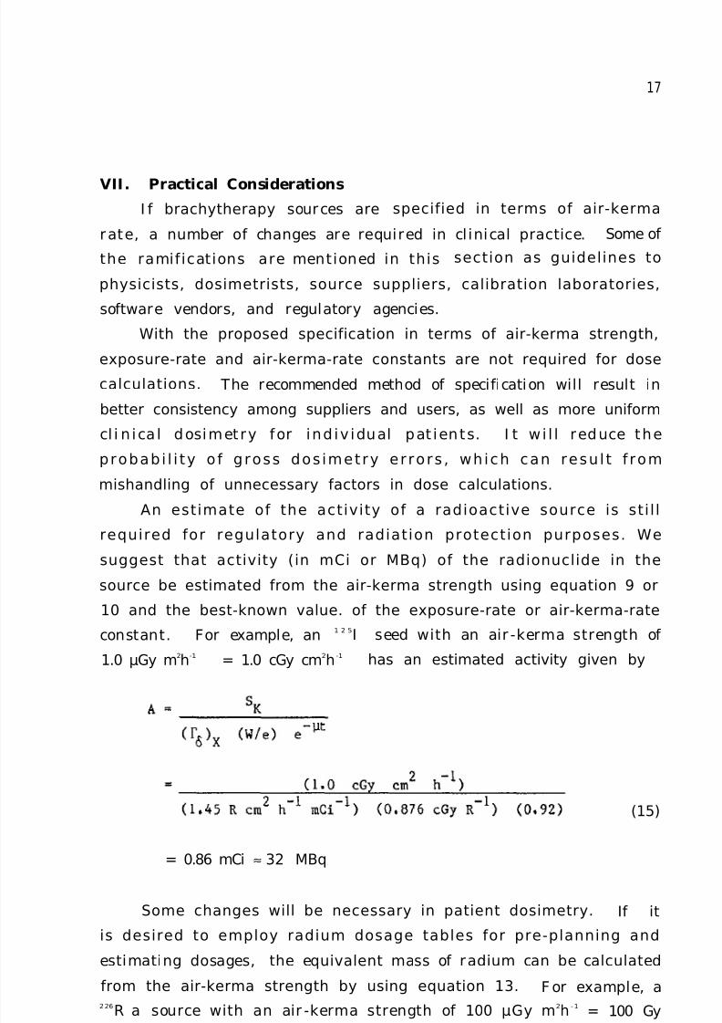
VII. Practical Considerations
I f brachytherapy sources are specified in terms of air-kerma
rate, a number of changes are required in cl inical practice. Some of
the ramifications are mentioned in this section as guidelines to
physicists, dosimetrists, source suppliers, calibration laboratories,
software vendors, and regulatory agencies.
With the proposed specification in terms of air-kerma strength,
exposure-rate and air-kerma-rate constants are not required for dose
calculations. The recommended method of specification will result in
better consistency among suppliers and users, as well as more uniform
cl i n ica l dosimetry for ind iv idual pat ients. I t w i l l reduce the
probabi l i ty o f gross dos imetry er rors , which can resu l t f rom
mishandling of unnecessary factors in dose calculations.
An estimate of the activity of a radioactive source is sti l l
required for regulatory and radiation protection purposes. Wesuggest that activity (in mCi or MBq) of the radionuclide in the
source be estimated from the air-kerma strength using equation 9 or
10 and the best-known value. of the exposure-rate or air-kerma-rate
constant. For example, an1 2 5I seed with an air -kerma strength of
1.0 µGy m2h-1 = 1.0 cGy cm2h -1 has an estimated activity given by
= 0.86 mCi ≈ 32 MBq
Some changes will be necessary in patient dosimetry.
(15)
If it
is desired to employ radium dosage tables for pre-planning and
estimating dosages, the equivalent mass of radium can be calculated
from the air-kerma strength by using equation 13. F or example, a226R a source with an air -kerma strength of 100 µGy m2h -1
= 100 Gy
m 2 h - 1has an equivalent mass of radium given by

7/28/2019 Aapm Report No. 21 Specification Of
http://slidepdf.com/reader/full/aapm-report-no-21-specification-of 21/24
18
(16)
= 13.8 mg
Currently, tr eatment planning systems speci fy the source
strength as the activity of the radionuclide or the equivalent mass
of radium and then employ approximate forms of equation 1 to compute
exposure rate. I t i s recommended that ai r -k erma str ength be
specified as input i n these algori thms, and that equation 11 or its
equivalent be employed for exposure computations. I t should be noted
that th i s r equi r es a minor change in the cur rent ly employed
algorithms for exposure-rate calculations: specifically the product
A ( Γ δ )X must be replaced by SK /(W/e). Since (W/e) is a constant, this
recommendation is replacing two variables A and ( Γ δ )X by a single
variable SK . Al l other aspects of the dose calculation programs such
as tissue attenuation factors, obliquity effect, Sievert integrals,
etc. are unaffected by this change.

7/28/2019 Aapm Report No. 21 Specification Of
http://slidepdf.com/reader/full/aapm-report-no-21-specification-of 22/24
19
Appendix: I nformation Recommended for Manufacturer’s Certi ficate
of Calibration.
Manufacturer’s name and address.
Certificate number.
Serial or lot number, model number.
Radionuclide, nominal activity, half- l i fe.
Surface contamination: method of test, date of test, resul t of test.
Leakage : method of test, date of test, result of test.
Active contents: chemical and physical form, mass, volume.
Encapsulati on: mater i a l s of const ruct i on, dimensions, diagrams
illustrating all dimensions.
Air-kerma strength, in µGy m2h-1. Date of calibration, and level of
traceability to NBS standards.
T he cer t i f i cate shoul d al so con tai n statements such as the
following:
Air-kerma strength is the product of air-kerma rate in free space and
the square of the distance of the calibration point from the source
center along the perpendicular bisector.
The ai r -kerma rate i s not i sot ropi c ar ound the source. The mean
source strength, averaged over all dir ecti ons, can be obtained by
applying the factor _____ to the air-kerma strength.
Documentation of the calibration procedures employed is available on
application to (name to be supplied by manufacturer).

7/28/2019 Aapm Report No. 21 Specification Of
http://slidepdf.com/reader/full/aapm-report-no-21-specification-of 23/24
20
References
1.
2.
3.
4.
5.
6.
7.
8.
9.
National Commission on Radiation Protection and Measurements,
Specification of Gamma-Ray Brachytherapy Sources, NCRP Report
No. 41, Washington, D.C. (1974).
A. Dutr ei x and A. Wambersie, "Specification of Gamma-Ray
Brachytherapy Sources," Bri e. J . Radiol. 48, 1034 (1975).
R. Gibb and J .B. Massey, "Radium Dosage: SI Units and the
Machester System," Br i t. J . Radiol. 53, 1100 (1980).
E.G.A. Aird and M.J . Day, "Quantities and SI Units for DoseRate Ca l cu l a t i ons f rom Rad ionucl i de Sources ," B r i e. J .
Radiol. 56, 978 (1983).
S. J ayaraman, L .H. Lanzl and S.K . Agarwal, "An Overvi ew of
Errors in L ine Source Dosimetry for Gamma-Ray Brachytherapy,"
Med. Phys. 10, 871 (1983).
C omit e' F r ancais M easure des R ayonnements I on i s a n t s,
"Recommendations Pour la Determination Des Doses Absorbees E n
Curietherapie," CFMRI Report No. 1 (1983).
R . L oevinger , "The Ro le of the S tandards L abora tory in
Brachytherapy," in Recent Advances in Brachytherapy Physics,
edited by D.R. Shearer, Medical Physics Monograph No. 7, American
Institute of Physics, New York (1981), pp. 22-31.
British Committee on Radiat. Units and Measurements, "Specifica-
tion of Brachytherapy Sources," Bri t. J . Radiol. 57, 941 (1984).
AAPM Task Group 21, "A Protocol for the Determination of Absorbed
Dose from High-Energy Photon and Electron Beams," Med. Phys. 10,
741 (1983).
10. National Council on Radiation Protection, "Dosimetry of X-Ray and
Gamma-Ray Beams for Radiation Therapy in the Energy Range 10 keV
to 50 MeV," NCRP Report No. 69, Washington, D.C. (1981).
11. T .P . L oftus, "Standardizat ion of 137Cs Gamma-Ray Sources in
Terms of E xposure Uni ts (Roentgens)," J . Res. Nat. Bur . Stand.
74A, 1 (1970).
12. T .P . Loftus, "Standardizat ion of 192I r Gamma-Ray Sources in
Terms of Exposure," J . Res. Nat. Bur. Stand, 85, 19 (1980).
13. T.P. Loftus, "Exposure Standardization of 1 2 5I Seeds Used for
Brachytherapy," J . Res. Nat. Bur. Stand. 89, 295 (1984).

7/28/2019 Aapm Report No. 21 Specification Of
http://slidepdf.com/reader/full/aapm-report-no-21-specification-of 24/24
21
14. J .F. Williamson, R.L. Morin and F.M. Khan, "Monte Carlo Evalution
of the Sievert I ntegral for B rachytherapy Dosimetry," Phys. Med.
Biol. 28, 1021 (1983).
15. K . J . Cassel l , "A F undamental A pproach to the Design of a
Dose-Rate Calculation Program for Use in Brachytherapy Planning,"
Bri t. J . Radiol . 56, 113 (1983).
16. G.M. Keyser, "Absorption Corrections for Radium Standardization,"
Can. J . Phys. 29, 301 (1951).
17. G.N. Whyte, "Attenuation of Radium Gamma Radiation in CylindricalGeometry," Bri t. J . Radiol . 28, 635 (1955).
18. C.C. L ing, E.D. Y orke, I .J . Spiro, D. K ubiatowicz and D. Bennett,
"Phys i ca l Dos imet ry o f 1 2 5I S eeds of a N ew D esi gn for
I ntersti ti al I mplant," I nt. J . Radiat. Oncol . B io. Phys. 9, 1747
(1983).
19. C .C . L ing , Z.C . Gromadzk i , S .N . Rus tgi and J .H . Cund i f f ,
"Di recti onal Dependence of R adiati on F luence fr om
1 9 2
I r a n d198Au Sour ces , "Radiol. 146, 791 (1983).
20. M.E .J . Young and H.F . Batho, "Dose Tables for L inear Radium
Sources Calculated by an Electronic Computer," Brit. J . Radiol.
37, 38 (1964).
21. L .L . Anderson, H.M. Kuan and I.Y. Ding, "Clinical Dosimetry
w i t h 1 2 5I S e ed s , " in Modern Interst it ial and Intracavitary
Radiation Cancer Management, edited by F .W. George I I I , MassonPublishing Co. (1981). pp. 9-15.
22. W.J . M eredith (E d.), Radium Dosage: The Manchester System,
L ivingstone Publishers, Edinburgh and London (1947).
23. L .L . Anderson, B.S. Hi laris and L.K . Wagner, "A N omograph for
P lanar I mplants P lanning," Endocurietherapy/Hyperthermia Oncol.
1, 9-15 (1985).
24. Int. Commission on Radiation Units and Measurements, "Dose
and V ol ume Speci fi cati on f or Reporting I ntracavitary Therapy
in Gynecology," ICRU Report No. 38, Bethesda, MD, 1985.
25. M. Boutillon and A.M. Perroche-Roux, "Re-evaluation of the W
Value for E lect rons in Dry A i r , " Phys . Med. B io l . 32, 213
(1987).