accepted manuscript - universidade de coimbra
TRANSCRIPT

Accepted Manuscript
Neurotoxic effect of oligomeric and fibrillar species of A<beta>1-42 peptide. Involvement of ER calcium release in oligomers-induced cell death.
Rosa Resende, Elisabete Ferreiro, Claudia Pereira, Catarina Resende de Oliveira
PII: S0306-4522(08)00884-1 DOI: 10.1016/j.neuroscience.2008.06.036 Reference: NSC 10409
To appear in: Neuroscience
Received date: 15 November 2007 Revised date: 5 June 2008 Accepted date: 5 June 2008 Please cite this article as: Resende, R., Ferreiro, E., Pereira, C., Resende de Oliveira, C., Neurotoxic effect of oligomeric and fibrillar species of A<beta>1-42 peptide. Involvement of ER calcium release in oligomers-induced cell death., Neuroscience (2008), doi: 10.1016/j.neuroscience.2008.06.036. This is a PDF file of an unedited manuscript that has been accepted for publication. As a service to our customers we are providing this early version of the manuscript. The manuscript will undergo copyediting, typesetting, and review of the resulting proof before it is published in its final form. Please note that during the production process errors may be discovered which could affect the content, and all legal disclaimers that apply to the journal pertain.

ACC
EPTE
D M
ANU
SCR
IPT
1
Neurotoxic effect of oligomeric and fibrillar species of Aβ1-42 peptide. Involvement of
ER calcium release in oligomers-induced cell death.
Rosa Resende, Elisabete Ferreiro, Cláudia Pereira, Catarina Resende de Oliveira
Institute of Biochemistry, Faculty of Medicine and Center for Neuroscience and Cell Biology,
University of Coimbra, 3004-504 Coimbra, Portugal
Corresponding author: Cláudia Pereira, Institute of Biochemistry, Faculty of Medicine and
Center for Neuroscience and Cell Biology, University of Coimbra, 3004-504 Coimbra,
Portugal
Tel: +351239820190; Fax: +351239822776; E-mail: [email protected].
Article type: Research Paper
Section Editor: Dr. Constantino Sotelo, CNRS UMR 7102, Universite Pierre et Marie Curie,
6eme etage, Bat B, Case 12, 9 Quai St. Bernard, 75005 Paris, France
ACCEPTED MANUSCRIPT

ACC
EPTE
D M
ANU
SCR
IPT
2
Abbreviations
Aβ, amyloid-beta peptide; AD, Alzheimer’s disease; ADDLs, amyloid-derived diffusible
ligands; APP, amyloid precursor protein; BSA, bovine serum albumine; CICR, Ca2+-induced
calcium release; DEVD-pNA, N-acetyl-Asp-Glu-Val-Asp-p-nitroanilide; DMSO, dimethyl
sulfoxide; DT, dantrolene; DTT, 1,4-dithiotreitol; ECF, enhanced chemifluorescence; ER,
endoplasmic reticulum; Fura-2/AM, Fura-2 acetoxymethyl ester; HFIP, 1,1,1,3,3,3-
hexafluoro-2-propanol; Indo-1/AM, indo-1 acetoxymethyl ester; IP3R, inositol 1,4,5-
triphosphate receptor; LTP, long-term potentiation; MTT, 3-(4,5-dimethylthiazol-2-yl)-2,5-
diphenyl tetrazolium bromide; MAP-2, microtubule-associated protein 2; NMDA, N-methyl-
D-aspartate; PBS, phosphate-buffered saline; PLC, phospholipase C; PMSF,
phenylmethylsulfonyl fluoride; PVDF, polyvinylidene difluoride; RyR, ryanodine receptors;
RT, room temperature; TBS, tris-buffered saline, ThS, thioflavin S; ThT, thioflavin T.
ACCEPTED MANUSCRIPT
ACC
EPTE
D M
ANU
SCR
IPT
3
ABSTRACT
The nature of the toxic form of amyloid-β peptide (Aβ) early involved in Alzheimer’s disease
(AD) pathology and whether it is the fibrillar or the oligomeric peptide the most deleterious to
neurons still remain controversial issues. This work was aimed to compare the neurotoxicity
of different Aβ1-42 assemblies, using “fresh” and “aged” samples enriched in oligomeric and
fibrillar species, respectively, and also isolated oligomers and fibrils. The results obtained
with “fresh” and “aged” Aβ1-42 preparations suggested that oligomeric species are more
toxic to cortical neurons in culture than fibrillar forms, which was confirmed by using isolated
oligomers and fibrils. In order to further elucidate the mechanisms involved in soluble Aβ
toxicity, the involvement of endoplasmic reticulum (ER) calcium release in oligomers-
induced apoptosis was evaluated. We observed that oligomeric Aβ1-42 depletes ER Ca2+
levels leading to intracellular Ca2+ dyshomeostasis involving phospholipase C activation.
Moreover, in the presence of dantrolene, an inhibitor of ER Ca2+ release through ryanodine
receptors, the oligomers-induced apoptosis was prevented demonstrating the involvement of
ER Ca2+ release.
KEYWORDS
Alzheimer’s disease, amyloid-beta peptide, oligomers, fibrils, endoplasmic reticulum, calcium
homeostasis.
ACCEPTED MANUSCRIPT

ACC
EPTE
D M
ANU
SCR
IPT
4
The in vitro and in vivo neurotoxicity induced by fibrillar amyloid-beta peptide (Aβ) ( Pike et
al., 1993; Lorenzo and Yankner, 1994) has supported the amyloid cascade hypothesis in
which seeding of insoluble Aβ1-42 is a causative factor in the pathogenesis of Alzheimer’s
disease (AD) (Hardy and Selkoe, 2002). However, amyloid plaques do not always correlate
with neurodegeneration and cognitive decline (Masliah et al., 1994; Mucke et al., 2000; Klein
et al., 2001). In vivo, small stable oligomers of Aβ1-42 have been isolated from the brain of
AD patients (Gong et al., 2003) and the levels of soluble Aβ are well correlated with
cognitive deficits (McLean et al., 1999; Naslund et al., 2000). Recent studies in animals have
established a link between natural, as well as synthetic, soluble Aβ oligomers and cognitive
impairment (Richardson et al., 2003; Cleary et al., 2005; Lesné et al., 2006). In vitro,
oligomeric and protofibrillar forms of Aβ have been shown to be directly neurotoxic (Lambert
et al., 1998; Hartley et al., 1999; Deshpande et al., 2006). Despite the significant advances in
AD research made in the last decade, the nature of the toxic form of Aβ early involved in AD
pathology and also whether it is the fibrillar or the non-fibrillar peptides that are the most
deleterious to neurons still remain controversial issues. The above mentioned studies
emphasize the necessity to clarify the initial response of neurons to the non-fibrillar and
fibrillar Aβ, in particular to the Aβ1-42 peptide.
The endoplasmic reticulum (ER) has several important functions including the regulation of
intracellular Ca2+ homeostasis. Under various conditions, ER function is disturbed leading to
the accumulation of unfolded proteins and activation of a sporadic ER stress response, also
known as the unfolded protein response (UPR). When cells are subjected to severe or
prolonged ER stress, the transcriptional factor CHOP/Gadd153 is induced and apoptotic cell
death occurs (Kaufman, 1999; Paschen, 2001). ER stress has been implicated in many
important diseases, including AD (Lindholm et al., 2006). Ferreiro and colleagues (2006)
ACCEPTED MANUSCRIPT

ACC
EPTE
D M
ANU
SCR
IPT
5
have recently demonstrated that ER Ca2+ release through ryanodine receptors (RyR) and
inositol 1,4,5-triphosphate receptor (IP3R) contributes to the early increase in intracellular
Ca2+ levels and to the activation of apoptosis induced by fibrillar Aβ peptide. The present
paper was aimed to compare the neurotoxic effects of different Aβ1-42 assemblies, namely
oligomers and fibrils, using “fresh” and “aged” preparations of synthetic Aβ1-42 which are
enriched in oligomeric or fibrillar species, respectively, and also isolated oligomers and
fibrils. Furthermore, in this study we investigated the role of ER dysfunction in apoptosis
induced by oligomeric Aβ1-42 in order to further elucidate the molecular mechanisms
involved in Aβ oligomers-induced neurodegeneration. We have shown that oligomers are
more toxic to cortical neurons than fibrils and induce apoptotic cell death through a
phospholipase C- (PLC) mediated mechanism that involves ER Ca2+ release through channels
associated with the RyR. Taken together, our findings support the hypothesis that oligomeric
Aβ plays an important role in AD pathogenesis, being responsible for the early changes that
lead to neuronal death.
ACCEPTED MANUSCRIPT

ACC
EPTE
D M
ANU
SCR
IPT
6
EXPERIMENTAL PROCEDURES
Materials
Neurobasal medium and B27 supplement were purchased from GIBCO BRL, Life
Technologies (Paisley, UK). Trypsin, trypsin inhibitor type-II-soybean, deoxyribonuclease I
(DNase I), 3-(4,5-dimethylthiazol-2-yl)-2,5-diphenyl tetrazolium bromide (MTT), protease
inhibitors, phenylmethylsulfonyl fluoride (PMSF), bovine serum albumine (BSA),
1,1,1,3,3,3-hexafluoro-2-propanol (HFIP), U-73122 and Thioflavin T (ThT) were obtained
from Sigma Chemical Co. (St. Louis, MO, USA). Acetoxymethyl ester of Fura-2 (Fura-
2/AM) and Indo-1 (Indo-1/AM), Hoechst 33342 and Alexa Fluor 594 goat anti-mouse IgG
conjugate were purchased from Molecular Probes (Leiden, Netherlands). The synthetic Aβ1-
42 peptide was from Bachem (Bubendorf, Switzerland). Phenol red-free Ham’s F-12 medium
was purchased from Cambrex Bio Science (Walkersville, USA). In Situ Cell Death Detection
Kit, Fluorescein was obtained from Roche Applied Science (Mannheim, Germany). The
caspase-3 colorimetric substrate N-acetyl-Asp-Glu-Val-Asp-p-nitroanilide (DEVD-pNA),
ZVAD-fmk and MK801 were obtained from Calbiochem (Darmstadt, Germany). Mouse
monoclonal antibody 6E10, reactive against residues 1-17 of Aβ was obtained from Signet
(Deshman, MA, USA). Reagents and apparatus used in immunoblotting assays were obtained
from Bio-Rad (Hercules, CA, USA), whereas polyvinylidene difluoride (PVDF) membrane,
goat alkaline phosphatase-linked anti-mouse secondary antibody and the enhanced
chemifluorescence (ECF) reagent were from Amersham Pharmacia Biotech
(Buckinghamshire, UK). Low-Range Rainbow prestained protein standard was from
Amersham Biosciences (USA). The Dako fluorescent mounting medium was purchased from
DakoCytomation Inc. (Carpinteria, CA, USA). All the others chemicals were obtained from
Sigma Chemical Co. (St. Louis, MO, USA) or from Merck kgaA (Damstadt, Germany).
ACCEPTED MANUSCRIPT

ACC
EPTE
D M
ANU
SCR
IPT
7
Primary rat embryo cortical neuronal cultures
Cortical neurons were isolated from E15-E16 Wistar rat embryos according to the
method described by Hertz and collaborators (1989) slightly modified (Resende et al., 2007).
Briefly, removed cortices were aseptically dissected and washed in Ca2+- and Mg2+-free Krebs
buffer (in mmol/L): NaCl 120, KCl 4.8, KH2PO4 1.2, glucose 13, Hepes 10 (pH 7.4) and then
incubated in Krebs solution supplemented with BSA (0.3 g/L), containing trypsin (0.5 g/L)
and DNase I (0.04 g/L), for 10 min at 37 ºC. The tissue digestion was stopped by the addition
of trypsin inhibitor (type II-S) (0.75 g/L) in Krebs buffer containing DNase I (0.04 g/L),
followed by a centrifugation at 140 x g for 5 min. After washing the pellet once with Krebs
buffer, the cells were dissociated mechanically and ressuspended in fresh Neurobasal medium
with 2 mmol/L L-glutamine, 2% (v/v) B27 supplement, penincillin (100,000 U/L), and
streptomycin (100 mg/L).
The cells were seeded on poly-L-lysine (0.1 g/L)-coated dishes at a density of 0.25 x
106 cells/cm2 for measurement of caspase-3-like activity, 0.125 x 10⁶ cells/cm2 for the MTT
assay or 0.33 x 10⁶ cells/cm2 for western blotting. For fluorescence studies, neurons were
mounted on poly-L-lysine-coated glass coverslips at a density of 0.1 x 10⁶ cells/ cm2 or on
poly-L-lysine-coated dishes at 0.4 x 10⁶ cells/cm2. The cultures were maintained in serum-
free Neurobasal medium supplemented with B27, at 37 °C in a humidified atmosphere of 5%
CO2/95% air for 5-7 days before treatments in order to allow neuronal differentiation. Under
these conditions, glial growth is less than 10% (Ferreiro et al., 2006).
Preparation of amyloid-β (Aβ) peptide solutions and treatment protocols
“Fresh and aged” peptide
ACCEPTED MANUSCRIPT

ACC
EPTE
D M
ANU
SCR
IPT
8
The synthetic peptide Aβ1-42, corresponding to the neurotoxic amino acid residues of
the human amyloid-beta protein (Aβ), was dissolved in sterile water, or in a diluted ammonia
solution to facilitate peptide solubilization, at a concentration of 1 g/L (221.5 µmol/L). Aβ1-
42 aliquots were then stored at -20 ºC until being used (“fresh” Aβ1-42 solution), or were
incubated for 7 days at 37 ºC (“aged” Aβ1-42 solution).
The fibril content of both preparations (“fresh” and “aged” Aβ1-42) was analyzed by
different criteria including the thioflavin T (ThT) fluorimetric assay and immunoblotting with
the 6E10 antibody. To study the effect of the aggregation state of Aβ1-42 peptide on cell
viability and apoptosis, different concentrations (0.5-5 µmol/L) of “fresh” or “aged” Aβ1-42
were added to the culture medium of cortical neuronal cells for 6-24 hr.
Aβ oligomers and fibrils
Synthetic Aβ1-42 peptide was dissolved in 1,1,1,3,3,3-hexafluoro-2-propanol (HFIP) to 1
mmol/L. The HFIP was then removed in a Speed Vac (Ílshin Lab. Co., Ltd, Ede, The
Netherlands) and the dried HFIP film was stored at -20 ºC. The peptide film was resuspended
to make a 5 mmol/L solution in anhydrous dimethyl sulfoxide (DMSO) (Dahlgren et al.,
2002). Aβ1-42 oligomers were prepared by diluting the solution in phenol red-free Ham’s F-
12 medium without glutamine to a 100 µmol/L final concentration and incubated overnight at
4 ºC (Lambert et al., 1998). The preparation was centrifuged at 15,000 x g for 10 min at 4 ºC
to remove insoluble aggregates, and the supernatant containing soluble oligomers was
transferred to clean tubes and stored at 4 ºC. The fibrils were prepared by diluting 5 mmol/L
Aβ1-42 in DMSO to 200 µmol/L in 100 mmol/L Hepes buffer (pH 7.5) and aged at 37 ºC for
7 days. The preparation was then centrifuged during 10 min at 15,000 x g at RT, and the
supernatant containing soluble oligomers was discarded. The pellet containing Aβ fibrils (and
possibly proto-fibrils) was ressuspended in 100 mmol/L Hepes buffer (pH 7.5). Protein
ACCEPTED MANUSCRIPT

ACC
EPTE
D M
ANU
SCR
IPT
9
concentrations of Aβ oligomers and fibrils were determined using the Bio-Rad protein dye
assay reagent.
Aggregation state of Aβ peptide
The fibril content in Aβ1-42 stock solutions was evaluated by a ThT fluorimetric
assay as previously described (Hashioka et al., 2005) with some modifications. “Fresh” or
“aged” Aβ1-42 peptide (221.5 µmol/L) was added to 3 µmol/L ThT in 50 mmol/L glycine-
NaOH buffer (pH 8.5). Fluorescence was monitored at 450 nm excitation and 482 nm
emission using a Perkin-Elmer LS50B spectrofluorometer. A time scan was performed and
fluorescence values were measured after the decay reached a plateau and the background
fluorescence of 3 µmol/L ThT was subtracted. The data from three identical samples in
separate experiments were then averaged to obtain the final values.
The presence of different assembly forms (monomers, oligomers and fibrils) of Aβ1-
42 in “fresh” and “aged” preparations and the purity of isolated oligomers and fibrils was
evaluated by gel electrophoresis and western blot. Aβ samples containing 10 µg of protein,
were diluted (1:2) with sample buffer [40% (w/v) glycerol, 2% (w/v) SDS, 0.2 mol/L Tris-
HCl, pH 6.8 and 0.005% (w/v) Comassie G-250] and were separated by electrophoresis on a
4-16% Tris-Tricine SDS gel (Klafki et al., 1996). Samples were not boiled to minimize
disaggregation prior to electrophoresis. To facilitate the identification of proteins a Low-
Range Rainbow prestained protein standard was used. Proteins were then transferred to PVDF
membranes, which were further blocked for 1 hr at RT with 5% (w/v) fat-free milk in Tris-
buffered saline (150 nmol/L NaCl, 50 nmol/L Tris, pH 7.6) with 0.1% (w/v) Tween 20 (TBS-
T). The membranes were next incubated overnight at 4 ºC with 6E10 mouse monoclonal
primary antibody against Aβ diluted in TBS-T with 0.5% (w/v) fat-free milk (1:1000). After
washing in TBS-T with 0.5% (w/v) fat-free milk, membranes were further incubated for 1 hr
ACCEPTED MANUSCRIPT
ACC
EPTE
D M
ANU
SCR
IPT
10
at RT with an alkaline phosphatase-conjugated anti-mouse secondary antibody (1:20,000). Aβ
bands were visualized after membrane incubation with ECF reagent for 5 min, on a Versadoc
Imaging System.
Cell viability assay
After treatment of cortical neurons with different concentrations of the Aβ peptides,
cell viability was evaluated by the MTT assay (Mosmann, 1983), which measures the ability
of metabolic active cells to form formazan through cleavage of the tetrazolium ring of MTT.
Neurons were washed in sodium medium (in mmol/L: NaCl 132, KCl 4, NaH2PO4 1.2, MgCl2
1.4, glucose 6, Hepes 10, and CaCl2 1, pH 7.4) and incubated with MTT (0.5 g/L) for 2 hr at
37 ºC. The blue formazan crystals formed were dissolved in an equal volume of HCl 0.04
mol/L in isopropanol and quantified spectrophotometrically by measuring the absorbance at
570 nm using a microplate reader (Spectra max Plus 384, Molecular Devices).
MAP2 immunocytochemistry
Cortical neurons grown in glass coverslips, in the presence or absence of Aβ1-42,
were washed with PBS buffer (pH 7.4), and were fixed with 4% paraformaldehyde (w/v) for
15 min at RT. Then, the cells were permeabilized for 2 min at with 0.2% (v/v) Triton X-100
in PBS buffer (pH 7.4) and blocked for 1 hr and 30 min in PBS containing 3% (w/v) BSA.
The cells were incubated for 1 hr with a mouse anti-microtubule-associated protein-2 (MAP-
2) monoclonal antibody (1:500 dilution in 3% BSA/PBS) and then washed and incubated with
Alexa Fluor 594 goat anti-mouse IgG antibody conjugate (1:200 dilution in 3% BSA/PBS) for
1 hr. Finally, coverslips were mounted in Dako fluorescent mounting medium on a
microscope slide and neurons were visualized in an inverted fluorescence microscope
Axiovert 200 (Zeiss, Germany).
ACCEPTED MANUSCRIPT

ACC
EPTE
D M
ANU
SCR
IPT
11
Caspase-3 activity
Cultured cortical neurons, which were either treated or untreated with the Aβ peptide,
were scrapped in cold (4 ºC) lysis buffer (in mmol/L): HEPES-Na 25, MgCl2 2, EDTA 1,
EGTA 1, PMSF 0.1, DTT 2, supplemented with a protease inhibitor cocktail containing
leupeptin, pepstatin A, chymostatin and antipain (1 g/L each). The cellular extract was rapidly
frozen and thawed three times, and then centrifuged for 10 min at 20,200 x g at 4 ºC. The
supernatant was collected and assayed for protein content using the Bio-Rad protein dye assay
reagent. Aliquots of cellular extracts containing 30 µg of protein were reacted with 100
µmol/L Ac-DEVD-pNA, a chromogenic substrate for caspase-3, in a reaction buffer
containing 25 mmol/L Hepes-Na, 10 mmol/L DTT, 10% (wt/vol) sucrose, and 0.1% (wt/vol)
CHAPS (pH 7.4), for 2 hr at 37 ºC (Cregan et al., 1999). Caspase-3-like activity was
determined by measuring substrate cleavage at 405 nm in a microplate reader.
Neuronal apoptosis
Apoptotic cells were identified and quantified based on nuclear DNA morphology by
staining neurons with the cell-permeable DNA dye Hoechst 33342, as described previously
by Kruman et al. (1997). Cortical neurons, incubated in the absence or in the presence of
“fresh” Aβ, “aged” Aβ, oligomers or fibrils isolated from synthetic Aβ1−42, were washed
with PBS buffer (pH 7.4) and fixed with 4% (wt/vol) paraformaldehyde for 15 min at RT. The
cells were then incubated for 5 min with Hoechst 33342 (15 mg/L in PBS, pH 7.4), in the
dark. Cells with homogeneously stained nuclei were considered to be viable, whereas the
presence of condensed and fragmented nuclei was indicative of apoptotic cells. Cell death was
also evaluated by TUNEL staining performed using an In Situ Cell Death Detection Kit,
Fluorescein according to the manufacturer's directions. Fixed cells were permeabilized in
ACCEPTED MANUSCRIPT

ACC
EPTE
D M
ANU
SCR
IPT
12
0.1% Triton X-100, supplemented with 0.1% sodium citrate in PBS, for 2 min, on ice. After
washing, cells were incubated in a mixture of the enzymatic solution with the label solution,
for 1 hr at 37 °C, in the dark. Finally, cells were washed with PBS and coverslips mounted in
Dako fluorescent mounting medium on a microscope slide. DNAse pretreated cells were used
as a positive control. The number of apoptotic cells was counted by using an inverted
AxiovertMicroscope 200 (Zeiss, Germany).
Ca2+ homeostasis
The free intracellular cytosolic Ca2+ concentration ([Ca2+]i) was measured using Indo-
1/AM. Untreated or treated cultured cortical neurons were incubated with 3 µmol/L Indo-
1/AM in sodium medium (in mmol/L: NaCl 132, KCl 4, CaCl2 1, MgCl2 1.4, glucose 6,
HEPES-Na 10, pH 7.4) for 45 min at 37 ºC, in the dark. The cells were then incubated in
Indo-1/AM-free sodium medium for 15 min to ensure the complete hydrolysis of the dye. The
Indo-1 fluorescence was measured spectrofluorimetrically using a microplater reader
(SpectraMax Gemini EM, Molecular Devices) with 350 nm excitation and 410 nm emission.
Calibration of cytosolic Ca2+ levels was performed using the Ca2+ ionophore ionomycin (3
µmol/L) and MnCl2 (3 mmol/L). The free [Ca2+]i was calculated as previously described by
Bandeira-Duarte et al. (1990).
Changes in cytosolic Ca2+ concentration in individual control or treated cells were
analysed by calcium imaging using the fluorescent probe Fura-2/AM. Cells plated in
coverslips were washed 2 times in Krebs buffer (pH 7.4) (in mmol/L: NaCl 132, KCl 4,
MgCl2 1.4, Glucose 6, HEPES 10, NaHCO3 10, CaCl2 1) and loaded with 5 µmol/L Fura-
2/AM supplemented with 0.2% (w/v) pluronic acid, in Krebs buffer for 40 min at 37 °C, in
the dark. Afterwards, cells were washed 3 times and the coverslip was assembled to the
perfusion chamber filled with 500 µL of Krebs buffer, in an inverted fluorescence microscope
ACCEPTED MANUSCRIPT
ACC
EPTE
D M
ANU
SCR
IPT
13
axiovert 200 (Zeiss, Germany). Cells were alternately excited at 340 and 380 nm using a
Lambda DG4 apparatus (Sutter Instruments company, Nocato, Ca, USA), and emitted
fluorescence was collected with a 40x objective and was driven to a coll SNAP digital camera
(Roper Scientific, Trenton, NJ, USA). After a baseline was established, cells were stimulated
with Aβ oligomers (0.5 µmol/L). To assess extracellular Ca2+ contribution, recording of
Fura2-AM fluorescence was also performed in Ca2+- free Krebs buffer, supplemented with 50
µmol/L EGTA. To evaluate the involvement of PLC, cells were pre-incubated with PLC
inhibitor U-73122 (5 µmol/L) for 10 min, before Fura2-AM fluorescence recording.
ER Ca2+ content was assessed by single cell Ca2+ imaging, according to the method
described by Ferreiro and colleagues (2008). After a baseline was established in free Ca2+ -
free Krebs buffer, cells were stimulated with thapsigargin (2.5 µmol/L final concentration), to
empty [Ca2+]ER stores. Acquired values were processed using the MetaFluor software
(Universal Imaging Corporation, Buckinghamshire, UK). The peak amplitude of Fura-2
fluorescence (ratio at 340/380 nm) was used to evaluate cytosolic Ca2+ content.
Statistical analysis
Data were expressed as the means ± SEM of the indicated number of experiments.
Statistical significance was determined by using one-way ANOVA followed by Tukey post-
hoc tests. The differences were considered significant for p values < 0.05.
ACCEPTED MANUSCRIPT

ACC
EPTE
D M
ANU
SCR
IPT
14
RESULTS
“Fresh” and “aged” Aβ1-42 peptide solutions have different fibril content
In this paper, we analyzed the effect of different Aβ1-42 assemblies on neuronal cell
viability and apoptotic death. For that purpose, we prepared “fresh” and “aged” Aβ1-42
peptide solutions, which were characterized by ThT fluorescence and western blotting. The β-
sheet content and, therefore, of fibrils of “aged” Aβ1-42, prepared after incubation of the
peptide solution for 7 days at 37ºC, were significantly higher in comparison with that
determined in “fresh” Aβ1-42, used immediately after peptide solubilization (Fig. 1A). Low-
molecular weight oligomers of Aβ1-42 (~14 kDa) were preferentially detected in “fresh”
samples while fibrillar Aβ1-42 (high molecular weight bands) was the predominant species
detected in “aged” samples (Fig. 1B). Taken together, our results clearly show that “fresh”
and “aged” Aβ1-42 preparations have a significantly different fibril content, the former being
enriched in oligomeric forms and the later in fibrillar Aβ. Furthermore, the preformed fibrils
of the “aged” peptide are not dissociated when the peptide is diluted into the culture medium
as evaluated by fluorescence microscopy using thioflavin S (ThS) (data not shown). Thus,
“fresh” and “aged” Aβ1-42 preparations are suitable to evaluate the differential effect of
Aβ assemblies.
“Fresh” Aβ1-42 peptide is more toxic in comparison with the “aged” peptide
After 6 hr incubation of primary cortical neuronal cultures, in the absence or in the
presence of increasing concentrations (0.5-5 µmol/L) of “fresh” or “aged” Aβ1-42 peptide,
cell viability was analyzed by the MTT reduction assay. It was observed that at all tested
concentrations, both “fresh” and “aged” Aβ1-42 are toxic to cortical neurons, significantly
decreasing (p<0.001) the reduction of the tetrazolium salt (Fig. 2A). However, for the same
ACCEPTED MANUSCRIPT

ACC
EPTE
D M
ANU
SCR
IPT
15
concentration (1 and 5 µmol/L), the “fresh” peptide was shown to be more toxic as compared
to the “aged” peptide (p<0.05 and p<0.001).
Similar results were obtained when the number of apoptotic cells was determined in
“fresh” or “aged” Aβ1-42-treated neurons (Fig. 2B). After 24 hr incubation, “fresh” Aβ1-42
was shown to be more effective than the “aged” peptide, significantly increasing the number
of cells exhibiting apoptotic morphology as evaluated by using Hoechst 33342. These results
were further confirmed using Tunel staining (Fig.4). Taken together, the results obtained
demonstrate that Aβ1-42-induced toxicity and cell death correlate with the aggregation state
of the peptide and suggest that the soluble oligomeric forms are the main toxins against
cortical neurons.
“Fresh” Aβ1-42 peptide impairs Ca2+ homeostasis and leads to dendritic dystrophy and
caspase-3 activation
As the “fresh” Aβ1-42 is more toxic than the “aged” peptide, we decided to perform
some experiments using this peptide preparation enriched in oligomers in order to understand
the mechanisms that underly the toxicity of soluble Aβ species. To investigate the effect of
soluble Aβ on the intracellular Ca2+ homeostasis, the basal [Ca2+]i concentration was
measured in control and in cortical neurons treated with “fresh” Aβ1-42 during 6 or 24 hr.
Cytosolic Ca2+ levels were significantly higher (p<0.05) in cortical neurons treated with Aβ1-
42 for 6 hr comparing with the control and this Ca2+ rise persisted 24 hr after addition of
“fresh” Aβ1-42 (Fig. 3).
The analysis of dendritic morphology gives important information about neuronal
health and synaptic integrity. We evaluated dendritic integrity in Aβ1-42-treated cortical
neurons immunostained against MAP2 which was compared with that obtained in neurons
treated with “aged” Aβ peptide (Fig. 4). Untreated cortical cells (control) showed a complex
ACCEPTED MANUSCRIPT

ACC
EPTE
D M
ANU
SCR
IPT
16
dendritic network with smooth and long dendritic arborization (Fig. 4a). On the other hand,
cultured neurons exposed to 5 µmol/L Aβ1-42 for 24 hr showed loss and retraction of the
dendritic arborization (Fig. 4b, c). This effect is more pronounced in “fresh” Aβ-treated
neurons (Fig. 4b).
The activity of caspase-3, an effector caspase in the apoptotic cell death pathway was
measured in cortical neurons treated with 5 µmol/L “fresh” Aβ1-42 during 12 or 24 hr (Fig.
5A). Results obtained have demonstrated that “fresh” Aβ induces caspase-3 activation upon
12 hr treatment (p<0.05) which persists after 24 hr treatment. In order to evaluate the
functional involvement of caspases in Aβ1-42-induced toxicity, ZVAD-fmk, a broad range
caspase inhibitor was used. In the presence of 25 µmol/L ZVAD-fmk the increase in [Ca2+]i
was prevented (Fig. 5B) suggesting the involvement of caspases in Aβ-induced Ca2+
dyshomeostasis. Furthermore, in the presence of ZVAD-fmk the increase in the number of
apoptotic cells triggered by “fresh” Aβ1-42, determined by fluorescence microscopy using
Hoechst 33342, was shown to be abolished (Fig. 5C).
Aβ1-42 oligomers are more toxic than fibrils
Our results obtained using “fresh” and “aged” Aβ1-42, enriched in oligomers and
fibrils respectively suggest that soluble oligomeric forms of the Aβ peptide are more potent
neurotoxins than fibrils. In order to validate these results, we isolated oligomers and fibrils
accordingly to procedures previously published (Dahlgren et al., 2002; Lambert et al., 1998)
which were characterized by immunoblotting with the 6E10 antibody. As shown in Fig. 6,
oligomeric preparation (Aβ1-42O) is composed only of low-n oligomers (~14 kDa) and the
fibrillar preparation (Aβ1-42f) is mainly composed of fibrils although smaller amounts of
oligomers are also detected.
ACCEPTED MANUSCRIPT

ACC
EPTE
D M
ANU
SCR
IPT
17
In cortical neurons, a significant decrease in cell viability was observed after 6 or 24
hr treatment with Aβ oligomers (p<0.01), as demonstrated by the MTT assay (Fig. 7A). At 0.5
and 1 µmol/L, Aβ1-42 oligomers were shown to be more toxic than fibrils (Fig 7B). At the
same concentration, Aβ1-42 fibrils significantly decreased cell viability only after 24 hr
treatment (p<0.05). Moreover, oligomers decreased cell survival more effectively than fibrils
at all time points tested (p<0.05; p<0.001). The same results were obtained when the
apoptotic cell number was determined after 24 hr treatment with oligomeric or fibrillar Aβ1-
42 species (Fig. 7C). The increase in the number of apoptotic cells observed in oligomers-
treated neurons was more pronounced (p<0.05) than that measured in cells incubated with
fibrils. These data are in agreement with the results obtained with “fresh” and “aged” Aβ1-42
preparations.
Aβ oligomers-induced apoptosis is mediated through endoplasmic reticulum Ca2+ release
In cortical neurons treated with oligomeric Aβ1-42 (0.5 µmol/L) during 6 or 24 hr, the
intracellular Ca2+ concentration was measured spectrofluorimetrically using Indo-1/AM. A
significant increase in cytosolic Ca2+ levels was observed after 6 hr treatment with oligomeric
Aβ compared with controls, which persisted until 24 hr incubation (Fig. 8). Dantrolene (DT),
an inhibitor of ER Ca2+ release through channels associated to RyR, was shown to prevent the
Aβ1-42 oligomers-induced rise of [Ca2+]i, suggesting the involvement of Ca2+ release by ER.
To further investigate the involvement of ER on the impairment of Ca2+ homeostasis
triggered by Aβ oligomers, ER Ca2+ content was analyzed indirectly using thapsigargin, an
inhibitor of the ER Ca2+ ATPase that releases Ca2+ from ER stores, in the absence of
extracellular Ca2+. A representative trace of Fura-2 fluorescence ratio at 340 nm and 380 nm
is presented in Fig. 9A. After a baseline was established, cells were stimulated with
thapsigargin, to empty [Ca2+]ER stores. In control neurons, there was an increase in free
ACCEPTED MANUSCRIPT

ACC
EPTE
D M
ANU
SCR
IPT
18
[Ca2+]i after the addition of thapsigargin. In treated neurons, this increase was not observed
since Aβ oligomers have already induced the leak of Ca2+ from this intracellular organelle.
The content of ER Ca2+ was evaluated by the calculation of the peak over baseline, obtained
after thapsigargin addition and the results normalized to values determined in untreated
cortical neurons. Figure 9B show that treatment with Aβ-42 oligomers for 1 hr induce a
significant decrease in the ER Ca2+ content (p<0.001), which was more pronounced 24 hr
after the addition of oligomers.
The inhibition of Ca2+ release from ER with DT (10 µmol/L) also prevented the
apoptotic cell death that was observed in cortical neurons after exposure to Aβ1-42 oligomers
(0.5 µmol/L) for 24 hr (Fig. 10). Together, these results demonstrate that the early release of
Ca2+ from ER in cortical neurons exposed to oligomeric Aβ1-42 species leads to the
perturbation of Ca2+ homeostasis and subsequently to apoptotic cell death.
IP3-PLC pathway is involved in the perturbation of ER Ca2+ homeostasis induced by
Aβ oligomers
To analyze whether the entry of Ca2+ through Ca2+ channels present in the plasma
membrane also contributes to the increase of cytosolic Ca2+ concentration upon treatment
with Aβ oligomers, cortical neurons were loaded with the Ca2+-sensitive fluorescent dye Fura-
2/AM and the Ca2+ content in the cytosol was analyzed by single cell Ca2+ imaging in the
presence or in the absence of extracellular Ca2+. After 4 min recording, Aβ oligomers (0.5
µmol/L) were added to the cells. Few seconds after Aβ addition, Fura-2 fluorescence ratio at
340 nm and 380 nm (F340/F380) significantly increased in cells treated either in the presence
or in the absence of Ca2+ in the medium. However, in the absence of Ca2+, this increase was
smaller than in the presence of Ca2+ (Fig. 11A). The increase in cytosolic Ca2+ content was
determined by measuring the peak over baseline obtained after Aβ oligomers addition. Figure
ACCEPTED MANUSCRIPT

ACC
EPTE
D M
ANU
SCR
IPT
19
11B depicts that the increase of cytosolic Ca2+ content is significantly higher in Aβ-treated
cells in the presence of extracellular Ca2+ than in Ca2+- free medium (p<0.01). These results
indicate that the entry of Ca2+ through channels at the plasma membrane also contribute to the
increase of intracellular Ca2+ induced by Aβ oligomers. To further investigate this hypothesis,
we evaluated the involvement of NMDA receptors by using the MK801 which is an
antagonist of these receptors. In the presence of 2 µmol/L MK801, the increase in the [Ca2+]i
was partially prevented (Fig. 12A) but it does not affect the Aβ oligomers-induced apoptotic
cell death (Fig. 12B) .
The increase in cytosolic Ca2+ content upon ER Ca2+ depletion induced by Aβ
oligomers can be related to the activation of PLC, leading to the formation of diacylglycerol
(DAG) and inositol 1,4,5-trisphosphate (IP3) that subsequently mediates the release of Ca2+
from ER through IP3R and potentiates Ca2+ release by RyR (Ca2+-induced Ca2+ release,
CICR) (Mikoshiba, 2006). To evaluate whether IP3-PLC pathway is involved in the [Ca2+]i
increase induced by Aβ oligomers, cortical neurons were pre-incubated with the PLC
inhibitor U-73122, loaded once again with Fura-2/AM, and stimulated with Aβ oligomers (0.5
µmol/L). The increase in F340/F380 ratio observed in Aβ oligomers-treated cells was
prevented by U-73122 in the presence or absence of extracellular Ca2+, suggesting the
involvement of IP3-PLC signalling in the intracellular Ca2+ mobilization by Aβ oligomers
(Fig. 11 A and B, p<0.05).
ACCEPTED MANUSCRIPT

ACC
EPTE
D M
ANU
SCR
IPT
20
DISCUSSION
Aβ peptide exists in several different conformations, including monomers, oligomers,
amyloid-derived diffusible ligands (ADDLs), protofibrils and fibrils. A strong correlation
between soluble Aβ levels, and the extent of synaptic loss and cognitive impairment was
described (Mclean et al., 1999; Klein et al., 2001; Lacor et al., 2004), suggesting that soluble
oligomers of Aβ are the toxic assembly of the peptide that causes the synaptic dysfunction
and dementia associated with the disease. However, the relative contribution of fibrillar and
soluble oligomeric Aβ to neurodegeneration and dementia in AD has not yet been clearly
established.
We here addressed the possibility that soluble oligomers and fibrillar (plaque-forming)
Aβ1-42 peptide exert a differential effect on neurons. To test this hypothesis, we investigated
the conformation-dependent effect on neuronal viability and activation of apoptotic cell death
of different Aβ assemblies in vitro. Cortical neurons were exposed to “fresh” or “aged” Aβ1-
42, which are enriched in low-molecular-weight oligomers (and possibly protofibrils) or have
a significant high content in amyloid fibrils, respectively, as revealed by ThT staining and
western blotting. By doing this, we intended to mimic what happens in the brain of AD
patients in which the concentration of different Aβ1-42 assemblies varies depending on brain
region, proximity to plaques and the stage of disease. The fact that the Aβ peptide exists in
vivo as a mixture of peptides with different oligomerization states supports the use of our
preparations (“fresh” and “aged” Aβ1-42) to directly compare the effects of distinct
assemblies of Aβ. Our data, obtained using primary embryonic cortical neurons in culture
treated with “fresh” or “aged” Aβ1-42, demonstrate that both assemblies of Aβ1-42 are toxic
to neurons. However, in comparison with fibrils-enriched Aβ1-42 preparations, Aβ samples
enriched in oligomers caused a significantly greater decrease in cell viability and a higher
increase in the extent of apoptotic death than fibrils. In addition, an early activation of
ACCEPTED MANUSCRIPT

ACC
EPTE
D M
ANU
SCR
IPT
21
apoptosis was shown to occur upon exposure to “fresh” Aβ1-42. Although the monomeric
content is higher in “fresh” Aβ which was shown to be more toxic, it does not seem
reasonable to attribute the toxicity to the presence of monomers. It has been shown that unlike
the oligomers, the monomeric form of Aβ peptide does not increase intracellular Ca2+ levels
(Demuro et al., 2005) and does not affect neither LTP (long term potentiation) (Walsh et al.,
2002; Townsend et al., 2006) nor EPSC (excitatory post-synaptic current) activity (Cleary et
al., 2005). Indeed, monomeric Aβ peptide has been described as antioxidant and
neuroprotective (Zou et al., 2002). Therefore, the differential results obtained with “fresh” or
“aged” Aβ1-42 can be attributed solely to the presence of soluble oligomers or fibrils,
respectively. These results were validated using oligomers (low-n oligomers, probably
trimers) and fibrils isolated from synthetic Aβ1-42 being the oligomers more toxic than
fibrils. The fibrils preparation, obtained accordingly to previously published protocols, were
shown to have not only fibrils but also oligomers. However, this preparation was not as toxic
as the oligomeric Aβ preparation. We cannot exclude the hypothesis that the toxicity induced
by the different samples of the Aβ peptide is due to the presence of the oligomers and if so,
the fibrils can be protective. Indeed, Shimmyo and collegues (2007) recently demonstrated
that structural changes in Aβ1-42 from a random coil to a beta-sheet-rich structure protect
cortical neurons from apoptotic cell death and caspase-3 activation.
The neurotoxic effect of aggregated Aβ has been associated with oxidative stress
(Behl et al., 1994; Pereira et al., 1999; Ferreiro et al., 2006; Resende et al., 2007),
mitochondrial damage (Pereira et al., 1998; Cardoso et al., 2001; Casley et al., 2002) and
intracellular Ca2+ dyshomeostasis (Mattson et al., 2002, Ferreiro et al., 2006). The fibrillar Aβ
peptide has been shown to induce apoptosis in neurons involving both the mitochondrial-
mediated and the ER stress-mediated apoptotic pathways (Ferreiro et al., 2004, 2006, in
press) which may contribute to the neuronal degeneration in AD. In addition, neuritic
ACCEPTED MANUSCRIPT
ACC
EPTE
D M
ANU
SCR
IPT
22
dystrophy and synaptic loss were shown to be induced by fibrillar Aβ (Grace et al., 2002;
Grace and Busciglio, 2003; Resende et al., 2007). Deposition of fibrillar Aβ was also
described to be associated with neurite breakage and permanent disruption of neuronal
connections (Tsai et al., 2004). However, because insoluble protein aggregates are likely
surrounded by oligomers it is difficult to ascertain whether the large aggregates are
responsible for local neuronal dysfunction. Several in vitro studies as well as studies using
animal models of AD show that soluble oligomeric forms (and protofibrils) of Aβ,
intermediates in the formation of fibrils by synthetic Aβ, are the main neurotoxins for neurons
which are responsible for the synaptic dysfunction that occurs in the early stages of the
disease (Lambert et al., 1998; Hartley et al., 1999; Walsh et al., 2002). Naturally secreted low-
n Aβ oligomers (dimers and trimers) induce progressive loss of hippocampal synapses
(Shankar et al., 2007) and disrupt cognitive function (Cleary et al., 2005). Aβ oligomers
disrupt Ca2+ homeostasis in primary neuronal cultures (De Felice et al, 2007b; Kelly et al.,
2006). It has also been demonstrated that soluble oligomeric Aβ induces oxidative stress (De
Felice et al., 2007) and tau phosphorylation (De Felice et al., 2007b; Resende et al., 2008).
The oligomers that we obtained are low-n oligomers, probably trimers. Shankar and
colleagues (2007) showed that naturally secreted Aβ dimers and trimers induce progressive
loss of hippocampal synapses. Demuro and colleagues (2005) demonstrated the involvement
of these forms in Ca2+ dyshomeostasis and proposed that a rise in intracellular
Ca2+concentration is mainly due to the oligomer-induced increase of the permeability of the
plasma membrane. The present study demonstrate that “fresh” Aβ1-42, which is enriched in
low molecular weight oligomeric forms (~14 kDa) perturbs Ca2+ homeostasis causing neuritic
dystrophy and apoptotic cell death involving caspase-3 activation.
ACCEPTED MANUSCRIPT

ACC
EPTE
D M
ANU
SCR
IPT
23
One major finding of this work is the involvement of ER Ca2+ release in oligomers-
induced toxicity in cortical neurons. Here, we demonstrated that Aβ oligomers induce the
early release of Ca2+ from ER, leading to the depletion of ER Ca2+ content after 1 hr treatment.
In addition, using dantrolene, an inhibitor of ryanodine (RyR) receptors present in the ER, we
also demonstrated that ER Ca2+ release is involved in the oligomeric Aβ-induced apoptotic
cell death observed in cortical neurons. Furthermore, we show that Aβ oligomers also induce
the increase of cytosolic Ca2+ through a process dependent of phospholipase C (PLC)
signalling. The intracellular Ca2+ levels and the ER Ca2+ content is guaranteed by two
different processes: the pumping of Ca2+ into the ER by SERCA ATPases and the release of
Ca2+ by the opening of inositol 1,4,5-triphosphate receptor (IP3R) or RyR Ca2+ releasing
channels (Berridge et al., 2000), which participate in the signal transduction pathway of
apoptosis (Guo et al, 1997; Jayaraman and Marks, 1997; Pan et al, 2000). The activation of
IP3R is indirectly linked to the activity of PLC. When G-protein coupled receptors are
activated, phospholipase C causes the hydrolysis of phosphatidylinositol (4,5) biphosphate
(PIP2) to release IP3 and diacylglycerol (DAG) (Dutta, 2000). IP3 further reports the signal to
the ER by activating IP3R to release ER Ca2+. Both IP3R and RyR channels can be sensitized
by cytosolic Ca2+, resulting in a process called Ca2+-induced Ca2+ release (Finch et al., 1991;
Yao et al., 1992; Friel et al., 1992). CICR through RyR can also be triggered and amplified by
Ca2+ released through IP3R. It was previously shown that PLC is activated by Aβ25-35, the
fragment that mimics full length Aβ peptide toxicity, and to be a Ca2+-independent
stimulation (Singh et al., 1997; Hedin et al., 2000) possibly due to a receptor-mediated event,
activation of G-protein coupled receptors or direct interaction of Aβ with PLC. These data are
in accordance with the results presented in this work, since the inhibitor of PLC was shown to
have the same effect on the cytosolic Ca2+ increase induced by Aβ oligomers in the presence
or in the absence of extracellular Ca2+. Further studies are needed to elucidate how Aβ
ACCEPTED MANUSCRIPT

ACC
EPTE
D M
ANU
SCR
IPT
24
oligomers activate PLC to release Ca2+ from the ER. Nevertheless, it appears that the
production of IP3 induced by Aβ oligomers leads to the release of Ca2+ through IP3R that may
induce CICR and activation of RyR, culminating in exaggerated intracellular Ca2+ levels.
The contribution of Ca2+ influx through channels at the plasma membrane to the
increase in cytosolic Ca2+ content cannot be excluded since the rise in [Ca2+]i induced by Aβ
is attenuated in Ca2+-free medium. The excitoxicity induced by excessive Ca2+ influx through
channels associated to glutamate NMDA receptors (NMDARs) can be one of the many
mechanisms through which the Aβ peptide exerts its toxicity. Indeed, the involvement of
NMDARs on perturbation of Ca2+ homeostasis triggered by Aβ peptides have been published
(Domingues et al., 2007; Resende et al., 2007). In this work we also evaluated the
involvement of NMDARs on Aβ oligomers-induced toxicity and results have shown that
these Aβ species can induce Ca2+ influx through this subtype of glutamate receptors.
However, blocking the NMDARs with the competitive antagonist MK801 did not protected
from apoptotic cell death, suggesting that the extracellular Ca2+ has a relative contribution to
the Aβ oligomers-induced toxicity.
The in vitro results presented here, obtained using “fresh” and “aged” Aβ1-42
preparations enriched in oligomers or fibrils, respectively, as well as isolated oligomeric and
fibrillar species, are consistent with the view that soluble oligomeric forms of the Aβ peptide
are the main neurotoxic species in AD that are able to initiate neuronal dysfunction in the
early stages of the disease. Finally, we were able to demonstrate that the neurotoxicity
induced by oligomeric forms of the Aβ peptide (Aβ1-42 isoform) involves the release of Ca2+
from the ER through channels associated with RyR. Oligomers can also activate IP3-PLC
pathway leading to the release of Ca2+ through IP3R that may induce CICR and activation of
RyR, culminating in exaggerated intracellular Ca2+ levels.
ACCEPTED MANUSCRIPT

ACC
EPTE
D M
ANU
SCR
IPT
25
ACKNOWLEDGEMENTS
The work was supported by the Research Support Division from the Faculty of
Medicine, University of Coimbra, Portugal, and by the Portuguese Research Council (FCT).
Rosa Resende and Elisabete Ferreiro are PhD fellows from FCT (SFRH/BD/11005/2002 and
SFRH/BD/14108/2003, respectively).
ACCEPTED MANUSCRIPT

ACC
EPTE
D M
ANU
SCR
IPT
26
REFERENCES
Bandeira-Duarte, C., Carvalho, C.A., Cragoe Júnior, E.J., Carvalho, A.P., 1990. Influence of
isolation media on synaptosomal properties: intracellular pH, pCa, and Ca2+ uptake.
Neurochem. Res. 15, 313-320.
Bahr, B.A., Hoffman, K.B., Yang, A.J., Hess, U.S., Glabe, C.G., Lynch, G., 1998. Amyloid β
protein is internalized selectively by hippocampal field CA1 and causes neurons to
accumulate amyloidogenic carboxyterminal fragments of the amyloid precursor protein. J.
Comp. Neurol. 397, 139-147.
Behl, C., Davis, J.B., Lesley, R., Schubert, D., 1994. Hydrogen peroxide mediates amyloid
beta protein toxicity. Cell 77, 817-827.
Billings, L.M., Oddo, S., Green, K.N., McGaugh, J.L., LaFerla, F.M., 2005. Intraneuronal
abeta causes the onset of early Alzheimer’s disease-related cognitive deficits in transgenic
mice. Neuron 45, 675-688.
Berridge, M.J., Lipp, P., Bootman, M.D., 2000. Signal transduction. The calcium entry pas de
deux. Science. 287, 1604-1605.
Cardoso, S.M., Santos, S., Swerdlow, R.H., Oliveira, C.R., 2001. Functional mitochondria are
required for amyloid beta-mediated neurotoxicity. FASEB J. 15, 1439-1441.
Casley, C.S., Land, J.M., Sharpe, M.A., Clark, J.B., Duchen, M.R., Canevari, L., 2002. Beta-
amyloid fragment 25-35 causes mitochondrial dysfunction in primary cortical neurons.
Neurobiol. Dis. 10, 258-267.
Cleary, J.P., Walsh, D.M., Hofmeister, J.J., Shankar, G.M., Kuskowski, M.A., Selkoe, D.J.,
Ashe, K.H., 2005. Natural oligomers of the amyloid-beta protein specifically disrupt
cognitive function. Nat. Neurosci. 8, 79-84.
ACCEPTED MANUSCRIPT

ACC
EPTE
D M
ANU
SCR
IPT
27
Cregan, S.P., MacLaurin, J.G., Craig C.G., Robertson, G.S., Nicholson, D.W., Park, D.S.,
Slack, R.S., 1999. Bax-dependent caspase-3 activation is a key determinant in p-53-
induced apoptosis in neurons. J. Neurosci. 19, 7860-7869.
Deshpande, A, Mina, E, Glabe, C, Busciglio, J., 2006. Different conformations of amyloid
beta induce neurotoxicity by distinct mechanisms in human cortical neurons. J. Neurosci.
26, 6011-8.
Dahlgren, K.N., Manelli, A.M., Stine, W.B., Baker, L.K., Krafft, G.A., LaDu, M.J., 2002.
Oligomeric and fibrillar species of amyloid-β peptides differentially affect neuronal
viability. J. Biol. Chem. 277, 32046-32053.
De Felice, F.G., Velasco, P.T., Lambert, M.P., Viola, K., Fernandez, S.J., Ferreira, S.T.,
Klein, W.L., 2007. Abeta oligomers induce neuronal oxidative stress through an N-
methyl-D-aspartate receptor-dependent mechanism that is blocked by the Alzheimer drug
memantine. J. Biol. Chem. 282, 11590-11601.
De Felice, F.G., Wu, D., Lambert, M.P., Fernandez, S.J., Velasco, P.T., Lacor, P.N., Bigio,
E.H., Jerecic, J., Acton, P.J., Shughrue, P.J., Chen-Dodson, E., Kinney, G.G., Klein, W.L.,
2007b. Alzheimer's disease-type neuronal tau hyperphosphorylation induced by Abeta
oligomers. Neurobiol. Aging In press. doi:10.1016/j.neurobiolaging.2007.02.029.
Demuro, A., Mina, E., Kayed, R., Milton, S.C., Parker, I., Glabe, C.G., 2005. Calcium
dysregulation and membrane disruption as a ubiquitous neurotoxic mechanism of soluble
amyloid oligomers. J. Biol. Chem. 280, 17294-17300.
Dodart, J.C., Bales, K.R., Gannon, K.S., Greene, S.J., DeMattos, R.B., Mathis, C., DeLong,
C.A., Wu, S., Wu, X., Holtzman, D. M., Paul, S.M., 2002. Immunization reverses
memory deficits without reducing brain Aβ burden in Alzheimer’s disease model. Nat.
Neurosci. 5, 452-457.
ACCEPTED MANUSCRIPT

ACC
EPTE
D M
ANU
SCR
IPT
28
Domingues, A., Almeida, S., da Cruz e Silva, E.F., Oliveira, C.R., Rego, A.C., 2007.
Toxicity of beta-amyloid in HEK293 cells expressing NR1/NR2A or NR1/NR2B N-
methyl-D-aspartate receptor subunits. Neurochem. Int. 50, 872-880.
Dutta, D., 2000. Mechanism of store-operated calcium entry. J. Biosci. 25, 397-404.
Ferreiro, E., Costa, R., Marques, S., Cardoso, S.M., Oliveira, C.R., Pereira, C.M., 2008.
Involvement of mitochondria in endoplasmic reticulum stress-induced apoptotic cell death
pathway triggered by the prion peptide PrP(106-126). J. Neurochem. 104, 766-776.
Ferreiro, E., Oliveira, C.R., Pereira C. The release of calcium from the Endoplasmic reticulum
induced by amyloid-beta and prion peptides activates the mitochondrial apoptotic
pathway. Neurobiol. Dis. in press
Ferreiro, E., Oliveira, C.R., Pereira, C., 2004. Involvement of endoplasmic reticulum Ca2+
release through ryanodine and inositol 1,4,5-triphosphate receptors in the neurotoxic
effects induced by the amyloid-beta peptide. J. Neurosci. Res. 76, 872-880.
Ferreiro, E., Resende, R., Costa, R., Oliveira, C.R., Pereira, C.M., 2006. An endoplasmic-
reticulum-specific apoptotic pathway is involved in prion and amyloid-beta peptides
neurotoxicity. Neurobiol Dis. 23, 669-78.
Finch, E.A., Turner, T.J., Goldin, S.M., 1991. Calcium as a coagonist of inositol 1,4,5-
trisphosphate-induced calcium release. Science. 252, 443-6.
Friel, D.D., Tsien, R.W., 1992. A caffeine- and ryanodine-sensitive Ca2+ store in bullfrog
sympathetic neurones modulates effects of Ca2+ entry on [Ca2+]i. J. Physiol. 450,217-246.
Gong, Y., Chang, L., Viola, K.L., Lacor, P.N., Lambert, M.P., Finch, C.E., Krafft, G.A.,
Klein, W.L., 2003. Alzheimer’s disease-affected brain: presence of oligomeric Aβ ligands
ACCEPTED MANUSCRIPT
ACC
EPTE
D M
ANU
SCR
IPT
29
(ADDLs) suggests a molecular basis for reversible memory loss. Proc. Natl. Acad. Sci.
USA 100, 10417-10422.
Grace, E.A., Busciglio, J., 2003. Aberrant activation of focal adhesion proteins mediates
fibrillar amyloid beta-induced neuronal dystrophy. J. Neurosci. 23, 493-502.
Grace, E.A, Rabiner, C.A, Busciglio, J., 2002. Characterization of neuronal dystrophy
induced by fibrillar amyloid beta: implications for Alzheimer's disease. Neuroscience.
114, 265-273.
Guo, Q., Sopher, B.L., Furukawa, K., Pham, D.G., Robinson, N., Martin, G.M., Mattson,
M.P., 1997. Alzheimer's presenilin mutation sensitizes neural cells to apoptosis induced
by trophic factor withdrawal and amyloid beta-peptide: involvement of calcium and
oxyradicals. J. Neurosci. 17, 4212-4222.
Gylys, K.H., Fein, J.A., Tan, A.M., Cole, G.M.., 2003. Apolipoprotein E enhances uptake of
soluble but not aggregated amyloid-beta protein into synaptic terminals. J. Neurochem.
84, 1442-1451.
Hardy, J., Selkoe, D.J., 2002. The amyloid hypothesis of Alzheimer’s disease: progress and
problems on the road to therapeutics. Science 297, 353-356.
Hartley, D.M., Walsh, D.M., Ye, C.P., Diehl, T., Vasquez, S., Vassilev, P.M., Teplow, D.B.,
Selkoe, D.J., 1999. Protofibrillar intermediates of amyloid β-protein induce acute
electrophysiological changes and progressive neurotoxicity in cortical neurons. J.
Neurosci. 19, 8876-8884.
Hashioka, S., Monji, A., Ueda, T., Kanba, S., Nakanishi, H., 2005. Amyloid-β fibril
formation is not necessarily required for microglial activation by the peptides.
Neurochem. Int. 47, 369-376.
ACCEPTED MANUSCRIPT

ACC
EPTE
D M
ANU
SCR
IPT
30
Hedin, H.L., Eriksson, S., Fowler, C.J., 2000. Human platelet calcium mobilisation in
response to beta-amyloid (25-35): buffer dependency and unchanged response in
Alzheimer's disease. Neurochem. Int. 38, 145-151.
Hertz, E., Yua, C.H., Hertz, L., Juurlink, B.H.J., Schousboe, A., 1989. Preparation of primary
cultures of mouse cortical neurons. In A dissection and tissue culture manual of the
nervous system (Shahar, A., De Vellis, J., Vernadakis, A., and Haber, B., eds), pp. 183-
186, Alan R., Liss Inc., New York.
Jayaraman, T., Marks, A.R., 1997. T cells deficient in inositol 1,4,5-trisphosphate receptor are
resistant to apoptosis. Mol. Cell Biol. 17, 3005-3012.
Kaufman, R.J., 1999. Stress signaling from the lumen of the endoplasmic reticulum:
coordination of gene transcriptional and translational controls. Genes Dev. 13, 1211-1233.
Kim, H.J., Chae, S.C., Lee, D.W., Chromy, B., Lee, S.C., Park, Y.C., Klein, W.L., Krafft,
G.A., Hong S.T., 2003. Selective neurodegeneration induced by soluble oligomeric
amyloid beta-protein. FASEB J. 17, 118-120.
Klafki, H.-W., Wiltfang, J., Staufenbiel, M., 1996. Electrophoretic separation of βA4
peptides (1-40) and (1-42). Anal. Biochem. 237, 24-29.
Klein, W.L., Krafft, G.A., Finch, C.E., 2001. Targeting small Abeta oligomers: the solution to
an Alzheimer’s disease conundrum? Trends Neurosci. 24, 219-224.
Klyubin, I., Walsh, D.M., Lemere, C.A., Cullen, W.K., Shankar, G.M., Betts, V., Spooner,
E.T., Jiang, L., Anwyl, R., Selkoe, D.J., Rowan, M.J., 2005. Amyloid β protein
immunotherapy neutralizes Aβ oligomers that disrupt synaptic plasticity in vivo. Nat.
Med. 11, 556-561.
Kriem, B., Sponne, I., Fifre, A., Malaplate-Armand, C., Lozac’h-Pillot, K., Koziel, V., Yen-
Potin, F.T., Bihain, B., Oster, T., Olivier, J.-L., Pillot T., 2005. Cytosolic phospholipase
ACCEPTED MANUSCRIPT

ACC
EPTE
D M
ANU
SCR
IPT
31
A2 mediates neuronal apoptosis induced by soluble oligomers of the amyloid-β peptide.
FASEB J. 19, 85-7.
Kruman, I., Bruce-Keller, A.J., Bredesen, D.W., Waeg, G., Mattson, M.P., 1997. Evidence
that 4-hydroxynonenal mediates oxidative stress-induced neuronal apoptosis. J. Neurosci.
17, 5089-5100.
Lacor, P.N., Buniel, M.C., Chang, L., Fernandez, S.J., Gong, Y., Viola, K.L., Lambert, M.P.,
Velasco, P.T., Bigio, E.H., Finch, C.E., Krafft, G.A., Klein, W.L., 2004. Synaptic
targeting by Alzheimer’s related amyloid β oligomers. Neurobiol. Dis. 24, 10091-10200.
Lacor, P.N., Buniel, M.C., Furlow, P.W., Clemente, A.S., Velasco, P.T., Wood, M., Viola,
K.L., Klein, W.L., 2007. Abeta oligomer-induced aberrations in synapse composition,
shape, and density provide a molecular basis for loss of connectivity in Alzheimer's
disease.J. Neurosci. 27, 796-807.
Lambert, M.P., Barlow, A.K., Chromy, B.A., Edwards, C., Freed, R., Liosatos, M., Morgan,
T.E., Rozovsky, I., Trommer, B., Viola, K.L., Wals, P., Zhang, C., Finch, C.E., Krafft,
G.A., Klein W.L., 1998. Diffusible, nonfibrillar ligands derived from Abeta1-42 are
potent central nervous system neurotoxins. Proc. Natl. Acad. Sci. USA 98, 6448-6453.
Lesné, S., Koh, M.T., Kotilinek, L., Kayed, R., Glabe, C.G., Yang, A., Gallagher, M., Ashe,
K.H., 2006. A specific amyloid-β protein assembly in the brain impairs memory. Nature
440, 352-357.
Lindholm, D., Wootz, H., Korhonen, L., 2006. ER stress and neurodegenerative diseases. Cell
Death Differ. 13, 385-392.
Lorenzo, A, Yankner, B., 1994. β-Amyloid neurotoxicity requires fibril formation and is
inhibited by Congo red. Proc. Natl. Acad. Sci. USA 91, 12243-12247.
ACCEPTED MANUSCRIPT

ACC
EPTE
D M
ANU
SCR
IPT
32
Manelli, A.M., Bulfinch, L.C., Sullivan, P.M., LaDu, M.J., 2007. Abeta42 neurotoxicity in
primary co-cultures: effect of apoE isoform and Abeta conformation. Neurobiol. Aging.
28, 1139-47.
Masliah, E., Honer, W.G., Mallory, M., Voigt, M., Kushner, P., Hansen, L., Terry, R., 1994.
Topographical distribution of synaptic-associated proteins in the neuritic plaques of
Alzheimer's disease hippocampus. Acta Neuropathol (Berl). 87, 135-42.
Mattson, M.P., Cheng, B., Davis, D., Bryant, K., Lieberburg, I., Rydel, R.E., 2002. β-
Amyloid peptides destabilize calcium homeostasis and render human cortical neurons
vulnerable to excitotoxicity. J. Neurosci. 12, 376-389.
Mclean C.A., Cherny R.A., Fraser F.W., Fuller S.J., Smith M.J., Beyreuther K., Bush A.I. and
Masters, C.L., 1999. Soluble pool of Aβ amyloid as a determinant of severity of
neurodegeneration in Alzheimer’s disease. Ann. Neurol. 46, 860-866.
Mosmann, T., 1983. Rapid colorimetric assay for cellular growth and survival. J. Immunol.
Meth. 65, 55-63.
Mucke, L., Masliah, E,, Yu, G.Q., Mallory, M., Rockenstein, E.M., Tatsuno, G., Hu, K.,
Kholodenko, D., Johnson-Wood, K., McConlogue, L., 2000. High-level neuronal
expression of Aβ 1-42 in wild-type human amyloid protein precursor transgenic mice:
synaptotoxicity without plaque formation. J. Neurosci. 20, 4050-4058.
Nagele, R.G., D’Andrea, M.R., Anderson, W.J., Wang, H.-Y., 2002. Intracellular
accumulation of β-amyloid1-42 in neurons is facilitated by the α7 nicotinic acetylcholine
receptor in Alzheimer’s disease. Neuroscience 110, 199-211.
Naslund, J., Haroutunian, V., Mohs, R., Davis, K.L., Davies, P., Greengard, P., Buxbaum,
J.D., 2000. Correlation between elevated levels of amyloid beta-peptide in the brain and
cognitive decline. JAMA 283, 1571-1577.
ACCEPTED MANUSCRIPT

ACC
EPTE
D M
ANU
SCR
IPT
33
Nutt, L.K., Chandra, J., Pataer, A., Fang, B., Roth, J.A., Swisher, S.G., O'Neil, R.G.,
McConkey, D.J., 2002. Bax-mediated Ca2+ mobilization promotes cytochrome c release
during apoptosis. J. Biol. Chem. 277, 20301-20308.
Pan, Z., Damron, D., Nieminen, A.L., Bhat, M.B., Ma, J., 2000. Depletion of intracellular
Ca2+ by caffeine and ryanodine induces apoptosis of chinese hamster ovary cells
transfected with ryanodine receptor. J. Biol. Chem. 275, 19978-19984.
Paschen, W., 2001. Dependence of vital cell function on endoplasmic reticulum calcium
levels: implications for the mechanisms underlying neuronal cell injury in different
pathological states. Cell Calcium 29, 1-11.
Pereira, C., Santos, M.S., Oliveira, C., 1998. Mitochondrial function impairment induced by
amyloid beta-peptide on PC12 cells. NeuroReport 9, 1749-1755.
Pereira, C., Santos, M.S., Oliveira, C., 1999. Involvement of oxidative stress on the
impairment of energy metabolism induced by Abeta peptides on PC12 cells: protection
by antioxidants. Neurobiol. Dis. 6, 209-219.
Pike, C.J., Burdick, D., Walencewicz, A.J., Glabe, C.G., Cotman, C.W., 1993.
Neurodegeneration induced by β-amyloid peptides in vitro: the role of peptide assembly
state. J. Neurosci. 13, 1676-1687.
Pillot, T., Drouet, B., Queillé, S., Labeur, C., Vandekerckhove, J., Rosseneu, M., Pinçon-
Raymond, M., Chambaz, J., 1999. The non-fibrillar amyloid β-peptide induces apoptotic
neuronal cell death: involvement of its C-terminal fusogenic domain. J. Neurochem. 73,
1626-1634.
Resende, R., Ferreiro, E., Pereira, C., Oliveira, C.R. ER stress is involved in Abeta-induced
GSK-3beta activation and tau phosphorylation. J. Neurosci. Res. DOI: 10.1002/jnr.2164.
ACCEPTED MANUSCRIPT

ACC
EPTE
D M
ANU
SCR
IPT
34
Resende, R., Pereira, C., Agostinho, P., Vieira, A.P., Malva, J.O., Oliveira, C.R., 2007.
Susceptibility of hippocampal neurons to Abeta peptide toxicity is associated with
perturbation of Ca2+ homeostasis. Brain Res. 1143, 11-21.
Richardson, J.C., Kendal, C.E., Anderson, R. et al., 2003. Ultrastructural and behavioural
changes precede amyloid deposition in a transgenic model of Alzheimer’s disease.
Neuroscience 122, 213-228.
Roselli, F., Tirard, J.L., Hutzler, P., Lamberti, P., Livrea, P., Morabito, M., Almeida, O.F.X.,
2005. Soluble β-amyloid1-40 induces NMDA-dependent degradation of postsynaptic
density -95 at glutamatergic synapses. J. Neurosci. 25, 11061-11070.
Saavedra, L., Mohamed, A., Ma, V., Kar, S., Posse de Chaves, E., 2007. Internalization of
beta amyloid peptide by primary neurons in the absence of apoE. J Biol Chem. In press.
Shankar, G.M, Bloodgood, B.L, Townsend, M., Walsh, D.M., Selkoe, D.J., Sabatini, B.L.,
2007. Natural oligomers of the Alzheimer amyloid-beta protein induce reversible synapse
loss by modulating an NMDA-type glutamate receptor-dependent signaling pathway. J.
Neurosci. 27:2866-2875.
Shimmyo, Y., Kihara, T., Akaike, A., Niidome, T., Sugimoto, H. 2007. Multifunction of
myricetin on Abeta Neuroprotection via a conformational change of Abeta and reduction
of Abeta via the interference of secretases. J Neurosci Res. In press.
Singh, I.N, Sorrentino, G., Kanfer, J.N., 1997. Amyloid beta protein (25-35) stimulation of
phospholipase C in LA-N-2 cells. J Neurochem. 69, 252-258.
Sponne, I., Fifre, A., Drouet, B., Klein, C., Koziel, V., Pinçon-Raymond, M., Olivier, J.L.,
Chambaz, J., Pillot, T., 2003. Apoptotic neuronal cell death induced by the non-fibrillar
amyloid-β peptide proceeds through an early ROS-dependent cytoskeleton perturbation. J.
Biol. Chem. 278, 3437-3445.
ACCEPTED MANUSCRIPT

ACC
EPTE
D M
ANU
SCR
IPT
35
Townsend, M., Shankar, G.M., Mehta, T., Walsh, D.M., Selkoe, D.J., 2006. Effects of
secreted oligomers of amyloid beta-protein on hippocampal synaptic plasticity: a potent
role for trimers. J. Physiol. 572, 477-492.
Tsai, J., Grutzendler, J., Duff, K., Gan, W.B., 2004. Fibrillar amyloid deposition leads to local
synaptic abnormalities and breakage of neuronal branches. Nat. Neurosci. 11, 1181-1183.
Walsh, D.M., Klyubin, I., Fadeeva, J.V., Cullen, W.K., Anwyl, R., Wolfe, M.S., Rowan, M.J.,
Selkoe, D.J., 2002. Naturally secreted oligomers of amyloid β protein potently inhibit
hippocampal long-term potentiation in vivo. Nature 416, 535-539.
Wang, H.W., Pasternak, J.F., Kuo, H., Ristic, H., Lambert, M.P., Chromy, B., Viola, K.L.,
Klein, W.L., Stine, W.B., Krafft, G.A., Trommer, B.L., 2002. Soluble oligomers of beta
amyloid (1-42) inhibit long-term potentiation but not long-term depression in rat dentate
gyrus. Brain Res. 924, 133-40.
White, J.A., Manelli, A.M., Holmberg, K.H., Van Eldik, L.J., LaDu, M.J., 2005. Differential
effects of oligomeric and fibrillar amyloid-β1-42 on astrocyte-mediated inflammation.
Neurobiol. Dis. 18, 459-465.
Yao, Y., Parker, I., 1992. Potentiation of inositol trisphosphate-induced Ca2+ mobilization in
Xenopus oocytes by cytosolic Ca2+. J. Physiol. 458,319-338.
Zou, K., Gong, J.S., Yanagisawa, K., Michikawa, M., 2002. A novel function of monomeric
amyloid beta-protein serving as an antioxidant molecule against metal-induced oxidative
damage. J. Neurosci. 22, 4833-4841.
ACCEPTED MANUSCRIPT

ACC
EPTE
D M
ANU
SCR
IPT
36
LEGENDS
FIG. 1. Analysis of Aβ1-42 peptide fibrillogenesis in “fresh” and “aged” samples. (A)
ThT fluorimetric assay in a cell-free system. Increased ThT fluorescence in “aged” peptide
revealed greater β-sheet content in comparison with the “fresh” peptide. “Fresh” or “aged”
(221.5 µmol/L) Aβ1-42 peptide was added to 3 µmol/L ThT in 50 mM glycine-NaOH buffer
(pH 8.5) and fluorescence was monitored at 450 nm excitation and 482 nm emission. The data
from three identical samples in separate experiments were then averaged to obtain the final
values. *** p<0.001, significantly different from ThT fluorescence measured in “fresh” Aβ1-
42 samples. (B) Western blotting. “Fresh” and “aged” Aβ1-42 samples were subjected to
SDS-PAGE and the different assembly states of the Aβ peptide were detected, based on the
molecular weight, using the monoclonal antibody (6E10) against Aβ. The analysis of the
representative gel from three independent experiments shows that “fresh” and “aged” Aβ1-42
samples are enriched in oligomers or fibrils, respectively.
FIG. 2. “Fresh” Aβ1-42 peptide is more toxic to cortical neurons than “aged” peptide.
A) Cultured cortical neurons were treated for 6 hr with increasing concentrations (0.5-5
µmol/L) of synthetic Aβ1-42 in a soluble or fibrillar assembly form (peptide added from
“fresh” or “aged” solution, respectively). After that, cell viability was evaluated by measuring
the capacity of the cells to reduce MTT. The results were presented as the percentage of the
absorbance determined for control conditions and represent the means ± SEM of at least three
independent experiments performed in duplicate. B) Primary cortical neurons cultured in
glass coverslips were incubated for 24 hr with 1 or 5 µmol/L of both forms (“fresh” or
“aged”) of the synthetic Aβ1-42 peptide. Thereafter, apoptotic cells were quantified by nuclei
morphology using Hoechst 33342. Values are means ± SEM of at least three independent
ACCEPTED MANUSCRIPT
ACC
EPTE
D M
ANU
SCR
IPT
37
experiments performed in duplicate and correspond to the number of apoptotic cells as a
percentage of total cells analyzed per sample. ***p<0.001, significantly different from
control, in the absence of Aβ treatment. #p<0.05, ##p<0.01 significantly different from
cortical cultures exposed to the same concentration of “aged” Aβ peptide.
FIG. 3. Basal [Ca2+]i is increased upon treatment with “fresh” Aβ1-42. Aβ peptide
increases basal intracellular Ca2+ levels. Primary cortical neurons treated with 5 µmol/L Aβ1-
42 for 6 or 24 hr and the controls were incubated with Indo-1/AM for 45 min. After a 15 min
post-loading period, the fluorescence of this Ca2+ indicator was measured and the [Ca2+]i was
calculated. *P<0.05, significantly different from control, in the absence of Aβ treatment.
FIG. 4. Dendritic network dystrophy induced by Aβ1-42. Control neurons exhibit long and
smooth dendrites, while in Aβ-treated neurons dendritic loss and retraction and the presence
of dystrophic dendrites is evident with “fresh” Aβ-treated neurons showing a more
pronounced effect. Primary neurons cultured in glass coverslips (control or treated with 5
µmol/L “fresh” or “aged” Aβ1−42 for 24 hr) were fixed and immunostained with a mouse
anti-MAP2 monoclonal antibody (red). Apoptotic cells are stained with Tunel (green).
Representative images of control and of Aβ1-42-treated neurons were obtained from three
independent experiments. 630 X.
FIG. 5. “Fresh” Aβ1-42 peptide induces caspase-3 activation. A) After treatment with 5
µmol/L Aβ1-42 (“fresh” form) for 12 or 24 hr, cultured cortical neurons were lysed and the
cytosolic proteins were extracted. Caspase-3-like activity was determined by measuring
cleavage of the chromogenic substrate Ac-DEVD-pNA at 405 nm and was expressed as
arbitrary units of absorbance and the results represent the means ± SEM of at least three
ACCEPTED MANUSCRIPT
ACC
EPTE
D M
ANU
SCR
IPT
38
independent experiments. B) Basal intracellular Ca2+ levels were measured in primary
cortical neurons using the fluorescent Ca2+ indicator Indo-1/AM. Controls and neurons treated
with 5 µmol/L “fresh”Aβ for 6 hr, in the absence or in the presence of 25 µmol/L ZVAD-fmk
were incubated with Indo-1/AM for 45 min, followed by a 15 min post-loading period.
Finally, Indo-1 fluorescence was recorded and the cytosolic Ca2+ concentration was calculated
upon calibration with ionomycin and MnCl2. C) Primary cortical neurons cultured in glass
coverslips were incubated for 24 hr with 5 µmol/L of “fresh” Aβ1-42 peptide in the presence
or in the absence of 25 µmol/L ZVAD-fmk. Thereafter, apoptotic cells were quantified by
nuclei morphology using Hoechst 33342. Values are means ± SEM of at least three
independent experiments performed in duplicate and correspond to the number of apoptotic
cells as a percentage of total cells analyzed per sample. *P<0.05; ***p<0.001, significantly
different from control, in the absence of Aβ treatment. ##p<0.01; ###p<0.001, significantly
different from cortical cultures exposed to 25 µmol/L ZVAD-fmk.
FIG. 6. Aggregation state analysis of isolated Aβ1-42 oligomers and fibrils. Fibrils and
oligomers isolated from synthetic Aβ1-42 were subjected to SDS-PAGE. Proteins were
immunoblotted with an anti-Aβ monoclonal antibody (6E10) and immunopositive bands were
visualized by chemifluorescence. The gel depicts one of three independent experiments that
gave similar results.
FIG. 7. Aβ1-42 oligomers are more toxic to cortical neurons than Aβ fibrils. A-B)
Cultured cortical neurons were treated for 6 or 24 hr with oligomers (AβO) or fibrils (Aβf)
isolated from synthetic Aβ1-42 (0.5 or 1 µmol/L). After that, cell viability was evaluated by
measuring the ability of the cells to reduce MTT. The results were presented as the percentage
of the absorbance determined for control conditions (no treatment with Aβ peptide) and
ACCEPTED MANUSCRIPT

ACC
EPTE
D M
ANU
SCR
IPT
39
represent the means ± SEM of at least three independent experiments performed in duplicate.
C) Primary cortical neurons cultured in glass coverslips were incubated for 24 hr with
oligomeric (AβO) or fibrillar (Aβf) Aβ1-42 (0.5 µmol/L). Thereafter, apoptotic cells were
quantified by nuclei morphology analysis using Hoechst 33342. Values are means ± SEM of
at least three independent experiments performed in duplicate and correspond to the number
of apoptotic cells as a percentage of total cells analyzed per sample. *p<0.05; **p<0.01;
***p<0.001, significantly different from control, in the absence of Aβ treatment. #p<0.05,
##p<0.01; ###p<0.001 significantly different from cortical cultures exposed to the same
concentration of AβO or Aβf.
FIG. 8. Dantrolene prevents the Aβ1-42 oligomers-induced increase on basal [Ca2+]i.
Basal intracellular Ca2+ levels were measured using the fluorescent Ca2+ indicator Indo-1/AM
in primary cortical neurons. Controls and neurons treated with 0.5 µmol/L Aβ oligomers
(AβO) for 6 or 24 hr, in the absence or in the presence of 10 µmol/L dantrolene (DT) were
incubated with Indo-1/AM for 45 min, followed by a 15 min post-loading period. Finally,
Indo-1 fluorescence was recorded and [Ca2+]i was calculated upon calibration with ionomycin
and MnCl2. *P<0.05; **P<0.01, significantly different from control, in the absence of Aβ
treatment. #p<0.05, significantly different from cortical cultures exposed to 10 µmol/L
dantrolene.
FIG. 9. Depletion of ER Ca2+ content is induced by Aβ1-42 oligomers. Controls and
cortical neurons treated with 0.5 µmol/L oligomers (AβO) for 1 or 24 hr were loaded with 5
µmol/L Fura-2/AM for 40 min at 37 °C, in the absence of extracellular Ca2+, and changes in
fluorescence ratio (F340/F380) were monitored during 2 min. Thapsigargin (2.5 µmol/L) was
then added to empty ER Ca2+ stores and fluorescence ratio was monitored for an additional 10
ACCEPTED MANUSCRIPT

ACC
EPTE
D M
ANU
SCR
IPT
40
min period. A) Representative trace of Fura-2 fluorescence ratio is presented. B) The peak
amplitude of Fura-2 fluorescence was used to evaluate ER Ca2+ content. Data from at least
three independent experiments were expressed as the decrease above control values. Data
were expressed as the means ± SEM. ***p<0.001, significantly different from control.
FIG. 10. Apoptosis induced by Aβ1-42 oligomeric species is prevented by dantrolene.
Cultured cortical neurons were treated for 24 hr with 0.5 µmol/L of Aβ1-42 oligomers (AβO)
in the presence or in the absence of 10 µmol/L dantrolene (DT). The number of apoptotic
cells was quantified in untreated and treated cortical neurons by fluorescence microscopy
using Hoechst 33342. The results were presented as the percentage of the total number of
cells and represent the means ± SEM of at least three independent experiments performed in
duplicate. ***p<0.001, significantly different from control, in the absence of AβΟ treatment.
###p<0.001 significantly different from cortical cultures exposed to AβΟ alone.
FIG. 11. Plasma membrane Ca2+ channels and phospholipase C activation contributes to
Aβ oligomers-induced increase in [Ca2+]i. Cortical neurons were loaded with 5 µmol/L
Fura-2/AM for 40 min at 37 °C, either in the absence (-Ca2+) or in the presence (+Ca2+) of
extracellular Ca2+, and changes in fluorescence ratio (F340/F380) were monitored during 4
min. Aβ oligomers (AβO, 0.5 µmol/L) were added to the cells and fluorescence ratio was
monitored for an additional 8 min period. A group of cells were also pre-incubated with PLC
inhibitor U-73122 (5 µmol/L) for 10 min, before Fura2-AM fluorescence recording. A)
Representative trace of Fura-2 fluorescence ratio is presented. B) The peak amplitude of Fura-
2 fluorescence was used to evaluate cytosolic Ca2+ content. Data from at least three
independent experiments were expressed as the increase above control values. Data were
expressed as the means ± SEM. **p<0.01, significantly different from control. # p<0.05
ACCEPTED MANUSCRIPT

ACC
EPTE
D M
ANU
SCR
IPT
41
significantly different from AβO-treated cells. § p<0.05 significantly different from AβO-
treated cells in the presence of extracellular Ca2+.
FIG. 12. MK801 partially prevents increase on basal [Ca2+]i but does not prevent
apoptosis induced by Aβ1-42 oligomeric species. Cultured cortical neurons were treated for
6 or 24 hr with 0.5 µmol/L of Aβ1-42 oligomers (AβO) in the presence or in the absence of
25 µmol/L ZVAD-fmk. A) Basal intracellular Ca2+ levels were measured using fluorescent
Ca2+ indicator Indo-1/AM. Primary cortical neurons were incubated with Indo-1/AM for 45
min, followed by a 15 min post-loading period. Finally, Indo-1 fluorescence was recorded and
the cytosolic Ca2+ concentration was calculated upon calibration with ionomycin and MnCl2.
B) The number of apoptotic cells was quantified in untreated and treated cortical neurons by
fluorescence microscopy using Hoechst 33342. The results were presented as the percentage
of the total number of cells and represent the means ± SEM of at least three independent
experiments performed in duplicate. **p<0.01 ***p<0.001, significantly different from
control, in the absence of AβΟ treatment.
ACCEPTED MANUSCRIPT

ACCEPTED MANUSCRIPT
FigureClick here to download high resolution image
ACCEPTED MANUSCRIPT
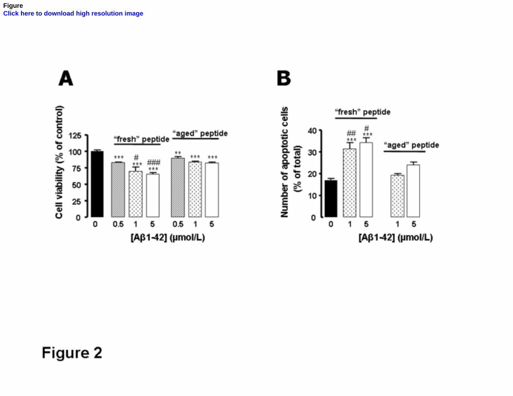
ACCEPTED MANUSCRIPT
FigureClick here to download high resolution image
ACCEPTED MANUSCRIPT
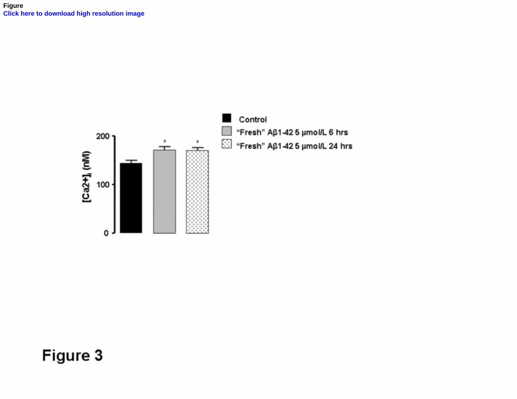
ACCEPTED MANUSCRIPT
FigureClick here to download high resolution image
ACCEPTED MANUSCRIPT

ACCEPTED MANUSCRIPT
FigureClick here to download high resolution image
ACCEPTED MANUSCRIPT

ACCEPTED MANUSCRIPT
FigureClick here to download high resolution image
ACCEPTED MANUSCRIPT
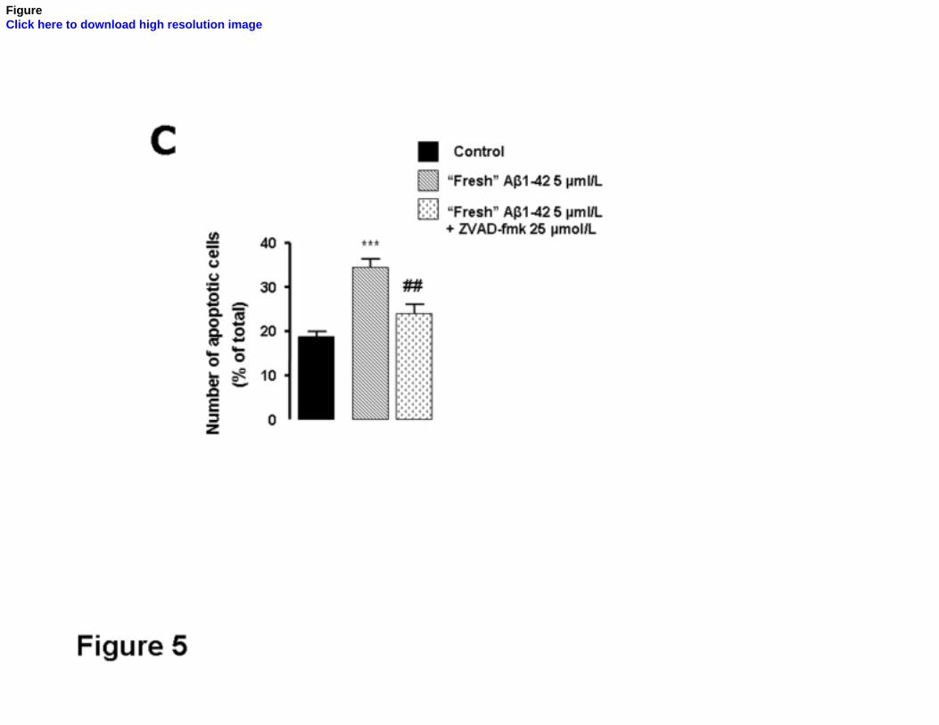
ACCEPTED MANUSCRIPT
FigureClick here to download high resolution image
ACCEPTED MANUSCRIPT

ACCEPTED MANUSCRIPT
FigureClick here to download high resolution image
ACCEPTED MANUSCRIPT

ACCEPTED MANUSCRIPT
FigureClick here to download high resolution image
ACCEPTED MANUSCRIPT

ACCEPTED MANUSCRIPT
FigureClick here to download high resolution image
ACCEPTED MANUSCRIPT

ACCEPTED MANUSCRIPT
FigureClick here to download high resolution image
ACCEPTED MANUSCRIPT

ACCEPTED MANUSCRIPT
FigureClick here to download high resolution image
ACCEPTED MANUSCRIPT

ACCEPTED MANUSCRIPT
FigureClick here to download high resolution image
ACCEPTED MANUSCRIPT
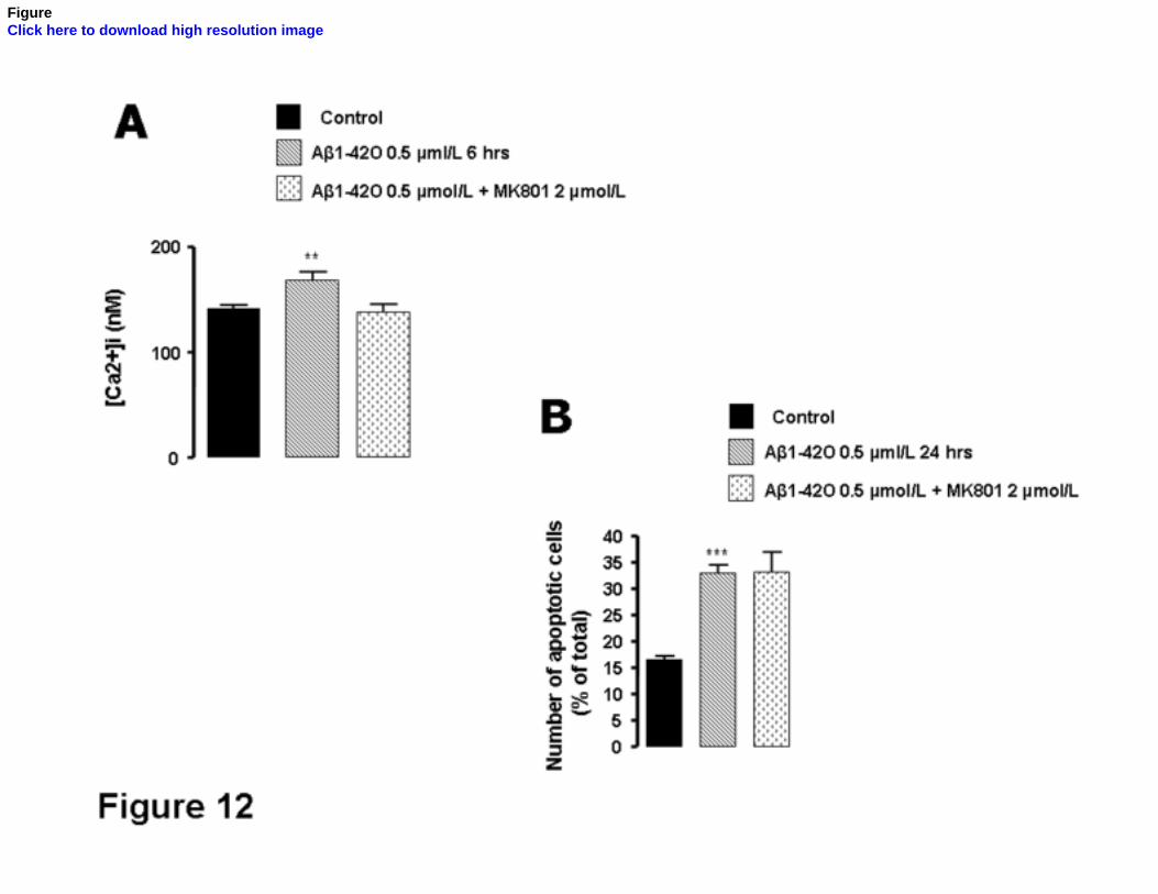
ACCEPTED MANUSCRIPT
FigureClick here to download high resolution image
ACCEPTED MANUSCRIPT