accreditation in adult tte echocardiography information … · · 2018-01-17this document...
TRANSCRIPT

Page 1 Contents
Accreditation in Adult TTE Echocardiography
Information Pack
This pack is for the use of all candidates undergoing the accreditation process and
becomes effective as of January 2018.
This document supersedes all previous versions.
Page 2 Contents
Contents Welcome message from Accreditation Chair 3 Introduction and Aims 4 Summary of process requirements 4 Details of written assessment and practical assessments 5 Written section 5 Theory Section 6 Reporting (imaging) 6 Practical assessment 6 Logbook 7 Viva Case Submission 9 Appendix 1: Suggested Reading List 12 Appendix 2: Training syllabus for BSE accreditation 14 Appendix 3: Proficiency Examination: Example Theory Questions 30 Appendix 4: Proficiency Examination: Example Reporting Questions 31 Appendix 5: Pearson VUE guidance notes 32 Appendix 6: Curriculum Based Competency Assessment Tool 34 Appendix 7: Suggested format for a report 38 Appendix 8: Report Format 39 Appendix 9: Transthoracic Proficiency: Summary Sheet 41 Appendix 10: Examples of Station 1 Practical Assessment Mark Sheets 42 Appendix 11: Examples of Station 2 Practical Assessment Mark Sheets 43 Appendix 12: Getting the Viva Cases right 46 Appendix 13: Examples of Station 3 Practical Assessment Mark Sheets 47 Appendix 14: Mentor statement to accompany the Practical Assessment 52 Appendix 15: BSE Policy on the Non-Anonymisation of Patient Data 53
Appendix 16: Final Checklist for candidates 56
Page 3 Contents
Welcome message from Accreditation Chair
Dear Candidate, Welcome to the British Society of Echocardiography. The process underlying Accreditation is set up to assist the echocardiographer in training, and it is important that you read all the information carefully before commencing your specific speciality logbook. The written section of the Adult Transthoracic Assessment is held twice each year in several Pearson VUE centres around the UK, Republic of Ireland and South Africa. The practical assessment will be held at least five times per year in a variety of locations. Full details and online registration information is available on the website www.bsecho.org. We would like every BSE member to undertake the relevant Accreditation process, which has, as its ultimate aim, the achievement and maintenance of high standards of clinical echocardiography for the benefit of our patients. A list of Accredited members is maintained on the BSE website. The process has to be regulated, and the standard of proficiency required for each specific Accreditation has to be set at a high enough level to command the respect of our professional colleagues. We want to make it possible for as many members as possible to obtain Accreditation, and not to put any unnecessary barriers in their way. Please let us know if we can assist you in this process in any specific way, or if you have constructive feedback to offer the accreditation committee then please just get in touch. Good luck with your accreditation process. Best wishes,
Dr Claire L Colebourn Chair, BSE Accreditation Committee

Page 4 Contents
Introduction and Aims
• Accreditation is run as a service for members of the British Society of Echocardiography and is not a compulsory or regulatory certificate of competence or excellence.
• Accredited members are expected to be able to perform and report echocardiographic studies unsupervised.
• Accreditation is a minimum requirement and cannot be regarded as a guarantee of competence.
• The Accreditation process comprises a written exam, (theory and case reporting sections) and a practical assessment comprising demonstration of selected echo views on a normal volunteer in an exam setting, review of the required log-book and a review of selected Viva echo cases performed to a high standard.
• Echo skills can only be maintained by continued education and practical involvement in echocardiography. The importance of this is underlined by limiting Accreditation to five years after which reaccreditation must be sought.
Summary of process requirements
• You must be a member of the British Society of Echocardiography
• You should address all queries regarding accreditation to:
BSE Accreditation Administrator, address details are available on www.bsecho.org. Tel: 020 7345 5185 (lines open 10am-4pm Mon-Fri), Fax: 020 7345 5186, Email: [email protected].
• You should register for the written and practical assessments by visiting the accreditation section of www.bsecho.org. This will advise the dates and location of the next examinations.
• You must pass the written assessment before attending the practical assessment.
• The practical assessment cases should be collected over a period of no more than 24 months from the written examination with the practical assessment being taken no later than two months after the end of the collection period.
• You must submit:
➢ Five full cases accompanied by reports signed by yourself ➢ A logbook containing 250 reports of a specific case mix (or 150 cases if you
hold BSE or EACVI TOE Accreditation. There is no reduction in numbers for holding EACVI TTE Accreditation)
➢ The full mentor sheet -appendix 6. ➢ Transthoracic Proficiency: Summary Sheet- appendix 9 ➢ Mentor statement signed- appendix 14 ➢ Final checklist – appendix 16

Page 5 Contents
• Extensions to the 24-month deadline may be granted only following periods of parental or extended sick leave or in exceptional circumstances. Extension requests forms must be submitted before the case COLLECTION deadline. Extension request forms can be obtained by visiting FAQ section of accreditation of www.bsecho.org. Requests received after the case deadline may not be reviewed. We strongly advise that requests are supported by documents such as doctors letter or letter from employer confirming the reasons for an extension.
• Extensions are not guaranteed. A non-refundable charge of £100 will be made for each extension request regardless of the outcome.
• A fee of £250 is charged for the complete Accreditation process. This fee is payable, in advance upon registration for the written section of the examination and will also cover the Practical assessment.
• Candidates who are unsuccessful in the written section of the examination will be charged a reduced fee of £125 to re-sit this section. This reduced fee only applies to candidates who re-sit the examination within two sittings of the unsuccessful attempt. A re-attempt at the Practical assessment is also subject to a fee of £125.
• Candidates are entitled to one re-attempt at the practical assessment, after which the entire process.
• The full training syllabus is available in appendix 2.
• Appeals - Please see the Appeal document available on FAQs section of www.bsecho.org.
Details of written assessment and practical assessments
Written section
• The written assessment is held on two occasions each year. The Spring and Autumn examinations are held at various Pearson VUE centres (online) in the UK, Republic of Ireland and South Africa.
• Please follow instruction on written examination dates section of www.bsecho.org or see appendix 5 for further information on registrations for the written exams.
• In the written assessment it is necessary to pass both the multiple choice and imaging questions at the same exam sitting. The approximate pass mark for the Theory Section is 95/125 marks (76%) and for the Reporting Section 30/50 (60%). These may vary slightly at the discretion of the Chief Examiner following moderation.
• There is no bar to re-sitting the written assessment.
• Accreditation will only be awarded once a candidate has also successfully completed the practical assessment (logbook and cases). A satisfactory performance at the written assessment alone does not allow ‘partial accreditation.’
Page 6 Contents
Theory Section
• This consists of 25 questions which must be answered within 60 minutes. The questions test knowledge of echocardiographic findings with some additional questions on basic cardiology and up to five questions on physics.
• The subject matter reflects the spectrum of clinical practice according to both frequency and technical complexity. Thus valve disease is more frequently represented than ischaemic disease since, though seen less commonly in clinical practice, it presents a greater challenge to the echocardiographer.
• This part of the examination will be marked +1 for correct answers, 0 for incorrect or unanswered questions (no negative marking).
• There are no ‘trick’ questions.
• Each question comprises a brief statement followed by five questions relating to the statement. Candidates are required to say whether each question is ‘true’ or ‘false’ a blank response is used for ‘don’t know.’ Some example questions are provided in Appendix 3.
• There are no fixed number of correct answers, i.e. for each question it is possible for every answer to be false or every answer to be true or any combination of true or false.
• The maximum possible mark is 125.
Reporting (imaging)
• This will consist of 50 questions, typically five questions on each of 10 case studies. Each question will have four possible answers and candidates will be asked to select the best answer. These reflect the range of clinical material seen in routine echocardiographic practice. Normal or near-normal studies may be presented.
• Each case will have five associated MCQ questions asking the candidate to select the best response from four answers (single best answer). The clips and stills will last one to three minutes and will contain sufficient information to answer the questions. One minute reading time will be provided before the first showing. Each case is replayed once following a two-minute pause to allow the candidate to answer and read the questions again.
• An example question is provided in Appendix 4. Each case is worth a total of five marks giving a total of 50.
Practical assessment
• All candidates will be required to attend a practical assessment within 26 months of beginning to collect their cases (i.e. within two months of their case collection
Page 7 Contents
deadline). The written examination must have been passed before attending. The Practical assessment will be held five times per year.
• Dates and locations will be announced on the Practical Registrations section of www.bsecho.org. Candidates will need to select an available date and register online (full instructions provided on website). Registrations will open at least three months before the assessment date. Upon a confirmed placement, candidates will be given an assessment time.
• The assessment will consist of three Stations.:
The first station will be the Logbook station. ➢ Logbooks and cases must be fully anonymised – please read the BSE Policy
on the Non-Anonymisation of Patient data in appendix. 15 major breach of this policy will result in a fail.
➢ Logbook submission: The Logbook should be submitted in a ring binder folder (do not use plastic pockets) with the different categories separated by dividers or ideally via the online logbook portal. Further details regarding the logbook submission can be found on page 8.
➢ If the candidate is successful at station 1, they will progress to Station 2. If
unsuccessful at station 1, the candidate can still proceed to the following station for the purpose of receiving formative feedback.
➢ Station 2 will consist of a practical assessment. The candidate will be asked
to acquire a number of views on a normal volunteer. The assessor will be present in the room and may help adjust the echo machine buttons as directed by the candidate if the machine is unfamiliar. This will be done in a specified timescale.
➢ If the candidate is successful at obtaining the views to the required standard at Station 2, the candidate will progress to Station 3. If unsuccessful at station 2, the candidate can still proceed to the following station for the purpose of receiving formative feedback.
➢ Station 3 will be a Viva assessing the video cases. Further details can be found on Page 8. If the candidate is successful at station 3, they will be deemed to have passed the Accreditation process and will receive their certificate prior to leaving the practical assessment.
➢ Logbook and video submissions should be checked by an experienced echocardiographer prior to attendance at the Practical Assessment.
Logbook
• The Logbook should comprise details of 250 transthoracic cases personally
Page 8 Contents
performed and reported by you during the specified period of 24 months (or 150 cases if you hold BSE or EACVI TOE Accreditation). It is not acceptable to include cases reported by you that have been performed by someone else.
• The format for the Logbook is a set of copies of actual clinical reports enclosed in a folder or binder, or submitted via the BSE online logbook portal. The reports should ensure:
➢ All patient data has been removed including full date of birth, name or address. See appendix 14
➢ All cases have been collected in accordance with local requirements for data protection, i.e. your trust policy.
➢ The inclusion of cavity and Doppler measurements, objective observations and a comment - appendices 7 and 8.
➢ The signature and full name of the candidate is included. At least the final 150 cases should be reported primarily by the candidate alone although they may be checked by another operator.
➢ In some cases, Trust Policy dictates that reports are signed by Accredited Echocardiographers only. In this case, reports signed by the supervising echocardiographer may be included in the logbook but should be countersigned by both the candidate and the supervising echocardiographer to confirm that the trainee has produced the report. An advisory letter from the head of department or supervisor should also be included in the submission.
• The studies should reflect the normal case-load of a general adult department with the following constraints:
➢ At least 25 cases should be for left ventricular function assessment. ➢ At least 50 cases should be for valve disease assessment. ➢ At least 10 should show replacement valves. ➢ At least 10 should be for assessment of the right ventricle. ➢ At least 5 should show pericardial disease/effusion. ➢ At least 5 should show diseases of the aorta (e.g. aortic root or ascending aorta
dilatation, aortic dissection). ➢ There should be at least 5 cases of suspected endocarditis. ➢ At least 5 should show left ventricular hypertrophy. ➢ There should be at least 5 cases of cardiomyopathy including at least two with
hypertrophic cardiomyopathy. ➢ There should be some cases showing mass or thrombus. ➢ There should be some cases of simple congenital disease (e.g. ASD) ➢ No more than 20 studies should be specialised studies (e.g. Stress or Bubble
Contrast). These studies are not a compulsory inclusion in your logbook, however, if you do include any, there should be no more than 20 in total.
➢ No more than 25 studies should be completely normal.
• The different categories of echoes should be separated by dividers.
➢ A tally of the primary diagnosis assigned to each case must be entered on the
Page 9 Contents
appropriate enclosed summary sheet - appendix 9. ➢ If possible there should be one or more examples of unusual diagnoses such as
myxoma. More than one candidate from the same institution is permitted to study the same patient if the diagnosis is unusual but each candidate must independently scan and do their own report.
➢ If you have problems finding enough specific cases, discuss this with your mentor who may consider arranging for you to attend a larger centre.
Viva Case Submission
• Five full studies with reports (paper copies) must be brought to the Practical Assessment. The cases must be anonymised. This is the section that is often done least well and is where many candidates fail. It is worth spending extra time doing this to make sure the submission is as good as it can be. Remember that it is assumed you will submit your best cases, so we will expect the studies to be complete and of a high standard. Also, remember we are assessing your echo skills, not the pathology you are sending in. The following diagnoses and minimum criteria are required:
➢ A normal study demonstrating appropriate use of machine settings for optimal imaging and correct use of standard views as per BSE minimum dataset (parasternal, apical, subcostal, suprasternal), M Mode (minimum Ao/LA, MV, LV) and 2D, CW, PW and Colour Doppler to assess chambers and valves. It is essential to demonstrate accurate measurement of the LV dimensions (minimum IVSd, LVEDd, PWd and LVEDs) in at least one case. This would normally be in this case but if this is not possible it is acceptable to provide this in at least one of the other cases.
➢ Moderate or severe aortic stenosis (you must include a good demonstration of the use of the stand-alone CW Doppler probe from apical or right parasternal or suprasternal). You should also calculate the valve area using the continuity equation and show all measurements used in the calculation.
➢ Moderate or severe mitral or aortic regurgitation carefully demonstrating quantification of the degree of regurgitation as per BSE guidelines.
➢ Previous or recent myocardial infarct describing regional wall motion abnormalities and carefully quantifying overall ejection fraction using the Simpson’s Biplane method. You must show the measurements taken in diastole and systole in both apical 4 chamber and 2 chamber views. Please carefully demonstrate any complications arising from the demonstrated regional wall motion abnormality (RWMA).
• The fifth case should show an example of one of the following:
a) Prosthetic valve with size and type noted and reference to normal values for that specific valve b) Mass or thrombus with differential diagnoses
Page 10 Contents
c) Simple congenital e.g. ASD, VSD or PS d) Significant LVH including Amyloid, HCM
There will be a limited number of PCs available to review these cases. In order to ensure that your cases play properly and remain anonymised at the assessment, it is recommended that you bring your own laptop to the centre having checked that the cases play on this. Please ensure that each clip plays automatically on click and loops continuously.
• The studies must demonstrate all appropriate echocardiographic views and must show the methods of measuring all dimensions on M-mode or 2D and all parameters on Doppler echocardiography.
• All cases must have patient data removed. Some machinery cannot do this post-examination so please ensure due care is taken to put ‘case 1’ instead of patient’s name or patient’s personal details. Alternatively, you may wish to use descriptions of pathology such as “aortic stenosis”. It does not matter so long as it is very clear to the marker.
• Please ensure that each case and accompanying report is clearly labelled in the same manner so that the marker is able to match the case with the report easily.
• Reports should include quantitative measurements, observations and a conclusion or summary.
• The cases must be submitted as digital loops and stills within a PowerPoint presentation.
• A guide to getting the cases right is available in appendix 11.
Cases that are of high quality may be copied to be used in subsequent BSE written exams.
Outcomes and process for re-attempts If you are successful at all three stations, you will be deemed to have passed the accreditation process and will receive your certificate prior to leaving the assessment.
• If you are unsuccessful at any station, you will be deemed to have been
unsuccessful at this sitting of the practical assessment. You will be provided with constructive feedback to facilitate a re-attempt, and offered the opportunity to continue on to experience the next station. This will be a formative assessment only – once a station has been failed it is not possible to pass further stations at the same sitting. However, constructive feedback will be given to help you to understand the requirements of the station and facilitate a subsequent re-attempt.
➢ Please note this feedback is for guidance only and does not necessarily represent the opinion of the deciding assessor at your next attempt. To re-attempt, you will need to attend another practical assessment and begin at

Page 11 Contents
the station at which you were unsuccessful and complete all remaining stations successfully. The timescale allowed for re-attempts will depend on which stations were not passed and the number of Viva cases required to be resubmitted. This will be discussed with you in the assessment.
A second attempt at the practical assessment is subject to a fee of £125. Candidates are entitled to one re-attempt at the practical assessment, after which the entire process must be undertaken again.
Page 12 Contents
Appendix 1: Suggested Reading List The syllabus is set by the Accreditation Committee of the British Society of Echocardiography and is presented as a guide to candidates. The reading list is provided by the Accreditation Committee of the British Society of Echocardiography. There are many excellent books on echocardiography, and some examples are listed below. In addition to those listed, there are many small basic texts which are a useful introduction to the subject.
• Authoritative textbooks (starting with the simpler texts as a suggestion)
• Echo made Easy Sam Kaddoura Churchill Livingstone 2001 ISBN 0443061882
• Echocardiography: Guidelines for reporting – a practical handbook Helen Rimington and John Chambers Taylor & Francis 1998 ISBN 1850700117
• Cardiac Ultrasound Leonard M Shapiro & Antoinette Kenny Manson Publishing 1999 ISBN 1874545081
• Feigenbaum‘s Echocardiography H.Feigenbaum Lippicott, Williams & Wilkins 2004 ISBN 0781731984
• Textbook of Clinical Echocardiography Catherine Otto W. B. Saunders 2004 ISBN 0721607896
Useful review articles:
• Wranne B, Baumgartner H, Flachskampf FA, Hasenkam M, Pinto F. Stenotic lesions (editorial). Heart 1996; 75 (Suppl.2);36-42. Downloadable from http://heart.bmjjournals.com/supplements.shtml
• M Enriquez-Sarano, C Tribouilloy. Quantitation of mitral regurgitation: rationale, approach, and interpretation in clinical practice. Heart 2002; 88 (Suppl 4): iv1-iv3. Downloadable from http://heart.bmjjournals.com/supplements.shtml
• S Y Ho Anatomy of the mitral valve. Heart 2002; 88 (Suppl 4): iv5-iv10. Downloadable from http://heart.bmjjournals.com/supplements.shtml
• T Irvine, X K Li, D J Sahn, A Kenny. Assessment of mitral regurgitation. Heart 2002; 88 (Suppl 4): iv11-iv19. Downloadable from http://heart.bmjjournals.com/supplements.shtml
• D Pellerin, S Brecker, and C Veyrat. Degenerative mitral valve disease with emphasis on mitral valve prolapse. Heart 2002; 88 (Suppl 4): iv20-iv28. Downloadable from http://heart.bmjjournals.com/supplements.shtml
• Gardin JM, Adams DB, Douglas PS, Feigenbaum H, Forst DH, Fraser AG, Grayburn PA, Katz AS, Keller AM, Kerber RE, Khandheria BK, Klein AL, Lang RM, Pierard LA, Quinones MA, Schnittger I; American Society of Echocardiography. Recommendations for a standardized report for adult transthoracic echocardiography: a report from the American Society of Echocardiography's

Page 13 Contents
Nomenclature and Standards Committee and Task Force for a Standardized Echocardiography Report. J Am Soc Echocardiogr. 2002 Mar;15(3):275-90. Downloadable from www.asecho.org
• Zoghbi WA, Enriquez-Sarano M, Foster E, Grayburn PA, Kraft CD, Levine RA, Nihoyannopoulos P, Otto CM, Quinones MA, Rakowski H, Stewart WJ, Waggoner A, Weissman NJ. Recommendations for Evaluation of the Severity of Native Valvular Regurgitation with Two-dimensional and Doppler Echocardiography. J Am Soc Echocardiogr 2003;16:777-802. Downloadable from http://www.asecho.org/freepdf/valvularregurg.pdf
• Cheitlin MD, Armstrong WF, Aurigemma GP, Beller GA, Bierman FZ, Davis JL, Douglas PS, Faxon DP, Gillam LD, Kimball TR, Kussmaul WG, Pearlman AS, Philbrick JT, Rakowski H, Thys DM. ACC/AHA/ASE 2003 guideline update for the clinical application of echocardiography—summary article: a report of the American College of Cardiology/American Heart Association Task Force on Practice Guidelines (ACC/AHA/ASE Committee to Update the 1997 Guidelines on the Clinical Application of Echocardiography). Circulation.2003
• http://www.acc.org/clinical/guidelines/echo/summary_article.pdf
• BSE website www.bsecho.org, downloads, BSE Echo report – Recommendations.
Page 14 Contents
Appendix 2: Training syllabus for BSE accreditation Topics that maybe included in the multiple choice examination General Concepts 1. The place of echocardiography Clinical role of echocardiography and Doppler
• Information that echocardiography can, and cannot provide
• ‘Ruling out’ pathology (sensitivity, specificity & Baye’s theorem)
• Likelihood of findings influencing patient management
• Undesirable outcomes: inaction while waiting for results, clinical ‘red herrings’
• Indications for echocardiography
• Competing and complementary technology
• Cardiac catheterisation (ventriculography and coronary angiography)
• C-T imaging
• Magnetic resonance imaging
• Nuclear Cardiology
1.1 Service Provision
• Advantages/disadvantages of Physiologist-led versus physician-led service
• Costs: fixed and variable
• Provision and indication for specialised techniques, e.g. TOE. Stress echo, Contrast echo
• Availability and access
• Controlling workload
• Training & motivation of staff
• Audit, Quality Control, Clinical Governance
• Infection control 1.2 Relationship with patients
• Explaining the procedure in terms relevant to the particular patient
• Respect for patients’ dignity and cultural backgrounds
• Relationships with colleagues
• Handling requests for information about the study findings 1.3 Reporting and Documentation
• Standard methods & terminology
• Distinction between Technical and Clinical reports
• Responsibility for reporting - Medico-legal considerations (Data Protection Act)
2. Imaging Physics & Instrumentation
Page 15 Contents
2.1 Concepts and Terminology
• Concept of compression waves
• Definitions: frequency, wavelength, propagation velocity
• Units of measurement: Hz and MHz
• Decibel Comparison of Ultrasound with audible sound. 2.2 Propagation of ultrasound through tissues
• Speed of sound in different body tissues.
• Frequency range used for diagnostic imaging
• Distinction between specular reflection and backscatter
• Principles of attenuation and scattering 2.3 Ultrasound Transducers
• Piezo-electric effect
• General concepts of 2D and 3D transducer construction
• Characteristics of the ultrasound beam: Far (Fraunhofer) & Near (Fresnel) zones, side lobes
• Beam steering methods: mechanical & electronic
• Focusing methods, including dynamic receive focusing
• Focus position and use of dual focus
• The role of intracardiac echocardiography 2.4 Imaging physics
• Factors affecting choice of imaging frequency: typical practical values for adults & children
• Broad-band imaging
• Harmonic imaging
• B mode and M Mode methods.
• Curved Anatomical M Mode
• Scanning speed limitations, relationships between pulse repetition frequency, frame rate, lines per frame, field of view, depth to be imaged.
• Concept of Parallel Processing and its influence on frame rate and image quality
• Effect on evaluation of rapid motion
• Temporal resolution.
• Grey scale and dynamic range
• Measurement and optimisation of Resolution: axial, azimuthal and elevation
• Lateral resolution and grating artefacts
• Reverberation artefacts
• Limiting factors for detecting small targets
2.5 Echo Instrumentation
• Function of machine controls: Transmit power; overall gain; time gain compensation; reject, logarithmic compression, signal processing, dynamic range, pre-processing; post-processing
• Optimisation of imaging parameters, including transducer frequency, scan angle,
Page 16 Contents
gamma correction, spatial and temporal smoothing
• Optimisation of 3D volume acquisitions including cropping and manipulation of viewing plane
• The advantages of 3D echocardiography over 2D echocardiography e.g. appreciation of mitral valve pathology, elimination of geometric assumptions in cardiac chamber volume estimations
2.6 Optimising Images
• Use of gel (infection risk from transducer, operator)
• Positioning of the subject
• Standard views: Parasternal, apical (4, 5 and 2-chamber, long axis), subcostal, suprasternal, right parasternal, long and short axis.
• Use of non-standard views
• Adapting for subjects with difficult echo windows, ventilated patients, ward-based echos, emergency room echos
2.7 Storage and Display of Images
• Basic concept of digital acquisition and storage systems. Scan converters and digital memories.
• Display devices and controls, recording techniques 3. Doppler Physics & Fluid Dynamics 3.1 Basic Fluid Dynamics
• Fluid flow: significance of peak & mean velocities
• Determination of volumetric flow, Continuity equation
• Laminar & turbulent flow: Reynolds’ equation (qualitative)
• Transition from Laminar to turbulent flow: inlet jet Bernoulli equation 3.2 Principles of Doppler
• Interaction of ultrasound waves with moving blood: The Doppler effect
• The Doppler equation: factors influencing magnitude of Doppler shift
• Spectral analysis: fast Fourier transform (qualitative)
• The spectral Doppler display: determination of mean, modal and peak velocities
• Limitation of CW Doppler caused by lack of depth discrimination
• Audible range of Doppler shift frequencies
• The effect of beam angle errors on Doppler velocities
• Aliasing: how it is caused and how it manifests in practice: The Nyquist limit
• Influence on aliasing of: transducer frequency; sample depth (range x velocity product); and beam angle
• High pulse repetition frequency (extended range) PW Doppler and the phenomenon of range ambiguity
• Relative advantages and disadvantages of CW, PW and HPRF modes
• Concept of colour flow imaging as multi-sampled PW
• Velocity estimation, by moving target indication and autocorrelation (qualitative)

Page 17 Contents
• Limitations of mean velocity: use of velocity variance to show high velocities/turbulence
• Aliasing in colour Doppler
• The principles of pulse wave tissue Doppler
• Packet size, colour mode and sector size and their effect on frame rate and aliasing
4. Deformation Analysis 4.1 Principles of Myocardial Deformation
• The definition of displacement, velocity, strain and strain rate
• The cardiac ultrasound co-ordinate system for describing motion and deformation: longitudinal, radial, circumferential and rotational axes
• Quantifying myocardial deformation as opposed to velocity or displacement
• Concept of shear deformation; rotation of the base and apex of the left ventricle, and the resultant twisting deformation or torsion
4.2 Quantifying myocardial strain and strain rate by tissue Doppler
• The concept of the myocardial velocity gradient
• The concept of strain and strain rate to define deformation
• Tissue Doppler imaging for deriving strain and strain rate: practical parameters in measuring strain and strain rate (e.g. sample size and shape, offset distance, drift compensation, spatial and temporal averaging, tracking of sample volume)
• Reproducibility issues 4.3 Speckle Tracking Echocardiography/2D strain
• Familiarity with the concept of speckles and speckle tracking in greyscale 2D loops
• Speckle tracking for angle-independent derivation of velocities, displacement, strain and strain rate, in 2 dimensions
• The impact of frame rates on the quality of speckle tracking
• Speckle tracking vs. tissue Doppler techniques for assessing myocardial motion and deformation
• Speckle tracking for measuring left ventricular rotation and torsion
• Kindred technologies 5. Doppler instrumentation 5.1 Spectral Doppler Instrumentation
• Duplex Doppler using imaging transducers
• The ‘Stand-alone’ Doppler probe
• Features of the spectral display: positive & negative velocities; scale & baseline controls.
• Effect of high-and low-pass filter and intensity threshold (‘reject’) settings
• Pulsed Doppler sample volume: influence of gate length and distance (beam width)
• Representation of signal strength by image intensity
Page 18 Contents
• How aliasing manifests on the spectral display 5.2 Colour Flow Instrumentation
• The colour display: BART convention
• Colour maps to show velocity scales
• Image domination and additive colour modes
• Difference between velocity and power (signal amplitude) displays
• Basic principles of Tissue Doppler Imaging, including optimisation of filters for detecting tissue versus blood velocities, sample volume and size, impact of interrogation angle on measured velocities, minimising aliasing, and maximising frame rates to detect short duration myocardial motion
• Differences between colour Doppler tissue Doppler Imaging and pulsed wave tissue Doppler imaging
• Minimisation of myocardial translational movements during acquisition.
• The concept of tracking on colour Doppler tissue Doppler imaging to ensure that sample volume remains in the region of interest
• Parametric (curved M-mode) display of tissue Doppler images
• The relevance of importing cardiac cycle time points, such as aortic valve closure, into tissue Doppler traces
TOE Instrumentation
• Transducer types: single plane, biplane, multiplane
• Optimising machine settings for TOE Patient monitoring for TOE and general safety considerations
• Control of infection Safety of ultrasound
• Potential hazardous biological effects: heating, resonance and cavitation effects
• Measurement of beam intensity (SPTA)
• Practical precautions: power levels, use of colour and CW Doppler Recording methods
• Advantages/disadvantages of recording on videotape and digitally
• Basic understanding of digital image processing and recording methods: pixel density, volume of data, the DICOM standard, concept of data compression (JPEG, AVI, etc.) archiving of echocardiographic studies on magneto-optical discs, CD/DVD, portable solid-state memories, ECG-gated acquisitions vs. continuous recording, facility to review acquired loop prior to storage, facility to choose the number and type of cardiac cycles to be recorded, facility for offline image properties adjustment and further quantitative analysis.
6. Cardiac Anatomy and Physiology

Page 19 Contents
6.1 Anatomy of the thorax
• Thorax contained by rib cage & diaphragm
• Lungs & pleura; heart & pericardium; mediastinum
• Blood vessels within the thorax
6.2 Gross anatomy of the heart
• Basic cardiac embryology
• Nomenclature of chambers and valves
• Major relationships of chambers, valves and blood vessels
• Distinguishing features of valves and chambers as related to echocardiography
• The pericardial sac 6.3 Cardiac anatomy and physiology as demonstrated by echocardiography
• Detailed structural anatomy of the heart, great vessels and pericardium
• Visualisation of normal cardiac anatomy and normal variants in standard echocardiographic planes
• Normal valve function, normal Doppler parameters and normal variants
• The phases of atrial function: reservoir, conduit and contractile phases
• The LV remodelling process in response to disease: eccentric (chronically elevated preload) vs. concentric hypertrophy (chronically elevated afterload)
6.4 The Cardiac Cycle
• Temporal relationships of the ECG, chamber pressures and valve movements
• Typical values for intracardiac pressures
• Relationship of valve movements to heart sounds
• Identification of valve opening and closure signals on Doppler recordings
• The timing of aortic valve closure as a marker of end-ejection, as derived from M-mode, blood flow Doppler or tissue Doppler
7. Cardiac functional parameters 7.1 Measurements and calculations
• On-screen measurement of length, slope, area, volume and time interval, and their significance for 2-D, 3D images, M-mode and spectral Doppler displays
• Standard M-mode measurements (including MAPSE) and calculations, both using machine software and manual methods
• Derivation of Stroke Volume, Ejection Fraction and LV Mass
• Methods of measuring LV volume, including biplane area, area-length, Simpson’s rule methods and 3D.
• Limitations of single plane estimations of LV ejection fraction e.g. Teicholtz formula method
• Limitations of single plane measurements of LA size
• Geometric assumptions used in estimation of cardiac chamber volumes with M mode and 2D imaging
• The advantages of deriving volumes and ejection fraction by 3D echocardiography
• Limitations of measurement and/or calculation validity in the presence of poor
Page 20 Contents
quality and/or off-axis images 7.2 Doppler determination of cardiac output, ejection time and velocity acceleration
• Methods of measuring diastolic dysfunction: E/A ratio, deceleration time, pulmonary venous flow patterns, the ratio of the peak early diastolic transmitral velocity and the peak early diastolic tissue velocity of the mitral valve annulus (the E/E’ or E/Ea) ratio for estimating LV filling pressures, the mitral valve propagation velocity
• Peak and Mean pressure gradient measurements by Doppler and their relationship to catheterisation data
• Measurement of pulmonary pressures from tricuspid and pulmonary regurgitant flow velocities and assessment of inferior vena cava contraction during inspiration
8. Contrast Studies
• Significance of spontaneous echo contrast
• Optimisation of machine control settings for detecting contrast
• Main indications for a bubble contrast study: diagnosis of intracardiac shunts and PFO, diagnosis of left sided SVC
• Manoeuvres to provoke right –to-left passage of bubbles during assessment for PFO
• Relevance of injecting bubble contrast through upper arm vein vs. femoral vein for detecting PFO
• Technique for performing a hand-agitated contrast study
• Clinical precautions 8.1 Awareness of encapsulated contrast agents and techniques
• Interaction of ultrasound with encapsulated agents
• Generation of harmonic energy by bubble distortion and fracture
• Doppler signals generated by bubbles (Power Mode)
• Main indications for LV and RV opacification: enhancing endocardial definition for assessment of regional contractility and accurate cardiac volume estimations, detection of intracardiac masses, distinguishing thrombus from a vascular tumour, diagnosis of cardiomyopathies e.g. non-compaction, arrhythmogenic right ventricular dysplasia, Doppler enhancement
• Use of contrast in stress echocardiography for improving detection of wall motion abnormalities and for assessment of myocardial perfusion
9. Pathology 9.1 Mitral Valve Disease, 2D, 3D, M-mode and Doppler features of the normal mitral valve
• Mitral Stenosis
• Recognition of rheumatic mitral stenosis
• Qualitative description of valve and sub-valve calcification and fibrosis
• Measurement of orifice area by planimetry
• Factors favouring successful balloon valvuloplasty
Page 21 Contents
• Doppler assessment of mean and end-diastolic gradient
• Doppler assessment of area by ‘pressure half-time’: technique and limitations
• Role of exercise echocardiography in assessing the change in transmitral gradient and pulmonary systolic pressures with exercise, as decision aid in the timing of surgery/balloon valvuloplasty
9.2 Mitral regurgitation
• Aetiologies and typical echocardiographic features of Rheumatic Mitral annular calcification
• ‘Floppy MV’/myxomatous mitral valve
• Ischaemic functional
• Infective endocarditis Assessment of severity by:
• Chamber sizes and volume overload
• CW Doppler – shape and density of contour of Doppler signal
• Vena contracta, PISA and effective regurgitant orifice area
• Size of colour jet relative to atrial size by colour flow Doppler Regurgitant fraction, regurgitant volume
• Pulmonary vein flow patterns
• Indirect effects on LV and LA
• Role of echocardiography in determining timing of surgery for primary mitral valve disease: ejection fraction, end-systolic LV diameter, EROA
• Role of TOE in assessing mitral valve pathology and in determining likelihood of repair as opposed to replacement
10. Aortic Valve Disease 10.1 2D, 3D, M-mode and Doppler features of the normal aortic valve 10.2 Aortic Stenosis Aetiologies and echocardiographic features:
• Rheumatic
• Bicuspid
• Senile degenerative
• Sub-and supra-valve obstruction
• Assessment by CW Doppler
• Peak and Mean gradients
• Apical, right parasternal and suprasternal positions
• Continuity equation
• Assessment of left ventricular hypertrophy and use of stress echocardiography for distinguishing fixed anatomical stenosis from pseudostenosis in low flow aortic stenosis and for assessing LV contractile reserve
• Difference between transaortic pressure gradients derived from echocardiography and from cardiac cathetherisation

Page 22 Contents
10.3 Aortic Regurgitation Aetiologies and typical echocardiographic features of:
• Rheumatic
• Bicuspid valve
• Aortic root disease
• Infective endocarditis (including root abscesses) Assessment of severity by:
• Chamber sizes/volume overload (regurgitant volume, regurgitant fraction)
• CW Doppler – shape and density of contour of Doppler signal, pressure half time
• Colour Doppler – size of jet relative to left ventricular outflow tract diameter
• Vena Contracta
• Effective regurgitant orifice area
• Diastolic flow reversal in descending aorta
• Indirect effects on LV
• Role of echo in determining timing of surgery
• Role of TOE in assessing aetiology and severity 11. Tricuspid Valve Disease
11.1 2D, M-mode and Doppler features of the normal tricuspid valve 11.2 Rheumatic tricuspid valve stenosis
• Echocardiographic features
• Assessment of severity by imaging and Doppler
11.3 Tricuspid Regurgitation Aetiologies and echocardiographic features of:
• Rheumatic
• Prolapse
• Congenital
• Endocarditis
• Carcinoid
• Functional Assessment of severity by:
• 2D imaging and M-mode
• CW Doppler – shape and density of contour of Doppler signal
• Colour Doppler
• Hepatic vein flow pattern
• Indirect effects on RV and RA 12. Pulmonary Valve Disease 12.1 2D, M-mode and Doppler features of the normal pulmonary valve

Page 23 Contents
12.2 Pulmonary Valve Stenosis
• Echocardiographic features
Assessment of severity by:
• Spectral Doppler
• Detection of infundibular obstruction by spectral Doppler Pulmonary Regurgitation
• Aetiologies and echocardiographic features Assessment of severity by
• CW Doppler – deceleration rate, density and contour of Doppler signal
• Colour Doppler
• Indirect effects Infective Endocarditis – Rick factors for I.E
• Typical echocardiographic appearance of vegetations in bacterial and fungal endocarditis
• Preferred locations for vegetations
• ‘Jet’, ‘kissing’ lesions
• Endocarditis associated with congenital disease and HCM
• Complications: abscess, fistula, perforation, valve regurgitation
• Role of TOE in suspected endocarditis
• Monitoring of IE 13. Prosthetic Valves 13.1 2D, M-Mode and Doppler features of the main types of replacement valves
• Tilting Disc
• Bi-leaflet
• Ball & cage
• Bioprostheses (stented and stentless)
• Age-related deterioration of bioprostheses
• Role of TOE in examining normal and malfunctioning prosthetic valves
13.2 Prosthetic valve stenosis
• Assessment by 2D, M-mode and Doppler
• Normal ranges
• Use of Continuity Equation for aortic prostheses
• The phenomenon of pressure recovery
• The diagnosis of patient-prosthesis mismatch 13.3 Prosthetic valve regurgitation
• Trans-versus para-valvar regurgitation
• Normal versus abnormal regurgitation
Page 24 Contents
• Assessment by CW, PW and Colour
• Doppler Colour artefacts from mechanical prostheses
14. Cardiomyopathies Dilated Cardiomyopathy 14.1 2D, M-mode and Doppler features of dilated cardiomyopathy
• Detection and assessment of associated lesions:
• Functional valve regurgitation
• Thrombus in cardiac chambers
• Pericardial effusions
• Role of echocardiography in assessment and follow-up 14.2 Hypertrophic Cardiomyopathy
• 2D, M-mode and Doppler features of Hypertrophic Cardiomyopathy
• Differentiation from other causes of hypertrophy, e.g. hypertension, athletic heart’, amyloidosis, Fabry‘s disease, Friedreich‘s ataxia cardiomyopathy
• Techniques for measurement of left ventricular wall thickness, detection of left ventricular outflow tract obstruction and intracavity gradient
• Assessment of right ventricular involvement
• Associated abnormalities, e.g. systolic anterior motion mitral valve 14. 3 Restrictive Cardiomyopathy
• Causes e.g. primary amyloidosis, sarcoidosis, idiopathic, endomyocardial fibrosis
• 2D, Doppler & TDI features of impaired ventricular filling – increased ventricular wall thickness, dilated atria, increased E/A ratio, reduced deceleration time, increased E/E’ ratio, reduced S’
14.4 Main features of LV non-compaction 14.5 Intracardiac Masses
• Typical locations for formation of intracardiac thrombus
• Echocardiographic features of typical LA Myxoma
• Differentiation of myxoma from other cardiac tumours
• Features suggestive of malignancy
• Role of TOE in assessment of intracardiac masses
• Role of contrast in the assessment of intracardiac masses
15. Pericardial Disease 15.1 Anatomy of the normal pericardium
• Relationships of serous pericardium to heart and great vessels
• Transverse and oblique sinuses of the pericardium 15.2 Echocardiographic features of pericardial fluid
• Location of fluid in relation to patient position and fluid volume
Page 25 Contents
• Differentiation from pleural effusion
• Assessment of volume of pericardial fluid
• Role of echocardiography in pericardiocentesis 15.3 Features of tamponade
• Collapse of RA and/or RV walls
• Effect on IVC and hepatic vein flow pattern
• Effect on A-V valve flow velocities during respiratory cycle 15.4 Features of pericardial constriction
• Pericardial thickening/appearance
• Effect on A-V valve flow velocities
• Effect of respiration
• SVC/hepatic vein flow
• Differentiation from restrictive cardiomyopathy including use of tissue Doppler 16. Coronary Artery Disease and Systolic LV function
• Anatomy & nomenclature of the major branches of the coronary arteries
• Relationship of coronary anatomy to standard echocardiographic imaging planes
• Nomenclature for describing myocardial segments (16 & 17 segment model)
• Analysis of segmental systolic myocardial function
• Use of stress echo to assess for myocardial ischaemia
• Diastolic dysfunction in coronary artery disease Global measures of LV function:
• Ejection Fraction
• Stroke Distance
• Stroke Volume and Cardiac output
• Use of tissue Doppler and speckle tracking echocardiography for assessment of regional myocardial velocities and deformation in ischaemic heart disease, at rest and with stress
• Longitudinal function of the left ventricle, as assessed by M-mode (MAPSE) and tissue
• Doppler of the mitral valve annulus
• The concept of post-systolic contraction
• The concept of isovolumic acceleration by tissue Doppler
• Left ventricular torsion and its implications for systolic function of the LV 17. Diastolic function of the LV
• The 4 stages of diastolic dysfunction as assessed by transmitral flow Doppler (including DT); impaired filling pattern and restrictive flow pattern
• The limitations of transmitral flow Doppler for assessing diastolic dysfunction:
• Effect of LA
• Pressures and pseudonormalisation, effect of mitral regurgitation
Page 26 Contents
• The use of Valsalva manoeuvre in reducing LA pressures to differentiate normal from pseudonormal transmitral
• Flow Doppler patterns
• The use of left atrial size, IVRT, tissue Doppler (diastolic longitudinal velocities of the mitral valve annulus, the E/E’ ratio), pulmonary vein flow pattern and mitral propagation velocity for assessing diastolic function
• The importance of untwisting in left ventricular filling 18. LV dyssynchony and assessment by echocardiography
• Techniques for measuring interventricular and intraventricular dyssynchrony for predicting response to cardiac resynchronisation treatment
• Tissue Doppler quantitation of intraventricular dyssynchrony and their limitations
• Techniques for optimising settings of the cardiac resynchronisation device after implantation
19. Stress Echocardiography
• Indications and basic knowledge of techniques for exercise, dobutamine or vasodilator stress echocardiography
• Exercise or pharmacological stress echocardiography for diagnosis of ischaemic heart disease and myocardial
viability
• The concept of viable and hibernating myocardium, and the relevance of the various responses of the myocardium to stress
• The concept of contractile reserve
• The American Society of Echocardiography regional wall motion scoring system
• Dobutamine stress echo in ‘low flow’ aortic stenosis
• Exercise stress echo in valvular heart disease and pulmonary hypertension 20. Myocardial Infarction and its sequelae
• 2D, 3D, M-mode and Doppler features of: post-infarction VSD
• Mitral papillary muscle rupture
• Tamponade
• Mural thrombus
• Myocardial scarring
• Dressler’s syndrome
• Left ventricular aneurysm – true aneurysm vs. pseudoaneurysm
• Main features of stress-induced (Takotsubo) cardiomyopathy as differential diagnosis of acute myocardial infarction
21. Pulmonary Hypertension and functional assessment of RV
• 2-D, M-mode and Doppler features of pulmonary hypertension
• Aetiologies: primary; post pulmonary embolism; secondary to left-sided lesions; lung disease
• Assessment of global systolic function of the RV: Tricuspid annular peak systolic
• excursion by M-mode (TAPSE), fractional area change of the RV, tissue Doppler of

Page 27 Contents
the RV
• Right ventricular dysfunction in pulmonary embolism, chronic pulmonary diseases, cardiomyopathy, Eisenmonger‘s syndrome, and systemic right ventricle
22. Diseases of the Aorta Technique for examining the ascending and descending thoracic aorta Echocardiographic features of the normal aortic root, sinuses of Valsalva, ascending aorta and aortic arch 2-D, M-mode and Doppler features of:
• Marfan syndrome
• sinus of Valsalva aneurysm
• thoracic aortic aneurysm
• aortic dissection
• additional features related to aortic dissection:
• aortic cusp prolapse
• aortic regurgitation
• fluid in pericardium Role of transoesophageal echocardiography in the diagnosis of aortic dissection Assessment of aortic root for patients undergoing transcutaneous aortic valve replacement 23. Adult Congenital Heart Disease Anatomy, pathophysiology and natural history of common congenital lesions present in adults:
• 2-D, M-mode and Doppler features of the following, pre-operatively and post-operatively, as seen in the older child or adult
• Ostium Secundum Atrial septal defects
• Perimembranous and muscular ventricular septal defects
• Partial and complete atrio-ventricular septal defects
• Persistent ductus arteriosus
• Bicuspid aortic valve and associated aortopathy
• Sub-and supra-valve aortic stenosis
• Aortic coarctation
• Pulmonary stenosis
• Ebstein‘s anomaly
• Fallot‘s tetralogy
• Transposition and corrected transposition of the great arteries
• Role of contrast echocardiography in evaluating shunts in adults
• Calculation of shunts
• Role of TOE in adult congenital disease 24. Likely echocardiographic findings for common clinical presentations:
• Heart failure or breathlessness
• Arrhythmia
• Ejection systolic murmur
• Hypertension
Page 28 Contents
• Collagen abnormalities (including systemic sclerosis)
• Renal failure
• Stroke 25. Emergency and ICU Echo 25.1 General
• Constrained environment (multiple arterial/venous lines, ventilator, lighting issues etc.)
25.2 The hypotensive/shocked patient and post-cardiac arrest
• Role of focused peri-arrest study and appreciation of limited echo windows
• Evaluation of LV (systolic and diastolic) and RV function.
• Exclusion of severe valve disease (e.g. severe AS, endocarditis) and acute aortic dissection
• Assessment for pericardial effusion and cardiac tamponade, hypovolaemia and underfilling, and high output cardiac failure
• Septic shock – assess for LV systolic/diastolic dysfunction
• Value of repeated echo studies to assess any deterioration/improvement in underlying state
25.3 Suspected acute pulmonary embolus
• Echocardiographic evaluation of RV size and function, tricuspid regurgitation and pulmonary artery systolic pressure assessment, IVC size and respiratory variation, thrombus presence in IVC/RA
25.4 Blunt and penetrating cardiac trauma
• Typical echocardiographic features including pericardial effusion, right and left ventricular contusion, acute valve lesions, aortic dilation and dissection/transection, VSD, pleural effusion
25.5 Echo in the ventilated patient
(e.g. exclude PFO, proximal PE)
• Acute arrhythmias such as fast AF (assessment for chamber abnormalities, valve
• disease, LV impairment, pericardial effusion)
• Cardiac source of embolus – CVA/peripheral embolic event in ventilated patients (LV/LA thrombus, endocarditis, myxoma)
• Value of TOE in ventilated patients (if poor transthoracic echo window) 25.6 Post surgery patient
• Appreciation of effects of general anaesthesia and cardio-pulmonary bypass on LV function
• Assessment of post-surgery haemodynamic compromise/ acute deterioration e.g.
• cardiac surgery (tamponade, wall motion abnormalities, valvular dysfunction),
Page 29 Contents
general surgery (air/fat embolism, venous thromboembolism, acute MI, volume overload)
25.7 Assessment of filling status
• Awareness of the role of echocardiography in assessing filling using LV and RV systolic and diastolic function, IVC, SVC and hepatic vein size and reactivity, atrial septal motion, chamber sizes and variation in Doppler velocities.
• Role of repeated echo studies in assessing effects of fluid challenge and inotropes 26. Additional topics The level of knowledge expected is that of a competent echocardiographer performing transthoracic studies and sustaining knowledge through the BSE and other educational resources, including issues relevant to clinical scanning and practice raised in the BSE Newsletter
Page 30 Contents
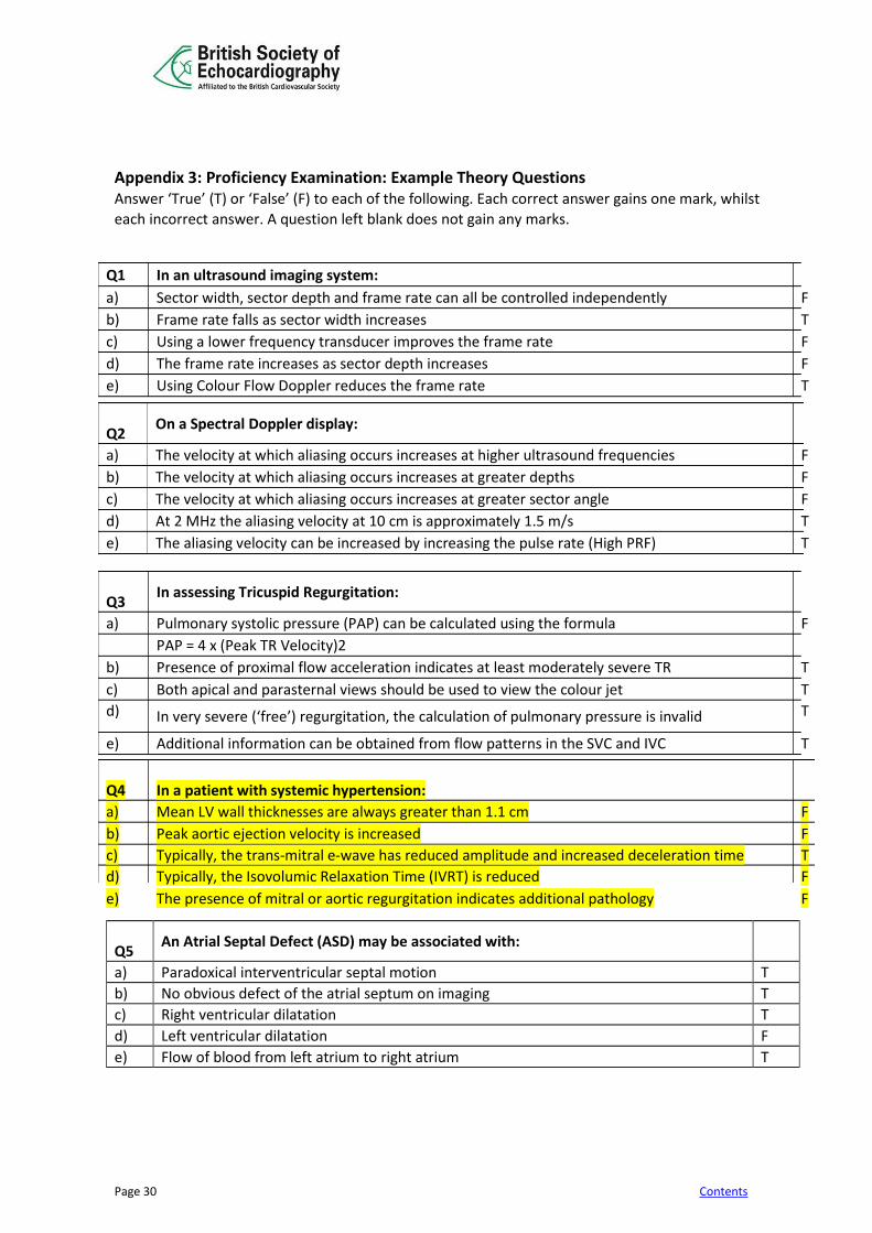
Appendix 3: Proficiency Examination: Example Theory Questions Answer ‘True’ (T) or ‘False’ (F) to each of the following. Each correct answer gains one mark, whilst
each incorrect answer. A question left blank does not gain any marks.
Q5
An Atrial Septal Defect (ASD) may be associated with:
a) Paradoxical interventricular septal motion T
b) No obvious defect of the atrial septum on imaging T
c) Right ventricular dilatation T
d) Left ventricular dilatation F
e) Flow of blood from left atrium to right atrium T
Q1 In an ultrasound imaging system:
a) Sector width, sector depth and frame rate can all be controlled independently F
b) Frame rate falls as sector width increases T
c) Using a lower frequency transducer improves the frame rate F
d) The frame rate increases as sector depth increases F
e) Using Colour Flow Doppler reduces the frame rate T
Q2
On a Spectral Doppler display:
a) The velocity at which aliasing occurs increases at higher ultrasound frequencies F
b) The velocity at which aliasing occurs increases at greater depths F
c) The velocity at which aliasing occurs increases at greater sector angle F
d) At 2 MHz the aliasing velocity at 10 cm is approximately 1.5 m/s T
e) The aliasing velocity can be increased by increasing the pulse rate (High PRF) T
Q3
In assessing Tricuspid Regurgitation:
a) Pulmonary systolic pressure (PAP) can be calculated using the formula F
PAP = 4 x (Peak TR Velocity)2
b) Presence of proximal flow acceleration indicates at least moderately severe TR T
c) Both apical and parasternal views should be used to view the colour jet T
d) In very severe (‘free’) regurgitation, the calculation of pulmonary pressure is invalid T
e) Additional information can be obtained from flow patterns in the SVC and IVC T Q4
In a patient with systemic hypertension:
a) Mean LV wall thicknesses are always greater than 1.1 cm F
b) Peak aortic ejection velocity is increased F
c) Typically, the trans-mitral e-wave has reduced amplitude and increased deceleration time T
d) Typically, the Isovolumic Relaxation Time (IVRT) is reduced F
e) The presence of mitral or aortic regurgitation indicates additional pathology F

Page 31 Contents
Appendix 4: Proficiency Examination: Example Reporting Questions Clips and stills will be shown lasting 1-3 mins and below is an example of a question with all relevant information provided. SELECT THE SINGLE BEST ANSWER There is no negative marking. One mark added for a correct answer, no mark deducted for an incorrect answer. Case 1 Male age 46 Request: Systolic murmur Data: IVS 1cm; LVIDd 4.2cm; PWT 1cm; LVIDs 3cm; LA 3.6cm; Ao valve 2.7cm; PA 2.2cm; Pulmonary FVI 32; LVOT 2.2cm; LVOT FVI 29; LV to RV pressure gradient 118mmHg; BP 150/88 1. Describe the main abnormality a. Apical VSD b. Endocardial cushion defect c. Subaortic VSD X d. ASD 2. What is the likely aetiology? a. Congenital X b. Inferior myocardial infarction c. Anterior myocardial infarction d. Endocarditis 3. What is the pulmonary to systemic flow ratio a. 0.9 b. 1.0 c. 1.1 X d. 1.3 4. What is the RV systolic pressure? a. 20mmHg b. 32mmHg X c. 38mmHg d. 42mmHg 5. Which of these is correct? a. RV and LV are normal X b. RV is dilated and LV is normal c. LV is dilated and RV is normal d. LV and RV are both dilated

Page 32 Contents
Appendix 5: Pearson VUE guidance notes
BSE written exams are delivered in partnership with Pearson VUE. Candidates will be able to sit the exam at local centres throughout the UK, Republic of Ireland and in South Africa. Each candidate will have their own monitor and will be able to replay videos during the examination. Full instructions will be provided on the day of the exam.
Pre-Registration
• Candidate must register their interest to sit the written exam by completing an online pre-registration form via accreditation section of www.bsecho.org. BSE will transfer your data and requirements to Pearson VUE who will contact all pre-registered candidates with further information on confirming placements for the exam.
• All registration and payments will be managed by Pearson VUE after the stage of pre-registration.
• Candidates with special requirements or conditions should notify the BSE during the pre-registration stage.
On the day of the exam
• Instructions will be given on the day of the exam via a video tutorial at the test centre. Candidates will complete the exam on a computer at the test centre.
• A basic calculator is already built in to the online exam. An erasable sheet will be given to candidates by the examining centre.
• Candidates are required to bring photo ID that reflects on the registration as booked.
• Candidates are not required to bring any stationery to the exam.
• Any last-minute requests of special accommodations will not be facilitated by the test centre.
Part 1 Theory Section A. Time The theory section will last 60 minutes. B. Format The theory section will consist of multiple choice questions. C. Answers For one part the answers will be either TRUE or FALSE There will be NO negative marking for this paper – each correct answer will receive a score of 1. Incorrect or unanswered questions/stems will receive a score of 0. Part 2 Digital Reporting Section

Page 33 Contents
A. Time The reporting section will last 90 minutes B. Format
The section will consist of 10 cases, each with 4 single best answer questions relating to it
C. Answers For each question there is only one correct answer, a choice of A B C or D
There will be NO negative marking for this paper – each correct answer will receive a score of 1. Incorrect or unanswered questions/stems will receive a score of 0. Please watch the demo available via Pearson VUE; http://www.pearsonvue.com/demo/
D. Additional Information Candidates are advised to check the security proceedures in the “What to expect section” of the Pearson VUE/BSE guide page; https://home.pearsonvue.com/test-taker/security.aspx

Page 34 Contents
Appendix 6: Curriculum Based Competency Assessment Tool (This may also be completed in digital form on the online logbook portal)
MENTOR TO COMPLETE DURING CANDIDATE’S TRAINING PERIOD How to use this document: You should keep it with you throughout your training period At each hospital, you must have a mentor who should be a senior and experienced echocardiographer. Someone holding BSE Accreditation is encouraged but not mandatory. Your mentor should initial and date each entry once he or she is satisfied that you are competent to perform and report it unsupervised. This competency checklist should be submitted with your logbook. The theory component will be self-taught. Your department should have suitable text-books 1. BASIC ECHOCARDIOGRAPHY Knowledge Basic principles of ultrasound Basic principles of spectral Doppler Basic principles of colour flow Doppler Basic instrumentation Ethics and sensitivities of patient care Basic anatomy of the heart Basic echocardiographic scan planes Parasternal long axis standard, RV inflow, RV outflow Parasternal short axis including aortic valve, mitral valve and papillary muscles Apical views, 4- and 5-chamber, 2-chamber and long-axis. Subcostal and suprasternal views Indications for transthoracic and transoesophageal echocardiography Normal variants and artefacts Practical competencies Interacts appropriately with patients Understands basic instrumentation Cares for machine appropriately Can obtain standard views Can optimise gain setting, sector width, depth, harmonics, focus, sweep speed, Doppler baseline and scale, colour gain Can obtain standard measurements using 2D or M-mode Can recognise normal variants; Eustachian valve, chiari work, LV tendon Can use colour examination in at least two planes for all valves optimising gain and box-size Can obtain pulsed Doppler at a) left ventricular inflow (mitral valve) b) left ventricular outflow tract (LVOT) c) right ventricular inflow (tricuspid valve) d) right ventricular outflow tract, pulmonary valve & main pulmonary artery Initials and date 2. LEFT VENTRICLE Knowledge Coronary anatomy and correlation with 2D views of left ventricle. Segmentation of the left ventricle (16 and 17 segment models) Wall motion

Page 35 Contents
Measurements of global systolic function. (LVOT VTI, stroke volume, fractional shortening, ejection fraction using Simpson’s rule) Doppler mitral valve filling patterns & normal range Appearance of complications after myocardial infarction Aneurysm, pseudoaneurysm, Ventricular septal and papillary muscle rupture Ischaemic mitral regurgitation Features of dilated, and hypertrophic cardiomyopathy Common differential diagnosis Athletic heart, hypertensive disease Practical competencies Can differentiate normal from abnormal LV systolic function Can recognise large wall motion abnormalities Can describe wall motion abnormalities and myocardial segments Can obtain basic measures of systolic function VTI, FS, LVEF Understands & can differentiate diastolic filling patterns Can detect and recognise complications after myocardial infarction Understands causes of a hypokinetic left ventricle Can recognise features associated with hypertrophic cardiomyopathy Can recognise hypertensive heart disease Initials and date 3. MITRAL VALVE DISEASE Knowledge Normal anatomy of the mitral valve, and the subvalvar apparatus and their relationship with LV function Causes of mitral stenosis and regurgitation Ischaemic, functional, prolapse, rheumatic, endocarditis Practical competencies Can recognise rheumatic disease Can recognise mitral prolapse Can recognise functional mitral regurgitation Can assess mitral stenosis 2D planimetry, pressure half-time, gradient Can assess severity of regurgitation, chamber size, signal density, proximal flow acceleration & vena contracta Initials and date 4. AORTIC VALVE DISEASE and AORTA Knowledge Causes of aortic valve disease Causes of aortic disease Methods of assessment of aortic stenosis and regurgitation Basic criteria for surgery to understand reasons for making measurements Practical competencies Can recognise bicuspid, rheumatic, and degenerative disease Can recognise a significantly stenotic aortic valve Can derive peak & mean gradients using continuous wave Doppler Can measure valve area using the continuity equation Can recognise severe aortic regurgitation Can recognise dilatation of the ascending aorta Knows the echocardiographic signs of dissection Initials and date

Page 36 Contents
5. RIGHT HEART Knowledge Causes of tricuspid and pulmonary valve disease Causes of right ventricular dysfunction Causes of pulmonary hypertension The imaging features of pulmonary hypertension The estimation of pulmonary pressures Practical competencies Recognises right ventricular dilatation Can estimate PA systolic pressure Can estimate right atrial pressure from the appearance of the IVC Initials and date 6. REPLACEMENT HEART VALVES Knowledge Types of valve replacement Criteria of normality Signs of failure Practical competencies Can recognise broad types of replacement valve Can recognise paraprosthetic regurgitation Can recognise prosthetic obstruction Initials and date 7. INFECTIVE ENDOCARDITIS Knowledge Duke criteria for diagnosing endocarditis Echocardiographic features of endocarditis Criteria for TOE Practical competencies Can recognise typical vegetations Can recognise an abscess Can recognise complications just on valve regurgitation Initials and date 8. INTRACARDIAC MASSES Knowledge Types of mass found in the heart Features of a mxyoma Differentiation of atrial mass Normal variants and artifacts Practical competencies Can recognise a LA myxoma Can differentiate LV thrombus and trabeculation Initials and date 9. PERICARDIAL DISEASE Knowledge Features of tamponade RV collapse, effect on IVC, A-V valve flow velocities and respiratory variation. Features of pericardial constriction Differentiation of pericardial constriction from restrictive myopathy

Page 37 Contents
Practical competencies Can differentiate a pleural and pericardial effusion Can recognise the features of tamponade Can judge the route for pericardiocentesis Can recognise restrictive physiology Differentiation of pericardial constriction from restrictive myopathy Initials and date 10. ADULT CONGENITAL HEART DISEASE Knowledge Anatomy and echo features of basic congenital disease: ASD, VSD, partial & complete atrio-ventricular defects Patent ductus arteriosus Sub and supravalvar aortic stenosis Sub valvar, valvar and supra-valvar pulmonary stenosis Ebstein’s anomaly Fallot’s tetralogy Role of contrast Shunt calculation Estimation of pulmonary artery pressure Practical competencies Can recognise a secundum ASD Can calculate a shunt Initials and date
Mentor Name _____________________________________________ Signature___________________________________________
Page 38 Contents
Appendix 7: Suggested format for a report This is a basic framework for a report; Appendix 4 includes further details for candidates to look through. Guidelines are also available on the BSE website: A report should have a section for objective M-mode or 2D dimensions and Doppler measurements. There should be a section for describing observations and a short conclusion. Please see “Minimum Data set for Transthoracic Echocardiography” at www.bsecho.org. Measurements - Measurements of intracardiac dimensions can be useful in monitoring, disease progression. These can be made using M-mode or 2D and must be interpreted in the light of the size and sex of the patient. Many pragmatic normal ranges are outdated and modern data based on large populations include upper dimensions previously regarded as abnormal. Doppler measurements should be listed (see normal valves chart on BSE Website). Text - This should include a description of observations made in a logical order. The order will vary for the operator and the study. The most important feature might be described first. Alternatively, each anatomical region might be discussed in turn. Interpretation should not be a part of this section and even minor abnormalities are best described. These can be put into context in the conclusion. It is usually not advisable to describe each modality in turn or to describe findings at each window as is sometimes done. This is confusing since small differences can emerge between different windows or repetitions occur. It is better to integrate all windows and all modalities. Normal findings should also be stated and if a region could not be imaged this should also be admitted. This gives the reader the confidence that a systematic study has been undertaken rather than a study focused on only a region of interest. Conclusion - This should summarize the whole study and be easily understood by a non-echocardiographer. It should identify any abnormality, its cause and any secondary effect. No interpretation should be offered that is not derived from the recorded study, and no medical advice should normally be given.
Page 39 Contents
Appendix 8: Report Format THIS IS A SUGGESTED FORMAT FOR A REPORT WITHIN THE WORKPLACE. PLEASE NOTE – ALL REPORTS SUBMITTED IN THE LOGBOOK AND ACCOMPANYING THE CASES MUST BE ANONYMISED AS PER APPENDIX 14
The report should comprise the following sections: Demographic and other Identifying Information
Obligatory information Patient’s name Medical record number, NHS number or other unique identifier Age Gender Indications for test Referring clinician identification Interpreting echocardiographer identification Date of study Additional, optional information Location of the patient (e.g. outpatient, inpatient, etc.) Location where study was performed Study classification (routine, urgent, emergency) Date on which the study was requested, reported Height and weight Blood pressure Videotape or disk number/identifier) Echocardiographic study This covers the main content of the report. For each cardiac structure, the report is divided as follows:
• Descriptive terms: phrases that are used to construct the text content of a report, describing morphology (e.g. mitral leaflet -thickened tips) and function (e.g. mitral leaflet –reduced mobility of the PMVL) of cardiac structures.
• Measurements/analysis: (e.g. peak gradient, mean gradient, MVA) – recommended measurements and calculations are included in Section 3 of this document (also, please refer to BSE Minimum Dataset2)
• Diagnostic statements: phrases that add echocardiographic interpretation to descriptive terms (e.g. appearance of rheumatic mitral valve disease, suitable for commissurotomy)
Page 40 Contents
Summary This important section should contain final comments that address the clinical question posed by the TTE request. This may comprise simple repetition of key descriptive terms from within the main part of the report (e.g. “severe LV dysfunction”). It may add clinical context to the technical aspects of the report, particularly with respect to abnormal findings. Where possible, comparison with previous echocardiographic studies or reports should be made and important differences (or similarities) highlighted. Technical limitations of the study or its interpretation should be included.
Page 41 Contents
Appendix 9: Transthoracic Proficiency: Summary Sheet
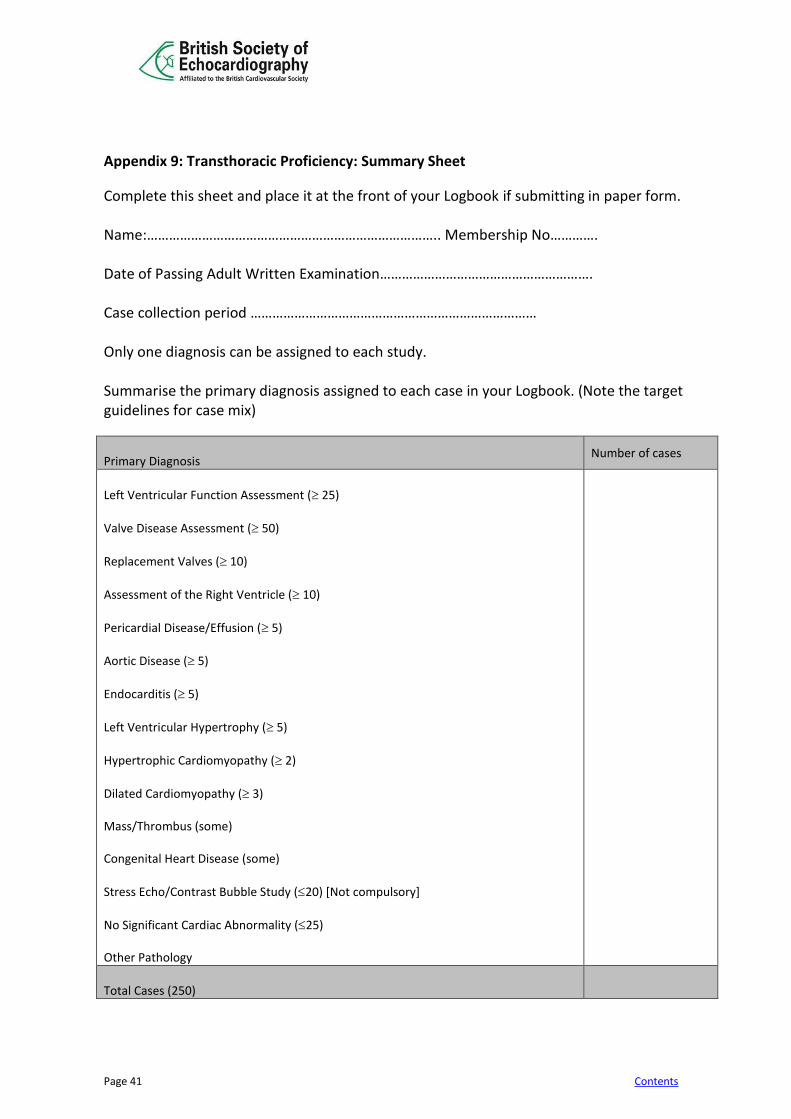
Complete this sheet and place it at the front of your Logbook if submitting in paper form. Name:…………………………………………………………………….. Membership No…………. Date of Passing Adult Written Examination…………………………………………………. Case collection period …………………………………………………………………… Only one diagnosis can be assigned to each study. Summarise the primary diagnosis assigned to each case in your Logbook. (Note the target guidelines for case mix)
Primary Diagnosis
Number of cases
Left Ventricular Function Assessment ( 25)
Valve Disease Assessment ( 50)
Replacement Valves ( 10)
Assessment of the Right Ventricle ( 10)
Pericardial Disease/Effusion ( 5)
Aortic Disease ( 5)
Endocarditis ( 5)
Left Ventricular Hypertrophy ( 5)
Hypertrophic Cardiomyopathy ( 2)
Dilated Cardiomyopathy ( 3) Mass/Thrombus (some) Congenital Heart Disease (some)
Stress Echo/Contrast Bubble Study (20) [Not compulsory]
No Significant Cardiac Abnormality (25) Other Pathology
Total Cases (250)

Page 42 Contents
Appendix 10: Examples of Station 1 Practical Assessment Mark Sheets
Reports 1 2 3 4 5 6 7 8 9 10 11 12 13 14 15 Comments
Fully Anonymised
Indication for echo present
2D /M Mode Measurements present
Appropriate measurements/Doppler calculations present
Do measurements/ Doppler Calculations match descriptions
All parts of heart described
Descriptions complete
Appropriate to request
Conclusion present
Pass or fail
Logbook YES NO Comments
Logbook submitted in one ring binder /file with dividers
All cases collected within 24-month period
250 TTE reports performed and reported by the candidate
150 cases reported by the candidate as 1st operator (can be countersigned)
All cases fully anonymised
Correct case mix
All reports with full name and signature
Summary sheet present
Supervisor/Mentor statement Present
Final check list present
Page 43 Contents
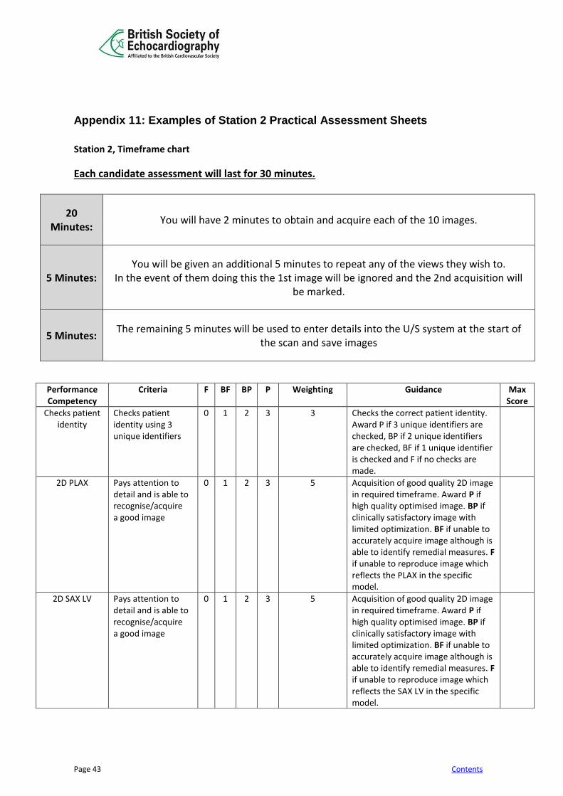
Appendix 11: Examples of Station 2 Practical Assessment Sheets
Station 2, Timeframe chart
Each candidate assessment will last for 30 minutes.
20 Minutes:
You will have 2 minutes to obtain and acquire each of the 10 images.
5 Minutes: You will be given an additional 5 minutes to repeat any of the views they wish to.
In the event of them doing this the 1st image will be ignored and the 2nd acquisition will be marked.
5 Minutes: The remaining 5 minutes will be used to enter details into the U/S system at the start of
the scan and save images
Performance Competency
Criteria F BF BP P Weighting Guidance Max Score
Checks patient identity
Checks patient identity using 3 unique identifiers
0 1 2 3 3 Checks the correct patient identity. Award P if 3 unique identifiers are checked, BP if 2 unique identifiers are checked, BF if 1 unique identifier is checked and F if no checks are made.
2D PLAX
Pays attention to detail and is able to recognise/acquire a good image
0 1 2 3 5 Acquisition of good quality 2D image in required timeframe. Award P if high quality optimised image. BP if clinically satisfactory image with limited optimization. BF if unable to accurately acquire image although is able to identify remedial measures. F if unable to reproduce image which reflects the PLAX in the specific model.
2D SAX LV
Pays attention to detail and is able to recognise/acquire a good image
0 1 2 3 5 Acquisition of good quality 2D image in required timeframe. Award P if high quality optimised image. BP if clinically satisfactory image with limited optimization. BF if unable to accurately acquire image although is able to identify remedial measures. F if unable to reproduce image which reflects the SAX LV in the specific model.
Page 44 Contents
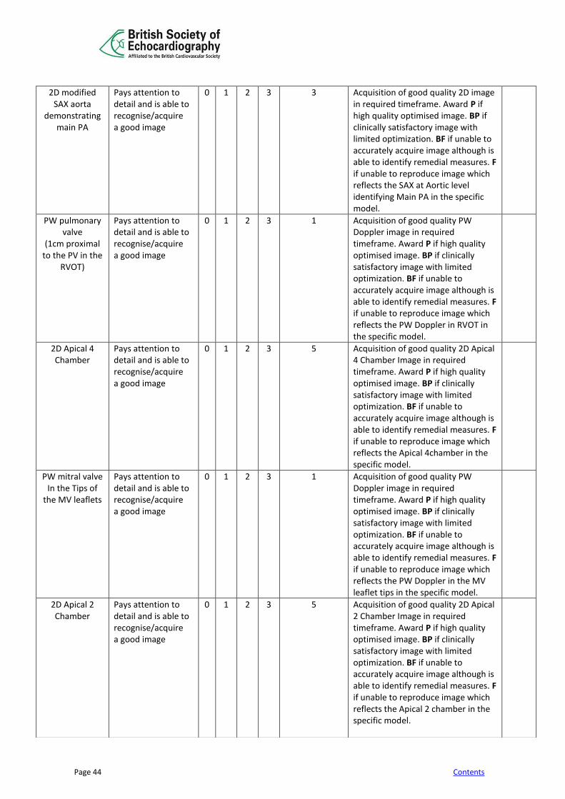
2D modified SAX aorta
demonstrating main PA
Pays attention to detail and is able to recognise/acquire a good image
0 1 2 3 3 Acquisition of good quality 2D image in required timeframe. Award P if high quality optimised image. BP if clinically satisfactory image with limited optimization. BF if unable to accurately acquire image although is able to identify remedial measures. F if unable to reproduce image which reflects the SAX at Aortic level identifying Main PA in the specific model.
PW pulmonary valve
(1cm proximal to the PV in the
RVOT)
Pays attention to detail and is able to recognise/acquire a good image
0 1 2 3 1 Acquisition of good quality PW Doppler image in required timeframe. Award P if high quality optimised image. BP if clinically satisfactory image with limited optimization. BF if unable to accurately acquire image although is able to identify remedial measures. F if unable to reproduce image which reflects the PW Doppler in RVOT in the specific model.
2D Apical 4 Chamber
Pays attention to detail and is able to recognise/acquire a good image
0 1 2 3 5 Acquisition of good quality 2D Apical 4 Chamber Image in required timeframe. Award P if high quality optimised image. BP if clinically satisfactory image with limited optimization. BF if unable to accurately acquire image although is able to identify remedial measures. F if unable to reproduce image which reflects the Apical 4chamber in the specific model.
PW mitral valve In the Tips of
the MV leaflets
Pays attention to detail and is able to recognise/acquire a good image
0 1 2 3 1 Acquisition of good quality PW Doppler image in required timeframe. Award P if high quality optimised image. BP if clinically satisfactory image with limited optimization. BF if unable to accurately acquire image although is able to identify remedial measures. F if unable to reproduce image which reflects the PW Doppler in the MV leaflet tips in the specific model.
2D Apical 2 Chamber
Pays attention to detail and is able to recognise/acquire a good image
0 1 2 3 5 Acquisition of good quality 2D Apical 2 Chamber Image in required timeframe. Award P if high quality optimised image. BP if clinically satisfactory image with limited optimization. BF if unable to accurately acquire image although is able to identify remedial measures. F if unable to reproduce image which reflects the Apical 2 chamber in the specific model.
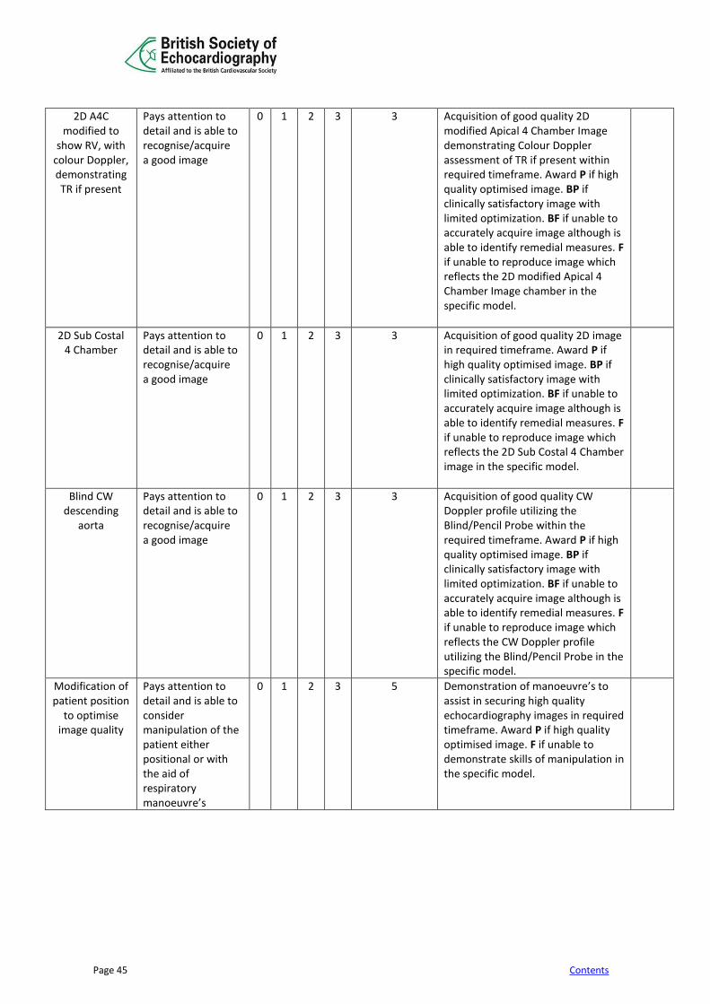
Page 45 Contents
2D A4C modified to
show RV, with colour Doppler, demonstrating TR if present
Pays attention to detail and is able to recognise/acquire a good image
0 1 2 3 3 Acquisition of good quality 2D modified Apical 4 Chamber Image demonstrating Colour Doppler assessment of TR if present within required timeframe. Award P if high quality optimised image. BP if clinically satisfactory image with limited optimization. BF if unable to accurately acquire image although is able to identify remedial measures. F if unable to reproduce image which reflects the 2D modified Apical 4 Chamber Image chamber in the specific model.
2D Sub Costal 4 Chamber
Pays attention to detail and is able to recognise/acquire a good image
0 1 2 3 3 Acquisition of good quality 2D image in required timeframe. Award P if high quality optimised image. BP if clinically satisfactory image with limited optimization. BF if unable to accurately acquire image although is able to identify remedial measures. F if unable to reproduce image which reflects the 2D Sub Costal 4 Chamber image in the specific model.
Blind CW descending
aorta
Pays attention to detail and is able to recognise/acquire a good image
0 1 2 3 3 Acquisition of good quality CW Doppler profile utilizing the Blind/Pencil Probe within the required timeframe. Award P if high quality optimised image. BP if clinically satisfactory image with limited optimization. BF if unable to accurately acquire image although is able to identify remedial measures. F if unable to reproduce image which reflects the CW Doppler profile utilizing the Blind/Pencil Probe in the specific model.
Modification of patient position
to optimise image quality
Pays attention to detail and is able to consider manipulation of the patient either positional or with the aid of respiratory manoeuvre’s
0 1 2 3 5 Demonstration of manoeuvre’s to assist in securing high quality echocardiography images in required timeframe. Award P if high quality optimised image. F if unable to demonstrate skills of manipulation in the specific model.

Page 46 Contents
Appendix 12: Getting the Viva Cases right This section of the submission will be reviewed in great detail by the assessors. Candidates are most often failed at this stage of the process. The 5 cases are assumed to be your best work. Do not choose patients who are poorly echogenic. All images must be recorded on the same patient on the same occasion. Each case should be a full study performed to the BSE minimum dataset and not a focussed study. Optimise the 2D and Doppler images. Make sure you have optimised the gain setting, sector width, depth, harmonics, focus, sweep speed, Doppler baseline and scale, colour gain. Candidates may be failed on just this aspect. If these points are not done well in your submission it may be assumed that you will have poor quality images on your routine cases. Read the mark sheet that the assessors will be using to assess the study. You need to show all standard views, including the subcostal and suprasternal views. Only include loops and stills that you wish to be assessed. Ensure that loops and stills with measurements shown match the parameters quoted in the report. Include images demonstrating all of the measurements quoted in the report. Ensure that the M-mode is correctly aligned. If it is too difficult take your measurements from the 2D images or the measurements may be very inaccurate. M-mode measurement of the left ventricular dimensions performed accurately is essential in at least one case. Get the Doppler right. Place the cursor correctly and alter the Doppler scale and sweep speed appropriately. Get the best signal before recording. Optimise the colour Doppler gains before recording. Use the stand alone probe for the aortic stenosis case (and other cases, if appropriate) and demonstrate you are able to obtain a CW Doppler signal from the apical, right parasternal and/or suprasternal windows in your search for the peak gradient. You must be able to use the common Doppler equations e.g. continuity equation, calculation of a shunt, estimation of pulmonary artery systolic pressure. Each case should be accompanied by a full and comprehensive report. This should include a summary that can be understood by any non-echocardiographer. GOOD LUCK
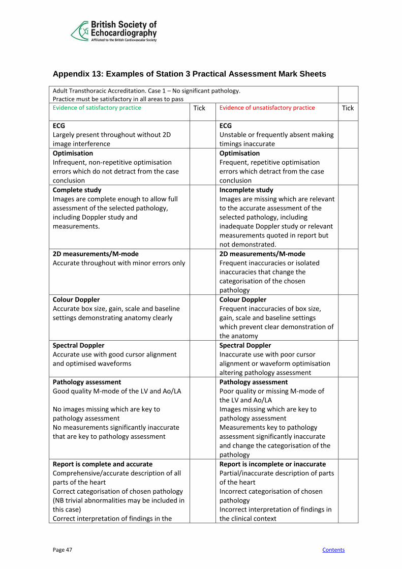
Page 47 Contents
Appendix 13: Examples of Station 3 Practical Assessment Mark Sheets
Adult Transthoracic Accreditation. Case 1 – No significant pathology. Practice must be satisfactory in all areas to pass
Evidence of satisfactory practice
Tick Evidence of unsatisfactory practice
Tick
ECG Largely present throughout without 2D image interference
ECG Unstable or frequently absent making timings inaccurate
Optimisation Infrequent, non-repetitive optimisation errors which do not detract from the case conclusion
Optimisation Frequent, repetitive optimisation errors which detract from the case conclusion
Complete study Images are complete enough to allow full assessment of the selected pathology, including Doppler study and measurements.
Incomplete study Images are missing which are relevant to the accurate assessment of the selected pathology, including inadequate Doppler study or relevant measurements quoted in report but not demonstrated.
2D measurements/M-mode Accurate throughout with minor errors only
2D measurements/M-mode Frequent inaccuracies or isolated inaccuracies that change the categorisation of the chosen pathology
Colour Doppler Accurate box size, gain, scale and baseline settings demonstrating anatomy clearly
Colour Doppler Frequent inaccuracies of box size, gain, scale and baseline settings which prevent clear demonstration of the anatomy
Spectral Doppler Accurate use with good cursor alignment and optimised waveforms
Spectral Doppler Inaccurate use with poor cursor alignment or waveform optimisation altering pathology assessment
Pathology assessment Good quality M-mode of the LV and Ao/LA No images missing which are key to pathology assessment No measurements significantly inaccurate that are key to pathology assessment
Pathology assessment Poor quality or missing M-mode of the LV and Ao/LA Images missing which are key to pathology assessment Measurements key to pathology assessment significantly inaccurate and change the categorisation of the pathology
Report is complete and accurate Comprehensive/accurate description of all parts of the heart Correct categorisation of chosen pathology (NB trivial abnormalities may be included in this case) Correct interpretation of findings in the
Report is incomplete or inaccurate Partial/inaccurate description of parts of the heart Incorrect categorisation of chosen pathology Incorrect interpretation of findings in the clinical context
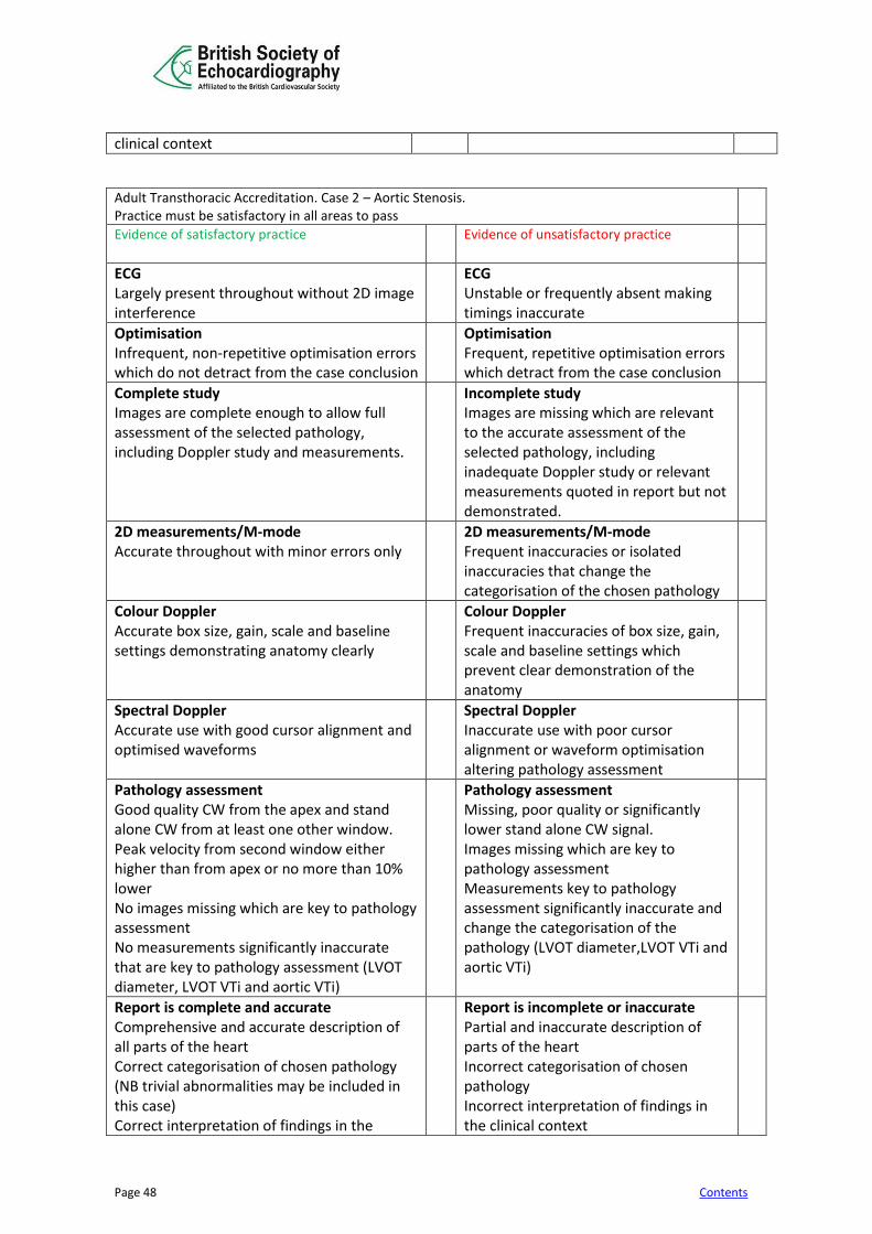
Page 48 Contents
clinical context
Adult Transthoracic Accreditation. Case 2 – Aortic Stenosis. Practice must be satisfactory in all areas to pass
Evidence of satisfactory practice
Evidence of unsatisfactory practice
ECG Largely present throughout without 2D image interference
ECG Unstable or frequently absent making timings inaccurate
Optimisation Infrequent, non-repetitive optimisation errors which do not detract from the case conclusion
Optimisation Frequent, repetitive optimisation errors which detract from the case conclusion
Complete study Images are complete enough to allow full assessment of the selected pathology, including Doppler study and measurements.
Incomplete study Images are missing which are relevant to the accurate assessment of the selected pathology, including inadequate Doppler study or relevant measurements quoted in report but not demonstrated.
2D measurements/M-mode Accurate throughout with minor errors only
2D measurements/M-mode Frequent inaccuracies or isolated inaccuracies that change the categorisation of the chosen pathology
Colour Doppler Accurate box size, gain, scale and baseline settings demonstrating anatomy clearly
Colour Doppler Frequent inaccuracies of box size, gain, scale and baseline settings which prevent clear demonstration of the anatomy
Spectral Doppler Accurate use with good cursor alignment and optimised waveforms
Spectral Doppler Inaccurate use with poor cursor alignment or waveform optimisation altering pathology assessment
Pathology assessment Good quality CW from the apex and stand alone CW from at least one other window. Peak velocity from second window either higher than from apex or no more than 10% lower No images missing which are key to pathology assessment No measurements significantly inaccurate that are key to pathology assessment (LVOT diameter, LVOT VTi and aortic VTi)
Pathology assessment Missing, poor quality or significantly lower stand alone CW signal. Images missing which are key to pathology assessment Measurements key to pathology assessment significantly inaccurate and change the categorisation of the pathology (LVOT diameter,LVOT VTi and aortic VTi)
Report is complete and accurate Comprehensive and accurate description of all parts of the heart Correct categorisation of chosen pathology (NB trivial abnormalities may be included in this case) Correct interpretation of findings in the
Report is incomplete or inaccurate Partial and inaccurate description of parts of the heart Incorrect categorisation of chosen pathology Incorrect interpretation of findings in the clinical context
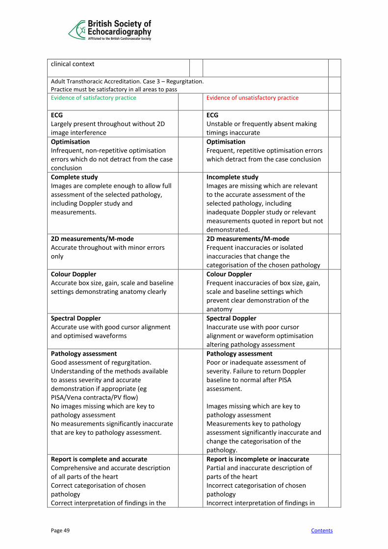
Page 49 Contents
clinical context
Adult Transthoracic Accreditation. Case 3 – Regurgitation. Practice must be satisfactory in all areas to pass
Evidence of satisfactory practice
Evidence of unsatisfactory practice
ECG Largely present throughout without 2D image interference
ECG Unstable or frequently absent making timings inaccurate
Optimisation Infrequent, non-repetitive optimisation errors which do not detract from the case conclusion
Optimisation Frequent, repetitive optimisation errors which detract from the case conclusion
Complete study Images are complete enough to allow full assessment of the selected pathology, including Doppler study and measurements.
Incomplete study Images are missing which are relevant to the accurate assessment of the selected pathology, including inadequate Doppler study or relevant measurements quoted in report but not demonstrated.
2D measurements/M-mode Accurate throughout with minor errors only
2D measurements/M-mode Frequent inaccuracies or isolated inaccuracies that change the categorisation of the chosen pathology
Colour Doppler Accurate box size, gain, scale and baseline settings demonstrating anatomy clearly
Colour Doppler Frequent inaccuracies of box size, gain, scale and baseline settings which prevent clear demonstration of the anatomy
Spectral Doppler Accurate use with good cursor alignment and optimised waveforms
Spectral Doppler Inaccurate use with poor cursor alignment or waveform optimisation altering pathology assessment
Pathology assessment Good assessment of regurgitation. Understanding of the methods available to assess severity and accurate demonstration if appropriate (eg PISA/Vena contracta/PV flow) No images missing which are key to pathology assessment No measurements significantly inaccurate that are key to pathology assessment.
Pathology assessment Poor or inadequate assessment of severity. Failure to return Doppler baseline to normal after PISA assessment. Images missing which are key to pathology assessment Measurements key to pathology assessment significantly inaccurate and change the categorisation of the pathology.
Report is complete and accurate Comprehensive and accurate description of all parts of the heart Correct categorisation of chosen pathology Correct interpretation of findings in the
Report is incomplete or inaccurate Partial and inaccurate description of parts of the heart Incorrect categorisation of chosen pathology Incorrect interpretation of findings in
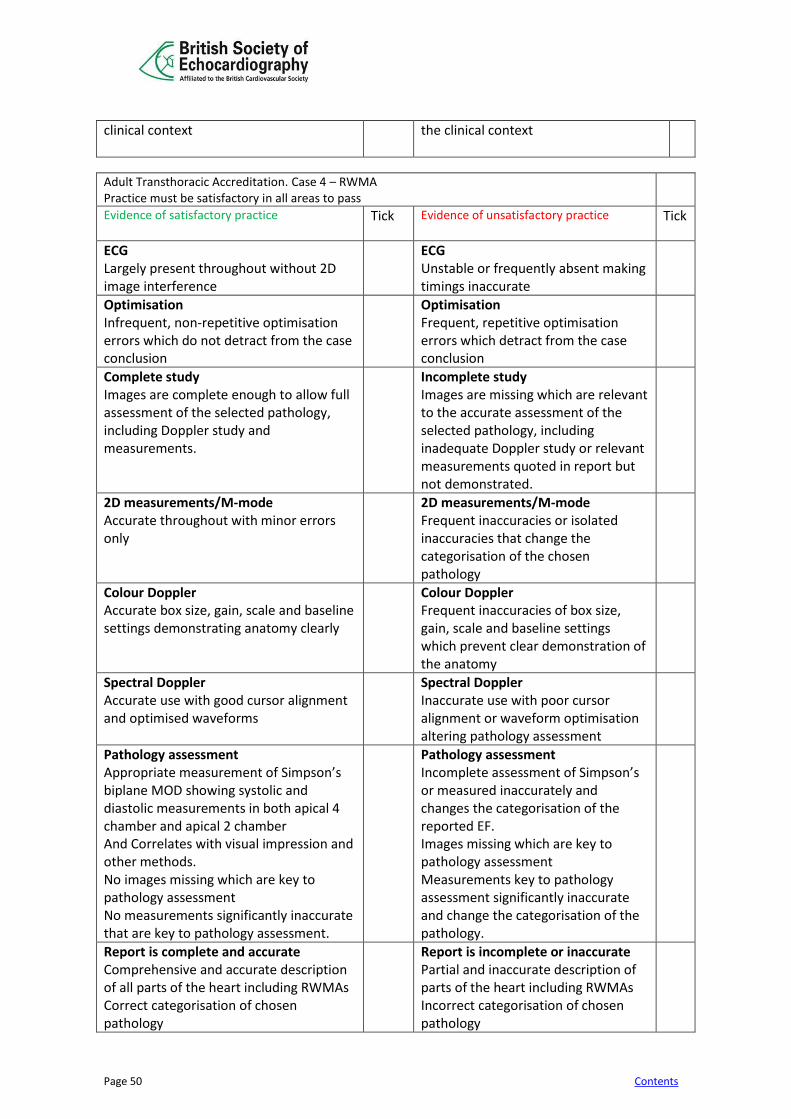
Page 50 Contents
clinical context
the clinical context
Adult Transthoracic Accreditation. Case 4 – RWMA Practice must be satisfactory in all areas to pass
Evidence of satisfactory practice
Tick Evidence of unsatisfactory practice
Tick
ECG Largely present throughout without 2D image interference
ECG Unstable or frequently absent making timings inaccurate
Optimisation Infrequent, non-repetitive optimisation errors which do not detract from the case conclusion
Optimisation Frequent, repetitive optimisation errors which detract from the case conclusion
Complete study Images are complete enough to allow full assessment of the selected pathology, including Doppler study and measurements.
Incomplete study Images are missing which are relevant to the accurate assessment of the selected pathology, including inadequate Doppler study or relevant measurements quoted in report but not demonstrated.
2D measurements/M-mode Accurate throughout with minor errors only
2D measurements/M-mode Frequent inaccuracies or isolated inaccuracies that change the categorisation of the chosen pathology
Colour Doppler Accurate box size, gain, scale and baseline settings demonstrating anatomy clearly
Colour Doppler Frequent inaccuracies of box size, gain, scale and baseline settings which prevent clear demonstration of the anatomy
Spectral Doppler Accurate use with good cursor alignment and optimised waveforms
Spectral Doppler Inaccurate use with poor cursor alignment or waveform optimisation altering pathology assessment
Pathology assessment Appropriate measurement of Simpson’s biplane MOD showing systolic and diastolic measurements in both apical 4 chamber and apical 2 chamber And Correlates with visual impression and other methods. No images missing which are key to pathology assessment No measurements significantly inaccurate that are key to pathology assessment.
Pathology assessment Incomplete assessment of Simpson’s or measured inaccurately and changes the categorisation of the reported EF. Images missing which are key to pathology assessment Measurements key to pathology assessment significantly inaccurate and change the categorisation of the pathology.
Report is complete and accurate Comprehensive and accurate description of all parts of the heart including RWMAs Correct categorisation of chosen pathology
Report is incomplete or inaccurate Partial and inaccurate description of parts of the heart including RWMAs Incorrect categorisation of chosen pathology
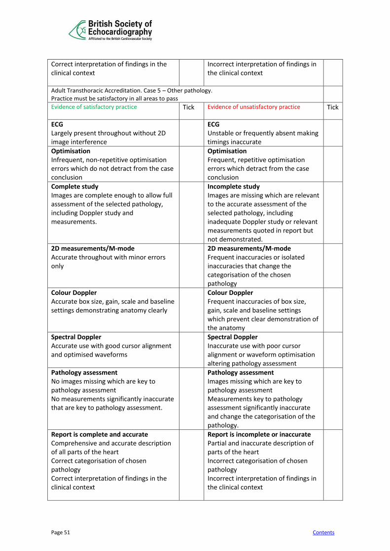
Page 51 Contents
Correct interpretation of findings in the clinical context
Incorrect interpretation of findings in the clinical context
Adult Transthoracic Accreditation. Case 5 – Other pathology. Practice must be satisfactory in all areas to pass
Evidence of satisfactory practice
Tick Evidence of unsatisfactory practice
Tick
ECG Largely present throughout without 2D image interference
ECG Unstable or frequently absent making timings inaccurate
Optimisation Infrequent, non-repetitive optimisation errors which do not detract from the case conclusion
Optimisation Frequent, repetitive optimisation errors which detract from the case conclusion
Complete study Images are complete enough to allow full assessment of the selected pathology, including Doppler study and measurements.
Incomplete study Images are missing which are relevant to the accurate assessment of the selected pathology, including inadequate Doppler study or relevant measurements quoted in report but not demonstrated.
2D measurements/M-mode Accurate throughout with minor errors only
2D measurements/M-mode Frequent inaccuracies or isolated inaccuracies that change the categorisation of the chosen pathology
Colour Doppler Accurate box size, gain, scale and baseline settings demonstrating anatomy clearly
Colour Doppler Frequent inaccuracies of box size, gain, scale and baseline settings which prevent clear demonstration of the anatomy
Spectral Doppler Accurate use with good cursor alignment and optimised waveforms
Spectral Doppler Inaccurate use with poor cursor alignment or waveform optimisation altering pathology assessment
Pathology assessment No images missing which are key to pathology assessment No measurements significantly inaccurate that are key to pathology assessment.
Pathology assessment Images missing which are key to pathology assessment Measurements key to pathology assessment significantly inaccurate and change the categorisation of the pathology.
Report is complete and accurate Comprehensive and accurate description of all parts of the heart Correct categorisation of chosen pathology Correct interpretation of findings in the clinical context
Report is incomplete or inaccurate Partial and inaccurate description of parts of the heart Incorrect categorisation of chosen pathology Incorrect interpretation of findings in the clinical context

Page 52 Contents
Appendix 14: Mentor statement to accompany the Practical Assessment
Candidate’s name ____________________________________________
Initial
I certify that the candidate has undergone a programme of training in echocardiography
I certify I have observed the candidate scanning and I am satisfied that he/she is competent at completing a full transthoracic echo study.
I certify that the candidate has reached a standard of training to be able to independently perform and report a transthoracic echocardiographic study. He/she has reached all of the mandated competencies. I have signed off the candidate’s competency sheet.
I certify that the candidate above has performed and reported the cases included in the accompanying Log Book within a 24-month period.
I certify that all cases are fully anonymised (no patient’s personal details such as names, full date of births or addresses) as per Appendix 14
I certify that all cases are signed with name printed of the candidate
I certify that these cases are being handed in as per Trust Policy Guidelines
Mentor’s name: ____________________________________________ Signature: ______________________________ Date: _______________________ I am satisfied that the candidate above has performed and reported the cases included in the accompanying Log Book within a 24-month period and five cases are also enclosed. Medical/Technical Head of Echocardiography’s name: __________________________ Signature: _______________________________ Date: _________________________ Notes: The Head of Echocardiography is usually the lead clinician or consultant cardiologist with overall responsibility for echocardiography. This may be a representative from a local Cardiology department who has personally observed the candidate scanning and is satisfied that they have the ability to perform and report echoes independently.
Page 53 Contents
Appendix 15: BSE Policy on the Non-Anonymisation of Patient Data
Introduction
The duty of confidentiality arises out of the common law of confidentiality, professional obligations and also staff employment contracts. Breach of confidence may lead to disciplinary measures, bring into question professional reputation and possibly result in legal proceedings.
Guidance is provided to NHS staff in the ‘NHS Code of Practice on Confidentiality’ (November 2003). http://www.dh.gov.uk/prod_consum_dh/groups/dh_digitalassets/@dh/@en/documents/digitalasset/dh_40 69254.pdf
Patient information that can identify individual patients is confidential and must not be used or disclosed. In contrast, anonymised information is not confidential and may be used.
Key identifiable information includes:
Patient’s name, address, full post code, date of birth; NHS number and local identifiable codes; Anything else that may be used to identify a patient directly or indirectly. For example, rare diseases, drug treatment or statistical analyses which have very small numbers within a small population may allow individuals to be identified.
Anonymisation requires the removal of such information from all reports and images.
For accreditation purposes, BSE Administrators and BSE Assessors must not be able to identify the patient from the detail or combination of details given.
Speakers presenting on behalf of the BSE at meetings and speakers on courses/meetings awarded BSE re- accreditation points must ensure that all presentation material is anonymised.
Guidance to candidates submitting Logbooks and Cases for Accreditation
The NHS Code of Practice on confidentiality means that evidence submitted for the practical part of the Accreditation process must have all patient identification removed.
In order for evidence to be considered to have been anonymised, BSE Administrators and BSE Assessors must not be able to see any of the identifiers listed above. As age is relevant to the assessment either the age or year of birth must be provided however a full date of birth must not be shown.
Page 54 Contents
Reports Please note that correction fluid may still allow data to be visible if you look at the back of the page, as does placing a sticker over the patient data. Marker pen often fades so that data may be correctly disguised at the point of anonymisation but not when brought to a Practical Assessment session
We therefore advise: Use the online portal and electronically delete all patient information except age and gender Or: cutting out the patient data Or: Deleting data electronically prior to printing Or: Using corrective fluid or marker pen, then photocopying the sheet
Cases In order for cases to be classed as anonymous BSE Administrators and BSE Markers must not be able to gain personal information about the patient that is not directly relevant to the echocardiogram. This means that name, address, NHS/Hospital number and full date of birth must not be visible on the report that is enclosed with the images nor on the images themselves. If the age is not given separately the year of birth must be left visible on the report. Please see the notes above about correctly removing patient ID from the paper report that is enclosed with the cases. We appreciate that the removal of patient ID from cases may be difficult depending on the machine being used, we therefore advise that the cases are specifically collected for the BSE and the data inputs are made relevant to your cases. E.g. Patient Name could be ‘BSE Case 1’ or ‘Aortic Stenosis’, Patient Number could be your membership number followed by case number, ‘1111-1’
Explanatory notes for the inclusion of patient identifiable data in any medium are NOT acceptable. Breach of NHS Code of Practice on Confidentiality Major breach: One or more examples of detailed patient demographics (e.g. name and address) OR One or more examples of patient data sufficient to allow a patient to be traced in any way
Minor breach: Examples of patient identifiable information found within the logbook. These might include, for example, first name or date of birth but insufficient information to identify the patient. In the event of a major breach: The candidate will automatically fail and candidate will be informed of the fail and notified of the reason for it. The Chair of the Accreditation Committee will be notified of all major breaches and will make the decision as to whether the Head of Information Governance at the candidate’s place of employment should be informed.

Page 55 Contents
In the event of a minor breach: The candidate will be informed of the breach and notified of the reason for it. This will be taken into account in the marking scheme. The final decision remains at the discretion of the Chair of the Accreditation Committee.

Page 56 Contents
Appendix 16: Final Checklist for candidates. Please complete and bring to your Practical Assessment. Some of these documents are included in the online logbook.
App
YES
NO Comments
Logbook submitted in one ring binder /file with dividers
All cases collected within 24-month period
250 TTE reports performed and reported by the candidate All reports with full name and signature
200 cases for Community. 150 if holding previous TOE
BSE/EACVI Accreditation.
All cases fully anonymised
Correct case mix
Curriculum based assessment (Appendix 6)
Summary sheet present (Appendix 9)
Mentor statement Present (Appendix 14)