activity-dependent app trafficking by mints mint proteins are
TRANSCRIPT
Activity-dependent APP trafficking by Mints
1
Mint Proteins are Required for Synaptic Activity-Dependent APP Trafficking and Aβ Generation*
Sarah E. Sullivan, Gregory M. Dillon, Josefa M. Sullivan and Angela Ho
From the Department of Biology, Boston University, Boston, MA 02215.
*Running title: Activity-dependent APP trafficking by Mints To whom correspondence should be addressed: Angela Ho, Department of Biology, Boston University. 24 Cummington Mall, Boston MA, USA, Tel.: (617) 353-2093; Fax: (617) 353-6340; E-mail: [email protected] Keywords: Adaptor proteins, Mint/X11, Amyloid precursor protein, Synaptic activity, Trafficking, Alzheimer’s disease Background: Activity-dependent amyloid-β (Aβ) generation requires endosomal proteolytic cleavage of the amyloid precursor protein (APP). Results: Mints are adaptor proteins that regulate APP endocytosis and insertion at the plasma membrane upon activity induction or inhibition. Conclusion: Mints are necessary for activity-induced APP trafficking and Aβ generation. Significance: Insight into the cell biology and molecules controlling APP trafficking is essential in understanding Aβ pathogenesis. ABSTRACT
Aberrant amyloid-β (Aβ) production plays a causal role in Alzheimer’s disease (AD) pathogenesis. A major cellular pathway for Aβ generation is the activity-dependent endocytosis and proteolytic cleavage of the amyloid precursor protein (APP). However, the molecules controlling activity-dependent APP trafficking in neurons are less defined. Mints are adaptor proteins that directly interact with the endocytic sorting motif of APP and are functionally important in regulating APP endocytosis and Aβ production. We analyzed neuronal cultures from control and Mint knockout neurons that were treated with either glutamate or tetrodotoxin (TTX) to stimulate an increase or decrease in neuronal activity, respectively. We found that neuronal activation by glutamate increased APP endocytosis followed by elevated APP insertion into the
cell surface stabilizing APP at the plasma membrane. Conversely, suppression of neuronal activity by TTX decreased APP endocytosis and insertion. Interestingly, we found that activity-dependent APP trafficking and Aβ generation were blocked in Mint knockout neurons. We showed that wild-type Mint1 can rescue APP internalization and insertion in Mint knockout neurons. In addition, we found that Mint overexpression increased excitatory synaptic activity and APP was predominantly internalized to endosomes associated with APP processing. We demonstrated that presenilin 1 (PS1) endocytosis requires interaction with the PDZ domains of Mint1 and that this interaction facilitates activity-dependent colocalization of APP and PS1. These findings demonstrate that Mints are necessary for activity-induced APP and PS1 trafficking and provide insight into the cellular fate of APP in endocytic pathways essential for Aβ production.
Synaptic activity modulates the formation and secretion of Aβ (1-3). Previous evidence indicates that enhanced neuronal activity increases Aβ production, whereas blocking neuronal activity decreases Aβ production (1). Neuronal activity-dependent Aβ production is largely mediated by clathrin-dependent endocytosis of surface APP, endosomal processing of APP and Aβ release (3,4). The sorting signal that regulates endocytic processing of APP required for Aβ generation is
http://www.jbc.org/cgi/doi/10.1074/jbc.M113.541003The latest version is at JBC Papers in Press. Published on April 17, 2014 as Manuscript M113.541003
Copyright 2014 by The American Society for Biochemistry and Molecular Biology, Inc.
by guest on February 5, 2018http://w
ww
.jbc.org/D
ownloaded from

Activity-dependent APP trafficking by Mints
2
the highly conserved YENPTY consensus sequence for clathrin-coated pit internalization located in the cytoplasmic region of APP (5). Knock-in mice lacking the YENPTY endocytic motif of APP or mice in which clathrin-mediated endocytosis is inhibited have reduced Aβ levels in the brain, indicating that endocytosis is critical for Aβ production (6,7). Thus, understanding the cell biology and molecules controlling APP trafficking is of great significance for the mechanistic understanding of AD.
We propose that Mint proteins are critical players in regulating activity-dependent APP trafficking, which in turn, affects Aβ generation. Mints 1-3, also referred to as X11α/β/γ, X11/L1/L2 or APBA1/2/3, are multidomain adaptor proteins that interact with a variety of synaptic and Alzheimer’s-related proteins. In the variable N-terminal region, neuronal Mints 1 and 2 bind to Munc18-1, an essential synaptic fusion protein, linking Mints to synaptic vesicle exocytosis (8). In the conserved C-terminus, all Mints contain a phosphotyrosine-binding (PTB) domain which directly binds with the YENPTY endocytic motif of APP (9) and two PDZ domains that bind a number of proteins including PS1, the catalytic core of γ-secretase which cleaves APP (10,11). Several lines of evidence suggest Mints play an important role in activity-dependent APP trafficking: (i) Mints directly bind to APP and regulate Aβ production as deletion of Mints decreases Aβ plaque production in the brains of aging mice and mouse models of AD (12); (ii) APP endocytosis is attenuated in Mint knockout neurons, revealing a role for Mints in APP trafficking (13); (iii) Mints are regulators of synaptic transmission as deletion of Mints decreases spontaneous miniature currents and neurotransmitter release at excitatory synapses (14,15); (iv) Mints are upregulated and found in Aβ plaques of post-mortem AD brains (16,17); and (v) MINT2 was recently identified as a core regulatory mediator of APP endocytosis and metabolism associated with late-onset AD based on whole-transcriptome cerebral cortex gene expression (18). Herein, we examined the cellular mechanism by which Mints regulate
activity-induced APP trafficking that is relevant for Aβ pathogenesis. EXPERIMENTAL PROCEDURES
Plasmids-For lentivirus production, individual full-length rat Mints 1-3 cDNAs were inserted into pEGFP-C3 (Clontech) and subsequently inserted into the pFUW lentiviral vector to generate pFUW-EGFP-Mint1, pFUW-EGFP-Mint2, and pFUW-EGFP-Mint3 plasmids. Generation of pFUW-Mint1∆PDZ1/2 was previously described (31).
Neuronal cultures and lentiviral infection-We established two mouse lines that are homozygous for the floxed mutant alleles of all three Mint genes (Mint triple-floxed, MTF) and MTF carrying the double transgene of mutant APPswe/PS1∆E9 that overproduce human Aβ (MTFtg). High-density MTF and MTFtg hippocampal neurons were prepared from newborn mice of either sex and infected with lentiviruses as previously described (12-14). Briefly, neuronal cultures were infected with lentivirus at 3 days in vitro (DIV) and sustained until 13-15 DIV for analyses. The Mint1 rescue was performed by superinfection of cre-infected cultures with Mint1 expressing lentiviruses. All rescue experiments were performed with 3% lentivirus expressing GFP-tagged Mint1 protein. Wild-type neurons overexpressing individual Mints 1-3 were infected with 20% lentivirus expressing GFP-tagged Mint1, 2 or 3 proteins.
Live cell internalization and immunocytochemistry in primary neurons-Neurons at 13-15 DIV were treated with either 5 min pulse of 25 µM glutamate prior to internalization, 150 µM picrotoxin (PTX) or 2 µM tetrodotoxin (TTX) for 1 h at 37oC prior to antibody-labeling. Live neurons were labeled with an antibody against the N-terminal epitope of APP (clone 22C11; Millipore), PS1 (MAB5232; Millipore), or GluR1 (Millipore) at 14 DIV. Briefly, neurons were washed with cold artificial-cerebral spinal fluid (ACSF in mM) 150 NaCl, 10 HEPES, 30 KCl, 2.6 CaCl2, 1 MgCl2, 10 D-glucose, pH 7.4 and incubated at 37oC for 15 min to induce internalization as described previously (13). The remaining surface-bound antibodies were stripped in acid stripping buffer (0.5 M NaCl, 0.2 M acetic acid)
by guest on February 5, 2018http://w
ww
.jbc.org/D
ownloaded from
Activity-dependent APP trafficking by Mints
3
and fixed with 4% paraformaldehyde. Neurons were permeabilized in 0.3% Triton-X-100 and incubated with goat anti-mouse secondary antibodies conjugated to Alexa Fluor 546 (Invitrogen). For colocalization studies, neurons following PS1 internalization assay were incubated with primary anti-APP antibodies (U955) overnight at 4oC prior to incubation with an Alexa Fluor 633 conjugated secondary antibody. For surface-expressed APP, neurons were fixed and the surface-expressed APP was labeled with anti-APP antibodies (clone 22C11; Millipore) and an Alexa Fluor 546 conjugated secondary antibody. All immunofluorescence images were captured by FluoView FV10i scanning confocal microscope using a 60x objective with 3x optimal zoom with laser gain and threshold filters kept constant between conditions. Z-stacks were acquired from top to bottom of neurons using a 0.25 µm step with a numerical aperture of 1.5. Image analysis was performed using ImageJ software (NIH). Internalization was measured by manual selection of the most central image from the Z-stack and pixel intensity was quantified. The total soma areas were manually outlined from a non-experimental channel and overlaid onto the experimental channel prior to measuring intensity. The mean pixel intensity was divided by the soma area to eliminate variability due to varying soma sizes. Surface and insertion quantifications were taken from compressing the Z-stack into a single image and measuring pixel intensity of total captured soma area. For colocalization of internalized PS1 and APP, images were acquired as above with crosstalk correction activated to prevent signal bleed. Images were acquired at 512x512 pixels with a single pixel that represents 0.137 µm. JaCoP plugin for ImageJ was applied to unprocessed, center-selected images from acquired Z-stacks and the degree of signal overlap was determined by Mander’s coefficient.
Insertion assay-The insertion assay was performed at 14 DIV as previously described (13). Briefly, live neurons were first incubated with the N-terminal epitope of APP (22C11; Millipore) for 15 min followed by a cold (non-conjugated) secondary antibody for additional 15 min to block preexisting cell surface APP.
Neurons were then incubated for 15 min at 37oC and fixed with 4% paraformaldehyde and stained with the same APP antibody and an Alexa Fluor 546 secondary antibody. Newly inserted APP was visualized by scanning confocal microscope with constant laser and threshold settings and pixel intensity was quantified using ImageJ software.
Electrophysiological analysis-Whole-cell voltage-clamp was performed in cultured hippocampal neurons at 13-16 DIV as previously described (14,19). Briefly, synaptic recordings were filtered in electrode solution containing (in mM) 105 Cs-MeSO3, 10 CsCl, 5 NaCl, 10 HEPES, 0.2 EGTA, 4 Mg-ATP, and 0.3 Na2GTP, pH 7.4 at 300 mOsm. Spontaneous event recordings were recorded in the presence of 1 µM TTX and 50 µM D-2-amino-5-phosphonovaleric acid plus 20 µM bicuculline for miniature excitatory postsynaptic currents (mEPSCs). Glutamate-evoked recordings were recorded in the presence of 50 µM cyclothiazide to additionally block AMPA receptor desensitization. Glutamate at 100 µM was perfused into the bath until the evoked current stabilized. The total glutamate-evoked current was calculated as the difference between the peak and leak-evoked current which was determined from the current before and after glutamate application. Recordings were acquired with an Axopatch 200A amplifier and Clampex 10.0 software (Molecular Devices).
Aβ peptide measurement-MTFtg neurons at 13-15 DIV were treated with either 150 µM PTX or 2 µM TTX for 1 h. Conditioned medium was collected and centrifuged at 15,000 x g for 10 min at 4oC. The supernatant was subjected to ELISA measurement for human Aβ42 according to the manufacturer’s instructions (27711, ImmunoBiological Laboratories).
Antibodies-APP 22C11 (MAB348, Millipore), APP U955, GluR1-NT (MAB2263, Millipore), Mint1 P730, Mint2 (M3319, Sigma), Mint3 (PA1-072, Thermo Scientific), PS1 (MAB5232, Millipore), tubulin DM1A (T6199, Sigma). APP U955 and Mint1 P730 are generous gifts from Dr. T.C. Südhof.
Statistical analysis-We used a Tukey’s test to determine statistical significance. * p<0.05,
by guest on February 5, 2018http://w
ww
.jbc.org/D
ownloaded from
Activity-dependent APP trafficking by Mints
4
** p<0.01, *** p<0.001. RESULTS
Mints are required for activity-induced APP trafficking. We first examined whether Mints play a role in activity-dependent APP endocytosis. A live cell endocytosis assay was performed to quantify intracellular APP in cultured neurons from established mouse lines that is homozygous for the floxed mutant alleles of all three Mint genes (Mint triple-floxed, MTF) and MTF carrying the double transgene of mutant APP and PS1 (APPswe/PS1∆E9) that overproduce human Aβ (MTFtg). We have previously shown that cultured neurons from newborn mice infected with lentiviruses that expressed a functional cre recombinase showed complete deletion of all Mints, whereas neurons infected with inactive cre recombinase retain endogenous Mint expression [Fig. 6B; (12-14)]. Following glutamate application at 13-15 DIV, control MTF neurons showed an approximately 17% and 33% increase in internalized APP in the somas and processes, respectively, demonstrating that enhanced synaptic activity increases endogenous APP endocytosis (Fig. 1A-C).
We next examined whether activity-induced APP endocytosis requires Mints. Under basal conditions, Mint knockout neurons showed a 23-28% reduction in internalized APP compared to control neurons demonstrating that Mints regulate APP internalization which supports our previous studies (13). Interestingly, Mint knockout neurons showed no additional changes in APP internalization following glutamate application. As a control, no visible staining was observed in non-permeabilized cells or cells treated with the endocytic inhibitor dynasore (data not shown).
In supporting experiments, addition of PTX, a noncompetitive GABAA receptor antagonist to increase synaptic transmission caused a 31-34% increase in internalized APP in control MTF neurons (Fig.1D-F). In contrast, Mint knockout neurons prevented the increase in APP internalization following PTX application suggesting that Mints play an important role in activity-induced APP internalization.
Because studies have shown differences in APP sorting pathways as a result of mutations
that overproduce Aβ levels (20, 21), we examined APP endocytosis using MTFtg neurons carrying the APPswe/PS1∆E9 transgene. Consistent with our findings, we found an increase in internalized APP in MTFtg neurons following glutamate treatment (Fig. 1G-I). Mint knockout neurons showed a significant reduction in internalized APP compared to MTFtg control neurons and were not affected upon glutamate application. To determine whether Mints regulate activity-induced APP surface levels, both MTF and MTFtg neurons were fixed following a 5 min treatment with glutamate and the surface-expressed APP was immunolabeled with anti-APP antibodies. Surface APP staining showed no difference between control and Mint knockout neurons and glutamate stimulation fails to alter APP surface staining in both MTF and MTFtg neurons (Fig. 2A-D). The finding that activity controls APP endocytosis without significantly affecting steady-state surface expression suggests that membrane insertion of APP might also be regulated by activity. Newly inserted APP could arise from stable intracellular pools or by recycling of recently internalized APP. To examine whether neuronal activity affects APP insertion into the plasma membrane and to measure the effect of synaptic activity on insertion, we performed insertion assays. We found newly inserted APP staining on the surface of somas that represent a rapid, constitutive insertion of APP into the plasma membrane in control neurons. Following glutamate application, control neurons showed an increase in APP insertion, demonstrating that enhancing synaptic activity accelerated the amount of newly inserted APP in the plasma membrane (Fig. 2E-H). Under basal conditions, Mint knockout neurons showed a significant reduction in APP insertion compared to control neurons, demonstrating that Mints are necessary for the facilitation of APP insertion. Mint knockout neurons did not show any additional changes in APP insertion following glutamate application, suggesting that Mints are essential for glutamate-induced APP insertion into the plasma membrane.
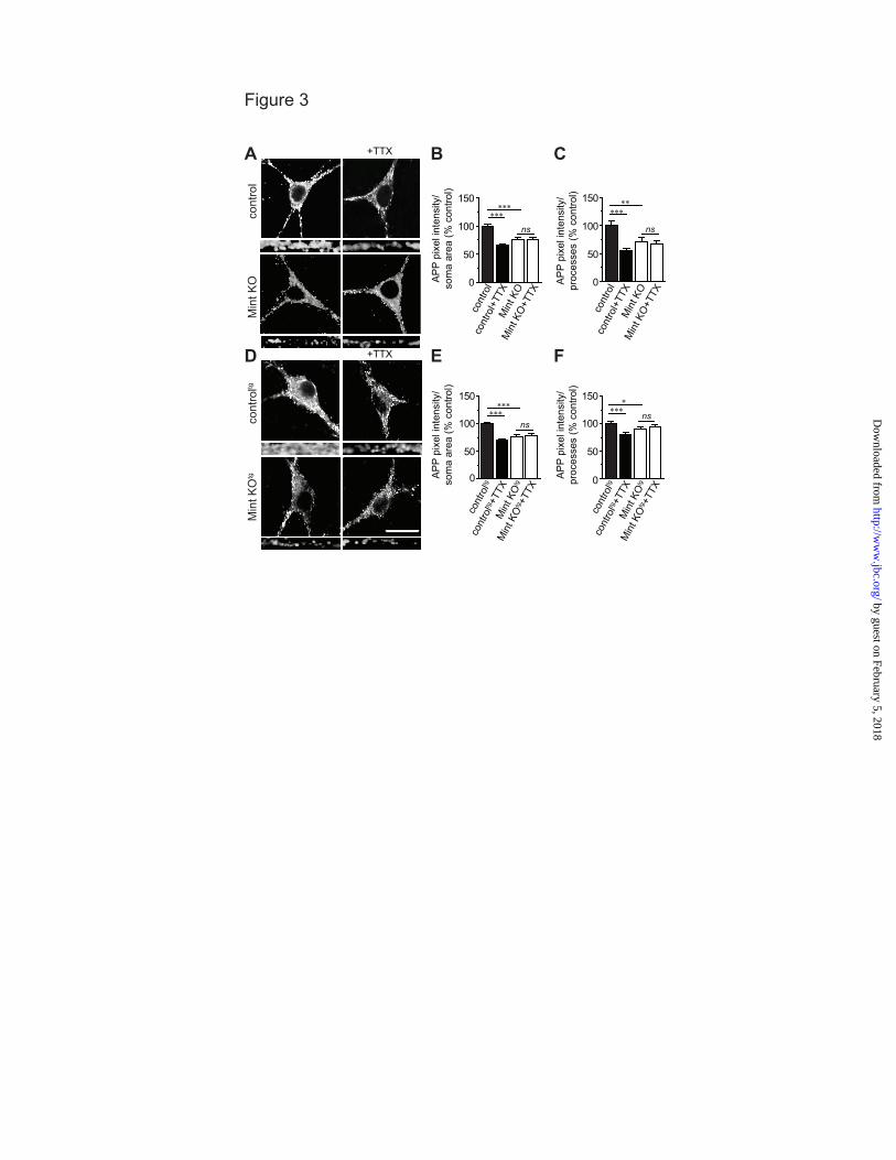
Mints are required for TTX-induced APP trafficking. To examine whether decreasing
by guest on February 5, 2018http://w
ww
.jbc.org/D
ownloaded from
Activity-dependent APP trafficking by Mints
5
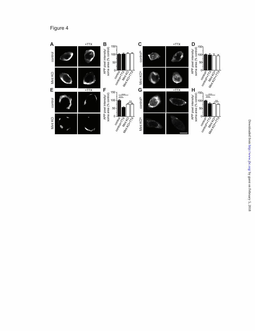
synaptic activity alters APP internalization, cultured MTF and MTFtg neurons were treated with tetrodotoxin (TTX), a sodium channel blocker that inhibits action potentials. TTX dramatically reduced APP internalization in the somas and processes of control MTF and MTFtg neurons, demonstrating that inhibiting activity can decrease APP endocytosis (Fig. 3). As expected, Mint knockout neurons showed a reduction in internalized APP compared to control neurons under basal conditions. In addition, TTX treatment fails to alter APP internalization in Mint knockout neurons. Also, TTX had no effect on the steady-state amount of APP at the cell surface in both control and Mint knockout neurons (Fig. 4A-D). However, the insertion of APP was significantly decreased in the presence of TTX in control MTF and MTFtg neurons (Fig. 4E-H). In addition, we found that TTX treatment does not affect APP insertion in Mint knockout neurons. Together, these experiments suggest that APP trafficking can be controlled by synaptic activity and that Mints are essential for activity-induced APP trafficking.
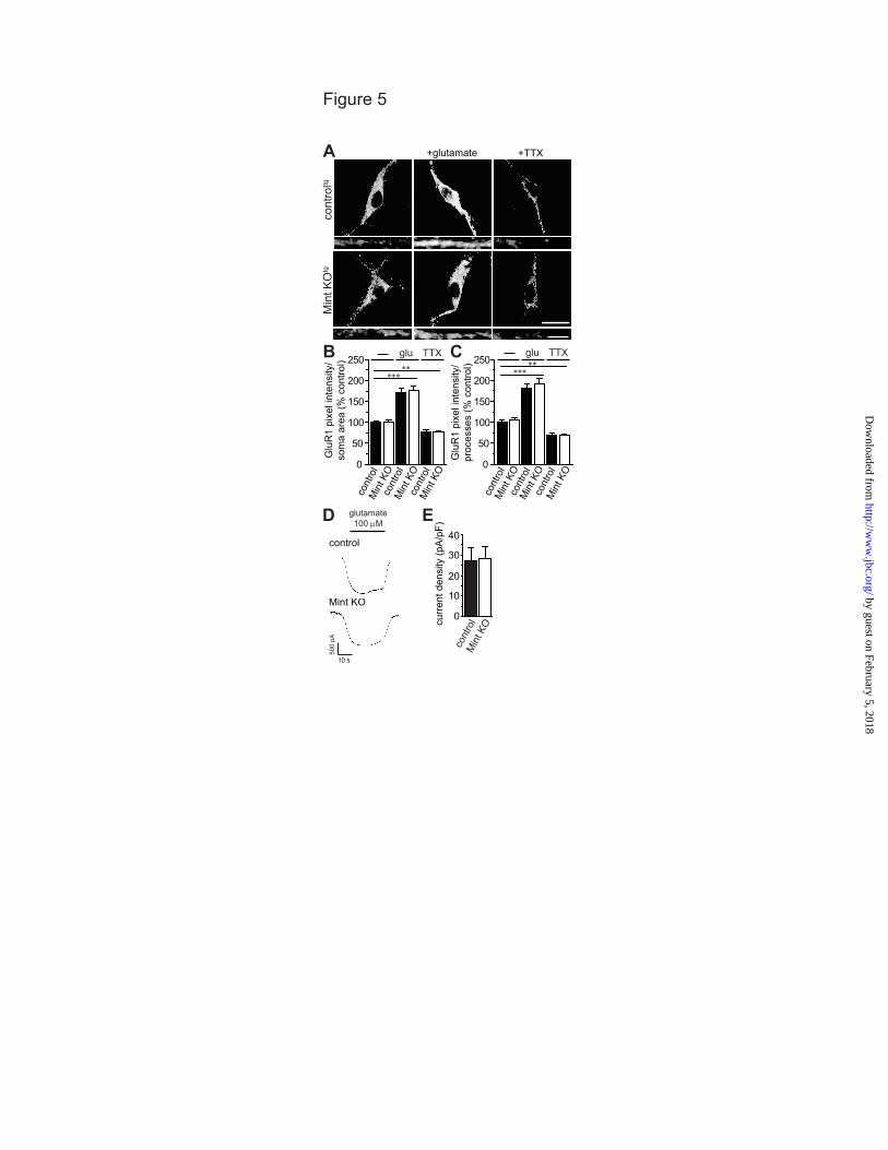
Mints specifically affect APP trafficking. To determine whether Mints have a specific effect on APP trafficking or cause a more general effect on endocytosis, we examined the internalization of the excitatory glutamate AMPA-type receptor GluR1, a protein that is internalized independently of Mints. Under basal conditions, GluR1 undergoes endocytosis and we found no changes in GluR1 internalization in Mint knockout neurons (Fig. 5A-C). Increasing synaptic activity with glutamate accelerated GluR1 internalization to 170% and 180% in the somas and processes of control MTFtg neurons, respectively. Conversely, blocking synaptic activity with TTX significantly reduced GluR1 internalization in control neurons. Under each pharmacological condition, GluR1 displayed similar endocytosis in Mint knockout neurons as compared to control, indicating that Mints do not affect GluR1 trafficking or endocytosis, in general.
To exclude the possibility that Mint proteins could affect glutamate receptor function, we measured whole-cell currents to exogenous glutamate application in control and Mint knockout MTFtg neurons (Fig. 5D-E).
Glutamate application induced an inward current at a holding potential of -70 mV. To eliminate differences in cell size, currents were normalized by cell capacitance and expressed as current densities. No difference in current density was detected after glutamate application in control and Mint knockout neurons.
Mints are essential and directly involved in activity-dependent APP trafficking and Aβ generation. To determine whether Mints are essential and directly involved in activity-dependent Aβ generation, we examined 13-15 DIV control and Mint knockout MTFtg neurons following PTX or TTX treatment, pharmacological agents to increase or decrease neuronal acitivty, respectively. We found Aβ42 levels were signficantly decreased in the conditioned medium of Mint knockout neurons, supporting our previous findings [Fig. 6A; (12)]. Addition of PTX increased Aβ generation in control neurons, as previously reported (1). Interestingly, PTX failed to increase Aβ generation from Mint knockout neurons. Furthermore, decreasing neuronal activity by TTX signficiantly decreased secreted Aβ levels in control but not in Mint knockout neurons. We next examined whether synaptic activity affects the overall protein expression of Mints and APP. Both TTX and glutamate had no effect on total protein expression of Mints 1-3 and APP (Fig. 6B). In addition, we did not detect any changes in APP protein expression in Mint knockout neurons.
To directly probe whether Mints are essential and directly involved in activity-dependent APP trafficking, we performed rescue experiments with full-length Mint1 in cultured MTFtg neurons. Rescue experiments were performed by co-infecting neurons with lentiviruses expressing GFP-tagged Mint1 proteins and active or inactive cre recombinase. Immunoblotting of Mint1 demonstrated that culture medium containing 3% lentivirus expressing GFP-tagged Mint1 in Mint knockout neurons showed similar protein expression level as endogenous control neurons (Fig. 6C). Therefore, all rescue experiments were performed with 3% lentivirus expressing GFP-tagged Mint1 protein. We found that increasing
by guest on February 5, 2018http://w
ww
.jbc.org/D
ownloaded from
Activity-dependent APP trafficking by Mints
6
the amount of lentivirus GFP-tagged Mint1 protein did not affect the overall protein expression of APP. Electrophysiologically, we found that deletion of Mint proteins caused an approximately 2-fold decrease in the frequency but not amplitude of spontaneous “minis” of excitatory postsynaptic currents (mEPSCs) as described previously (Fig. 6D-F) (14). Importantly, this decrease in excitatory synaptic transmission could be rescued to control levels with full-length Mint1.
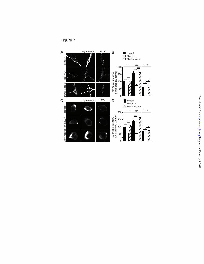
We next tested rescue of the Mint knockout phenotype on APP endocytosis and insertion following glutamate and TTX application. Expression of wild-type Mint1 fully reversed the impairment in APP internalization and insertion in Mint knockout neurons demonstrating that Mints directly function in activity-dependent APP trafficking (Fig. 7).
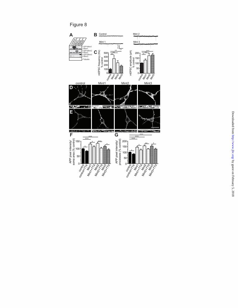
Overexpression of Mints enhanced excitatory synaptic transmission and accelerated APP endocytosis and insertion. To determine whether overexpression of Mints alters neurotransmitter release, we infected wild-type neurons with lentivirus for Mints 1, 2 or 3 (Fig. 8A) and monitored synaptic transmission. Neurons overexpressing Mints 1-3 showed a significant increase in the frequency and amplitude of spontaneous mEPSCs (Fig. 8B-C). These results suggest that the enhanced effects of Mint overexpression are due to an increase in the number of functional synapses at either presynaptic and/or postsynaptic sites.
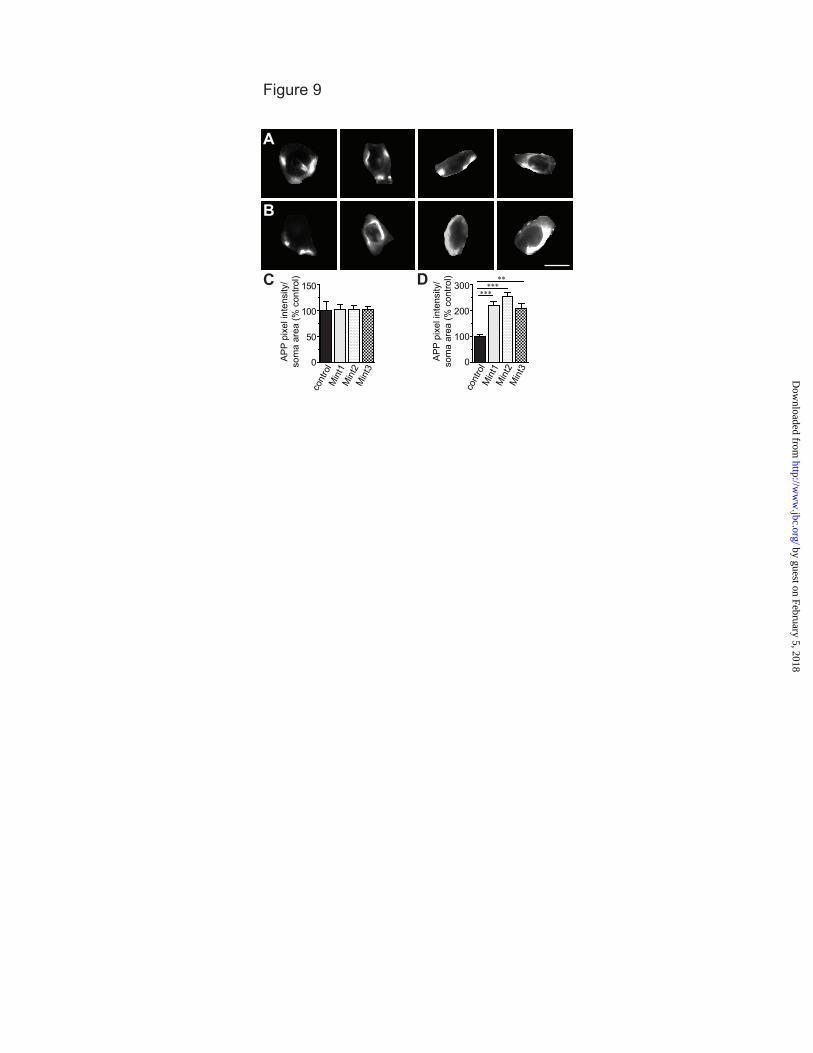
To examine the effect of Mint overexpression on APP trafficking, we performed live-cell immunostaining. Overexpression of individual Mints significantly enhanced APP internalization in the somas and processes of neurons compared to control neurons (Fig. 8D, F-G). Interestingly, the increased APP endocytosis in Mint overexpressed neurons was prevented by the addition of TTX, indicating that synaptic activity had a significant effect on the efficiency of APP internalization (Fig. 8E, F-G). We detected no significant differences between control and Mint overexpressing neurons in surface APP (Fig. 9A, C), but observed a large increase in APP insertion in Mint overexpressing neurons (Fig. 9B, D), suggesting a significant fraction of APP
is inserted into the plasma membrane and that the extent of APP endocytosis and insertion are increased by Mint overexpression.
Mints are required for PS1 internalization and activity-dependent APP/PS1 colocalization. Because the PDZ domains of Mint1 have been shown to interact with PS1 (10,11), we hypothesized that Mints also regulate PS1 trafficking. We therefore performed a live-cell endocytosis assay to quantify the relative amount of endocytosed PS1. MTFtg control and Mint knockout neurons were incubated with an antibody against the extracellular loop domain of PS1. Under basal conditions, Mint knockout neurons showed a 27% decrease in internalized PS1 suggesting that Mints regulate PS1 internalization (Fig. 10A-B). To determine whether Mints are essential and directly involved in PS1 internalization, we performed rescue experiments with full-length Mint1 by co-infecting neurons with lentiviruses expressing GFP-tagged Mint1 proteins and active cre recombinase. Expression of wild-type Mint1 fully reversed the impairment in PS1 internalization in Mint knockout neurons (Fig. 10A-B). We then tested whether PS1 binding to the Mint1 PDZ domains is required for Mint-dependent PS1 internalization by expressing a mutant Mint1 protein in which PDZ1 and 2 domains have been truncated (Mint1∆PDZ1/2). The Mint1∆PDZ1/2 impaired rescue, indicating that Mint1 interaction with PS1 is necessary to regulate PS1 trafficking.
Next, we examined whether synaptic activity regulates PS1 internalization. Following glutamate application, control neurons showed an approximately 25% increase in internalized PS1, demonstrating that enhanced synaptic activity increases endogenous PS1 endocytosis (Fig. 10C-D). Interestingly, we found that glutamate application failed to increase PS1 internalization in Mint knockout neurons.
To determine whether enhanced synaptic activity induce APP/PS1 colocalization, we immunolabeled with APP antibody after live-cell PS1 endocytosis assay upon glutamate stimulation. Quantitative analysis of confocal images revealed a 78% increase of internalized PS1 staining overlapping with APP in control neurons following glutamate induction (Fig.
by guest on February 5, 2018http://w
ww
.jbc.org/D
ownloaded from
Activity-dependent APP trafficking by Mints
7
10C, E). Mint knockout neurons showed no changes in APP/PS1 colocalization with glutamate stimulation suggesting that Mints may play an important role in activity-induced APP/PS1 colocalization favoring Aβ generation. DISCUSSION
The present study identifies Mint proteins as an essential mechanistic component of activity-dependent APP trafficking and Aβ generation. Neuronal stimulation by glutamate or PTX increases APP endocytosis and insertion, thus stabilizing APP at the plasma membrane (Figs. 1-2). Conversely, suppression of neuronal activity by TTX decreases APP endocytosis and insertion (Figs. 3-4). Importantly, we demonstrate that Mints are required for activity-dependent APP trafficking given that activity-dependent APP endocytosis and insertion were blocked in Mint knockout neurons. The effect of Mint deletion on APP internalization was not due to an overall effect on endocytosis since AMPA-type GluR1 internalization was unaffected in Mint knockout neurons (Fig. 5). In addition, we show that Mints are required for activity-dependent Aβ generation (Fig. 6). Also, we show that the decrease in APP endocytosis and insertion in Mint knockout neurons was fully rescued by viral expression of Mint1 (Fig. 7). We found that overexpression of individual Mints enhanced spontaneous synaptic transmission in excitatory neurons in addition to specifically altering APP trafficking (Figs. 8-9). Finally, we demonstrate a role for Mints in PS1 trafficking (Fig. 10). Mint knockout neurons display impaired PS1 endocytosis, which can be fully rescued by full-length Mint1. In contrast, the mutant Mint1∆PDZ1/2 fails to rescue suggesting that the Mint1 interaction with PS1 is important in regulating PS1 trafficking. Furthermore, we show that enhanced synaptic activity facilitate the convergence of APP/PS1 colocalization which require Mints. Together, these findings demonstrate that Mint-dependent mechanisms, which are known to control synaptic transmission, also control activity-dependent APP and PS1 trafficking.
Endocytic trafficking of APP is required for synaptic activity-dependent Aβ production and secretion (22). However, the molecular and cellular mechanism(s) by which activity
modulates APP endocytosis and Aβ production are less defined. Previous studies have demonstrated Arc protein, best known for its role in glutamate receptor trafficking and synaptic plasticity, is essential for activity-dependent Aβ production (23). It was shown that Arc does not directly affect APP endocytosis from the plasma membrane, instead Arc binds to PS1 and associates with recycling endosomes suggesting that Arc assists in sorting γ-secretase to early and recycling endosomes to process APP. Herein, we show that Mints are essential for activity-dependent APP endocytic trafficking. We analyzed primary neurons from three independent mouse lines to characterize the effect of Mints on activity-dependent APP trafficking: conditional deletion of Mints from MTF, MTFtg carrying the double transgene APPswe/PS1∆E9 and wild-type overexpression experiments. Since it has been shown that wild-type APP sorting pathways differ from that of mutant APPswe (24,25), it was important to compare endogenous APP and mutant APPswe trafficking pathways in response to changes in neuronal activity. We uncovered similar phenotypes in APP trafficking with endogenous APP and mutant APPswe. Neuronal stimulation increased APP endocytosis while inhibition of neuronal activity decreased APP endocytosis demonstrating a direct relationship between neuronal activity and APP endocytosis. The intracellular APP immunoreactive puncta were shown in both the somas and processes of primary neurons suggesting that APP is sorted to both axonal and somatodendritic compartments. Previous studies have shown that APP is sorted equally to both presynaptic and postsynaptic compartments in primary neurons (26,27). These changes were independent of alterations at the cell surface suggesting membrane insertion of APP could be regulated by neuronal activity. In fact, we found an increase in newly inserted APP staining on the surface, representing a rapid constitutive insertion of APP into the plasma membrane in response to glutamate activation, while TTX decreased APP insertion. The finding that activity-dependent APP endocytosis was blocked in Mint knockout neurons and was fully rescued with wild-type Mint1 strongly suggests
by guest on February 5, 2018http://w
ww
.jbc.org/D
ownloaded from
Activity-dependent APP trafficking by Mints
8
that Mints are essential for activity-induced APP trafficking.
Recent studies have shown activity-induced convergence of APP and BACE1 via an endocytosis-dependent pathway to facilitate APP processing and Aβ generation (28). Here, we show that APP and PS1 converge in an activity-dependent manner that requires Mints. In the past, adaptor proteins have been regarded to play a passive role in tethering proteins for cellular signaling and function; however, it is now clearly recognized that they play a dynamic role in finely tuning cellular information and responses (29,30). Mint1 and Mint2 play more sophisticated roles in achieving functional regulation through autoinhibitory mechanisms (31,32). We have previously shown that Mint1 undergoes a conformational switch between a ‘closed’ state that does not bind APP and an ‘open’ state involved in APP binding, and this switch plays a central role in regulating Aβ production (31). In addition, previous NMR studies observed intramolecular contacts between the Mint1 C-terminal tail and the PDZ1 domain which blocks it ability to bind exogenous targets such as PS1 (33). The discovery that Mint1 binding to APP and PS1 is intramolecularly regulated by autoinhibition generates valuable clues as to how a multi-domain adaptor protein controls diverse neuronal signaling processes. However, further work is needed to determine what regulates Mint1 autoinhibition. It is plausible that activity-driven mechanisms induce conformational changes in the Mint1 protein and promote APP and PS1 binding and trafficking convergence in endocytic compartments enhancing Aβ generation.
APP trafficking in response to direct stimulation of glutamate or TTX may differ from physiological changes. For example, we have shown that phosphorylation of Mints by src kinase stimulates APP trafficking (13). Phosphorylation of Mint2 accelerates APP endocytosis and sorts APP predominantly to LC3-positive autophagosomes whereas phospho-resistant Mint2 sorts APP to the recycling pathway, back to the plasma membrane to facilitate APP processing and enhance Aβ secretion. These studies
demonstrate src-mediated phosphorylation of Mint2 in regulating the APP endocytic sorting pathway, providing a mechanism for regulating Aβ generation. Recently, it has been reported that polarized trafficking of Drosophila APP-like protein (APPL) to axons is dependent on Drosophila Mint proteins through endocytosis in dendrites of the Drosophila mushroom body (34). This raises the question of whether Mints directly regulate endocytosis or act cooperatively with endocytosis to control specific membrane proteins that interact with Mints such as APP. Our results indicate that Mints specifically affect the endocytosis of cell-surface APP since activity-dependent AMPA-type GluR1 internalization was not affected in Mint knockout neurons. While previous studies have shown that elevated Aβ attenuates excitatory synaptic transmission by decreasing the number of surface glutamate AMPA and NMDA receptors via endocytosis (1,35,36), we did not detect any changes in AMPA-type GluR1 receptor trafficking in primary neurons carrying mutant APPswe compared to wild-type neurons (data not shown). This is partly due to the fact that neuronal cultures are derived from postnatal day 1 pups and mice at this age do not yet contain toxic amounts of Aβ species such as soluble oligomers or insoluble amyloid plaques that interfere with excitatory synaptic transmission.
Neurons overexpressing individual Mint proteins showed a significant increase in both miniature event frequency and amplitude in excitatory synapses compared with control neurons suggesting that overexpression of Mints alters the number of functional synapses either at presynaptic and/or postsynaptic sites. Interestingly, the levels of Mint1 are increased in the hippocampus of rats with epilepsy (37) and studies have shown that seizure activity is increased in AD and is associated with early amyloid deposition (38). It is thus conceivable that the increased expression of Mints found in AD patients (16,17) can lead to changes in synaptic activity, in addition to an increase in APP trafficking and Aβ generation. Therefore, a detailed delineation of APP trafficking is an invaluable tool for future targeted therapeutics.
by guest on February 5, 2018http://w
ww
.jbc.org/D
ownloaded from
Activity-dependent APP trafficking by Mints
9
REFERENCES 1. Kamenetz, F., Tomita, T., Hsieh, H., Seabrook, G., Borchelt, D., Iwatsubo, T., Sisodia, S., and
Malinow, R. (2003) APP processing and synaptic function. Neuron 37, 925-937 2. Cirrito, J. R., Deane, R., Fagan, A. M., Spinner, M. L., Parsadanian, M., Finn, M. B., Jiang, H.,
Prior, J. L., Sagare, A., Bales, K. R., Paul, S. M., Zlokovic, B. V., Piwnica-Worms, D., and Holtzman, D. M. (2005) P-glycoprotein deficiency at the blood-brain barrier increases amyloid-beta deposition in an Alzheimer disease mouse model. J Clin Invest 115, 3285-3290
3. Cirrito, J. R., Yamada, K. A., Finn, M. B., Sloviter, R. S., Bales, K. R., May, P. C., Schoepp, D.
D., Paul, S. M., Mennerick, S., and Holtzman, D. M. (2005) Synaptic activity regulates interstitial fluid amyloid-beta levels in vivo. Neuron 48, 913-922
4. Groemer, T. W., Thiel, C. S., Holt, M., Riedel, D., Hua, Y., Huve, J., Wilhelm, B. G., and
Klingauf, J. (2011) Amyloid precursor protein is trafficked and secreted via synaptic vesicles. PLoS One 6, e18754
5. Haass, C., Hung, A. Y., Selkoe, D. J., and Teplow, D. B. (1994) Mutations associated with a
locus for familial Alzheimer's disease result in alternative processing of amyloid beta-protein precursor. J Biol Chem 269, 17741-17748
6. Koo, E. H., and Squazzo, S. L. (1994) Evidence that production and release of amyloid beta-
protein involves the endocytic pathway. J Biol Chem 269, 17386-17389 7. Ring, S., Weyer, S. W., Kilian, S. B., Waldron, E., Pietrzik, C. U., Filippov, M. A., Herms, J.,
Buchholz, C., Eckman, C. B., Korte, M., Wolfer, D. P., and Muller, U. C. (2007) The secreted beta-amyloid precursor protein ectodomain APPs alpha is sufficient to rescue the anatomical, behavioral, and electrophysiological abnormalities of APP-deficient mice. J Neurosci 27, 7817-7826
8. Okamoto, M., and Sudhof, T. C. (1997) Mints, Munc18-interacting proteins in synaptic vesicle
exocytosis. J Biol Chem 272, 31459-31464 9. Borg, J. P., Ooi, J., Levy, E., and Margolis, B. (1996) The phosphotyrosine interaction domains
of X11 and FE65 bind to distinct sites on the YENPTY motif of amyloid precursor protein. Mol Cell Biol 16, 6229-6241
10. Lau, K. F., McLoughlin, D. M., Standen, C. L., Irving, N. G., and Miller, C. C. (2000) Fe65 and
X11beta co-localize with and compete for binding to the amyloid precursor protein. Neuroreport 11, 3607-3610
11. Biederer, T., Cao, X., Sudhof, T. C., and Liu, X. (2002) Regulation of APP-dependent
transcription complexes by Mint/X11s: differential functions of Mint isoforms. J Neurosci 22, 7340-7351
12. Ho, A., Liu, X., and Sudhof, T. C. (2008) Deletion of Mint proteins decreases amyloid production
in transgenic mouse models of Alzheimer's disease. J Neurosci 28, 14392-14400
by guest on February 5, 2018http://w
ww
.jbc.org/D
ownloaded from
Activity-dependent APP trafficking by Mints
10
13. Chaufty, J., Sullivan, S. E., and Ho, A. (2012) Intracellular amyloid precursor protein sorting and amyloid-beta secretion are regulated by Src-mediated phosphorylation of Mint2. J Neurosci 32, 9613-9625
14. Ho, A., Morishita, W., Atasoy, D., Liu, X., Tabuchi, K., Hammer, R. E., Malenka, R. C., and
Sudhof, T. C. (2006) Genetic analysis of Mint/X11 proteins: essential presynaptic functions of a neuronal adaptor protein family. J Neurosci 26, 13089-13101
15. Ho, A., Morishita, W., Hammer, R. E., Malenka, R. C., and Sudhof, T. C. (2003) A role for Mints
in transmitter release: Mint 1 knockout mice exhibit impaired GABAergic synaptic transmission. Proc Natl Acad Sci U S A 100, 1409-1414
16. Jacobs, E. H., Williams, R. J., and Francis, P. T. (2006) Cyclin-dependent kinase 5, Munc18a and
Munc18-interacting protein 1/X11alpha protein up-regulation in Alzheimer's disease. Neuroscience 138, 511-522
17. McLoughlin, D. M., Irving, N. G., Brownlees, J., Brion, J. P., Leroy, K., and Miller, C. C. (1999)
Mint2/X11-like colocalizes with the Alzheimer's disease amyloid precursor protein and is associated with neuritic plaques in Alzheimer's disease. Eur J Neurosci 11, 1988-1994
18. Rhinn, H., Fujita, R., Qiang, L., Cheng, R., Lee, J. H., and Abeliovich, A. (2013) Integrative
genomics identifies APOE epsilon4 effectors in Alzheimer's disease. Nature 500, 45-50 19. Beffert, U., Dillon, G. M., Sullivan, J. M., Stuart, C. E., Gilbert, J. P., Kambouris, J. A., and Ho,
A. (2012) Microtubule plus-end tracking protein CLASP2 regulates neuronal polarity and synaptic function. J Neurosci 32, 13906-13916
20. Perez, R. G., Squazzo, S. L., and Koo, E. H. (1996) Enhanced release of amyloid beta-protein
from codon 670/671 "Swedish" mutant beta-amyloid precursor protein occurs in both secretory and endocytic pathways. J Biol Chem 271, 9100-9107
21. Thinakaran, G., Teplow, D. B., Siman, R., Greenberg, B., and Sisodia, S. S. (1996) Metabolism
of the "Swedish" amyloid precursor protein variant in neuro2a (N2a) cells. Evidence that cleavage at the "beta-secretase" site occurs in the golgi apparatus. J Biol Chem 271, 9390-9397
22. Cirrito, J. R., Kang, J. E., Lee, J., Stewart, F. R., Verges, D. K., Silverio, L. M., Bu, G.,
Mennerick, S., and Holtzman, D. M. (2008) Endocytosis is required for synaptic activity-dependent release of amyloid-beta in vivo. Neuron 58, 42-51
23. Wu, J., Petralia, R. S., Kurushima, H., Patel, H., Jung, M. Y., Volk, L., Chowdhury, S., Shepherd,
J. D., Dehoff, M., Li, Y., Kuhl, D., Huganir, R. L., Price, D. L., Scannevin, R., Troncoso, J. C., Wong, P. C., and Worley, P. F. (2011) Arc/Arg3.1 regulates an endosomal pathway essential for activity-dependent beta-amyloid generation. Cell 147, 615-628
24. Haass, C., Koo, E. H., Capell, A., Teplow, D. B., and Selkoe, D. J. (1995) Polarized sorting of
beta-amyloid precursor protein and its proteolytic products in MDCK cells is regulated by two independent signals. J Cell Biol 128, 537-547
by guest on February 5, 2018http://w
ww
.jbc.org/D
ownloaded from
Activity-dependent APP trafficking by Mints
11
25. Huse, J. T., Pijak, D. S., Leslie, G. J., Lee, V. M., and Doms, R. W. (2000) Maturation and endosomal targeting of beta-site amyloid precursor protein-cleaving enzyme. The Alzheimer's disease beta-secretase. J Biol Chem 275, 33729-33737
26. Back, S., Haas, P., Tschape, J. A., Gruebl, T., Kirsch, J., Muller, U., Beyreuther, K., and Kins, S.
(2007) beta-amyloid precursor protein can be transported independent of any sorting signal to the axonal and dendritic compartment. J Neurosci Res 85, 2580-2590
27. Hoey, S. E., Williams, R. J., and Perkinton, M. S. (2009) Synaptic NMDA receptor activation
stimulates alpha-secretase amyloid precursor protein processing and inhibits amyloid-beta production. J Neurosci 29, 4442-4460
28. Das, U., Scott, D. A., Ganguly, A., Koo, E. H., Tang, Y., and Roy, S. (2013) Activity-induced
convergence of APP and BACE-1 in acidic microdomains via an endocytosis-dependent pathway. Neuron 79, 447-460
29. Good, R. L., Liang, L. P., Patel, M., and Radcliffe, R. A. (2011) Mouse strain- and age-dependent
effects of binge methamphetamine on dopaminergic signaling. Neurotoxicology 32, 751-759 30. Pawson, T. (2007) Dynamic control of signaling by modular adaptor proteins. Curr Opin Cell
Biol 19, 112-116 31. Matos, M. F., Xu, Y., Dulubova, I., Otwinowski, Z., Richardson, J. M., Tomchick, D. R., Rizo, J.,
and Ho, A. (2012) Autoinhibition of Mint1 adaptor protein regulates amyloid precursor protein binding and processing. Proc Natl Acad Sci U S A 109, 3802-3807
32. Xie, H., Hou, S., Jiang, J., Sekutowicz, M., Kelly, J., and Bacskai, B. J. (2013) Rapid cell death is
preceded by amyloid plaque-mediated oxidative stress. Proc Natl Acad Sci U S A 110, 7904-7909 33. Long, J. F., Feng, W., Wang, R., Chan, L. N., Ip, F. C., Xia, J., Ip, N. Y., and Zhang, M. (2005)
Autoinhibition of X11/Mint scaffold proteins revealed by the closed conformation of the PDZ tandem. Nat Struct Mol Biol 12, 722-728
34. Gross, G. G., Lone, G. M., Leung, L. K., Hartenstein, V., and Guo, M. (2013) X11/Mint genes
control polarized localization of axonal membrane proteins in vivo. J Neurosci 33, 8575-8586 35. Hsieh, H., Boehm, J., Sato, C., Iwatsubo, T., Tomita, T., Sisodia, S., and Malinow, R. (2006)
AMPAR removal underlies Abeta-induced synaptic depression and dendritic spine loss. Neuron 52, 831-843
36. Shankar, G. M., Bloodgood, B. L., Townsend, M., Walsh, D. M., Selkoe, D. J., and Sabatini, B.
L. (2007) Natural oligomers of the Alzheimer amyloid-beta protein induce reversible synapse loss by modulating an NMDA-type glutamate receptor-dependent signaling pathway. J Neurosci 27, 2866-2875
37. Scorza, C. A., Garrido Ydel, C., Arida, R. M., Amado, D., Cavalheiro, E. A., and Naffah-
Mazzacoratti Mda, G. (2003) Levels of the synaptic protein X11 alpha/mint1 are increased in hippocampus of rats with epilepsy. Epilepsy Res 57, 49-57
by guest on February 5, 2018http://w
ww
.jbc.org/D
ownloaded from
Activity-dependent APP trafficking by Mints
12
38. Amatniek, J. C., Hauser, W. A., DelCastillo-Castaneda, C., Jacobs, D. M., Marder, K., Bell, K., Albert, M., Brandt, J., and Stern, Y. (2006) Incidence and predictors of seizures in patients with Alzheimer's disease. Epilepsia 47, 867-872
by guest on February 5, 2018http://w
ww
.jbc.org/D
ownloaded from
Activity-dependent APP trafficking by Mints
13
Acknowledgments- We thank Drs. T.C. Südhof and H. Man for plasmids and antibodies. We thank Dr. U. Beffert, Dr. Z. Pang and M. Toomey for comments and suggestions and Dr. T. Blute and J.P. Gilbert for technical advice and assistance. FOOTNOTES *This work was supported by grants from the National Institute of Health (K01 AG027311 and R01 AG044499 to A.H.) and the Alzheimer’s Association (IIRG-09-130591 to A.H.). 1To whom correspondence should be addressed: Department of Biology, Boston University. 24 Cummington Mall, Boston MA, USA, Tel.: (617) 353-2093; Fax: (617) 353-6340; E-mail: [email protected] 2The abbreviations used are: ACSF, artificial cerebral spinal fluid; AD, Alzheimer’s disease; Aβ, amyloid-beta; APP, amyloid precursor protein; APPL, Drosophila APP-like protein; APPswe/PS1∆E9, APP Swedish and Presenilin1 exon 9 mutations; DIV, days in vitro; KO, knockout; mEPSC, miniature excitatory postsynaptic current; MTF, Mint triple-floxed; MTFtg, MTF carrying the double transgene of mutant APP and presenilin1; PTB, phosphotyrosine binding; PS1, presenilin1; TTX, tetrodotoxin. FIGURE LEGENDS FIGURE 1. Mint proteins regulate activity-dependent APP endocytosis. MTF and MTFtg neurons were infected with lentiviral inactive (control) or active cre-recombinase to delete all Mint proteins (Mint KO). Live cell endocytosis assay of neurons were labeled with an extracellular N-terminal APP antibody (22C11) and treated with 25 µM glutamate for 5 min or 150 µM of PTX for 1 h at 37oC to depolarize neurons. Proteins were internalized for 15 min at 37oC before stripping of excess surface antibody and fixation. A, Representative images of MTF control and Mint KO neurons following glutamate treatment. Bottom panels are representative images of processes. B, C, Internalized APP immunostaining was visualized by confocal microscopy and quantified as a measure of pixel intensity within defined somas or processes and expressed as percent control (n=2/132 represents number of independent experiments/total number of neurons assessed). D, Representative images of MTF control and Mint KO neurons following PTX treatment. E, F, Quantification of APP pixel intensity within defined somas or processes (n=3/373). G, Representative images of MTFtg control and Mint KO neurons following glutamate treatment. H, I, Quantification of APP pixel intensity within defined somas or processes (n=4/218). Scale bars represent 20 µm for somas and 5 µm for processes. FIGURE 2. Mints regulate activity-induced APP insertion at the plasma membrane. A-D, Surface APP immunostaining following glutamate stimulation in control and Mint KO neurons. A, B, MTF control and Mint KO neurons and quantification of APP pixel intensity within defined somas (n=2/65 represents number of independent experiments/total number of neurons assessed). C, D, MTFtg control and Mint KO neurons and quantification of APP pixel intensity within defined somas (n=3/74). E-H, Live cell recycling assay following glutamate treatment in MTF and MTFtg neurons. After blocking existing cell surface APP with primary antibody (22C11) and cold, (non-fluorescence conjugated) secondary antibody, neurons were incubated at 37oC for 15 min. Following fixation, newly inserted APP were labeled with the same APP antibody, visualized using fluorescent-conjugated secondary antibody, and APP pixel intensity was quantified within defined somas. E, F, MTF control and Mint KO neurons and quantification of APP pixel intensity within defined somas (n=2/136). G, H, MTFtg control and Mint KO neurons and quantification of APP pixel intensity within defined somas (n=2/120). Scale bars represent 20 µm for somas and 5 µm for processes. FIGURE 3. TTX failed to alter APP endocytosis in Mint knockout neurons. Neurons were treated with 2 µM of TTX for 1 h to reduce synaptic activity prior to live-cell immunostaining. A, Representative images of MTF control and Mint KO neurons. Bottom panels are representative images of processes. B, C, Internalized APP immunostaining was quantified as a measure of pixel intensity within defined somas
by guest on February 5, 2018http://w
ww
.jbc.org/D
ownloaded from
Activity-dependent APP trafficking by Mints
14
or processes, respectively, and expressed as percent control (n=2/118 represents number of independent experiments/total number of neurons assessed). D, MTFtg control and Mint KO neurons. E, F, Quantification of APP pixel intensity within defined somas or processes, respectively (n=4/295). Scale bars represent 20 µm for somas and 5 µm for processes. FIGURE 4. APP insertion is decreased in TTX-treated neurons. A-D, Surface APP immunostaining. A, B, MTF control and Mint KO neurons and quantification of APP pixel intensity within defined somas (n=2/160 represents number of independent experiments/total number of neurons assessed). C, D, MTFtg control and Mint KO neurons and quantification of APP pixel intensity within defined somas (n=2/86). E-H, Live cell recycling assay. E, F, MTF control and Mint KO neurons and quantification of APP pixel intensity within defined somas (n=2/52). G, H, MTFtg control and Mint KO neurons and quantification of APP pixel intensity within defined somas (n=3/95). Scale bars represent 20 µm for somas and 5 µm for processes. FIGURE 5. Mints does not affect glutamate receptor trafficking and function. Live cell endocytosis assay of MTFtg neurons incubated with an extracellular N-terminal GluR1 antibody and subsequently treated with 25 µM glutamate for 5 min at 37oC or with 2 µM of TTX for 1 h prior to live-cell immunostaining. A, Representative images of MTFtg control and Mint KO neurons treated with glutamate and TTX. Bottom panels are representative images of processes. B, C, Internalized GluR1 immunostaining was quantified as a measure of pixel intensity within defined somas or processes and expressed as percent control (n=3/307 represents number of independent experiments/total number of neurons assessed). Scale bars represent 20 µm for somas and 5 µm for processes. D, Whole-cell currents to exogenous glutamate application in MTFtg control and Mint KO neurons at a holding potential of -70 mV. E, No difference in current density was detected in glutamate-induced currents in control and Mint KO neurons. FIGURE 6. Mints are necessary for activity-dependent Aβ generation. A, Aβ42 ELISA measurement from conditioned medium of MTFtg control and Mint KO neurons following 1 h of PTX and TTX treatment at 37oC. B, Representative immunoblots of lysates from MTFtg control and Mint KO neurons treated with TTX or glutamate. Lysates probed for individual Mint proteins show efficient deletion of Mints 1-3 in Mint KO neurons. C, Expression of Mint1 in MTFtg Mint KO neurons examined by immunoblotting of neurons treated with increasing percentage of GFP-Mint1 lentivirus. D-F, Sample traces of mEPSC in MTF control, Mint KO and Mint1 rescue neurons and bar graphs indicating mEPSC frequency and mEPSC amplitude, respectively (n=3/55 represents number of independent experiments/total number of neurons assessed). FIGURE 7. Mints are directly involved in activity-dependent APP trafficking A, B, Live cell endocytosis assay. MTFtg control, Mint KO and Mint1 rescue neurons treated with glutamate or TTX. Internalized APP immunostaining was quantified as a measure of pixel intensity within defined somas and expressed as percent control (n=4/664). D, E, Live cell recycling assay. Representative images and quantification of APP pixel intensity within defined somas (n=2/337). All scale bars represent 20 µm. FIGURE 8. Mint overexpression increases excitatory synaptic transmission and regulates APP endocytosis. A, Representative immunoblots of lysates from wild-type neurons infected with lentiviral GFP-Mints 1, 2, or 3. Lysates probed for individual Mint proteins show expression of endogenous and overexpressed Mint proteins. B, Sample traces showing mEPSCs of neurons infected with individual Mint proteins in wild-type neurons. C, Bar graphs of mEPSC frequency and amplitude, respectively (n=3/77 represents number of independent experiments/total number of neurons assessed). D, Live cell endocytosis assay. Representative images of wild-type control, Mint1, 2 and 3 overexpressing neurons.
by guest on February 5, 2018http://w
ww
.jbc.org/D
ownloaded from
Activity-dependent APP trafficking by Mints
15
Bottom panels are representative images of processes. E, Wild-type control, Mint1, 2 and 3 overexpressing neurons treated with TTX for 1 h prior to live-cell immunostaining. Bottom panel represent images of processes. F, G, Internalized APP immunostaining was quantified as a measure of pixel intensity within defined somas or processes and expressed as percent control (n=2/343). Scale bars represent 20 µm for somas and 5 µm for processes. FIGURE 9. Mint overexpressing neurons increases APP insertion. A, C, Surface APP immunostaining. Wild type control, Mint1, 2 and 3 overexpressing neurons and quantification of APP pixel intensity within defined somas (n=3/87 represents number of independent experiments/total number of neurons assessed). B, D, Live cell recycling assay. Wild type control, Mint1, 2 and 3 overexpressing neurons and quantification of APP pixel intensity within defined somas (n=2/102). Scale bars represent 20 µm for somas and 5 µm for processes. FIGURE 10. Mints are required for PS1 internalization and activity-dependent colocalization with APP. A, B, Live cell endocytosis assay of MTFtg control, Mint KO, Mint 1 rescue and Mint1 truncated PDZ1/2 (Mint1∆PDZ1/2) mutant neurons were labeled with an extracellular PS1 antibody. Internalized PS1 immunostaining was quantified as a measure of pixel intensity within defined somas and expressed as percent control (n=2/199 represents number of independent experiments/total number of neurons assessed). C, Representative images of live cell PS1 endocytosis assay and immunostaining with APP following glutamate application. D, PS1 immunostaining was quantified as a measure of pixel intensity within defined somas and expressed as percent control (n=2/87). E, Colocalization of internalized PS1 with APP quantified within defined somas using Manders’ coefficient (n=2/168). Scale bars represent 20 µm for somas.
by guest on February 5, 2018http://w
ww
.jbc.org/D
ownloaded from
cont
rol
Min
t KO
+glutamate
50
100
150
0
cont
rol
cont
rol+
glu
Min
t KO
Min
t KO
+glu
ns
∗∗∗∗∗
∗∗∗
AP
P pi
xel i
nten
sity
/so
ma
area
(% c
ontro
l)
cont
roltg
cont
roltg +g
luM
int K
Otg
Min
t KO
tg +glu
cont
roltg
Min
t KO
tg
AP
P pi
xel i
nten
sity
/so
ma
area
(% c
ontro
l)
ns
∗∗∗∗∗
∗∗∗
50
100
150
0
+glutamate
cont
rol
cont
rol+
glu
Min
t KO
Min
t KO
+glu
AP
P pi
xel i
nten
sity
/pr
oces
ses
(% c
ontro
l)
50
100
150
0
ns
∗∗
∗∗∗
AP
P pi
xel i
nten
sity
/pr
oces
ses
(% c
ontro
l)
cont
roltg
cont
roltg +g
luM
int K
Otg
Min
t KO
tg +glu
∗∗∗∗∗∗
50
100
150
0
A B C
G H I
Figure 1
ns
cont
rol
cont
rol+
PTX
Min
t KO
Min
t KO
+PTX
AP
P pi
xel i
nten
sity
/so
ma
area
(% c
ontro
l)
50
100
150
0
cont
rol
Min
t KO
+PTXD E
ns
∗∗∗∗
∗∗∗
cont
rol
cont
rol+
PTX
Min
t KO
Min
t KO
+PTX
AP
P pi
xel i
nten
sity
/pr
oces
ses
(% c
ontro
l)
50
100
150
0
F∗
∗∗
ns
by guest on February 5, 2018http://w
ww
.jbc.org/D
ownloaded from
50
100
150
0
AP
P pi
xel i
nten
sity
/so
ma
area
(% c
ontro
l)
+glutamate
cont
rol
Min
t KO
cont
roltg
Min
t KO
tg
+glutamate
cont
rol
cont
rol+
glu
Min
t KO
Min
t KO
+gluA
PP
pixe
l int
ensi
ty/
som
a ar
ea (%
con
trol)
50
100
150
0
AP
P pi
xel i
nten
sity
/so
ma
area
(% c
ontro
l)
50
100
150
0
cont
roltg
cont
roltg +g
luM
int K
Otg
Min
t KO
tg +glu
ns
∗∗∗
∗∗∗
ns
∗∗∗∗∗
∗∗∗
cont
roltg
cont
roltg +g
luM
int K
Otg
Min
t KO
tg +glu
50
100
150
0
AP
P pi
xel i
nten
sity
/so
ma
area
(% c
ontro
l)
+glutamateco
ntro
lM
int K
O
cont
roltg
Min
t KO
tg
cont
rol
cont
rol+
glu
Min
t KO
Min
t KO
+glu
+glutamateA B C D
E F G H
Figure 2
by guest on February 5, 2018http://w
ww
.jbc.org/D
ownloaded from
cont
rol
Min
t KO
+TTX
50
100
150
0co
ntro
lco
ntro
l+TT
XM
int K
OM
int K
O+T
TX
AP
P pi
xel i
nten
sity
/so
ma
area
(% c
ontro
l)
cont
roltg
cont
roltg +T
TXM
int K
Otg
Min
t KO
tg +TTX
cont
roltg
Min
t KO
tg
AP
P pi
xel i
nten
sity
/so
ma
area
(% c
ontro
l)
50
100
150
0
+TTX
AP
P pi
xel i
nten
sity
/pr
oces
ses
(% c
ontro
l)
50
100
150
0
ns
∗∗∗∗∗
AP
P pi
xel i
nten
sity
/pr
oces
ses
(% c
ontro
l)
50
100
150
0
A B C
D E F
ns∗∗∗∗∗∗
cont
rol
cont
rol+
TTX
Min
t KO
Min
t KO
+TTX
cont
roltg
cont
roltg +T
TXM
int K
Otg
Min
t KO
tg +TTX
Figure 3
ns
∗∗∗∗∗∗
ns
∗∗∗∗
by guest on February 5, 2018http://w
ww
.jbc.org/D
ownloaded from
50
100
150
0
AP
P pi
xel i
nten
sity
/so
ma
area
(% c
ontro
l)
+TTX
cont
rol
Min
t KO
cont
roltg
Min
t KO
tg
+TTX
AP
P pi
xel i
nten
sity
/so
ma
area
(% c
ontro
l)50
100
150
0
AP
P pi
xel i
nten
sity
/so
ma
area
(% c
ontro
l)
50
100
150
0
ns
∗∗∗∗∗∗
50
100
150
0
AP
P pi
xel i
nten
sity
/so
ma
area
(% c
ontro
l)
+TTX
cont
rol
Min
t KO
cont
roltg
Min
t KO
tg
+TTXA B C D
E F G H
cont
rol
cont
rol+
TTX
Min
t KO
Min
t KO
+TTX
cont
roltg
cont
roltg +T
TXM
int K
Otg
Min
t KO
tg +TTX
cont
roltg
cont
roltg +T
TXM
int K
Otg
Min
t KO
tg +TTX
cont
rol
cont
rol+
TTX
Min
t KO
Min
t KO
+TTX
ns
∗∗∗∗∗∗
Figure 4
by guest on February 5, 2018http://w
ww
.jbc.org/D
ownloaded from
0
50
100
150
200
250
0
50
100
150
200
250
Glu
R1
pixe
l int
ensi
ty/
som
a ar
ea (%
con
trol)
Glu
R1
pixe
l int
ensi
ty/
proc
esse
s (%
con
trol)
cont
rol
Min
t KO
Min
t KO
Min
t KO
cont
roltg
Min
t KO
tg
+glutamateA +TTX
B C
Figure 5
Min
t KO
Min
t KO
Min
t KO
glu TTXglu TTX
cont
rol
cont
rol
cont
rol
cont
rol
cont
rol
∗∗∗∗∗ ∗∗∗
∗∗
500
pA
10 s
cont
rol
Min
t KO
40
30
20
10
0curr
ent d
ensi
ty (p
A/p
F)
Mint KO
control
glutamate100 µMD E
by guest on February 5, 2018http://w
ww
.jbc.org/D
ownloaded from
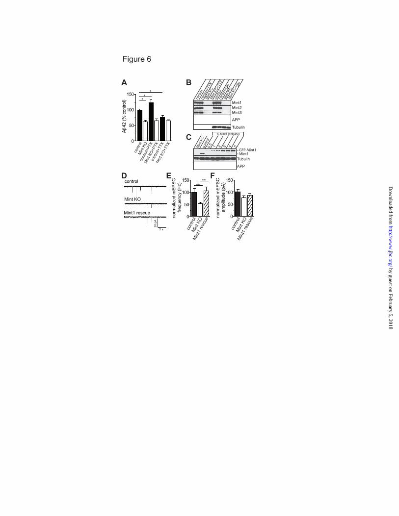
Figure 6
20 p
A
2 s
Mint KO
control
Mint1 rescue
D
0
50
100
150no
rmal
ized
mE
PS
C fr
eque
ncy
(Hz)
norm
aliz
ed m
EP
SC
am
plitu
de (p
A)
0
50
100
150∗∗
∗∗
cont
rol
Min
t KO
cont
rol
Min
t KO
E
cont
rol
Min
t KO
0.5 1 2 3 4 5
% Mint1 lentivirus
Tubulin
C
APP
Min
t1 re
scue
Min
t1 re
scue
F
Mint1Mint2Mint3
APP
Tubulin
cont
rol
cont
rol+
TTX
Min
t KO
Min
t KO
+TTX
cont
rol
cont
rol+
glu
Min
t KO
Min
t KO
+gluB
Aβ4
2 (%
con
trol)
0
50
100
150∗∗
∗
cont
rol
cont
rol+
TTX
Min
t KO
Min
t KO
+TTX
cont
rol+
PTX
Min
t KO
+PTX
A
Mint1GFP-Mint1
by guest on February 5, 2018http://w
ww
.jbc.org/D
ownloaded from
0
50
100
150
200
controlMint KOMint1 rescue
glu TTX
AP
P pi
xel i
nten
sity
/so
ma
area
(% c
ontro
l)
+glutamate +TTX
cont
roltg
Min
t KO
tgM
int1
resc
ue
A B
+glutamate +TTX
cont
roltg
Min
t KO
tgM
int1
resc
ue
C D
50
100
150
200A
PP
pixe
l int
ensi
ty/
som
a ar
ea (%
con
trol)
0
controlMint KOMint1 rescue
glu TTX
∗∗∗∗∗∗
∗∗∗∗∗∗
nsns
∗∗∗∗∗∗
∗∗∗∗∗
nsns
Figure 7
by guest on February 5, 2018http://w
ww
.jbc.org/D
ownloaded from
control Mint1 Mint2 Mint3
Figure 8
D
E
50
100
150
200
0100
200300
400500
cont
rol
Min
t1M
int2
Min
t3
0mE
PS
C fr
eque
ncy
(Hz)
% c
ontro
l
mE
PS
C a
mpl
itude
(pA
) %
con
trol
∗∗∗∗∗∗
∗∗∗∗∗∗
Control
Mint 1
2 s
Mint 3
Mint 2
25 p
A
A B
C
cont
rol
Min
t1M
int2
Min
t3
AP
P pi
xel i
nten
sity
/so
ma
area
(% c
ontro
l)
50
100
150
0
cont
rol
Min
t1
Min
t2M
int3
Min
t1+T
TXM
int2
+TTX
Min
t3+T
TX
∗∗∗∗∗∗
∗
∗∗
∗∗∗
AP
P pi
xel i
nten
sity
/pr
oces
ses
(% c
ontro
l)
50
100
150
0
200
cont
rol
Min
t1
Min
t2M
int3
Min
t1+T
TXM
int2
+TTX
Min
t3+T
TX
∗∗∗∗∗∗
∗∗∗
∗
∗∗∗∗
F G
cont
rol+
TTX
cont
rol+
TTX
cont
rol
Min
t1M
int2
Min
t3
Mint1GFP-Mint1
Mint2GFP-Mint2
Tubulin
Mint3GFP-Mint3
by guest on February 5, 2018http://w
ww
.jbc.org/D
ownloaded from
AP
P pi
xel i
nten
sity
/so
ma
area
(% c
ontro
l)
50
100
150
0
cont
rol
Min
t1M
int2
Min
t3
AP
P pi
xel i
nten
sity
/so
ma
area
(% c
ontro
l)
100
200
300
0
cont
rol
Min
t1M
int2
Min
t3
A
B
C D∗∗∗∗∗∗
∗∗
Figure 9
by guest on February 5, 2018http://w
ww
.jbc.org/D
ownloaded from

A
Figure 10
Controltg Mint KOtg
Mint1 rescue
Mint1∆PDZ1/2 0
50
100
cont
rol
Min
t KO
Min
t1 re
scue
PS
1 pi
xel i
nten
sity
/so
ma
area
(% c
ontro
l) ∗∗∗
Min
t1 ∆
PDZ1
/2
∗∗∗B
Controltg Mint KOtgControltg
+glutamateMint KOtg
+glutamate
PS
1A
PP
Mer
ge
C
PS
1 pi
xel i
nten
sity
/so
ma
area
(% c
ontro
l)
ns
∗∗∗∗
∗∗∗
50
100
150
0
D
PS
1 co
loca
lizat
ion
with
AP
P (%
con
trol)
100
200
300
0
400∗∗∗
∗∗∗
ns
E
cont
roltg
cont
roltg +g
luM
int K
Otg
Min
t KO
tg +glu
cont
roltg
cont
roltg +g
luM
int K
Otg
Min
t KO
tg +glu
by guest on February 5, 2018http://w
ww
.jbc.org/D
ownloaded from
Sarah E. Sullivan, Gregory M. Dillon, Josefa M. Sullivan and Angela HoGeneration
βMint Proteins are Required for Synaptic Activity-Dependent APP Trafficking and A
published online April 17, 2014J. Biol. Chem.
10.1074/jbc.M113.541003Access the most updated version of this article at doi:
Alerts:
When a correction for this article is posted•
When this article is cited•
to choose from all of JBC's e-mail alertsClick here
by guest on February 5, 2018http://w
ww
.jbc.org/D
ownloaded from