advanced biosensor-based strategy for specific and rapid
TRANSCRIPT
Exploratory Research and Hypothesis in Medicine 2018 vol. 3 | 61–67
Copyright: © 2018 Authors. This is an Open Access article distributed under the terms of the Creative Commons Attribution-Noncommercial 4.0 International License (CC BY-NC 4.0), permitting all non-commercial use, distribution, and reproduction in any medium, provided the original work is properly cited.
Innovation
Introduction and background
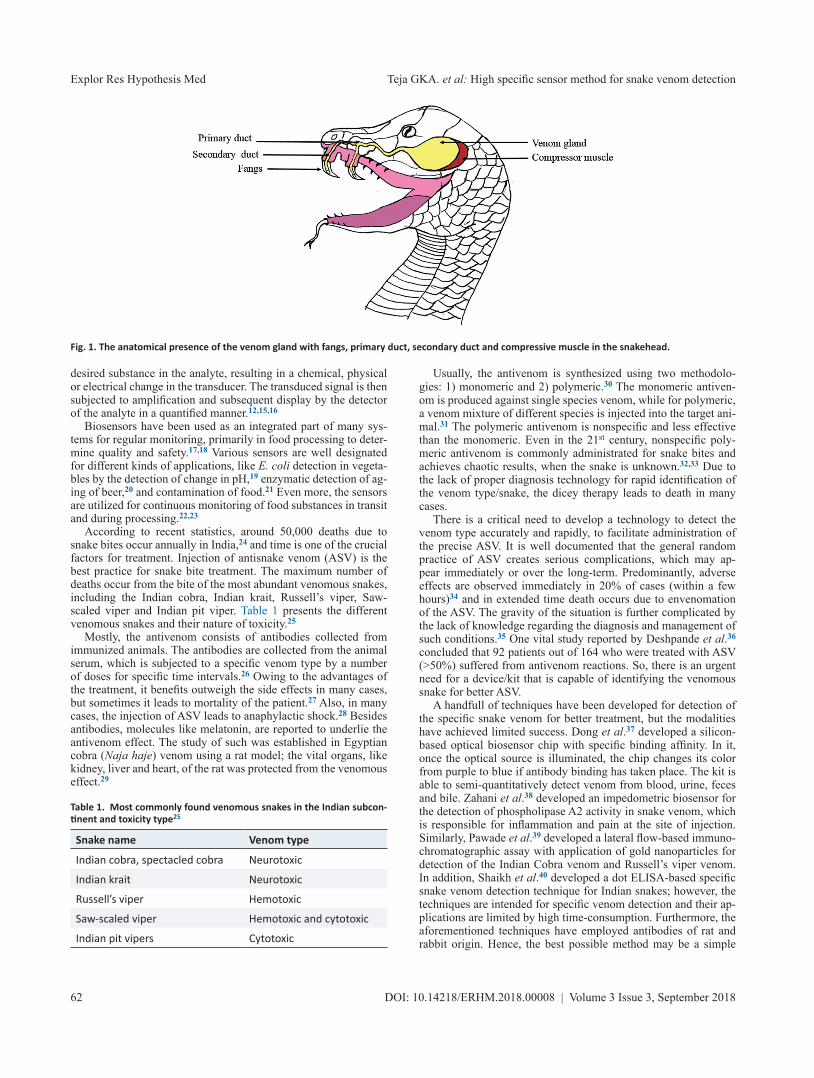
Snakes are a versatile species, made up of elongated, legless, car-nivorous reptiles of the suborder Serpentes.1 The most distinctive feature of the snakes are its fangs and, in some, venomous glands.2 The venom produced in venomous glands reaches the fangs through an anatomical tubing that is known as venomous ducts. The ducts open into the fangs, which are sharp and pointed tooth-like structures that help to inject the venom into a prey’s body upon biting (Fig. 1).
Venom is a clear, viscous fluid of amber or poisonous straw-colored fluid, comprised of many biologically active agents, such as proteases and hyaluronidase, metal ions, biogenic amines, lipids and free amino acids, etc. However, only 80 large and small pro-teins and polypeptides have been identified to date.3 Interestingly, most of the snake species are not venomous; although, for those that are, the venom is generally used for self-protection and obtain-ing food. The snake venoms are broadly classified into three types: 1) neurotoxic; 2) cytotoxic; and, 3) hemotoxic. The neurotoxins affect the central nervous system,4 while the cytotoxins kill the cells in a particular area, where the bite occurs5 and the hemotoxic
attacks the cardiovascular system.6Chemically, the toxins are composed of four main categories,
including enzymes, glycoproteins, polypeptides and low molecu-lar weight molecules. The enzymes, in particular, are represented by amino acid oxidase, thrombin-like procoagulant, kallikrein-like serine proteases metalloproteinases and phospholipase A2. Different types of toxins are present in the venom and the pro-file varies from species to species, primarily for α-bungarotoxin, α-cobratoxin, α-toxin, erabutoxin, notexin, ammodytoxin, cardio-toxin, cytotoxin, myotoxin-a, crotamine and peptides like pyro-glutamylpeptide.7–10 The toxins primarily participate in immuno-genic reactions when the venom is injected into the host body. In the case of snake bites, antivenom or antiserum immunoglobulins are employed to treat the patient. Antivenom includes a monova-lent antibody or commonly used polyvalent antibody against the venom.9
The biosensor is a tiny analytical device, capable of converting biological information into a detectable signal.11 As such, the de-vice is able to determine the concentration of substances and other parameters of biological interest. This noninvasive technique is highly advantageous for its high accurately and sensitivity.12 In modern-day medicine, the biosensor is widely used, for various applications, to determine a broad range of factors, such as blood glucose, cholesterol, catechol and bilirubin, etc.13 Examples in-clude the amperometric biosensor PDMS/glass capillary electro-phoresis biosensor microchip developed by Schoning et al.14 for the detection of catechol and dopamine, the biosensors employed in forensic science for the detection of DNA, and the microbial biosensors utilized for the detection of pathogenic microorgan-isms.15
Most significantly, any biosensor is very specific and accurate, and requires the smallest amount of analyte for detection. Basi-cally, the device is comprised of sensing material (bioreceptor), a transducer and a detector. The receptor may be an enzyme, an-tibody, microorganism or a cell, which senses the presence of the
Advanced Biosensor-based Strategy for Specific and Rapid Detection of Snake Venom for Better Treatment
Guduru KVVNSK Aditya Teja, Namdev More and Govinda Kapusetti*
Department of Medical Devices, National Institute of Pharmaceutical Education and Research (NIPER), Ahmedabad, Palaj, Gandhinagar, India
Abstract
Specific and rapid detection of snake venom type is a complex practice, even with the contemporary medical technology. Generally, in cases for which the species are not identified, the nonspecific polymeric antivenom is injected into the patient. Thus, the effectiveness of treatment is limited, as it acts arbitrarily on the target. Since most snakes are nonpoisonous and treatment is applied with a cautionary approach, the patient can experience severe side effects of a nonspecific agent and in some cases mortality. Therefore, there is an immediate need to develop a suitable medical methodology to avoid this arbitrary practice. The proposed hypothesis may be the best practice for rapid and specific determination of snake venom type by biosensor intervention.
Keywords: Snake venom; Electrochemical biosensor; Antibody and Quartz Crystal Microbalance.Abbreviations: ASV, Antisnake venom; DNA, Deoxyribonucleic acid; ELISA, Enzyme-Linked Immunosorbent Assay; PDMS, Polydimethylsiloxane; QCM, Quartz Crystal Microbalance.Received: May 21, 2018; Revised: September 19, 2018; Accepted: September 25, 2018*Correspondence to: Govinda Kapusetti, Department of Medical Devices, National Institute of Pharmaceutical Education and Research (NIPER), Ahmedabad, Palaj, Gandhinagar, India 382355. E-mail: [email protected] to cite this article: Teja GKA, More N, Kapusetti G. Advanced Biosensor-based Strategy for Specific and Rapid Detection of Snake Venom for Better Treatment. Ex-ploratory Research and Hypothesis in Medicine 2018;3(3):61–67. doi: 10.14218/ERHM.2018.00008.
DOI: 10.14218/ERHM.2018.00008 | Volume 3 Issue 3, September 201862
Teja GKA. et al: High specific sensor method for snake venom detectionExplor Res Hypothesis Med
desired substance in the analyte, resulting in a chemical, physical or electrical change in the transducer. The transduced signal is then subjected to amplification and subsequent display by the detector of the analyte in a quantified manner.12,15,16
Biosensors have been used as an integrated part of many sys-tems for regular monitoring, primarily in food processing to deter-mine quality and safety.17,18 Various sensors are well designated for different kinds of applications, like E. coli detection in vegeta-bles by the detection of change in pH,19 enzymatic detection of ag-ing of beer,20 and contamination of food.21 Even more, the sensors are utilized for continuous monitoring of food substances in transit and during processing.22,23
According to recent statistics, around 50,000 deaths due to snake bites occur annually in India,24 and time is one of the crucial factors for treatment. Injection of antisnake venom (ASV) is the best practice for snake bite treatment. The maximum number of deaths occur from the bite of the most abundant venomous snakes, including the Indian cobra, Indian krait, Russell’s viper, Saw-scaled viper and Indian pit viper. Table 1 presents the different venomous snakes and their nature of toxicity.25
Mostly, the antivenom consists of antibodies collected from immunized animals. The antibodies are collected from the animal serum, which is subjected to a specific venom type by a number of doses for specific time intervals.26 Owing to the advantages of the treatment, it benefits outweigh the side effects in many cases, but sometimes it leads to mortality of the patient.27 Also, in many cases, the injection of ASV leads to anaphylactic shock.28 Besides antibodies, molecules like melatonin, are reported to underlie the antivenom effect. The study of such was established in Egyptian cobra (Naja haje) venom using a rat model; the vital organs, like kidney, liver and heart, of the rat was protected from the venomous effect.29
Usually, the antivenom is synthesized using two methodolo-gies: 1) monomeric and 2) polymeric.30 The monomeric antiven-om is produced against single species venom, while for polymeric, a venom mixture of different species is injected into the target ani-mal.31 The polymeric antivenom is nonspecific and less effective than the monomeric. Even in the 21st century, nonspecific poly-meric antivenom is commonly administrated for snake bites and achieves chaotic results, when the snake is unknown.32,33 Due to the lack of proper diagnosis technology for rapid identification of the venom type/snake, the dicey therapy leads to death in many cases.
There is a critical need to develop a technology to detect the venom type accurately and rapidly, to facilitate administration of the precise ASV. It is well documented that the general random practice of ASV creates serious complications, which may ap-pear immediately or over the long-term. Predominantly, adverse effects are observed immediately in 20% of cases (within a few hours)34 and in extended time death occurs due to envenomation of the ASV. The gravity of the situation is further complicated by the lack of knowledge regarding the diagnosis and management of such conditions.35 One vital study reported by Deshpande et al.36 concluded that 92 patients out of 164 who were treated with ASV (>50%) suffered from antivenom reactions. So, there is an urgent need for a device/kit that is capable of identifying the venomous snake for better ASV.
A handfull of techniques have been developed for detection of the specific snake venom for better treatment, but the modalities have achieved limited success. Dong et al.37 developed a silicon-based optical biosensor chip with specific binding affinity. In it, once the optical source is illuminated, the chip changes its color from purple to blue if antibody binding has taken place. The kit is able to semi-quantitatively detect venom from blood, urine, feces and bile. Zahani et al.38 developed an impedometric biosensor for the detection of phospholipase A2 activity in snake venom, which is responsible for inflammation and pain at the site of injection. Similarly, Pawade et al.39 developed a lateral flow-based immuno-chromatographic assay with application of gold nanoparticles for detection of the Indian Cobra venom and Russell’s viper venom. In addition, Shaikh et al.40 developed a dot ELISA-based specific snake venom detection technique for Indian snakes; however, the techniques are intended for specific venom detection and their ap-plications are limited by high time-consumption. Furthermore, the aforementioned techniques have employed antibodies of rat and rabbit origin. Hence, the best possible method may be a simple
Table 1. Most commonly found venomous snakes in the Indian subcon-tinent and toxicity type25
Snake name Venom type
Indian cobra, spectacled cobra Neurotoxic
Indian krait Neurotoxic
Russell’s viper Hemotoxic
Saw-scaled viper Hemotoxic and cytotoxic
Indian pit vipers Cytotoxic
Fig. 1. The anatomical presence of the venom gland with fangs, primary duct, secondary duct and compressive muscle in the snakehead.

DOI: 10.14218/ERHM.2018.00008 | Volume 3 Issue 3, September 2018 63
Teja GKA. et al: High specific sensor method for snake venom detection Explor Res Hypothesis Med
blood test for a rapid detection technique.One commercial product is available for specific detection of
venom of five different kinds of Australia- and Papua New Guinea-originated snakes. The colorimetric enzyme immunoassay assists in selection of the monovalent antivenom to neutralize the snake venom involved in the bite.41 While the method provides high sensitivity to determine the venom type, it has some constraints. As per the manufacturer’s information, the assay method is highly time-consuming, needing at least 35–45 min to get the result; more significantly, the assay provides equivocal reactions in case of high concentration sample testing, involves multiple complicated sam-ple processing steps and stringent storage conditions, and needs a trained person with good laboratory practices for proper results. Therefore, there is a need for a simple methodology to identify the snake venom for administering an antidote with minimal time. Most importantly, the South Asian countries like India need a high-ly specific and simple kit for diagnosing venom type, since there is a lack of specialized labs and persons to process the analysis.
Hypothesis
The basic idea behind this hypothesis is to develop a device or kit which can detect the type of snake by analyzing its venom. Moreo-ver, the device can be designed in such a way that it can detect the venom type from a blood sample, which can be collected from either the bite site or the bloodstream. Even more, it will help to identify whether the bite is venomous or nonvenomous, so as to avoid unnecessary ASV administration and the subsequent trauma. The hypothesis is based on a chip-based biosensor to detect venom type for better treatment. The sensor will give the information by the formation of an immune complex by agglutination of the spe-cific antivenom antibodies with the venom. Initially, the selected antibodies of different antivenom types will be immobilized on the transducer surface of the sensor. Once the sample is collected from the patient, it is immediately analyzed by the sensor, which gener-ates a signal that will suggest the venom type by its specific bind-ing with immobilized antivenom antibody. The result will be ob-tained from the transducer (possibly a quartz crystal microbalance) in the form of electric signal generation by mass variation on the sensor surface through aggregation of the antigen of venom bind-ing with the immobilized antibody (Fig. 2).
Evaluation of the hypothesis
Isolation of specific antibodies
Antibodies against specific venom are obtained by immuniza-tion of hens with the particular venom collected from the selected snakes. Following the immunization, the antibodies will be iso-lated from the egg yolk.42,43
Immunization procedure
A handful of literature is available for the immunization of dif-ferent animal models with various methodologies. Conventionally, the antivenom antibodies are isolated from an immunized horse, goat or rabbit. Nevertheless, it exhibits the major constraints of an anti-compliment reaction,44,45 serum sickness46 and anaphylactic shock.47,48 Moreover, the isolation and standardization of the puri-fied antibodies is a tedious process.
As per the literature, the hen’s egg procedure is the best pos-sible, safe and easy isolation method. Briefly, the laying hens will be immunized with the interested (Indian-origin snake venoms in the present proposal) snake venoms in different groups for specific periods and the specific procedure shown in Figure 3. Initially, the chickens of a specific breed free-from-pathogens (fed and bred in a clean environment) are selected for the procedure. After specific growth, the hen is subjected to the administration of small doses of venom by injection into the pectoralis muscle.49 Before injection, the venom is exposed to radiation to reduce its toxicity.50,51 Fur-ther, the eggs of the immunized hens will be collected and the iso-lated yolk will be frozden at −20 °C for the subsequent procedure. The supernatant collected by centrifugation and filtered by various stages52 will be used to obtain the antibodies upon precipitation by addition of ammonium sulfate.53,54
The antibodies will be immobilized onto the transducer which detects the change in physical or chemical changes.55 The impor-tant factor to be optimized is the concentration of the antibodies and their orientation. Antibody orientation will be achieved by slight modification through adding bifunctional thiol containing reagents.56 Different antivenom antibodies will be collected from the eggs of different hens, which have been immunized with a unique venom type. The isolated antibodies are immobilized on
Fig. 2. Block diagram of the biosensor showing the immobilization of different antivenom antibodies on the transducer surface. Specific interaction of the venom to its counterpart will generate a detectable specific signal to identify the snake species.

DOI: 10.14218/ERHM.2018.00008 | Volume 3 Issue 3, September 201864
Teja GKA. et al: High specific sensor method for snake venom detectionExplor Res Hypothesis Med
the discrete receptor surfaces of the biosensors. The generated sig-nal during antigen-antibody interaction is amplified using ampli-fiers circuits, for better scrutiny.57
Bruce et al.58 isolated rattlesnake and scorpion antibodies from the aforementioned method. Similarly, Mayadevi et al.50 employed the same method, with slight modification in the lipid removal method to isolate antiviper antibodies. Paul et al.59 isolated and purified antibodies of anti-Echis carinatus venom from egg yolk by the water dilution method.
Detection principle
The collected blood sample from the wound site interacts with the biosensor. The antibodies present on the biosensor specifi-cally bind with the venom as a result of agglutination, changing the physical or chemical state at the receptor site. The change can be detected by various technologies; the highly sensitive quartz crystal microbalance (QCM) is the best recommend transducer and recognizes frequency change with mass variance.60 QCM pos-sesses very high accuracy rate and detects mass densities of 1µg/cm2.61 It can perform even under vacuum. A wide variety of im-mobilization techniques can be employed for fabrication of a bio-sensor in QCM. It can also detect the difference in nano-gram/unit area by measuring the change in resonant frequency.62 Structurally, the QCM quartz crystal is sandwiched between two “T” shaped electrodes and connected to electric terminals to supply voltage. The immobilized antibody on QCM selectively interacts with its counterpart and causes the mass change. The result of mass change can cause the resonance frequency shift, to help in quantification of the analyte. The frequency change depends on various factors, like mass, shape, structure and thickness of the analyte.62,63
In recent days, QCM has shown massive advancement in bio-sensor application. Park et al.60 fabricated a hemoglobin QCM biosensor with high sensitivity of detection (limit of 0.147%) for HbA1c (hemoglobin A1c) against hemoglobin. Similarly, Şerife et al.64 developed a QCM-based highly sensitive biosensor for detec-tion of ochratoxin A, with a reported detection limit of 17.2–200
ng/mL. Interestingly, the sensitivity of QCM can differentiate the normal to physiological conditions, like for C-reactive protein, which is a key biomarker for inflamed liver and related disorders.
The intended biosensor will exhibit high sensitivity, with linear-ity detection ranging from 0.04–100 µg/mL and lower detection limit of 0.02 µg/mL.65 Similarly, Lourdes et al.66 developed the High Fundamental Frequency QCM-based Immunosensor for de-tection of pesticides in honey. The sensor exhibits a limit of detec-tion of 0.035 µg/mL in a diluted honey sample. Furthermore, the QCM demonstrates high sensitivity, and is cost-effective, fast and reliable compared to the conventional techniques.
Rapidity
In case of life-threatening snake bites with narrow antidose periods to save the patient’s life, rapid detection time is an essential pa-rameter for sensor development. The response generation must be immediate for when the antibody interacts with a specific antigen in the sample. Ajeet et al.67 developed a biosensor with the aid of antibodies to detect the presence of ochratoxin, which is produced by Aspergillus species and found in foodstuffs. For this, the IgG antibodies are isolated and immobilized onto an indium tin oxide layer, with the help of chitosan and iron oxide composite. The elec-trodes have a very fast response time (18 s), with greater sensitivity and a minimum detection limit of 0.5 ng dL−1. An attempt has been made to measure the concentrations of cortisol and corticotrophin-releasing hormone by immobilizing the polyclonal antibodies on a platinum electrode. These probes are capable of giving a response within 30 s, with high sensitivity.68
Specificity
Although blood samples from bite regions have been exposed to a range of biosensors, accurate and specific results have only been obtained upon specific antibody-antigen interaction.69,70 The rest have shown negative results for suggesting proper treatment. The
Fig. 3. The detailed procedure of antibody generation in the hen’s egg model, followed by the development of a venom-specific biosensor.
DOI: 10.14218/ERHM.2018.00008 | Volume 3 Issue 3, September 2018 65
Teja GKA. et al: High specific sensor method for snake venom detection Explor Res Hypothesis Med
antibody-antigen reactions are very specific, even at very minute concentrations, and have been proven by various research groups to underlie the specificity of antibody-antigen reactions.71–73 The mouse immune globulin IgG is detected by using fluorophore-modified antibodies. This approach has been demonstrated as very useful for the detection of various antigens in different disease conditions.74 A rapid detection-capable amperometric biosensor has been developed for Streptococcus agalactiae detection, and a biotinylated antibody is utilized for the application. The antibody is conjugated with horseradish peroxidase-labeled streptavidin and the complex is immobilized on the carbon electrode.75
Perspectives
The proposed hypothesis is a potential methodology for the rapid and accurate identification of a snake that has bitten a victim. The proposed design offers greater specificity, selectivity and accuracy, in comparison to the current existing conventional techniques. The proposed device will be reusable and cost-effective. The main ad-vantage of this analysis will be bypassing sample preparation for analysis. The sensor will directly detect the analyte (venom) from the sample (blood collected from injury region or bloodstream). The device will be able to save the precious lives of many peo-ple and to prevent the occurrence of adverse effects of nonspecific ASV administration.
Conflict of interest
The authors have no conflict of interests related to this publication.
Author contributions
Guduru KVVNSK Aditya Teja and Namdev More have collected the necessary literature and prepared the manuscript to fulfill the hypothesis. Dr. Govinda Kapusetti has developed the hypothesis and review the final draft.
References
[1] Reeder TW, Townsend TM, Mulcahy DG, Noonan BP, Wood PL Jr, Sites JW Jr, et al. Integrated analyses resolve conflicts over squamate reptile phylogeny and reveal unexpected placements for fossil taxa. PLOS one 2015;10:e0118199. doi:10.1371/journal.pone.0118199.
[2] Jansen van Vuuren L, Kieser JA, Dickenson M, Gordon KC, Fraser-Mill-er SJ. Chemical and mechanical properties of snake fangs. J Raman Spectrosc 2016;47:787–795. doi:10.1002/jrs.4903.
[3] Koh DC, Armugam A, Jeyaseelan K. Snake venom components and their applications in biomedicine. Cell Mol Life Sci 2006;63:3030–3041. doi:10.1007/s00018-006-6315-0.
[4] Silva A, Maduwage K, Buckley NA, Lalloo DG, de Silva HJ, Isbis-ter GK. Antivenom for snake venom-induced neuromuscular pa-ralysis. Cochrane Database of Systematic Reviews 2017;(3):1–3. doi:10.1002/14651858.cd012604.
[5] Bradshaw MJ, Saviola AJ, Fesler E, Mackessy SP. Evaluation of cytotox-ic activities of snake venoms toward breast (MCF-7) and skin cancer (A-375) cell lines. Cytotechnology 2016;68:687–700. doi:10.1007/s10616-014-9820-2.
[6] Slagboom J, Kool J, Harrison RA, Casewell NR. Haemotoxic snake ven-oms: their functional activity, impact on snakebite victims and phar-maceutical promise. Br J Haematol 2017;177:947–959. doi:10.1111/bjh.14591.
[7] Naeem S. Snake venom toxins. Journal of Saidu Medical College 2018;7:1–3.
[8] Su MJ, Chang CC. Presynaptic effects of snake venom toxins which have phospholipase A2 activity (β-bungarotoxin, taipoxin, crotoxin). Toxicon 1984;22:631–640. doi:10.1016/0041-0101(84)90003-5.
[9] Ahmed SM, Ahmed M, Nadeem A, Mahajan J, Choudhary A, Pal J. Emergency treatment of a snake bite: Pearls from literature. J Emerg Trauma Shock 2008;1:97–105. doi:10.4103/0974-2700.43190.
[10] Strydom AJ, Botes DP. Snake venom toxins. Purification, properties, and complete amino acid sequence of two toxins from ringhals (he-machatus haemachatus) venom. J Biol Chem 1971;246(5):1341–1349.
[11] Shrivastava S, Jadon N, Jain R. Next-generation polymer nanocom-posite-based electrochemical sensors and biosensors: A review. Trends Analyt Chem 2016;82:55–67. doi:10.1016/j.trac.2016.04.005.
[12] Scheller FW, Schubert F, Renneberg R, Müller H-G, Jänchen M, Weise H. Biosensors: trends and commercialization. Biosensors 1985;1:135–160. doi:10.1016/0265-928x(85)80001-8.
[13] Wang J, Sun XW, Wei A, Lei Y, Cai X, Li CM, et al. Zinc oxide nanocomb biosensor for glucose detection. Appl Phys Lett 2006;88:233106. doi:10.1063/1.2210078.
[14] Schöning MJ, Jacobs M, Muck A, Knobbe D-T, Wang J, Chatrathi M, et al. Amperometric PDMS/glass capillary electrophoresis-based bio-sensor microchip for catechol and dopamine detection. Sens Actua-tors B Chem 2005;108:688–694. doi:10.1016/j.snb.2004.11.032.
[15] Turner A, Karube I, Wilson GS. Biosensors: fundamentals and applica-tions. Oxford university press; 1987, p. 13–29.
[16] Rapp BE, Gruhl FJ, Länge K. Biosensors with label-free detec-tion designed for diagnostic applications. Anal Bioanal Chem 2010;398:2403–2412. doi:10.1007/s00216-010-3906-2.
[17] Alocilja EC, Radke SM. Market analysis of biosensors for food safety. Biosens Bioelectron 2003;18:841–846. doi:10.1016/s0956-5663(03)00009-5.
[18] Patel P. Bio) sensors for measurement of analytes implicated in food safety: a review. Trends Analyt Chem 2002;21:96–115. doi:10.1016/s0165-9936(01)00136-4.
[19] Scognamiglio V, Arduini F, Palleschi G, Rea G. Biosensing technol-ogy for sustainable food safety. Trends Analyt Chem 2014;62:1–10. doi:10.1016/j.trac.2014.07.007.
[20] Ghasemi-Varnamkhasti M, Rodríguez-Méndez ML, Mohtasebi SS, Apetrei C, Lozano J, Ahmadi H, et al. Monitoring the aging of beers using a bioelectronic tongue. Food Control 2012;25:216–224. doi:10.1016/j.foodcont.2011.10.020.
[21] Arora P, Sindhu A, Dilbaghi N, Chaudhury A. Biosensors as innovative tools for the detection of food borne pathogens. Biosens Bioelectron 2011;28:1–12. doi:10.1016/j.bios.2011.06.002.
[22] Manz A, Graber N, Widmer Há. Miniaturized total chemical analy-sis systems: a novel concept for chemical sensing. Sens Actuators B Chem 1990;1:244–248. doi:10.1016/0925-4005(90)80209-i.
[23] Cheng N, Xu Y, Huang K, Chen Y, Yang Z, Luo Y, et al. One-step com-petitive lateral flow biosensor running on an independent quantifi-cation system for smart phones based in-situ detection of trace Hg (II) in tap water. Food Chem 2017;214:169–175. doi:10.1016/j.food-chem.2016.07.058.
[24] Menon JC, Joseph JK, Whitaker RE. Venomous snake bite in India-why do 50,000 Indians die every year? J Assoc Physicians India 2017;65:78–81.
[25] Gayathri SA, Jayashankar M, Avinash K. A pilot-survey to assess the diversity and distribution of reptilian fauna in Taralu Village, abutting the Bannerghatta National Park, Karnataka, India. Newsletter of the South Asian Reptile Network 2016;18:3.
[26] Bawaskar HS, Bawaskar PH. Envenoming by the common krait (Bun-garus caeruleus) and Asian cobra (Naja naja): clinical manifestations and their management in a rural setting. Wilderness Environ Med 2004;15:257–266. doi:10.1580/1080-6032(2004)015[0257:ebtckb]2.0.co;2.
[27] Joseph IM, Kuriakose CK, Dev AV, Philip GA. Low dose versus high dose anti-snake venom therapy in the treatment of haemato-toxic snake bite in South India. Trop Doct 2017;47(4):300–304. doi:10.1177/0049475517712804.
[28] Lalloo DG, Theakston RDG. Snake antivenoms: antivenoms. Journal

DOI: 10.14218/ERHM.2018.00008 | Volume 3 Issue 3, September 201866
Teja GKA. et al: High specific sensor method for snake venom detectionExplor Res Hypothesis Med
of Toxicology: Clinical Toxicology 2003;41:277–290. doi:10.1081/clt-120021113.
[29] Moneim AEA, Ortiz F, Leonardo-Mendonca RC, Vergano-Villodres R, Guerrero-Martínez JA, López LC, et al. Protective effects of me-latonin against oxidative damage induced by Egyptian cobra (Naja haje) crude venom in rats. Acta Trop 2015;143:58–65. doi:10.1016/j.actatropica.2014.12.007.
[30] Anil A, Singh S, Bhalla A, Sharma N, Agarwal R, Simpson ID. Role of neostigmine and polyvalent antivenom in Indian common krait (Bungarus caeruleus) bite. J Infect Public Health 2010;3:83–87. doi:10.1016/j.jiph.2010.01.002.
[31] Bhattacharya S, Chakraborty M, Mukhopadhyay P, Kundu P, Mishra R. Viper and cobra venom neutralization by alginate coated multicom-ponent polyvalent antivenom administered by the oral route. PLoS Negl Trop Dis 2014;8:e3039. doi:10.1371/journal.pntd.0003039.
[32] Ahuja T, Kumar D. Recent progress in the development of nano-structured conducting polymers/nanocomposites for sensor appli-cations. Sens Actuators B Chem 2009;136:275–286. doi:10.1016/j.snb.2008.09.014.
[33] Gutiérrez JM, León G, Burnouf T. Antivenoms for the treatment of snakebite envenomings: the road ahead. Biologicals 2011;39:129–142. doi:10.1016/j.biologicals.2011.02.005.
[34] Warrell DA. Snake bite. The Lancet 2010;375:77–88. doi:10.1016/S0140-6736(09)61754-2.
[35] Simpson ID, Norris RL. Snake antivenom product guidelines in India: The devil is in the details. Wilderness Environ Med 2007;18:163–168. doi:10.1580/07-weme-ed-099r.1.
[36] Deshpande RP, Motghare VM, Padwal SL, Pore RR, Bhamare CG, Deshmukh VS, et al. Adverse drug reaction profile of anti-snake venom in a rural tertiary care teaching hospital. J Young Pharm 2013;5:41–45. doi:10.1016/j.jyp.2013.02.003.
[37] Eng KH, Gopalakrishnakone P. Optical immunoassay for snake venom detection. Biosens Bioelectron 2004;19:1285–1294. doi:10.1016/j.bios.2003.11.020.
[38] Zehani N, Cheewasedtham W, Kherrat R, Jaffrezic-Renault N. Impedi-metric biosensor for the determination of phospholipase A2 activity in snake venom. Anal Lett 2018;51:401–410. doi:10.1080/00032719.2017.1312425.
[39] Pawade BS, Salvi NC, Shaikh IK, Waghmare AB, Jadhav ND, Wagh VB, et al. Rapid and selective detection of experimental snake enveno-mation–Use of gold nanoparticle based lateral flow assay. Toxicon 2016;119:299–306. doi:10.1016/j.toxicon.2016.10.009.
[40] Shaikh IK, Dixit PP, Pawade BS, Waykar IG. development of dot-elisa for the detection of venoms of major Indian venomous snakes. Toxi-con 2017;139:66–73. doi:10.1016/j.toxicon.2017.10.007.
[41] https://www.seqirus.com.au/s1/cs/aucb/1196562673365/Web_Product_C/1196562753754/ProductDetail.htm. Date accessed 09/ 08/2018.
[42] Sudjarwo SA, Eraiko K, Sudjarwo GW, Koerniasari. The potency of chicken egg yolk immunoglobulin (IgY) specific as immunotherapy to Mycobacterium tuberculosis infection. J Adv Pharm Technol Res 2017;8(3):91–96. doi:10.4103/japtr.JAPTR_167_16.
[43] Kovacs-Nolan J, Mine Y. Egg yolk antibodies for passive immunity. Annu Rev Food Sci Technol 2012;3:163–182. doi:10.1146/annurev-food-022811-101137.
[44] Sutherland SK. Serum reactions. An analysis of commercial antiven-oms and the possible role of anticomplementary activity in de-novo reactions to antivenoms and antitoxins. Med J Aust 1977;1:613–615.
[45] León G, Lomonte B, Gutiérrez JM. Anticomplementary activ-ity of equine whole IgG antivenoms: comparison of three frac-tionation protocols. Toxicon 2005;45:123–128. doi:10.1016/j.toxi-con.2004.07.025.
[46] de Silva HA, Ryan NM, de Silva HJ. Adverse reactions to snake an-tivenom, and their prevention and treatment. Br J Clin Pharmacol 2016;81:446–452. doi:10.1111/bcp.12739.
[47] Schaeffer TH, Khatri V, Reifler LM, Lavonas EJ. Incidence of imme-diate hypersensitivity reaction and serum sickness following ad-ministration of crotalidae polyvalent immune Fab antivenom: a meta-analysis. Academic Emergency Medicine 2012;19:121–131. doi:10.1111/j.1553-2712.2011.01276.x.
[48] Morais V, Massaldi H. Snake antivenoms: adverse reactions and pro-
duction technology. J Venom Anim Toxins Incl Trop Dis 2009;15:2–18. doi:10.1590/s1678-91992009000100002.
[49] da Rocha DG, Fernandez JH, de Almeida CMC, da Silva CL, Magnoli FC, da Silva OÉ, et al. Development of IgY antibodies against anti-snake toxins endowed with highly lethal neutralizing activity. Eur J Pharm Sci 2017;106:404–412. doi:10.1016/j.ejps.2017.05.069.
[50] Devi CM, Bai MV, Lal AV, Umashankar P, Krishnan LK. An improved method for isolation of anti-viper venom antibodies from chicken egg yolk. J Biochem Biophys Methods 2002;51:129–138. doi:10.1016/s0165-022x(02)00002-7.
[51] El-Missiry AG, Shaban EA, Mohamed MR, Ahmed AA, Abdallah NM, Moustafa MI. Influence of ionizing radiation on Echis pyramidium snake venom: biochemical and immunological aspects. Egyptian Journal of Hospital Medicine 2010;40:314–334.
[52] Jensenius JC, Andersen I, Hau J, Crone M, Koch C. Eggs: conveniently packaged antibodies. Methods for purification of yolk IgG. J Immunol Methods 1981;46:63–68. doi:10.1016/0022-1759(81)90333-1.
[53] Svendsen L, Crowley A, Ostergaard LH, Stodulski G, Hau J. Develop-ment and comparison of purification strategies for chicken antibod-ies from egg yolk. Lab Anim Sci 1995;45:89–93.
[54] Polson A, Coetzer T, Kruger J, Von Maltzahn E, Van der Merwe K. Improvements in the isolation of IgY from the yolks of eggs laid by immunized hens. Immunol Invest 1985;14:323–327. doi:10.3109/ 08820138509022667.
[55] Vashist SK, Lam E, Hrapovic S, Male KB, Luong JH. Immobilization of antibodies and enzymes on 3-aminopropyltriethoxysilane-function-alized bioanalytical platforms for biosensors and diagnostics. Chem Rev 2014;114:11083–11130. doi:10.1021/cr5000943.
[56] Karyakin AA, Presnova GV, Rubtsova MY, Egorov AM. Oriented immo-bilization of antibodies onto the gold surfaces via their native thiol groups. Anal Chem 2000;72:3805–3811. doi:10.1021/ac9907890.
[57] Funari R, Della Ventura B, Carrieri R, Morra L, Lahoz E, Gesuele F, et al. Detection of parathion and patulin by quartz-crystal microbalance functionalized by the photonics immobilization technique. Biosens Bioelectron 2015;67:224–229. doi:10.1016/j.bios.2014.08.020.
[58] Thalley BS, Carroll SB. Rattlesnake and scorpion antivenoms from the egg yolks of immunized hens. Nat Biotechnol 1990;8:934–938. doi:10.1038/nbt1090-934.
[59] Paul K, Manjula J, Deepa E, Selvanayagam Z, Ganesh K, Rao PS. Anti-Echis carinatus venom antibodies from chicken egg yolk: isolation, purification and neutralization efficacy. Toxicon 2007;50:893–900. doi:10.1016/j.toxicon.2007.06.017.
[60] Park HJ, Lee SS. A quartz crystal microbalance-based biosensor for enzymatic detection of hemoglobin A1c in whole blood. Sens Actua-tors B Chem 2018;258:836–840. doi:10.1016/j.snb.2017.11.170.
[61] Wang X, Ding B, Yu J, Wang M, Pan F. A highly sensitive humidity sensor based on a nanofibrous membrane coated quartz crystal mi-crobalance. Nanotechnology 2009;21:055502. doi:10.1088/0957-4484/21/5/055502.
[62] Vashist SK, Luong JH. Quartz crystal microbalance–based sensors. Handbook of Immunoassay Technologies: Elsevier; 2018. p. 333–357. doi:10.1016/b978-0-12-811762-0.00013-x.
[63] O’sullivan C, Guilbault G. Commercial quartz crystal microbalanc-es–theory and applications. Biosens Bioelectron 1999;14:663–670. doi:10.1016/s0956-5663(99)00040-8.
[64] Pirincci SS, Ertekin Ö, Laguna DE, Ozen FS, Oztürk ZZ, Oztürk S. Label-free QCM immunosensor for the detection of ochratoxin A. Sensors. 2018;18:1161. doi:10.3390/s18041161.
[65] Gao K, Cui S, Liu S. Development of an electrochemical quartz crys-tal microbalance-based immunosensor for C-reactive protein de-termination. Int J Electrochem Sci 2018;13:812–821. doi:10.20964/ 2018.01.49.
[66] Cervera-Chiner L, Juan-Borrás M, March C, Arnau A, Escriche I, Montoya Á, et al. High fundamental frequency quartz crystal micro-balance (HFF-QCM) immunosensor for pesticide detection in honey. Food Control 2018;92:1–6. doi:10.1016/j.foodcont.2018.04.026.
[67] Kaushik A, Solanki PR, Ansari AA, Ahmad S, Malhotra BD. Chitosan–iron oxide nanobiocomposite based immunosensor for ochratoxin-A. Electrochem commun 2008;10:1364–1368. doi:10.1016/j.ele-com.2008.07.007.
[68] Cook C. Measuring of extracellular cortisol and corticotropin-releas-
DOI: 10.14218/ERHM.2018.00008 | Volume 3 Issue 3, September 2018 67
Teja GKA. et al: High specific sensor method for snake venom detection Explor Res Hypothesis Med
ing hormone in the amygdala using immunosensor coupled micro-dialysis. J Neurosci Methods 2001;110:95–101. doi:10.1016/s0165-0270(01)00421-6.
[69] Conroy PJ, Hearty S, Leonard P, O’Kennedy RJ. Antibody production, design and use for biosensor-based applications. Seminars in cell & developmental biology: Elsevier; 2009. p. 10–26. doi:10.1016/j.sem-cdb.2009.01.010.
[70] North JR. Immunosensors: antibody-based biosensors. Trends Bio-technol 1985;3:180–186. doi:10.1016/0167-7799(85)90119-2.
[71] Luppa PB, Junker R, Schimke I, Stürenburg E. Immunological methods. Point-of-Care Testing: Springer; 2018. p. 69–79. doi:10.1007/978-3-662-54497-6_9.
[72] Xu S, Liu Y, Wang T, Li J. Positive potential operation of a cathodic
electrogenerated chemiluminescence immunosensor based on luminol and graphene for cancer biomarker detection. Anal Chem 2011;83:3817–3823. doi:10.1021/ac200237j.
[73] Skottrup PD, Nicolaisen M, Justesen AF. Towards on-site patho-gen detection using antibody-based sensors. Biosens Bioelectron 2008;24:339–348. doi:10.1016/j.bios.2008.06.045.
[74] Zhang W, Serpe MJ. Antigen detection using fluorophore-modified antibodies and magnetic microparticles. Sens Actuators B Chem 2017;238:441–446. doi:10.1016/j.snb.2016.07.070.
[75] Vásquez G, Rey A, Rivera C, Iregui C, Orozco J. Amperometric biosen-sor based on a single antibody of dual function for rapid detection of Streptococcus agalactiae. Biosens Bioelectron 2017;87:453–458. doi:10.1016/j.bios.2016.08.082.