an introduction to next-generation sequencing technology · table of contents i. welcome to...
TRANSCRIPT

An Introduction to Next-Generation Sequencing Technology
www.illumina.com/technology/next-generation-sequencing.html
Table of Contents
I. Welcome to Next-Generation Sequencing 3
a. The Evolution of Genomic Science 3
b. The Basics of NGS Chemistry 4
c. Advances in Sequencing Technology 5
Paired-End Sequencing 5
Tunable Coverage and Unlimited Dynamic Range 6
Multiplexing 6
Advances in Library Preparation 7
Flexible, Scalable Instrumentation 7
II. NGS Methods 8
a. Genomics 8
Whole-Genome Sequencing 8
Exome Sequencing 9
De Novo Sequencing 9
Targeted Sequencing 10
b. Transcriptomics 11
Total RNA and mRNA Sequencing 11
Targeted RNA Sequencing 12
Small RNA and Noncoding RNA Sequencing 12
c. Epigenomics 12
Methylation Sequencing 12
ChIP Sequencing 12
Ribosome Profiling 12
III. Illumina DNA-to-Data NGS Solutions 13
a. The Illumina NGS Workflow 13
b. Integrated Data Analysis 13
IV. Glossary 14
V. References 15

– 3 –
I. Welcome to Next-Generation Sequencing
a. The Evolution of Genomic Science
DNA sequencing has come a long way since the days of two-dimensional chromatography in the 1970s. With the advent of capillary electrophoresis (CE)-based sequencing in 1977, scientists gained the ability to sequence the full genome of any species in a reliable, reproducible manner.1 A decade later, Applied Biosystems introduced the first automated, CE-based sequencing instruments—the AB370 in 1987 and the AB3730xl in 1998—instruments that became the primary workhorses for the NIH-led and Celera-led Human Genome Projects.2 While these “first-generation” instruments were considered high throughput for their time, the Genome Analyzer emerged in 2005 and took sequencing runs from 84 kilobase (kb) per run to 1 gigabase (Gb) per run.3 The short read, massively parallel sequencing technique was a fundamentally different approach to sequencing that revolutionized sequencing capabilities and launched the “next-generation” in genomic science. From that point forward, the data output of next-generation sequencing (NGS) has outpaced Moore’s law—more than doubling each year (Figure 1).
$100,000
$1,000
$100
$10 0.1
10
100
1,000
10,000
1
0.01$12000 2006 200820042002 2010 2012 2014
/Gib
abas
etso
Ce O
utput/W
eeksa
bagi
G$1,000,000
$10,000,000
$100,000,000
$10,000
HiSeqX Ten
HiSeq 2500
Genome Analyzer IIxGenome
Analyzer
ABI 3730xl
Figure 1: Sequencing Cost and Data Output Since 2000—The dramatic rise of data output and concurrent falling cost of sequencing since 2000. The Y-axes on both sides of the graph are logarithmic.
In 2005, with the Genome Analyzer, a single sequencing run could produce roughly one gigabase of data. By 2014, the rate climbed to a 1.8 terabases of data in a single sequencing run—an astounding 1000× increase. It is remarkable to reflect on the fact that the first human genome, famously copublished in Science and Nature in 2001, required 15 years to sequence and cost nearly 3 billion dollars. In contrast, the HiSeq X™ Ten, released in 2014, can sequence over 45 human genomes in a single day for approximately $1000 each (Figure 2).4
Beyond the massive increase in data output, the introduction of NGS technology has transformed the way scientists think about genetic information. The $1000 dollar genome enables population-scale sequencing and establishes the foundation for personalized genomic medicine as part of standard medical care. Researchers can now analyze thousands to tens of thousands of samples in a single year. As Eric Lander, founding director of the Broad Institute of MIT and Harvard and principle leader of the Human Genome Project, states, “The rate of progress is stunning. As costs continue to come down, we are entering a period where we are going to be able to get the complete catalog of disease genes. This will allow us to look at thousands of people and see the differences among them, to discover critical genes that cause cancer, autism, heart disease, or schizophrenia.”5
– 4 –
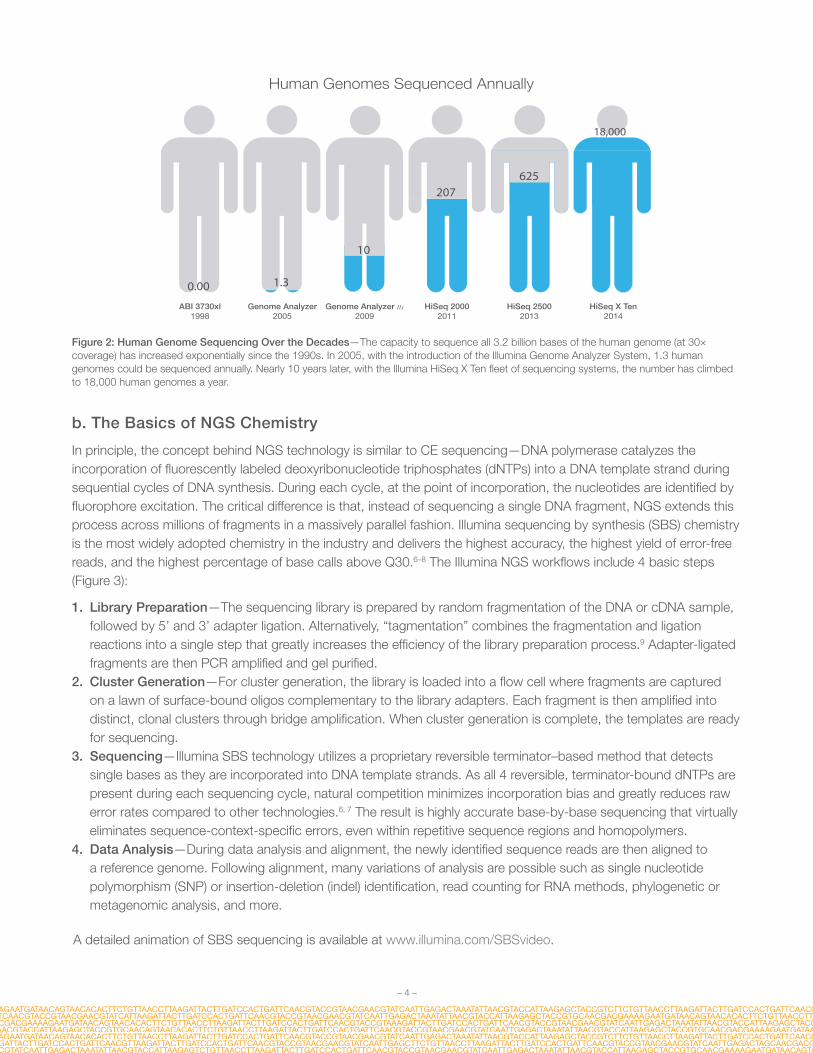
Human Genomes Sequenced Annually
ABI 3730xl1998
HiSeq 25002013
HiSeq 20002011
Genome Analyzer2005
Genome Analyzer IIx2009
HiSeq X Ten2014
0.00
10
207
18,000
1.3
625
Figure 2: Human Genome Sequencing Over the Decades—The capacity to sequence all 3.2 billion bases of the human genome (at 30× coverage) has increased exponentially since the 1990s. In 2005, with the introduction of the Illumina Genome Analyzer System, 1.3 human genomes could be sequenced annually. Nearly 10 years later, with the Illumina HiSeq X Ten fleet of sequencing systems, the number has climbed to 18,000 human genomes a year.
b. The Basics of NGS Chemistry
In principle, the concept behind NGS technology is similar to CE sequencing—DNA polymerase catalyzes the incorporation of fluorescently labeled deoxyribonucleotide triphosphates (dNTPs) into a DNA template strand during sequential cycles of DNA synthesis. During each cycle, at the point of incorporation, the nucleotides are identified by fluorophore excitation. The critical difference is that, instead of sequencing a single DNA fragment, NGS extends this process across millions of fragments in a massively parallel fashion. Illumina sequencing by synthesis (SBS) chemistry is the most widely adopted chemistry in the industry and delivers the highest accuracy, the highest yield of error-free reads, and the highest percentage of base calls above Q30.6–8 The Illumina NGS workflows include 4 basic steps (Figure 3):
1. Library Preparation—The sequencing library is prepared by random fragmentation of the DNA or cDNA sample, followed by 5’ and 3’ adapter ligation. Alternatively, “tagmentation” combines the fragmentation and ligation reactions into a single step that greatly increases the efficiency of the library preparation process.9 Adapter-ligated fragments are then PCR amplified and gel purified.
2. Cluster Generation—For cluster generation, the library is loaded into a flow cell where fragments are captured on a lawn of surface-bound oligos complementary to the library adapters. Each fragment is then amplified into distinct, clonal clusters through bridge amplification. When cluster generation is complete, the templates are ready for sequencing.
3. Sequencing—Illumina SBS technology utilizes a proprietary reversible terminator–based method that detects single bases as they are incorporated into DNA template strands. As all 4 reversible, terminator-bound dNTPs are present during each sequencing cycle, natural competition minimizes incorporation bias and greatly reduces raw error rates compared to other technologies.6, 7 The result is highly accurate base-by-base sequencing that virtually eliminates sequence-context-specific errors, even within repetitive sequence regions and homopolymers.
4. Data Analysis—During data analysis and alignment, the newly identified sequence reads are then aligned to a reference genome. Following alignment, many variations of analysis are possible such as single nucleotide polymorphism (SNP) or insertion-deletion (indel) identification, read counting for RNA methods, phylogenetic or metagenomic analysis, and more.
A detailed animation of SBS sequencing is available at www.illumina.com/SBSvideo.

– 5 –
Ligation
Adapters
Fragmentation
Genomic DNA
Sequencing Library
NGS library is prepared by fragmenting a gDNA sample and ligating specialized adapters to both fragment ends..
D. Alignment & Data Anaylsis
Bridge Ampli�cation Cycles
Sequencing Cycles
Data is exported to an output �le
G
1
T
2
A
4
C
G
TA
C
3
Cluster 1 > Read 1: GAGT...Cluster 2 > Read 2: TTGA...Cluster 3 > Read 3: CTAG...Cluster 4 > Read 4: ATAC...
Reads
Reference Genome
ATGGC
AGATGG ATTGCAATTTG ATGGCATTGCAATT
GCATTGCAATTTGACGATGGC
AGATGGT TGGC
C. Sequencing
A. Library Preparation
12
34
Digital Image
Flow Cell
Clusters
Reads are aligned to a reference sequence with bioinformatics software. After alignment, differences between the reference genome and the newly sequenced reads can be identi�ed.
AGATGGTATTGCAATTTGACAT
C
Text File
ATTGCAAATTGATTGCAATTTGATTGCAATTTGACAT
A. Cluster Ampli�cation
Library is loaded into a �ow cell and the fragments hybridizeto the �ow cell surface. Each bound fragment is ampli�ed intoa clonal cluster through bridge ampli�cation.
Sequencing reagents, including �uorescently labeled nucleo-tides, are added and the �rst base is incorporated. The �ow cell is imaged and the emission from each cluster is recorded.The emission wavelength and intensity are used to identify the base. This cycle is repeated “n” times to create a read length of “n” bases.
Figure 3: Next-Generation Sequencing Chemistry Overview.
c. Advances in Sequencing Technology
Paired-End SequencingA major advance in NGS technology occurred with the development of paired-end (PE) sequencing (Figure 4). PE sequencing involves sequencing both ends of the DNA fragments in a sequencing library and aligning the forward and reverse reads as read pairs. In addition to producing twice the number of reads for the same time and effort in library preparation, sequences aligned as read pairs enable more accurate read alignment and the ability to detect indels, which is simply not possible with single-read data.8 Analysis of differential read-pair spacing also allows removal of PCR duplicates, a common artifact resulting from PCR amplification during library preparation.

– 6 –
Furthermore, paired-end sequencing produces a higher number of SNV calls following read-pair alignment.8, 9 While some methods are best served by single-read sequencing, such as small RNA sequencing, most researchers currently use the paired-end approach.
Read 1
Read 2
Paired-End Reads Alignment to the Reference Sequence
Repeats
Reference
Figure 4: Paired-End Sequencing and Alignment—Paired-end sequencing enables both ends of the DNA fragment to be sequenced. Because the distance between each paired read is known, alignment algorithms can use this information to map the reads over repetitive regions more precisely. This results in much better alignment of the reads, especially across difficult-to-sequence, repetitive regions of the genome.
Tunable Coverage and Unlimited Dynamic RangeThe digital nature of NGS allows a virtually unlimited dynamic range for read-counting methods, such as gene expression analysis. Microarrays measure continuous signal intensities and the detection range is limited by noise at the low end and signal saturation at the high end, while NGS quantifies discrete, digital sequencing read counts. By increasing or decreasing the number of sequencing reads, researchers can tune the sensitivity of an experiment to accommodate various study objectives. Because the dynamic range with NGS is adjustable and nearly unlimited, researchers can quantify subtle gene expression changes with much greater sensitivity than traditional microarray-based methods. Sequencing runs can be tailored to zoom in with high resolution on particular regions of the genome, or provide a more expansive view with lower resolution.
The ability to easily tune the level of coverage offers several experimental design advantages. For instance, somatic mutations may only exist within a small proportion of cells in a given tissue sample. Using mixed tumor-normal cell samples, the region of DNA harboring the mutation must be sequenced at extremely high coverage, often upwards of 1000×, to detect these low frequency mutations within the mixed cell population. On the other side of the coverage spectrum, a method like genome-wide variant discovery usually requires a much lower coverage level. In this case, the study design involves sequencing many samples (hundreds to thousands) at lower resolution, to achieve greater statistical power within a given population.
Advances in Library PreparationLibrary preparation methods for NGS are more rapid and straightforward than for traditional CE-based Sanger sequencing. With Illumina NGS, library preparation has undergone rapid improvements in recent years. The first NGS library prep protocols involved random fragmentation of the DNA or RNA sample, gel-based size-selection, ligation of platform-specific oligonucleotides, PCR amplification, and several purification steps. While the 1–2 days required to generate these early NGS libraries were a great improvement over traditional cloning techniques, current NGS protocols, such as Nextera XT DNA Library Preparation, have reduced the library prep time to less than 90 minutes.10 PCR-free and gel-free kits are also available for sensitive sequencing methods. PCR-free library preparation kits result in superior

– 7 –
coverage of traditionally challenging areas such as high AT/GC-rich regions, promoters, and homopolymeric regions.11 To see a complete list of Illumina library preparation kits, visit support.illumina.com/sequencing/kits.html.
To advance the process even further, Illumina has combined the precision of digital microfluidics with its ease-of-use principles to create NeoPrep™ Library Prep System—a complete, fully automated library preparation instrument. Automation of library preparation will reduce opportunities for error, increase reproducibility, and reduce the amount of hands-on time required for a process that is often a bottleneck in the sequencing workflow. For more information on library prep automation developments, visit www.illumina.com/systems.html.
MultiplexingIn addition to the rise of data output per run, the sample throughput per run in NGS has also increased over time. Multiplexing allows large numbers of libraries to be pooled and sequenced simultaneously during a single sequencing run (Figure 5). With multiplexed libraries, unique index sequences are added to each DNA fragment during library preparation so that each read can be identified and sorted before final data analysis. With PE sequencing and multiplexing, NGS has dramatically reduced the time to data for multi-sample studies and enabled researchers to go from experiment to data faster and easier than ever before.
DNA Fragments
Sequencing Reads
Reference Genome
Library 1 Barcode
Library 2 Barcode
A B C D
Index 1
Index 2
E
(CATTCG)
(AACTGA)
CATTCGACGGATCG
CATTCGCAGTTCATT
AACTGAGTCCGATA
AACTGAGATTACAA
Library Preparation Pool Sequence Demultiplex Align
AACTGATCGGATCC
CATTCGTGGCAGTC
CATTCGAACTTCGA
CATTCGACGGATCG
CATTCGCAGTTCATTCATTCGTGGCAGTC
CATTCGAACTTCGA
AACTGAACCTGATG
AACTGAGTCCGATA
AACTGAGATTACAA
AACTGATCGGATCCAACTGAACCTGATGSequence Output
to Data File
Figure 5: Library Multiplexing Overview.
a. Two distinct libraries are attached to unique index sequences. Index sequences are attached during library preparation.
b. Libraries are pooled together and loaded into the same flow cell lane.
c. Libraries are sequenced together during a single instrument run. All sequences are exported to a single output file.
d. A demultiplexing algorithm sorts the reads into different files according to their indexes.
e. Each set of reads is aligned to the appropriate reference sequence.
Flexible, Scalable InstrumentationWhile the latest NGS platforms can produce massive data output, NGS technology is also highly scalable. Sequencing systems are available for every method and scale of study, from small laboratories to large genome centers (Figure 6). Illumina NGS instruments range from the desktop MiSeq® Series, with output ranging from 0.3–15 Gb for small genome, amplicon, or targeted sequencing studies, to the colossal HiSeq X Ten fleet, which can generate an impressive, 16–18 Tb per run* for population-scale studies.
* With the full suite of 10 HiSeq X Systems.

– 8 –
Flexible run configurations are also engineered into the design of Illumina NGS sequencers. For example, the HiSeq® 2500 System offers 2 run modes and single or dual flow cell sequencing while the NextSeq® Series offers 2 flow cell types to accommodate different throughput requirements. The HiSeq 3000/4000 Series uses the same patterned flow cell technology as the HiSeq X instruments for cost-effective production-scale sequencing. This flexibility allows researchers to configure runs tailored to their specific study requirements, with the instrument of their choice. For an in-depth comparison of Illumina platforms, visit www.illumina.com/systems/sequencing.html.
MiSeq Series NextSeq Series HiSeq Series HiSeq X Series
Small genome, ampliconand targeted gene panel sequencing.
Everyday genome, exometranscriptome sequencing,
and more.
Production-scale genome,exome, transcriptomesequencing and more.
Population- and production-scale human whole-genome
sequencing.
Figure 6: Sequencing Systems for Every Scale.
II. NGS MethodsNext-generation sequencing platforms enable a wide variety of methods, allowing researchers to ask virtually any question related to the genome, transcriptome, or epigenome of any organism. Sequencing methods differ primarily by how the DNA or RNA samples are obtained (eg, organism, tissue type, normal vs. affected, experimental conditions) and by the data analysis options used. After the sequencing libraries are prepared, the actual sequencing stage remains fundamentally the same regardless of the method. There are a number of standard library preparation kits that offer protocols for whole-genome sequencing, mRNA-Seq, targeted sequencing (such as exome sequencing or 16S sequencing), custom-selected regions, protein-binding regions, and more. Although the number of NGS methods is constantly growing, a brief overview of the most common methods is presented here.
a. Genomics
Whole-Genome SequencingMicroarray-based, genome-wide association studies (GWAS) have been the most common approach for identifying disease associations across the whole genome. While GWAS microarrays can interrogate over 4 million markers per sample, the most comprehensive method of interrogating the 3.2 billion bases of the human genome is with whole-genome sequencing (WGS). The rapid drop in sequencing cost and the ability of WGS to rapidly produce large volumes of data make it a powerful tool for genomics research. While WGS is commonly associated with sequencing human genomes, the scalable, flexible nature of the technology makes it equally useful for sequencing any species, such as agriculturally important livestock, plant genomes, or disease-related microbial genomes. This broad utility was demonstrated during the recent E. coli outbreak in Europe in 2011, which prompted a rapid scientific response. Using the latest NGS systems, researchers quickly sequenced the bacterial strain, enabling them to better track the origins and transmission of the outbreak as well as identify genetic mutations conferring the increased virulence.12
– 9 –
Exome SequencingPerhaps the most widely used targeted sequencing method is exome sequencing. The exome represents less than 2% of the human genome, but contains a majority of known disease-causing variants, making whole-exome sequencing a cost-effective alternative to whole-genome sequencing. With exome sequencing, the protein-coding portion of the genome is selectively captured and sequenced. It can efficiently identify variants across a wide range of applications, including population genetics, genetic disease, and cancer studies.
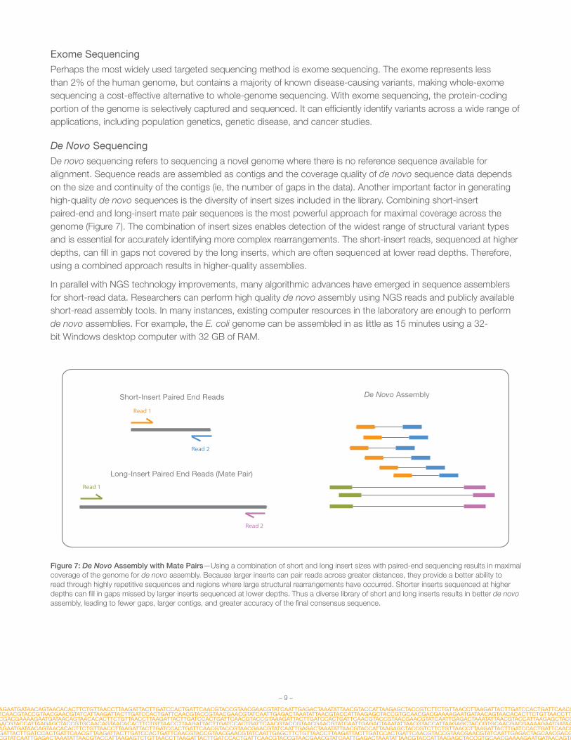
De Novo SequencingDe novo sequencing refers to sequencing a novel genome where there is no reference sequence available for alignment. Sequence reads are assembled as contigs and the coverage quality of de novo sequence data depends on the size and continuity of the contigs (ie, the number of gaps in the data). Another important factor in generating high-quality de novo sequences is the diversity of insert sizes included in the library. Combining short-insert paired-end and long-insert mate pair sequences is the most powerful approach for maximal coverage across the genome (Figure 7). The combination of insert sizes enables detection of the widest range of structural variant types and is essential for accurately identifying more complex rearrangements. The short-insert reads, sequenced at higher depths, can fill in gaps not covered by the long inserts, which are often sequenced at lower read depths. Therefore, using a combined approach results in higher-quality assemblies.
In parallel with NGS technology improvements, many algorithmic advances have emerged in sequence assemblers for short-read data. Researchers can perform high quality de novo assembly using NGS reads and publicly available short-read assembly tools. In many instances, existing computer resources in the laboratory are enough to perform de novo assemblies. For example, the E. coli genome can be assembled in as little as 15 minutes using a 32-bit Windows desktop computer with 32 GB of RAM.
Read 1
Read 2
Read 2
Read 1
Short-Insert Paired End Reads
Long-Insert Paired End Reads (Mate Pair)
De Novo Assembly
Figure 7: De Novo Assembly with Mate Pairs—Using a combination of short and long insert sizes with paired-end sequencing results in maximal coverage of the genome for de novo assembly. Because larger inserts can pair reads across greater distances, they provide a better ability to read through highly repetitive sequences and regions where large structural rearrangements have occurred. Shorter inserts sequenced at higher depths can fill in gaps missed by larger inserts sequenced at lower depths. Thus a diverse library of short and long inserts results in better de novo assembly, leading to fewer gaps, larger contigs, and greater accuracy of the final consensus sequence.

– 10 –
Targeted SequencingWith targeted sequencing, a subset of genes or regions of the genome are isolated and sequenced. Targeted sequencing allows researchers to focus time, expenses, and data analysis on specific areas of interest and enables sequencing at much higher coverage levels. For example, a typical WGS study achieves coverage levels of 30×–50× per genome, while a targeted resequencing project can easily cover the target region at 500×–1000× or higher. This higher coverage allows researchers to identify rare variants—variants that would be too rare and too expensive to identify with WGS or CE-based sequencing.
Targeted sequencing panels can be purchased with fixed, preselected content or can be custom designed. A wide variety of targeted sequencing library prep kits are available, including kits with probe sets focused on specific areas of interest such as cancer, cardiomyopathy, autism, or custom probe sets (Table 2). With custom designs, researchers can target regions of the genome relevant to their specific research interests. Custom targeted sequencing is ideal for examining genes in specific pathways, or for follow-up studies from GWAS or WGS. Illumina currently supports 2 methods for targeted sequencing—target enrichment and amplicon generation (Figure 8).
Denature double-stranded DNA library
Pooled Sample Library
Hybridize biotinylated probes to targeted regions
Biotin probes
Enrichment using streptavidin beads
Streptavidin beads
Elution from beads
Use DesignStudio to create custom oligo capture probes �anking each region of interest
CAT probes hybridize to �anking regions of interest in unfragmented gDNA
Extension/Ligation between Custom Probesacross regions of interest
PCR adds indices and sequencing primers
Uniquely tagged amplicon library ready for cluster generation and sequencing
P7 Index 2Index 1 P5
CAT (custom amplicon tube)
Region of interest
Custom Probe 1
CustomProbe 2
Custom Probe 1
CustomProbe 2
Index 1P7
Index 2 P5
Target Enriched Work�ow Amplicon Generation Work�ow
Figure 8: Target Enrichment and Amplicon Generation Workflows.

– 11 –
With target enrichment, specific regions of interest are captured by hybridization to biotinylated probes, then isolated by magnetic pulldown. Target enrichment captures between 20 kb–62 Mb regions depending on the library prep kit parameters. The second method, amplicon sequencing, involves the amplification and purification of regions of interest using highly multiplexed PCR oligo sets. Amplicon sequencing allows researchers to sequence 26–1536 targets at a time, spanning 150 bp–1.5 kb per target, depending on the library prep kit used. This highly multiplexed approach enables a wide range of applications for the discovery, validation, or screening of genetic variants. Amplicon sequencing is particularly useful for the discovery of rare somatic mutations in complex samples (eg, cancerous tumors mixed with germline DNA).13,14 Another common amplicon application is sequencing the bacterial 16S rRNA gene across multiple species, a widely used method for phylogeny and taxonomy studies, particularly in diverse metagenomic samples.15
For more information on Illumina targeted, WGS, exome, or de novo sequencing solutions, visit www.illumina.com/applications/sequencing/dna_sequencing.html.
b. Transcriptomics
Library preparation methods for RNA sequencing (RNA-Seq) typically begin with total RNA sample preparation followed by a ribosome removal step. The total RNA sample is then converted to cDNA before standard NGS library preparation. RNA-Seq focused on mRNA, small RNA, noncoding RNA, or microRNAs can be achieved by including additional isolation or enrichment steps before cDNA synthesis (Figure 9).
Targeted RNA Seq
Small RNASeq
RNA Access
Transcriptomics
mRNA Seq
ApoptosisPathway
miRNA
ncRNA
snRNA
P53Pathway
WntPathwayPlant
Human
FFPE
Mouse/ Rat
Stranded
Total RNA Seq
NFκBPathway
CustomTargets
Figure 9: A Complete View of Transcriptomics with NGS—A broad range of methods for transcriptomics with NGS have emerged over the past 10 years including total RNA-Seq, mRNA-Seq, small RNA-Seq, and targeted RNA-Seq.
Total RNA and mRNA SequencingTranscriptome sequencing is a major advance in the study of gene expression because it allows a snapshot of the whole transcriptome rather than a predetermined subset of genes. Whole-transcriptome sequencing provides a comprehensive view of a cellular transcriptional profile at a given biological moment and greatly enhances the power of RNA discovery methods. As with any sequencing method, an almost unlimited dynamic range allows identification and quantitation of both common and rare transcripts. Additional capabilities include aligning sequencing reads across splice junctions, as well as detection of isoforms, novel transcripts, and gene fusions. Library preparation kits that support precise detection of strand orientation are available for both total RNA-Seq and mRNA-Seq methods.

– 12 –
Targeted RNA Sequencing Targeted RNA sequencing is a method for measuring transcripts of interest for differential expression, allele-specific expression, as well as detection of gene-fusions, isoforms, cSNPs, and splice junctions. Illumina TruSeq® Targeted RNA Sequencing kits included preconfigured, experimentally validated panels focused on specific cellular pathways or disease states such as apoptosis, cardiotoxicity, NFkB pathway, and more. Custom content can also be designed and ordered for the analysis of specific genes of interest. Targeted RNA sequencing is a powerful method for the investigation of specific pathways of interest or for the validation of gene expression microarray or whole-transcriptome sequencing results.
Small RNA and Noncoding RNA SequencingSmall, noncoding RNA, or microRNA s are short, 18–22 bp nucleotides that play a role in the regulation of gene expression often as gene repressors or silencers. The study of microRNAs has grown as their role in transcriptional and translational regulation has become more evident.16,17
For more information regarding Illumina solutions for small RNA (noncoding RNA), targeted RNA, total RNA, and mRNA sequencing, visit www.illumina.com/applications/sequencing/rna.html.
c. Epigenomics
While genomics involves the study of heritable or acquired alterations in the DNA sequence, epigenetics is the study of heritable changes in gene activity caused by mechanisms other than DNA sequence changes. Mechanisms of epigenetic activity include DNA methylation, small RNA-mediated regulation, DNA–protein interactions, histone modification and more.
Methylation SequencingA critical focus in epigenetics is the study of cytosine methylation (5-mC) states across specific areas of regulation such as promotors or heterochromatin. Cytosine methylation can significantly modify temporal and spatial gene expression and chromatin remodeling. While there are many methods for the study of genetic methylation, methylation sequencing leverages the advantages of NGS technology and genome-wide analysis while assessing methylation states at the single-nucleotide level. Two methylation sequencing methods are widely used: whole-genome bisulfite sequencing (WGBS) and reduced representation bisulfite sequencing (RRBS). With WGBS-Seq, sodium bisulfite chemistry is converts nonmethylated cytosines to uracils, which are then converted to thymines in the sequence reads or data output. In RRBS-Seq, DNA is digested with MspI—a restriction enzyme unaffected by methylation status. Fragments in the 100–150 bp size range are isolated to enrich for CpG and promotor containing DNA regions. Sequencing libraries are then constructed using the standard NGS protocols.
ChIP SequencingProtein–DNA or protein–RNA interactions have a significant impact on many biological processes and disease states. These interactions can be surveyed with NGS by combining chromatin immunoprecipitation (ChIP) assays and NGS methods. ChIP-Seq protocols begin with the chromatin immunoprecipitation step (ChIP protocols vary widely as they must be specific to the species, tissue type, and experimental conditions).
Ribosome ProfilingRibosome profiling is a method based on deep sequencing of ribosome-protected mRNA fragments. Purification and sequencing of these fragments provides a “snapshot” of all the ribosomes active in a cell at a specific time point. This information can determine what proteins are being actively translated in a cell, and can be useful for investigating translational control, measuring gene expression, determining the rate of protein synthesis, or predicting protein abundance. Ribosome profiling enables systematic monitoring of cellular translation processes and prediction of

– 13 –
protein abundance. Determining what regions of a transcript are being translated can help define the proteome of complex organisms. With NGS, ribosome profiling allows detailed and accurate in vivo analysis of protein production.
To learn more about Illumina ribosome profiling, visit www.illumina.com/applications/sequencing/rna.html. For more on ChIP-Seq and methylation-Seq solutions, visit www.illumina.com/applications/epigenetics.html.
III. Illumina DNA-to-Data NGS Solutions
a. The Illumina NGS Workflow
Illumina offers a comprehensive, end-to-end solution for every step of the NGS sequencing workflow, from library preparation to final data analysis (Figure 10). Library preparation kits are available for all NGS methods including WGS, exome sequencing, targeted sequencing, RNA sequencing, and more. Illumina library preparation protocols can accommodate a range of throughput needs, from manual protocols for smaller laboratories to fully automated library preparation workstations for larger laboratories or genome centers. Likewise, Illumina offers a full portfolio of sequencing platforms, from the desktop MiSeq Series to the factory-scale HiSeq X Series that deliver the right level of speed, capacity, and cost for various laboratory or sequencing center requirements. For the last step in the NGS workflow, Illumina offers biologist-friendly bioinformatics tools that are easily accessible through the web, on instrument, or through onsite servers.
5−6 Hours 1−2 Days
NGS Library Preparation Kits
Illumina SequencingInstruments
Automated Data Analysis Tools
1−2 Days
Figure 10: Illumina DNA-to-Data Solutions—Illumina provides fully integrated, DNA-to-data solutions, with technology and support for every step of the NGS workflow including library preparation, sequencing, and final data analysis.
b. Integrated Data Analysis
BaseSpace® is a bioinformatics software solution for analyzing, storing, and sharing NGS data. BaseSpace can be accessed through the internet for data analysis and storage in the Illumina cloud or through an installed local server for data analysis and storage onsite. A major advantage of working in the BaseSpace environment is that it is directly integrated with Illumina sequencing systems. On-instrument access to BaseSpace enables the integration of many workflow steps, including library prep planning with BaseSpace Prep,† run set-up and chemistry validation, and real-time automatic data transfer to the BaseSpace computing environment.
The NGS workflow then proceeds seamlessly through alignment and subsequent data analysis steps with BaseSpace Apps. BaseSpace Apps offer a wide variety of analysis pipelines including analysis for de novo assembly, SNP and indel variant analysis, RNA expression profiling, 16S metagenomics, tumor-normal comparisons, epigenetic/gene regulation analysis, and many more. Illumina collaborates closely with commercial and academic software developers to create a full ecosystem of data analysis tools that address the needs of various research objectives. In the final stages of the NGS workflow, data can be shared with collaborators or delivered instantly to customers around the world.‡
To learn more about BaseSpace, visit www.illumina.com/basespace.
† Currently available with NextSeq 500 Sequencing Systems only. HiSeq and MiSeq Systems can use IEM for the same planning and validation functions.‡ Cloud-based environment only. Onsite BaseSpace restricts data sharing to local users.

– 14 –
IV. Glossaryadapters: The oligos bound to the 5’and 3’ end of each DNA fragment in a sequencing library. The adapters are complementary to the lawn of oligos present on the surface of Illumina sequencing flow cells.
bridge amplification: An amplification reaction that occurs on the surface of an Illumina flow cell. During flow cell manufacturing, the surface is coated with a lawn of 2 distinct oligonucleotides often referred to as “p5” and “p7.” In the first step of bridge amplification, a single-stranded sequencing library (with complementary adapter ends) is loaded into the flow cell. Individual molecules in the library bind to complementary oligos as they “flow” across the oligo lawn. Priming occurs as the opposite end of a ligated fragment bends over and “bridges” to another complementary oligo on the surface. Repeated denaturation and extension cycles (similar to PCR) results in localized amplification of single molecules into millions of unique, clonal clusters across the flow cell. This process, also known as “clustering,” occurs in an automated, flow cell instrument called a cBot™ or in an on-board cluster module within an NGS instrument.
clusters: A clonal grouping of template DNA bound to the surface of a flow cell. Each cluster is seeded by a single, template DNA strand and is clonally amplified through bridge amplification until the cluster has roughly 1000 copies. Each cluster on the flow cell produces a single sequencing read. For example, 10,000 clusters on the flow cell would produce 10,000 single reads and 20,000 paired-end reads.
contigs: A stretch of continuous sequence, in silico, generated by aligning overlapping sequencing reads.
coverage level: The average number of sequenced bases that align to each base of the reference DNA. For example, a whole genome sequenced at 30× coverage means that, on average, each base in the genome was sequenced 30 times.
digital microfluidics: Precise manipulation of droplets on a solid surface through applied voltages within a sealed microfluidic cartridge. For more information on digital microfluidics, see www.liquid-logic.com/technology.
flow cell: A glass slide with 1, 2, or 8 physically separated lanes, depending on instrument platform. Each lane is coated with a lawn of surface bound, adapter-complimentary oligos. A single library or a pool of up to 96 multiplexed libraries can be run per lane depending on application parameters.
indexes/barcodes/tags: A unique DNA sequence ligated to fragments within a sequencing library for downstream, in silico sorting and identification. Indexes are typically a component of adapters or PCR primers and are ligated to the library fragments during the sequencing library preparation stage. Illumina indexes are typically between 8–12 bp. Libraries with unique indexes can be pooled together, loaded into one lane of a sequencing flow cell, and sequenced in the same run. Reads are later identified and sorted via bioinformatic software. All together, this process is known as “multiplexing.”
insert: During the library preparation stage, the sample DNA is fragmented, and the fragments of a specific size (typically 200–500 bp, but can be larger) are ligated or “inserted” in between 2 oligo adapters. The original sample DNA fragments are also referred to as “inserts.”
mate pair library: A sequencing library with long inserts ranging in size from 2–5 kb typically run as paired-end libraries. The long gap length in between the sequence pairs is useful for building contigs in de novo sequencing, identification of indels, and other methods.
multiplexing: See “indexes/barcodes/tags.”
read: The process of next-generation DNA sequencing involves using sophisticated instruments to determine the sequence of a DNA or RNA sample. In general terms, a sequence “read” refers to the data string of A,T, C, and G bases corresponding to the sample DNA. With Illumina technology, millions of reads are generated in a single

– 15 –
sequencing run. In more specific terms, each cluster on the flow cell produces a single sequencing read. For example, 10,000 clusters on the flow cell would produce 10,000 single reads and 20,000 paired-end reads.
reference genome: A reference genome is a fully sequenced and assembled genome that acts as a scaffold against which new sequence reads are aligned and compared. Typically, reads generated from a sequencing run are aligned to a reference genome as a first step in data analysis. In the absence of a reference genome, the newly sequenced reads must be constructed by contig assembly (de novo sequencing).
sequencing by synthesis (SBS): SBS technology uses 4 fluorescently labeled nucleotides to sequence the tens of millions of clusters on the flow cell surface in parallel. During each sequencing cycle, a single labeled dNTP is added to the nucleic acid chain. The nucleotide label serves as a “reversible terminator” for polymerization: after dNTP incorporation, the fluorescent dye is identified through laser excitation and imaging, then enzymatically cleaved to allow the next round of incorporation. As all 4 reversible terminator-bound dNTPs (A, C, T, G) are present, natural competition minimizes incorporation bias. Base calls are made directly from signal intensity measurements during each cycle, which greatly reduces raw error rates compared to other technologies. The result is highly accurate base-by-base sequencing that eliminates sequence-context-specific errors, enabling robust base calling across the genome, including repetitive sequence regions and within homopolymers.
V. References1. Sanger F, Nicklen S, Coulson AR. DNA sequencing with chain-terminating inhibitors.
PNAS 1977;74:5463-5467.
2. Collins FS, Morgan M, Patrinos A. The human genome project: lessons from large-scale biology. Science. 2003;300:286-290.
3. Davies K. (2010) 13 years ago, a beer summit in an English pub led to the birth of Solexa. BioIT World (www.bio-itworld.com/) 28 Sept 2010.
4. Illumina (2014) HiSeqX Ten preliminary system specification sheet. (www.illumina.com/documents/products/datasheets/datasheet-hiseq-x-ten.pdf)
5. Fallows J. (2013) When will genomics cure cancer? A conversation with Eric S. Lander. The Atlantic (www.theatlantic.com/) 22 Dec 2013.
6. Ross MG, Russ C, Costello M, et al. Characterizing and measuring bias in sequence data. Gen Biol. 2013;14:R51.
7. Bentley DR, Balasubramanian S, Swerdlow HP, et al. Accurate whole human genome sequencing using reversible terminator chemistry. Nature. 2008;456:53-59.
8. Nakazato T, Ohta T, Bono H. Experimental design-based functional mining and characterization of high-throughput sequencing data in the sequence read archive. PLoS One. 2013;22;8(10):e77910.
9. Illumina (2014) Nextera DNA Library Preparation Kits data sheet. (www.illumina.com/documents/products/datasheets/datasheet_nextera_dna_sample_prep.pdf).
10. Illumina (2014) Nextera XT DNA Library Preparation Kit data sheet. (www.illumina.com/documents/products/datasheets/datasheet_nextera_xt_dna_sample_prep.pdf).
11. Illumina (2013) TruSeq DNA PCR-Free Library Preparation Kit data sheet. (www.illumina.com/documents/products/datasheets/datasheet_truseq_dna_pcr_free_sample_prep.pdf).
12. Grad YH, Lipsitch M, Feldgarden M, et al. Genomic epidemiology of the Escherichia coli O104:H4 outbreaks in Europe, 2011. PNAS. 2012;109:3065-3070.
13. McEllistrem MC. Genetic diversity of the pneumococcal capsule: implications for molecular-based serotyping. Future Microbiol. 2009;4:857-865.
14. Lo YMD, Chiu RWK. Next-generation sequencing of plasma/serum DNA: an emerging research and molecular diagnostic tool. Clin Chem. 2009;55:607-608.

Illumina • 1.800.809.4566 toll-free (US) • +1.858.202.4566 tel • [email protected] • www.illumina.com
For Research Use Only. Not for use in diagnostic procedures.
© 2015 Illumina, Inc. All rights reserved. Illumina, BaseSpace, cBot, HiSeq, MiSeq, NeoPrep, Nextera, NextSeq, TruSeq, and the pumpkin orange color are trademarks of Illumina, Inc. and/or its affiliate(s) in the U.S. and/or other countries. Pub. No. 770-2012-008 Current as of 21 April 2015
15. Ram JL, Karim AS, Sendler ED, and Kato I. Strategy for microbiome analysis using 16S rRNA gene sequence analysis on the Illumina sequencing platform. Syst Biol Reprod Med. 2011;57:117-118.
16. Wang Y, Kim S, Kim IM. Regulation of metastasis by microRNAs in ovarian cancer. Front Oncol. 2014;10:143.
17. Dior Up, Kogan L, Chill HH, Eizenberg N, Simon A. Emerging roles of microRNA in the embryo-endometrium cross talk. Semin Reprod Med. 2014;32:402-409.