ana luísa pequeno coelho dissertação de candidatura ao ... · doutor venceslau josé coelho...
TRANSCRIPT

Angiopoietin-2 and VEGF in non-small cell lung cancer
prognosis: can vessel co-option have a role here?
Ana Luísa Pequeno Coelho
Dissertação de candidatura ao grau de Doutor em Biomedicina
submetida à Faculdade de Medicina da Universidade do Porto
2016

Ana Luísa Pequeno Coelho
Angiopoietin-2 and VEGF in non-small cell lung cancer prognosis: can vessel co-option have a role here?
Dissertação de Candidatura ao grau de Doutor em
Biomedicina submetida à Faculdade de Medicina da
Universidade do Porto.
Orientador – Professor Doutor José Agostinho Marques
Lopes
Categoria – Professor Catedrático
Afiliação – Faculdade de Medicina da Universidade do
Porto; Director do Serviço de Pneumologia do Centro
Hospitalar de São João.
Co-orientador – Professor Doutor Rui Manuel Medeiros
Silva
Categoria – Professor Associado Convidado com
Agregação
Afiliação – Instituto de Ciências Biomédicas Abel Salazar
da Universidade do Porto; Instituto Português de
Oncologia FG Porto, EPE.
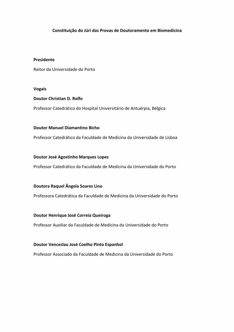
Constituição do Júri das Provas de Doutoramento em Biomedicina
Presidente
Reitor da Universidade do Porto
Vogais
Doutor Christian D. Rolfo
Professor Catedrático do Hospital Universitário de Antuérpia, Bélgica
Doutor Manuel Diamantino Bicho
Professor Catedrático da Faculdade de Medicina da Universidade de Lisboa
Doutor José Agostinho Marques Lopes
Professor Catedrático da Faculdade de Medicina da Universidade do Porto
Doutora Raquel Ângela Soares Lino
Professora Catedrática da Faculdade de Medicina da Universidade do Porto
Doutor Henrique José Correia Queiroga
Professor Auxiliar da Faculdade de Medicina da Universidade do Porto
Doutor Venceslau José Coelho Pinto Espanhol
Professor Associado da Faculdade de Medicina da Universidade do Porto
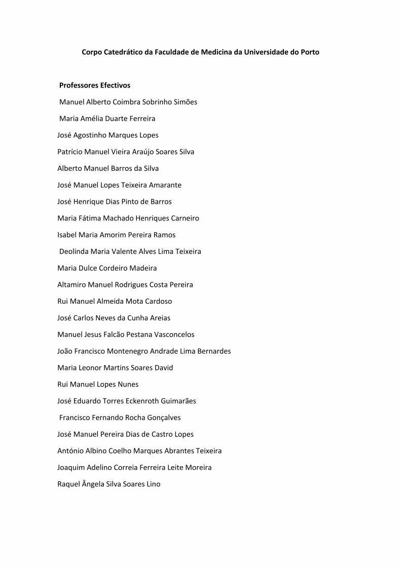
Corpo Catedrático da Faculdade de Medicina da Universidade do Porto
Professores Efectivos
Manuel Alberto Coimbra Sobrinho Simões
Maria Amélia Duarte Ferreira
José Agostinho Marques Lopes
Patrício Manuel Vieira Araújo Soares Silva
Alberto Manuel Barros da Silva
José Manuel Lopes Teixeira Amarante
José Henrique Dias Pinto de Barros
Maria Fátima Machado Henriques Carneiro
Isabel Maria Amorim Pereira Ramos
Deolinda Maria Valente Alves Lima Teixeira
Maria Dulce Cordeiro Madeira
Altamiro Manuel Rodrigues Costa Pereira
Rui Manuel Almeida Mota Cardoso
José Carlos Neves da Cunha Areias
Manuel Jesus Falcão Pestana Vasconcelos
João Francisco Montenegro Andrade Lima Bernardes
Maria Leonor Martins Soares David
Rui Manuel Lopes Nunes
José Eduardo Torres Eckenroth Guimarães
Francisco Fernando Rocha Gonçalves
José Manuel Pereira Dias de Castro Lopes
António Albino Coelho Marques Abrantes Teixeira
Joaquim Adelino Correia Ferreira Leite Moreira
Raquel Ângela Silva Soares Lino

Professores Jubilados ou Aposentados
Abel Vitorino Trigo Cabral
Alexandre Alberto Guerra Sousa Pinto
Álvaro Jerónimo Leal Machado de Aguiar
Amândio Gomes Sampaio Tavares
António Augusto Lopes Vaz
António Carlos de Freitas Ribeiro Saraiva
António Carvalho Almeida Coimbra
António Fernandes Oliveira Barbosa Ribeiro Braga
António José Pacheco Palha
António Manuel Sampaio de Araújo Teixeira
Belmiro dos Santos Patrício
Cândido Alves Hipólito Reis
Carlos Rodrigo Magalhães Ramalhão
Cassiano Pena de Abreu e Lima
Daniel Filipe de Lima Moura
Daniel Santos Pinto Serrão
Eduardo Jorge Cunha Rodrigues Pereira
Fernando Tavarela Veloso
Henrique José Ferreira Gonçalves Lecour de Menezes
Jorge Manuel Mergulhão Castro Tavares
José Carvalho de Oliveira
José Fernando Barros Castro Correia
José Luís Medina Vieira
José Manuel Costa Mesquita Guimarães
Levi Eugénio Ribeiro Guerra
Luís Alberto Martins Gomes de Almeida
Manuel António Caldeira Pais Clemente

Manuel Augusto Cardoso de Oliveira
Manuel Machado Rodrigues Gomes
Manuel Maria Paula Barbosa
Maria da Conceição Fernandes Marques Magalhães
Maria Isabel Amorim de Azevedo
Mário José Cerqueira Gomes Braga
Serafim Correia Pinto Guimarães
Valdemar Miguel Botelho dos Santos Cardoso
Walter Friedrich Alfred Osswald

Aos que nunca desistiram de mim…

“It is far more important to know what person the disease has
than what disease the person has.”
Hippocrates of Kos (460-377 BC)
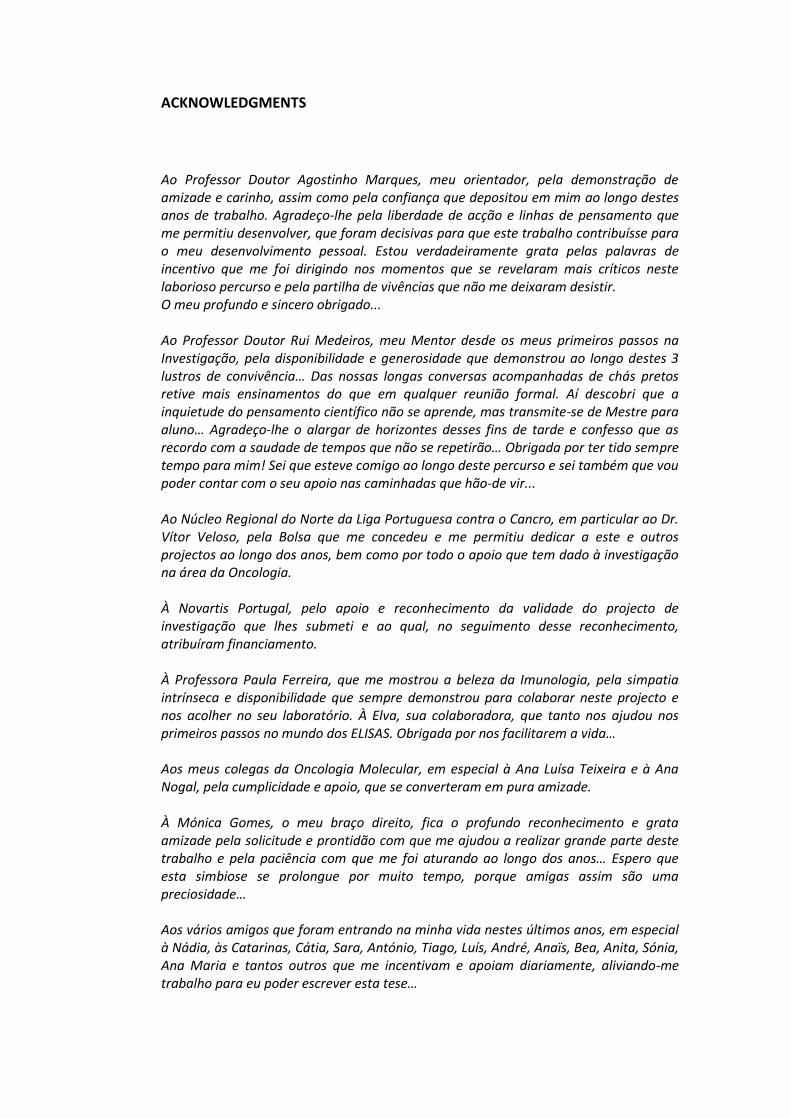
ACKNOWLEDGMENTS
Ao Professor Doutor Agostinho Marques, meu orientador, pela demonstração de amizade e carinho, assim como pela confiança que depositou em mim ao longo destes anos de trabalho. Agradeço-lhe pela liberdade de acção e linhas de pensamento que me permitiu desenvolver, que foram decisivas para que este trabalho contribuísse para o meu desenvolvimento pessoal. Estou verdadeiramente grata pelas palavras de incentivo que me foi dirigindo nos momentos que se revelaram mais críticos neste laborioso percurso e pela partilha de vivências que não me deixaram desistir. O meu profundo e sincero obrigado... Ao Professor Doutor Rui Medeiros, meu Mentor desde os meus primeiros passos na Investigação, pela disponibilidade e generosidade que demonstrou ao longo destes 3 lustros de convivência… Das nossas longas conversas acompanhadas de chás pretos retive mais ensinamentos do que em qualquer reunião formal. Aí descobri que a inquietude do pensamento científico não se aprende, mas transmite-se de Mestre para aluno… Agradeço-lhe o alargar de horizontes desses fins de tarde e confesso que as recordo com a saudade de tempos que não se repetirão… Obrigada por ter tido sempre tempo para mim! Sei que esteve comigo ao longo deste percurso e sei também que vou poder contar com o seu apoio nas caminhadas que hão-de vir... Ao Núcleo Regional do Norte da Liga Portuguesa contra o Cancro, em particular ao Dr. Vítor Veloso, pela Bolsa que me concedeu e me permitiu dedicar a este e outros projectos ao longo dos anos, bem como por todo o apoio que tem dado à investigação na área da Oncologia. À Novartis Portugal, pelo apoio e reconhecimento da validade do projecto de investigação que lhes submeti e ao qual, no seguimento desse reconhecimento, atribuíram financiamento. À Professora Paula Ferreira, que me mostrou a beleza da Imunologia, pela simpatia intrínseca e disponibilidade que sempre demonstrou para colaborar neste projecto e nos acolher no seu laboratório. À Elva, sua colaboradora, que tanto nos ajudou nos primeiros passos no mundo dos ELISAS. Obrigada por nos facilitarem a vida… Aos meus colegas da Oncologia Molecular, em especial à Ana Luísa Teixeira e à Ana Nogal, pela cumplicidade e apoio, que se converteram em pura amizade. À Mónica Gomes, o meu braço direito, fica o profundo reconhecimento e grata amizade pela solicitude e prontidão com que me ajudou a realizar grande parte deste trabalho e pela paciência com que me foi aturando ao longo dos anos… Espero que esta simbiose se prolongue por muito tempo, porque amigas assim são uma preciosidade… Aos vários amigos que foram entrando na minha vida nestes últimos anos, em especial à Nádia, às Catarinas, Cátia, Sara, António, Tiago, Luís, André, Anaïs, Bea, Anita, Sónia, Ana Maria e tantos outros que me incentivam e apoiam diariamente, aliviando-me trabalho para eu poder escrever esta tese…

À Teresa, minha irmã do coração, pelo apoio moral e constante preocupação, que se traduzem numa admirável e pura amizade, que dura há quarenta anos. Obrigada por não me deixares esquecer que estás sempre aí! À família Catarino, em cujo seio fui acolhida como “filha adoptiva”, fica o meu reconhecimento pela preocupação constante com o meu bem-estar… É bom ter uma casa fora da nossa casa… À Raquel, mana e companheira de viagem há 15 anos… Cada aventura que vivemos juntas foi única e irrepetível… Nunca conseguirei agradecer-lhe o suficiente, apenas posso dizer-lhe que o laço inquebrável, puro e sincero que nos une é mágico e um dos meus bens mais preciosos! Foi por causa dele que nunca mais me senti só… Obrigada do coração, Zucchini! À D. Margarida, que nestes 20 anos de convivência se tornou indispensável ao bom funcionamento do meu lar e cujo carinho que devota a cada um de nós a torna membro por direito desta família! À minha cunhada Fátima e aos meus meninos, João e Ricardo, agradeço a secreta alegria de os saber sempre do meu lado, apesar da distância física… Aos meus pais, nunca saberei ou conseguirei agradecer de forma adequada, tudo o que fizeram por mim… Aqui deixo apenas a minha sincera homenagem e a promessa de me continuar a esforçar para nunca os defraudar! À minha MÃE, em especial, agradeço as 50 vezes que me atende o telefone diariamente, para escutar os meus disparates e as minhas pequenas coisas, dando-me a segurança de que preciso para seguir em frente de cabeça sempre bem erguida e com os pés assentes no chão! Ao Miguel, agradeço o exemplo de tenacidade e invencibilidade, seja qual for o revés da sua vida… faço minhas as palavras que um dia ele escreveu: mais que meu sobrinho, meu filho, meu irmão, um dos amores da minha vida. “You’ll be in my heart!” To Miguel’s dear Rachel, I acknowledge the patience of the critical proofreading of the papers of this thesis, as well as the affection she demonstrates in all of our contacts. Ao meu irmão, por me ter amado incondicionalmente, desde o dia em que nasci… com ele aprendi, entre muitas outras coisas, que não há obstáculos grandes o suficiente para nos conseguirem parar! É e sempre será o meu porto de abrigo, aquele que nunca me deixou abandonar as minhas batalhas e que afugenta todos os meus demónios! Também te adoro, desde que me entendo por gente… Aos meus avós, cuja falta sinto todos os dias, desde que partiram… com eles aprendi a valorizar as pequenas grandes coisas da vida… Ao Tó, pelas aventuras e desventuras que vivemos juntos, que me fizeram crescer pessoal e profissionalmente. Profissionalmente, agradeço-lhe o incentivo para a realização desta tese, a ajuda na elaboração do projecto e recolha de amostras, bem como a leitura crítica e apurada de todas as minhas publicações. Pessoalmente, estou-lhe grata por estar presente “na alegria e na tristeza, na saúde e na doença,…”, nos meus melhores e piores momentos… Espero que a nossa seja uma história interminável!

Ao meu maravilhoso filho, a minha maior realização pessoal, agradeço a sobredose diária de felicidade que me proporciona… Espero que um dia me perdoe cada momento que lhe roubei para me dedicar a este projecto, que por isso também lhe pertence um bocadinho… Com ele aprendi a simplicidade que se esconde por detrás do verdadeiro amor! O seu olhar radioso e o seu pequeno espírito inquieto fazem-me desejar ser sempre melhor… A todos, reitero o meu apreço e a minha eterna gratidão.

XII
TABLE OF CONTENTS
List of Publications XIII
Abstract XIV
Resumo XVII
1. Introduction 1
1.1 Overview of lung cancer 2
1.2 Angiogenesis and cancer 3
1.2.1 The VEGF family 4
1.3 Anti-angiogenic therapy and lung cancer 5
1.4 Mechanisms of resistance to antiangiogenic therapy 6
1.4.1 Angiopoietin-Tie system (Ang-Tie system) 7
1.4.2 Ang-2 and Tie2 expressing-macrophages (TEMs) 9
1.4.3 Ang-2 involvement in alternative pathways of tumor
vascularization 10
1.4.4 VEGF and Ang-2-Tie2 axis inhibition 11
1.5 Biomarkers and antiangiogenic therapy 11
2. Objectives 13
3. Results / Publications 15
3.1 Circulating Ang-2 mRNA Expression Levels: Looking Ahead to a
New Prognostic Factor for NSCLC 16
3.2 Combined Ang-2 and VEGF serum levels: holding hands as a new
integral biomarker in non-small-cell lung cancers 21
3.3 Angiogenesis in NSCLC: is vessel co-option the trunk that sustains
the branches? 31
4. General Discussion and Conclusions 41
5. Future Perspectives 49
6. References 52
XIII
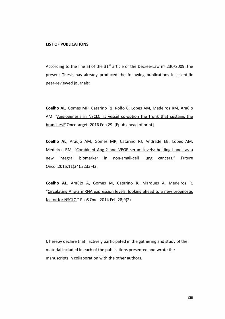
LIST OF PUBLICATIONS
According to the line a) of the 31st article of the Decree-Law nº 230/2009, the
present Thesis has already produced the following publications in scientific
peer-reviewed journals:
Coelho AL, Gomes MP, Catarino RJ, Rolfo C, Lopes AM, Medeiros RM, Araújo
AM. "Angiogenesis in NSCLC: is vessel co-option the trunk that sustains the
branches?”Oncotarget. 2016 Feb 29. [Epub ahead of print]
Coelho AL, Araújo AM, Gomes MP, Catarino RJ, Andrade EB, Lopes AM,
Medeiros RM. "Combined Ang-2 and VEGF serum levels: holding hands as a
new integral biomarker in non-small-cell lung cancers.” Future
Oncol.2015;11(24):3233-42.
Coelho AL, Araújo A, Gomes M, Catarino R, Marques A, Medeiros R.
“Circulating Ang-2 mRNA expression levels: looking ahead to a new prognostic
factor for NSCLC.” PLoS One. 2014 Feb 28;9(2).
I, hereby declare that I actively participated in the gathering and study of the
material included in each of the publications presented and wrote the
manuscripts in collaboration with the other authors.

XIV
ABSTRACT
XV
Introduction: Lung cancer remains the most common incident form of cancer
globally, with deaths exceeding those from any other type of malignancy, accounting
for nearly one in five deaths. Approximately 85% of those cases are non-small-cell lung
cancers (NSCLCs) and the vast majority of patients presents at advanced stages of
disease, resulting in an overall five-year survival around 15.9%. Lung cancer is the
result of a multistep process, in which angiogenesis assumes a major role, allowing
tumor access to oxygen and nutrients, growth factors and hormones. Antiangiogenic
strategies are becoming widely used in NSCLC treatment and the pathway involving
Vascular Endothelial Growth Factor (VEGF) and its receptors (VEGFRs) is the main
target of the approved antiangiogenic molecules (bevacizumab and ramucirumab).
Nevertheless, the overall survival (OS) benefits from these therapies are modest,
because a high fraction of tumors is intrinsically refractory to the therapy and a
substantial proportion of the remaining acquire resistance during treatment. Intensive
research in this field unveiled some of the mechanisms underneath these
disappointing results. These include: 1) upregulation of alternative pro-angiogenic
signalling circuits to overcome VEGF(R) inhibition; 2) recruitment of vascular
progenitor cells and pro-angiogenic monocytes from the bone marrow and 3)
alternative mechanism to angiogenesis during tumor development, such as vessel co-
option. Another obstacle in the quest for successful antiangiogenic strategies is the
lack of reliable predictive biomarkers that may help to tailor patients for whom these
therapies will be more suited. Angiopoietin-2 (Ang-2), a vessel destabilizing cytokine
expressed by the endothelial cells, that belongs to the angiopoietin/Tie (type I
transmembrane tyrosine kinase receptors) axis, works in concert with VEGF in tumor
angiogenesis, and is now one of the most promising therapeutic targets in
antiangiogenic strategies. It seems to be largely involved in the proposed escape
mechanisms to current antiangiogenics and has a major advantage over VEGF, since its
expression is seldom detected in healthy vasculature and highly regulated at mRNA
level. These particular characteristics also turn it into an interesting candidate as a
predictive and prognostic biomarker of outcome in antiangiogenic directed therapies.
Aims: Evaluation of the correlation between circulating Ang-2 mRNA levels and
NSCLC prognosis. Also, evaluation of the impact of combined serum levels of VEGF and
Ang-2 in the prognosis of NSCLC and its potential as diagnostic marker of disease.
XVI
Material and methods: An unselected cohort of 145 NSCLC cases admitted at
Portuguese Institute of Oncology of Porto was recruited to the study. 30 control
individuals, from the same geographical area as case subjects, were also recruited. A
peripheral blood sample was taken from each individual. mRNA extraction was
performed from the blood samples, and measured by the quantitative real-time
polymerase chain reaction (qRT-PCR) method. The serum levels of Ang-2 and VEGF of
each patient were determined by enzyme-linked immunosorbent assay (ELISA)
technique prior to treatment.
Results: There is an association between circulating Ang-2 mRNA levels and OS in
all stages of NSCLC and higher levels of Ang-2 mRNA correlate with poorer OS. This
relation is more pronounced when considering only patients eligible for antiangiogenic
therapies, with stage IV disease. Also, according to the results, circulating Ang-2 mRNA
levels independently determine OS in NSCLC patients.
Serum levels of Ang-2 and VEGF are significantly correlated. High serum levels of Ang-2
and VEGF isolated and both combined (HighAng-2/VEGF) correlate with likelihood of
presenting NSCLC. Serum levels of Ang-2 and HighAng-2/VEGF, but not VEGF alone, are
independent prognostic factors for NSCLC.
Conclusions: This study suggests that circulating Ang-2 mRNA levels could
successfully be included as predictive biomarkers of response in the design of clinical
trials involving antiangiogenic drugs targeting Ang-2 and that HighAng-2/VEGF serum levels
could be exploited as a new valuable integral biomarker in NSCLC. We hypothesise that
in some NSCLC, tumors obviate the need to generate angiogenesis by co-opting host
mature vessels and growing along them (vessel co-option). Tumor-co-opted vessel
interactions result in endothelial cells (ECs) activation and intense Ang-2 expression
and secretion, leading to vascular disruption and vessel regression, generating a
hypoxic core in the tumor that is rescued by an increased expression of VEGF, which
induces a robust angiogenic response. This gives the rationale for therapeutic
approaches of dual inhibition of Ang-2 and VEGF serving as a launchpad to more
successful NSCLC anti-vascular treatments.

XVII
RESUMO

XVIII
Introdução: O cancro do pulmão permanece a forma mais incidente e letal de
cancro a nível mundial, com uma taxa de mortalidade que representa
aproximadamente uma em cada cinco mortes (1.6 milhões de óbitos, no total). Cerca
de 85% destes casos correspondem a cancros de pulmão de não-pequenas células
(CPNPCs). O cancro do pulmão resulta de um processo que compreende várias etapas,
sendo que a angiogénese assume um papel central no desenvolvimento tumoral,
permitindo o aporte de oxigénio e nutrientes necessários para o crescimento do
tumor. Actualmente, a terapia com inibidores da angiogénese faz parte das guias de
tratamento do CPNPC, sendo a via que envolve o Factor de Crescimento do Endotélio
Vascular (VEGF) e os seus receptores (VEGFRs) o principal alvo dos inibidores
aprovados até à data (bevacizumab e ramucirumab). No entanto, no que concerne à
sobrevida global (SG) dos doentes, os benefícios obtidos na prática clínica com estes
tratamentos têm sido modestos, já que uma fracção importante dos tumores
apresenta resistência intrínseca à terapia e parte dos restantes adquire resistências ao
longo do tratamento. Alguns dos mecanismos propostos para justificar estes
resultados desapontantes incluem: 1) activação de vias de sinalização pro-angiogénicas
alternativas à via do VEGF(R); 2) recrutamento de células pró-angiogénicas para o
estroma do tumor e 3) utilização de mecanismos alternativos à angiogénese durante o
desenvolvimento tumoral, como a co-opção de vasos pré-existentes. Outro obstáculo
ao sucesso das terapias anti-angiogénicas é a ausência de biomarcadores preditivos
que possam ajudar a seleccionar doentes para quem estas terapias serão mais
eficazes. A angiopoietina-2 (Ang-2), expressa pelas células endoteliais (CEs) é
actualmente investigada como um dos mais promissores alvos da terapia anti-
angiogénica e parece estar envolvida nos mecanismos de evasão aos inibidores de
VEGF anteriormente descritos. Para além disso, a sua expressão é raramente
observada nos vasos sanguíneos saudáveis, sendo altamente regulada a nível do ARN
mensageiro (ARNm). Estas características particulares tornam a Ang-2 um candidato
interessante a biomarcador prognóstico de SG e preditivo nas terapias dirigidas a alvos
angiogénicos.
Objectivos: Avaliação da correlação entre os níveis circulantes de ARNm da Ang-2
e o prognóstico de doentes com CPNPC. Avaliação do impacto da combinação de níveis

XIX
séricos de Ang-2 e VEGF no prognóstico do CPNPC e do seu potencial como marcador
diagnóstico da doença.
Material e métodos: Participaram neste estudo 145 doentes com CPNPC,
admitidos no Instituto Português de Oncologia do Porto, e 30 indivíduos saudáveis da
mesma área geográfica. Foi colhida uma amostra de sangue periférico de cada doente
à data do diagnóstico, a partir da qual se extraiu ARNm, que foi doseado pelo método
quantitativo em tempo real da reacção da polimerase em cadeia (qRT-PCR). Os níveis
séricos de Ang-2 e VEGF de cada doente foram determinados por um método
enzimático (ELISA).
Resultados: Os níveis circulantes elevados de ARNm de Ang-2 estão associados a
menor SG em todos os estadios de CPNPC. Esta relação é ainda mais pronunciada
quando considerados apenas os doentes com estadio mais avançado (IV) da doença.
Também se verificou que níveis circulantes elevados de ARNm de Ang-2 constituem
um factor independente de prognóstico da SG.
Existe uma correlação directa entre níveis séricos de Ang-2 e VEGF. Quer
isoladamente quer quando combinados, níveis elevados correlacionam-se com a
probabilidade de desenvolvimento de CPNPC. Níveis séricos elevados de Ang-2
isoladamente ou combinados com níveis elevados de VEGF (HighAng-2/VEGF), mas não de
VEGF isoladamente, são factores de prognóstico para a SG de CPNPC.
Conclusões: Este trabalho sugere que níveis circulantes de ARNm da Ang-2
poderão ser incluídos como biomarcadores preditivos de resposta em ensaios clínicos
que envolvam terapias que tenham como alvo a Ang-2. Sugere também que a
combinação de níveis elevados de ANG-2 e VEGF no soro possam ser explorados como
biomarcadores integrais no tratamento de CPNPC com inibidores da angiogénese.
Também fica a sugestão de que, em alguns CPNPCs, o mecanismo de co-opção de
vasos pré-existentes do hospedeiro seja a forma preferencial de vascularização do
tumor, obviando a necessidade de angiogénese. A interacção entre as células tumorais
e os vasos do hospedeiro resulta na activação das CEs destes vasos, com intensa
expressão e secreção de Ang-2, conduzindo-os a um estado de dissociação que
culmina com a regressão dos vasos e com a geração de uma área de hipoxia central,
que despoleta a expressão de VEGF pelas células do estroma, induzindo uma robusta
resposta pró-angiogénica. Esta teoria serve de base ao racional para abordagens

XX
terapêuticas de inibição dupla de Ang-2 e VEGF, servindo de catapulta para terapias
anti-angiogénicas de maior sucesso clínico.

1
1. INTRODUCTION

2
1.1 Overview of lung cancer
Cancer is a major public health issue, representing a leading cause of morbidity
and mortality worldwide [1]. In 2013, the global incidence of cancer cases was 14.9
million, with 8.2 million cancer related deaths and, by 2030, an estimated 24 million
new cases are expected [1,2]. While advances in diagnostics and treatment have led to
a 3.4-fold increase in patient survival over the last 40 years, many disease settings
remain with little progress, and with a high rate of recurrence or fatality from
metastatic disease [3].
Lung cancer is the most common incident form of cancer worldwide, with an
estimated 1.8 million new cases in 2012, with deaths exceeding those from any other
type of malignancy, accounting for nearly one in five deaths (1.6 million deaths in
total) [4]. Approximately 85% of those cases are currently classified as Non-Small-Cell
Lung Cancers (NSCLCs), [5]. The remaining 15% are Small-Cell Lung Cancers (SCLCs),
extremely aggressive tumors, morphologically and histologically distinct from NSCLCs
and strongly correlated with cigarette smoking [6].
NSCLCs are currently divided in different subsets, according to its histopathological
characteristics [7]. The two main NSCLC histological phenotypes are adenocarcinoma
(ADC; ±50%), predominantly more peripheral tumours, thought to arise from the
alveolar or bronchiolar epithelium (pneumocytes or Clara cells) presenting often
glandular histology, and squamous cell carcinoma (SCC; ±40%), which typically arises
from the bronchial epithelium of the larger, more central airways [5]. Other subtypes
of NSCLC include large cell carcinoma (LCC; ±3%), which is essentially diagnosed by
exclusion if tumour cells do not appear glandular or squamous in shape or do not
express ADC or SCC biomarkers [5,7] and neuroendocrine (NE) neoplasms of the lung,
including typical and atypical carcinoid tumors and excluding SCLC [8].
The vast majority of patients with NSCLC presents at an advanced stage of disease,
when curative treatment is no longer a possibility, resulting in a poor prognosis and an
overall five year survival around 15.9% [7,9]. This number has only marginally
improved during the past few decades, despite the increased understanding and
appreciation of the complexity of NSCLC during this period [9].
Current knowledge on the biology of lung cancer shows that it is the result of a
multistep process, with intricate combinations of morphological, molecular and

3
genetic alterations, ultimately leading to a malignant cell agglomerate bearing the
phenotypic hallmarks of cancer, defined by Hanahan and Weinberg in 2011 [7,10].
Among these, angiogenesis seems to assume major importance, since gaining access
to the host vascular system and the generation of a tumour blood supply to obtain
oxygen and nutrients, growth factors and hormones, among others, are rate-limiting
steps in tumour progression [11].
1.2 Angiogenesis and cancer
The identification of massive vascularization in tumors dates to 1863 [12] and the
importance of tumor angiogenesis has been recognised since 1908 [13], but it was only
in the early 1970s, with the work of Folkman, that angiogenesis was acknowledged as a
potential target to inhibit cancer progression [14-17]. The therapeutic potential of anti-
angiogenic strategies boosted this field, and placed angiogenesis as one of the major
areas of cancer research nowadays.
It is now widely accepted that most tumors and metastases originate as small
avascular structures which must induce the development of new blood vessels from
pre-existing ones, in order to grow beyond a minimum size of 2-3 mm3 [11,18]. To
achieve this, tumors undergo an angiogenic switch, disrupting the equilibrium between
pro and anti-angiogenic regulators, favouring pro-angiogenic mechanisms, where
signalling molecules induce quiescent endothelial cells (ECs) to continually sprout from
existing blood vessels, forming new vessels that help to sustain expanding neoplastic
growth [11,19,20], according to the conventional model of angiogenesis, known as
angiogenic sprout [21].
Decades of research investigating the molecular basis of angiogenesis led to the
discovery of a number of angiogenic molecules that promote tumor angiogenesis [20].
Of all the identified angiogenic pathways, the most critical appears to be the one
involving the vascular endothelial growth factor (VEGF) family and its receptors
(VEGFR) [22,23].
4
1.2.1 The VEGF family
The VEGF family consists of five glycoproteins referred to as VEGFA, VEGFB,
VEGFC, VEGFD and placental growth factor (PIGF) and distinguishes itself from other
angiogenic super families by the largely non-redundant roles of its members [24]. The
VEGF ligands bind to and activate three structurally similar type III receptor tyrosine
kinases (TKR), designated VEGFR1, VEGFR2 and VEGFR3. The assortment of VEGF
ligands has distinctive binding specificities for each of these TKR, with consequent
diversity of function [25]. VEGFA and VEGFB have the greatest binding affinity to
VEGFR1 and 2, with the majority of angiogenic effects being attributable to VEGFA, the
best characterized of the VEGF family members (from now on referred to as VEGF),
which is expressed as various isoforms owing to alternative splicing [25]. It stimulates
angiogenesis in health and disease by signalling through VEGFR2, whose expression is
restricted primarily to the vasculature and is the key mediator of VEGF-induced
angiogenesis [25]. VEGFR1 can also bind VEGF, and might function as a decoy receptor
that sequesters VEGF from VEGFR2 and negatively regulates angiogenesis, [26],
although its precise role in angiogenesis it is still elusive [24]. The role of PIGF in
angiogenesis also remains controversial; it exclusively binds to VEGFR1 and it is
speculated that it may directly stimulate vessel growth and maturation and recruit
proangiogenic bone marrow-derived progenitors and monocyte-macrophage lineage
cells [27,28]. The remaining family members, VEGFC and VEGFD, are formed by
proteolytic processing (unlike VEGFA, B and PIGF, isoforms originated by alternative
splicing), and bind mainly to VEGFR3, appearing to be important contributors to
lymphangiogenesis [28].
Ever since the identification of VEGF as the first endothelium-acting specific
cytokine in 1983 [18,29,30], its overexpression has been found in several human
tumors, including NSCLC [31-35], probably due to its induction under the ischaemic
conditions that usually occur at the rim of necrotic and hypoxic regions of the tumor
[11]. A growing number of functions of VEGF in the tumor angiogenic process have
been unravelled [35]: it triggers multiple signalling networks that enhance ECs
proliferation and survival, increasing its migration and invasion capabilities, increases
vascular permeability and interstitial pressure of existing vessels and enhances

5
chemotaxis and mobilization of bone marrow derived endothelial progenitor cells
(EPCs) into the peripheral circulation [25,36].
The recognition of the central role of VEGF in tumor angiogenesis turned it into an
attractive target for therapeutic intervention in cancer and the VEGF pathway became
the main focus of research in the quest for effective targeted anti-angiogenic
strategies [29,37]. The extensive investigation in this field has led to the study of
several anti-angiogenic agents, including monoclonal antibodies to block VEGF and its
receptor VEGFR2 and VEGFR tyrosine kinase inhibitors (TKIs) [38].
1.3 Anti-angiogenic therapy and lung cancer
Presently, there are two anti-angiogenic compounds approved by the American
Food and Drug Administration (FDA) for the treatment of NSCLC. Bevacizumab, an anti-
VEGF recombinant monoclonal antibody that blocks the binding of VEGF to its high-
affinity receptors, was the first angiogenic inhibitor to complete clinical development,
showing clinical benefit in patients with metastatic colorectal cancer when combined
with chemotherapy [25,38]. It was approved in 2006 for the treatment of advanced
non-squamous NSCLC in the first line setting in combination with chemotherapy [35].
Later, in 2014, ramucirumab, a fully humanized monoclonal antibody that targets
angiogenesis by specifically binding to VEGFR-2 with higher affinity than its natural
ligand VEGF [39], has been approved for the treatment of patients with metastatic
NSCLC in second line setting, in combination with docetaxel [40]. Besides these two
anti-angiogenic compounds, many others are currently under clinical evaluation, in
different stages of clinical trials or waiting for approval for the treatment of metastatic
or recurrent NSCLC [41,42].
An important feature that distinguishes the antiangiogenic drugs from other
targeted therapies is that these agents are typically given to unselected NSCLC patients
for the approved indications [43] and despite the significant clinical achievements of
bevacizumab and ramucirumab in different NSCLC treatment settings, the overall
survival (OS) benefits from antiangiogenic therapies remain modest [44] and these
VEGF pathway inhibitors are failing to produce enduring clinical responses in most
patients [21,45]. In a high fraction of them, the tumor is intrinsically refractory to the
anti-angiogenic therapy such that disease progression continues ceaseless [46] and
6
when this is not the case, acquired resistance to therapy can rapidly occur and limit the
efficacy of the antiangiogenic treatments [24,47].
1.4 Mechanisms of resistance to antiangiogenic therapy
Tumor resistance to the antiangiogenic therapies (whether intrinsic or acquired),
makes the clinical use of VEGF/VEGFR blockers, in patients with advanced NSCLC, more
challenging than anticipated by the results of preclinical experiments [24]. The modest
success obtained in the clinical practice raised a number of questions, fuelling this
already active field of research, to improve anticancer treatment [47,48].
Researchers are aware that the mechanisms underlying tumor resistance to
angiogenic inhibitors are complex and diverse, depending upon the location of the
tumor, the nature of the tumor itself, the surrounding stroma, the dynamic nature of
the angiogenic process, the strong redundancy determined by a continuous cross-
interaction between main and alternative pathways and the ability to recruit
proangiogenic bone marrow-derived cells to the tumor site, just to mention a few.
Until this intricate network underlying tumor resistance is fully understood, and then
selectively inhibited, it will probably be difficult to achieve the full efficacy of anti-
VEGF(R) and other antiangiogenic therapies [49].
In the last few years, some of the explanations to resistance to current
antiangiogenic therapies have been unveiled and include: 1) upregulation of
alternative pro-angiogenic signalling circuits to overcome VEGF(R) inhibition; 2)
recruitment of vascular progenitor cells and pro-angiogenic monocytes from the bone
marrow to tumor stroma, where they promote tumor revascularization and growth
and increased capabilities of invasion without angiogenesis [21,23,24,46,47,50]; 3)
non-angiogenic tumor microenvironments, as is the case of tumors that do not need
angiogenic sprout to obtain an efficient blood supply and rather use alternative
vascularization mechanisms to support its growth [46,47,51]. These comprise NSCLCs
that grow using pre-existing vessels in the rich vascularized lung, through a vessel co-
option strategy [52].
Exhaustive research on this subject reveals that Angiopoietin-2 (Ang-2), a member
of the Angiopoietin (Ang) -Tie (type I transmembrane tyrosine kinase receptor) system,

7
participates or mediates, at least in part, all of the above proposed mechanisms of
resistance to angiogenic inhibitors [53].
1.4.1 Angiopoietin-Tie system (Ang-Tie system)
The human Ang-Tie system was identified in the mid-1990s as a family of growth
factors essential for blood vessel formation [54]. It consists of two type I
transmembrane tyrosine kinase receptors (Tie1 and Tie2) and three secreted ligands,
Ang-1, Ang-2 and Ang-4 [53]. Tie1 and Tie2, with its tyrosine kinase domain in the
cytoplasm, are preferentially expressed by vascular and lymphatic ECs, although Tie2
expression has been recorded in non-endothelial cells, both in normal tissue and
disease, including carcinoma cells and monocytes [55,56]. Unlike Tie1, Tie2 binds
directly to angiopoietins and has strong kinase activity [57]. Tie1 is currently
considered an orphan receptor, with no known ligand, but has been shown to bind
Tie2 and regulate its activity [53,58].
Ang-1 and Ang-2 are the most extensively characterized ligands of Tie2 and their
interactions with it comprise an important endothelial cell-specific receptor tyrosine
kinase signalling system in angiogenesis [54,55,59,60]. They are secreted glycoproteins
[54], which act primarily in the vasculature to control blood vessel development and
stability [61] and interact with Tie2 either in a paracrine (Ang-1) or autocrine (Ang-2)
manner. Although both Ang-1 and Ang-2 have important roles in angiogenesis, the
nature of their contributions is distinct [53].
Studies of loss-of-function have shown that Ang-1-deficient mice present
embryonic lethal phenotype, due to aberrant vessel remodelling and maturation,
phenocopying the early midgestation death of Tie2-deficient mice, suggesting that
Ang-1 is the single, non-redundant, agonist of Tie2 [5,59,62]. Ang-1 is produced
primarily by perivascular cells (smooth muscle cells, pericytes), although in the adult, it
is found in many types of tissues and is constitutively secreted in low levels throughout
the body [63]. Ang-1/Tie2 signaling promotes blood vessel maturation and stabilization
[64]; at low (basal) levels, Ang-1 engagement with Tie2 activates downstream
signalling, resulting in EC survival signals, in the maintenance of the endothelial barrier,
the quiescent state of vasculature and blood vessel assembly, thus ensuring the
8
resting, anti-thrombotic and anti-adhesive state of the vascular endothelium
[53,59,65].
Ang-2 is mainly synthesized by ECs and stored in Weibel-Palade bodies in its
cytoplasm, from where it can be rapidly released upon stimulation to act as an
autocrine regulator of ECs functions at sites of vascular remodeling [53]. Despite much
research in the last decade, the role of Ang-2 in the Ang-Tie signaling axis and in
vascular biology in general, is not as straightforward as the one of Ang-1 [57]. Genetic
manipulation experiments in mice showed that Ang-2 gain-of-function phenotype
resembles Ang-1 deficiency in embryonic development, suggesting that it functions as
a natural antagonist of Ang-1 [59,66], but unlike Ang-1, Ang-2 expression is expendable
for normal embryonic development, as shown by loss-of-function studies, although
Ang-2 deficiency leads to persistent vascular defects after birth [59].
Along with its prominent role in blood vessel biology during vascular remodeling,
Ang-2 seems to be an obligate partner of lymphatics maturation. An elegant study
performed by Gale and co-workers showed that Ang-2 is not required for the initiation
of lymphatic vascular development, but it is absolutely required for their remodeling
and normal functioning [67]. Moreover, they found that Ang-2 seemed to substitute
for Ang-1 agonistic functions in lymphatics in vivo, since lymphatic dysfunction in Ang-
2 deficient mice could be rescued with Ang-1 administration, suggesting that Ang-2
acts as an activating agonist of Tie2 in this situation [62,67].
The dynamic pattern of Ang-2 expression at sites of angiogenesis, such as cyclic
vessel regression in ovaries, tumor vascular co-option, and hyaloid vessel regression,
supports the concept that the dominant biologic role of Ang-2 is the control of vascular
remodeling through the interruption of Tie2 signaling [54,59,62]; Ang-2-Tie2
association allows for destabilization of established blood vessels through the
induction of vessel plasticity (e.g. by decreasing pericyte coverage), disrupting the
integrity of the blood vessel wall, thereby counteracting vascular normalization, a pre-
requisite for sprouting angiogenesis in the presence of other angiogenic molecules or
physiologic vascular regression in the absence of such stimuli [68,69]. Moreover, Ang-2
can exert a direct pro-angiogenic Tie2-independent role by directly binding integrins in
Tie2 negative ECs, promoting tumor invasion and metastases [61].
9
1.4.2 Ang-2 and Tie2 expressing-macrophages (TEMs)
Tie2-expressing monocytes/macrophages (TEMs) are a subpopulation of
circulating and tumor-infiltrating myeloid cells with inherent vascular growth
promoting activity, representing a reservoir of cells innately committed to a
proangiogenic function [70,71]. In human tumors, they are found mainly in
perivascular and avascular viable areas, but are largely absent in non-neoplastic tissues
adjacent to the tumor [72]. The preferential location of TEMs in the vicinity of tumor
blood vessels suggests that these cells may cross-talk with ECs and provide paracrine
support to nascent blood vessels in these areas during the angiogenic process [72,73].
This notion was reinforced by selective elimination of TEMs using a suicide gene
approach, showing that the absence of TEMs from tumors impairs angiogenesis and
delays tumor growth, suggesting that these cells have nonredundant, proangiogenic
activity in tumors [71,74].
Ang-2 levels in the tumor microenvironment have been directly correlated with
increased TEMs recruitment to tumor stroma, and it is well established that Ang-2 is
chemoattractant to TEMs, in a process mediated by Tie2 [70,73,75,76]. The
overexpression of Ang-2 by ECs and tumor cells exposed to hypoxia, results in greater
infiltration of TEMs into tumors [72] and in turn, hypoxia induces upregulation of cell-
surface expression of Tie2 in TEMs, increasing their responsiveness to Ang-2, in a
mechanism of feed-back loop used to amplify TEMs function in tumor
microenvironment [77]. It is described that stimulation with Ang-2 modulates the
cytokine profile expressed by TEMs [78], upregulating angiogenic mediators, such as
the proangiogenic enzymes thymidine phosphorylase (TP) and cathepsin B (CTSB) and
downregulating the expression of antiangiogenic and immunosuppressive cytokines,
such as pro-apoptotic tumor necrosis factor-α (TNF-α) and antiangiogenic interleukin-
12 (IL-12) [75,77].
Although TEMs exhibit some features of M2-polarized tumor-associated
macrophages (TAMs), a few differences have been found between them. TEMs express
lower levels of VEGF than TAMs, and do not home to hypoxic, avascular tumor areas.
Thus, it is likely that TEMs exert a requisite proangiogenic function by supporting
tumor angiogenesis downstream to VEGF-induced vascular activation, regulating blood
vessel formation by a VEGF independent pathway [76]. This could explain why the
10
presence of TEMs in the tumors counteracts the efficacy of antivascular treatments
[73], promoting vascular regrowth following therapy-induced vascular damage and its
association with the heightened invasive phenotype observed upon the use of anti-
angiogenesis therapies [79].
1.4.3 Ang-2 involvement in alternative pathways of tumor vascularization
It is fairly recognized that tumor progression is heavily dependent upon
angiogenesis. However, the model that angiogenesis is necessary for a tumor to
become larger than a few millimetres and become clinically detectable has been
challenged by the extensive research in this field, which has shown that angiogenesis is
not always a pre-requisite for tumor growth [80]. When tumors arise in well-
vascularized organs, their growth will rely on the invasion of host tissue. Enhancement
of invasion and metastasis facilitates access to normal tissue vasculature, and cancer
cells stay in close contact with the surface of blood vessels [42,81,82]. This allows
tumor cells to grow and migrate along quiescent normal vessels and take their oxygen
and essential nutrients without obligate neovascularization, in a process known as
vessel co-option [46,47,83]. This procedure has been recognized as an important
mechanism to establish tumor vasculature, especially in more aggressive tumors, and
represents a major route for a solid tumor to evade antiangiogenic therapy
[45,46,51,84].
The co-opted vessels are usually supported by pericytes, which stabilize it, while
promoting endothelial cell survival via induction of autocrine VEGF signalling [85]. In
vessel co-option, the first tumor-vessel interactions result in ECs activation and intense
Ang-2 expression and secretion [61]; Ang-2 then acts through an endogenous
autocrine loop mechanism that is context dependent [65,86]. When it binds to its Tie2
receptor, it functions as a vessel-destabilizing molecule that converts mature vessels to
a tenuous and plastic state by inducing loosening of endothelial cell interactions with
pericytes and smooth muscle cells, leading to the loss of vascular integrity and
increased vascular permeability that facilitates the infiltration of proteases, cytokines
and angiogenic myeloid cells, and thus, the priming of the vasculature for a robust
angiogenic response in the presence of growth factors, such as VEGF
[18,59,64,65,76,87,88].

11
In the absence of VEGF, as during the early phases of antiangiogenic treatment,
the expression of Ang-2 causes irreversible loss of vascular structures [87,89] with
marked regression of the co-opted vessels [53] in a very similar fashion to what
happens with primitive vessels during development [86]. This generates the hypoxic
core and the apoptotic tumor cell loss observed in nonangiogenic tumors [84,87], that
presumably act as de initial stimulus for the molecular changes (which are yet to be
clarified), that culminate in VEGF expression by the remaining tumor cells and in
neoangiogenesis [90], mediated both by VEGF and Ang-2 [84].
1.4.4 VEGF and Ang-2-Tie2 axis inhibition
Ang-2 seems to be a preferential partner of VEGF in the orchestration of tumor
angiogenesis. Similarly to what is seen with VEGF expression, elevated levels of Ang-2
have been associated with advanced disease, progression and poor prognosis in the
most diverse tumor models, including NSCLC [64,90], glioblastoma, gastric, colorectal,
breast, prostate, kidney and hepatocellular carcinomas, as well as multiple myeloma,
melanoma, and neuroendocrine tumors [91-99], but unlike VEGF, Ang-2 is seldom
detectable in healthy vasculature, making it the perfect target to tumor therapy [62].
Considering the multifaceted nature of Ang-2 in tumor angiogenesis promotion
and its proposed involvement in the mechanisms of resistance to VEGF targeted
therapies, it is not surprising that, nowadays, Ang-2/Tie2 system is regarded as one of
the most important therapeutic targets in antiangiogenic strategies. Its inhibition
would allow both the optimization of current antiangiogenic strategies and the
circumscription of acquired resistance to approved angiogenic inhibitors
[53,64,69,76,87,100-107]. Targeting Ang-2/Tie2 axis and VEGF(R) pathway holds the
promise of more clinically meaningful responses than monotherapies targeting VEGF
pathway alone [27,53].
1.5 Biomarkers and antiangiogenic therapy
The missing key to optimize the results obtained with angiogenic inhibitors would
be the distinction between patients that are likely to respond to these treatments
from those intrinsically refractory to angiogenic therapies. Predictive biomarkers are
greatly needed to achieve this goal [108-110]. Despite the several molecular mediators
12
of angiogenesis and inflammatory signalling that have been investigated as potential
biomarkers of antiangiogenic therapy in NSCLC, no biomarker has yet been
prospectively validated to correlate with outcomes [111-113].
In the future, it is essential that the selection of new antiangiogenic agents to
enter early pre-clinical trials is based on the availability of identified and validated
biomarkers predictive of future clinical efficacy [44,109,114].

13
2. OBJECTIVES

14
2.1 General objectives
- Evaluate the correlation between circulating Ang-2 mRNA levels and NSCLC
prognosis in an unselected cohort of NSCLC patients.
- Evaluate the prognostic relevance of serum Ang-2 and VEGF levels in an
unselected cohort of NSCLC patients.
2.2 Specific objectives
The aims of this study include:
- Determination of the independent prognostic value of circulating Ang-2 mRNA
levels in OS of NSCLC patients, by a quantitative real-time polymerase chain reaction
(qRT-PCR) method.
- Investigation of the correlation between serum Ang-2 and VEGF levels in NSCLC
patients by enzyme-linked immunosorbent assay (ELISA) technique.
- Assessment of the impact of the combined serum Ang-2/VEGF levels in the
prognostic of the disease.
- Evaluation of its potential value as a diagnostic cancer marker
- Assessment of the influence of the combined serum Ang-2/VEGF levels in the
susceptibility to NSCLC.
- Help to establish candidate markers for better prognostication for NSCLC
patients

15
3. RESULTS / PUBLICATIONS
Circulating Ang-2 mRNA Expression Levels: LookingAhead to a New Prognostic Factor for NSCLCAna L. Coelho1,2*, Antonio Araujo1,3,4, Monica Gomes1,4, Raquel Catarino1,4, Agostinho Marques2,5,
Rui Medeiros1,4,6
1 Molecular Oncology Group, Portuguese Institute of Oncology Porto, Porto, Portugal, 2 Faculty of Medicine, University of Porto, Porto, Portugal, 3 Medical Oncology
Department, Centro Hospitalar de Entre o Douro e Vouga, Santa Maria da Feira, Portugal, 4 Abel Salazar Institute for the Biomedical Sciences, University of Porto, Porto,
Portugal, 5 Pulmonology Department, Centro Hospitalar de S. Joao, Porto, Portugal, 6 Research Department, Portuguese League Against Cancer (NRNorte), Porto,
Portugal
Abstract
Non-small cell lung cancer (NSCLC) is the most common cancer and the leading cause of death from cancer worldwide.Antiangiogenic strategies directed towards tumor stroma are becoming gold standard in NSCLC treatment and researchershave been searching for biomarkers to identify patients for whom therapy with antiangiogenic inhibitors may be mostbeneficial and the importance of these as prognostic factors in NSCLC. The purpose of this study was to evaluate theprognostic value of circulating Ang-2 mRNA levels prior to treatment in NSCLC patients. The mRNA levels were determinedby quantitative real-time PCR in the peripheral blood of 92 NSCLC patients. Our results demonstrate that patients with highcirculating Ang-2 mRNA levels have diminished overall survival when compared to those with low mRNA levels (20.3months vs 34.3 months, respectively; Log Rank Test, p = 0.016), when considering all NSCLC stages and this difference iseven bigger when considering only patients with stage IV (15.9 months vs 31.3 months, respectively; Log Rank Test,p = 0.036). Moreover, circulating Ang-2 mRNA levels independently determine overall survival, and the concordance (c)index analysis showed that the definition of a nomogram that contains information regarding tumor stage, patients’smoking status and circulating Ang-2 mRNA levels present an increased capacity to predict overall survival in NSCLCpatients (c-index 0.798). These results suggest that this nomogram could serve as a unique and practical tool to determineprognosis in NSCLC, not relying on the availability of adequate surgical or biopsy specimens of NSCLC. Attending to ourresults, the circulating Ang-2 mRNA levels should also be included in the design of preclinical studies and clinical trialsinvolving antiangiogenic drugs targeting Ang-2, to guide adequate patient stratification and dose selection and increasingthe likelihood of benefit to a level that is acceptable to patients and clinicians.
Citation: Coelho AL, Araujo A, Gomes M, Catarino R, Marques A, et al. (2014) Circulating Ang-2 mRNA Expression Levels: Looking Ahead to a New PrognosticFactor for NSCLC. PLoS ONE 9(2): e90009. doi:10.1371/journal.pone.0090009
Editor: Anthony W. I. Lo, The Chinese University of Hong Kong, Hong Kong
Received December 16, 2013; Accepted January 24, 2014; Published February 28, 2014
Copyright: � 2014 Coelho et al. This is an open-access article distributed under the terms of the Creative Commons Attribution License, which permitsunrestricted use, distribution, and reproduction in any medium, provided the original author and source are credited.
Funding: The authors would like to thank the Liga Portuguesa Contra o Cancro — Centro Regional do Norte for the educational grant given to Ana L Coelho.This project was partially sponsored by a grant for basic research in molecular oncology from Novartis Portugal. The funders had no role in study design, datacollection and analysis, decision to publish, or preparation of the manuscript.
Competing Interests: The authors have the following interests. This project was partially sponsored by a grant for basic research in molecular oncology fromNovartis Portugal. Rui Medeiros is a PLOS ONE Editorial Board member. There are no patents, products in development or marketed products to declare. This doesnot alter the authors’ adherence to all the PLOS ONE policies on sharing data and materials, as detailed online in the guide for authors.
* E-mail: [email protected]
Introduction
Non-small-cell lung cancer is the most frequent type of lung
cancer and the most common cause of death from cancer [1]. In
2010, the number of deaths from lung cancer worldwide was 1?5
million, representing 19% of all cancer deaths that year. Most lung
cancers (,80%) are non-small-cell lung cancers (NSCLC) and of
these patients, more than 65% present with locally advanced or
metastatic disease [2].
Solid tumors, including NSCLC, require angiogenesis—the
formation of new blood vessels from existing vessels—for survival,
growth, and metastasis. These new tumor vessels are structurally
and functionally abnormal. They develop by sprouting or
intussusception from pre-existing vessels and exist in a constantly
dynamic state of sprout formation, proliferation, remodeling, or
regression [3,4].
In the last 9 years, antiangiogenic therapy has become part of
standard antitumor treatment. However, the clinical efficacy of
such therapies is limited, and it appears that the full therapeutic
potential of antiangiogenic intervention has not been fully
exploited [5].
It’s now known that there are various molecular players
involved in different mechanisms of vascular growth in solid
tumors, and among these, members of the Vascular Endothelial
Growth Factor (VEGF) and Angiopoietin (Ang) family have a
predominant role [3].
Angiopoietins, the bona fide ligands of Tie-2 receptor, form a
family of secreted 70 kDa glycoproteins acting primarily on the
vasculature to control blood vessel development and stability. Four
distinct angiopoietins have been described: Ang-1, Ang-2, Ang-3
and Ang-4. Angiopoietins bind the second immunoglobulin motif
of Tie-2 whereby they activate Tie-2 and, indirectly, Tie-1 in Tie-
1/Tie-2 heterodimers [6].
PLOS ONE | www.plosone.org 1 February 2014 | Volume 9 | Issue 2 | e90009

Ang-1 is expressed by pericytes, smooth muscle cells, and
fibroblasts and acts in a paracrine manner. In contrast, Ang-2 is
expressed by endothelial cells (EC) and stored in the Weibel-
Palade bodies from where it can be rapidly released on stimulation
to act as an autocrine regulator of EC functions [7].
Ang-1 and Ang-2 have been described to exert opposing
functions during vessel development. Ang-1–induced Tie2 activa-
tion transduces survival signals and leads to vessel stabilization and
maturation. In turn, Ang-2 acts as a vessel destabilizing agent that
induces permeability and leads to dissociation of cell-cell contacts
in cultured endothelial cells. Genetic experiments have solidly
established Ang-2 as an antagonistic Tie2 ligand [7]. Moreover,
Ang-2 can have a direct pro-angiogenic Tie-2-independent role by
directly binding integrins in Tie2 negative EC [6].
Ang-2 has been implicated in the remodeling of the tumor
vasculature in a process resembling its physiological actions [8,9].
Among the first steps of the angiogenic switch is the co-optive
engagement of the pre-existing host vasculature by the growing
tumor. This results in EC activation and intense Ang-2 expression,
which promotes the dissociation of pericytes from pre-existing
vessels and increases vascular permeability, which facilitates the
infiltration of proteases, cytokines and angiogenic myeloid cells,
and thus, the priming of the vasculature for a robust angiogenic
response in the presence of growth factors, such as VEGF-A [6].
Following the angiogenic switch, the Ang–Tie system contributes,
in concert with VEGF, to tumor angiogenesis [10].
Ang2 is strongly regulated at the transcriptional level. In fact,
almost any form of endothelial cell activation leads to upregulation
of Ang2 mRNA. The mRNA induction of Ang2 in tumor
endothelium has made Ang2 a very attractive circulating
biomarker of angiogenic activation [5].
Some studies have addressed the correlation between Ang-2
expression in tumor tissue and the protein circulating levels and
cancer development and metastasis. However, few clinical studies
have documented a correlation between this molecule and disease
clinical features or prognosis in lung cancer [11-15].
To the best of our knowledge, the present study is the first to
establish a correlation between circulating Ang-2 mRNA levels
and lung cancer prognosis.
Materials and Methods
Ethics statementThe study was conducted according to the principles of the
Helsinki Declaration. The study was approved by the local ethics
committee at the Portuguese Institute of Oncology of Porto
(Portugal). All individuals signed a written informed consent prior
to the inclusion in the study.
Study PopulationThe study included Caucasian patients from the North region of
Portugal. The inclusion criteria were histological or cytological
confirmed diagnosis of NSCLC, no previous treatment, an Eastern
Cooperative Oncology Group (ECOG) performance status#2
(with 0 indicating that the patient is fully active, 1 that the patient
is ambulatory but restricted in strenuous activity, and 2 that the
patient is ambulatory and capable of self-care but is unable to work
[16]), no prior oncologic disease and available clinical data.
Circulating mRNA levels quantificationCirculating Ang-2 mRNA levels were analyzed by quantitative
real-time PCR (qRT-PCR). Initially, the mRNA was isolated from
the cell fraction of peripheral blood samples by TriPure reagent
(Roche Applied Science), and after separation of the RNA
fraction, the samples were purified using the commercial kit
GeneJET RNA Purification Kit (Fermentas). RNA samples were
then used as templates for cDNA synthesis, using a High Capacity
RNA-to-cDNA Kit (Applied Biosystems), according to the
manufacturer’s instruction. Finally, qRT-PCR was carried out
on a StepOne TMOne qPCR equipment, containing 1x Master
Mix (Applied Biosystems), with 1x probe (TaqMan Gene
Expression Assay with reference number Hs 01048042_m1,
Applied Biosystems), cDNA sample, human GUSB (Beta Glucu-
ronidase) and human b-2M (b-2 Microglobulin) endogenous
controls (both from Applied Biosystems) according to manufac-
turer’s instructions.
To quantify the amplified transcripts, we used the comparative
CT (22DDCT) method [17]. In accordance with the method, the
mRNA amounts of the target gene (Ang-2) were normalized to two
endogenous controls and relatively to a calibrator. We used the
housekeeping genes GUSB (Applied Biosystems) and b-2M
(Applied Biosystems) as internal controls and commercial RNA
controls as calibrators (Applied Biosystems). DDCT represents the
difference between the mean DCT value of a patient blood sample
and the mean DCT value of the calibrator, both calculated after
the same PCR run, whereas DCT is the difference between the
CT of the target gene and the CT of the endogenous reference
gene of the same sample. The relative quantitative value was
expressed as 22DDCT. Relative quantification (RQ) based on the
Ct (the number of PCR cycles necessary to obtain the threshold
signal of fluorescence) values was analyzed using Applied
Biosystems StepOne Software v 2.2. All samples were run in
duplicate.
Statistical analysisAng-2 mRNA expression levels were considered as categorical
variables using the first quartile as cut-off point. We defined that
the values under the cut-off point should be included in the low
expression group and that all the other cases above the cut-off
point constituted the high expression group. Overall Survival (OS)
was calculated from the beginning of treatment to death from any
cause. Median OS was estimated with the Kaplan-Meier method
and compared with a two-sided log-rank test. Multivariate Cox
proportion analysis was performed to determine the influence of
age, gender, tumor stage, histological type, smoking status and
circulating Ang-2 mRNA levels on OS in NSCLC patients.
Hazard ratios (HRs) estimated from the Cox analysis were
reported as relative risks with corresponding 95% confidence
intervals (CIs). The extent of discrimination of the predictive
ability was quantified using the Harrel’s concordance index (c-
index), which estimates the probability of concordance between
predicted and observed responses. The interpretation of the C
index is similar to the interpretation of the area under a receiver
operating curve (ROC) curve. A value of 1.0 indicates that the
features of the model perfectly separate patients with different
outcomes while a value of 0.5 indicates the features contain
prognostic information equal to that obtained by chance alone.
All analyses were performed using Statistical Package for Social
Science (SPSS) for Windows version 18 (Chicago, IL). The level of
statistical significance was set at 5% (P#0.05).
Results
The study included 92 Caucasian individuals from the North
region of Portugal, with histopathological diagnosis of NSCLC,
with a mean age of 63.2 years610.7. The blood samples were
collected at the time of diagnosis, before treatment, and included
33 squamous cell carcinomas (SCC), 46 adenocarcinomas, 11
Circulating Ang-2 mRNA: A NSCLC Prognostic Factor
PLOS ONE | www.plosone.org 2 February 2014 | Volume 9 | Issue 2 | e90009
undifferentiated NSCLC, 1 large cells and 1 mixed carcinomas, of
which 77,0% were male and 75,8% smokers or former smokers,
divided in 42 non-metastatic and 50 metastatic cases (Table 1).
Our results demonstrate that patients with high circulating Ang-
2 mRNA levels have diminished overall survival when compared
to those with low mRNA expression (20.3 months vs 34.3 months,
respectively; Log Rank Test, p = 0.016) (Figure 1). Moreover,
when considering only stage IV patients, the most suitable
candidates to antiangiogenic treatment, the range interval between
overall survival in the high and low settings of Ang-2 mRNA
expression augments (15.9 months vs 31.3 months, respectively;
Log Rank Test, p = 0.036) (Figure 2).
To determine the independent prognostic value of circulating
Ang-2 mRNA levels for OS, a multivariate analysis using a Cox
proportional hazard model was performed. In the multivariate
analysis that included age, gender, tumor stage, histological type,
smoking status and circulating Ang-2 mRNA levels, we identified
tumor stage, smoking status and Ang-2 mRNA levels as
independent prognostic factors for OS in NSCLC patients
(Table 2).
Attending to these results, we performed an analysis considering
four different prognostic models to ascertain the predictive power
of circulating Ang-2 mRNA levels in the clinical outcome of
NSCLC patients (Table 3). In the first two models, we considered
the predictive ability of tumor stage and smoking status (c-index
0.657 for tumor stage and 0.522 for smoking status) (Model 1 and
Model 2). Model 3 addressed the question whether circulating
Ang-2 mRNA levels could also be considered a prognostic factor
in NSCLC, with a c-index of 0.629 (Model 3). In model 4, we
created a three variables prognostic nomogram congregating the
three aforementioned models. The prognostic predictive ability
was increased when adding circulating Ang-2 mRNA levels, with a
c-index of 0.798 (Model 4).
Discussion
Many predictive and prognostic markers have been assessed in
NSCLC but, until the discovery of the importance of Epidermal
Growth Factor Receptor gene (EGFR) [18], no single molecular
marker had proven to be useful for either patient selection or
selection of specific drugs. The TNM classification (lung cancer
staging) has stood the test of time and to date, no other prognostic
factors beyond it have been prospectively validated, remaining the
most powerful prognostic instrument in lung cancer [19,20].
Hence, the identification of other prognostic factors that can be
integrated with TNM to create a composite prognostic index for
NSCLC would be clinically useful.
Many clinical trials have demonstrated the importance of
evaluating several molecular biomarkers of NSCLC tumor
specimens to allow a personalized medicine, enhancing progres-
sion free survival and overall survival times, diminishing side
effects, giving patients a better quality of life and enabling to
perform more cost-effectiveness treatments [21]. Moreover, we
now know that in addition to predictors of response, these genes
can also be regarded as prognostic factors for NSCLC [21],
making the evaluation of these biomarkers the state of the art of
the advanced or metastatic NSCLC treatment. However, only
patients with lung adenocarcinoma with EGFR mutations or
anaplastic lymphoma kinase (ALK) rearrangements have an FDA-
approved therapy available [18,21–23]. Unfortunately, for squa-
mous cell NSCLC the scenario is even more shadowy. Although
this is an important field where novel targeted therapies are
currently under investigation, the disease prognosis still remains
disappointing.
As the field of lung cancer moves further into the age of
personalized medicine, alternative targets continue to be investi-
gated, since it has become clear that it will be imperative to target
the tumor stroma and surrounding environment and not merely
the genes’ mutations within the cancer cells itself. The main goal of
this quest is to identify a pan-NSCLC prognostic factor that can
also help to predict treatment response and monitor tumor
progression. One of the most extensive studied of such alternative
targets is angiogenesis, a necessary process in the growth and
metastasis of all solid tumors [23]. Preclinical models and selected
clinical trials showed benefits for targeting angiogenesis in lung
cancer, with antiangiogenic treatment emerging as the first
effective anti-stroma therapy to complement the established
antitumor therapies [7]. So far, only VEGF, the master switch
of the angiogenic cascade, has been validated as a therapeutic
target for antiangiogenic intervention [7] but no published clinical
study has proved that circulating levels of this target are prognostic
factors in patients with NSCLC subjected to antiangiogenic
therapy [24].
Despite of the advances into the elucidation of the tumor milieu
in general and tumor angiogenesis in particular, there is a
significant knowledge deficit in the understanding of the molecular
basis of antiangiogenic therapy and the related adverse events seen
with these agents [23]. Researchers have been searching for
potential biomarkers to identify patients for whom therapy with
antiangiogenic inhibitors may be most beneficial and the
importance of these as prognostic factors in NSCLC.
Whereas VEGF is abundantly expressed by the tumor cells in
most tumors, Ang-2 is mostly expressed by the tumor-associated
endothelium [7]. Moreover, unlike VEGF, Ang-2 shows limited
Table 1. NSCLC patients’ characteristics.
Patients(n = 92)
n %
Gender
Female 21 22.8
Male 71 77.2
Age
Mean6S.D. 63.2610.7
Histology
Adenocarcinoma 46 50.0
SCC 33 35.8
NSCLC NOS 11 12.0
Large CellsMixed
1 1.10
1 1.10
Staging
I 3 3.30
II 1 1.10
III 38 41.3
IV 50 54.3
Smoking status
Non-smoker 22 23.9
Smoker 48 52.2
Former smoker 22 23.9
doi:10.1371/journal.pone.0090009.t001
Circulating Ang-2 mRNA: A NSCLC Prognostic Factor
PLOS ONE | www.plosone.org 3 February 2014 | Volume 9 | Issue 2 | e90009
postnatal expression in normal tissues and its broad expression and
prominent upregulation in tumor milieu turns it in the perfect
candidate to help to define prognosis in solid tumors, besides being
a suitable suspect in the game of antiangiogenic strategies [7,25].
In the present study, we aimed to evaluate the prognostic
significance of Ang-2 mRNA detection in the cell fraction of
peripheral blood of patients with NSCLC prior to treatment, using
qRT-PCR. Moreover, we wanted to assess the possibility of using
it as a prognostic factor that could be adjoined to NSCLC staging
to create a composite prognostic index for NSCLC and created a
nomogram that predicts the influence of circulating Ang-2 mRNA
levels in NSCLC clinical outcome.
Our results demonstrate that high circulating Ang-2 mRNA levels
are a significantly unfavorable prognostic factor in NSCLC overall
survival. Patients with high circulating Ang-2 mRNA levels have
diminished overall survival when compared to those with low mRNA
expression, when considering all NSCLC stages and this difference is
even bigger when considering only patients with distant metastasis,
the most suitable candidates to antiangiogenic therapies. Moreover,
mRNA levels independently determine survival, and its prognostic
predictive ability increases when modeled in a simple and easy to
apply nomogram with NSCLC staging, patients’ smoking status and
Ang-2 mRNA levels (c-index 657 vs c-index 0.798, respectively).
Figure 1. Association of high and low circulating levels of Ang-2 mRNA with overall survival in NSCLC by Kaplan-Meier curves.doi:10.1371/journal.pone.0090009.g001
Figure 2. Association of high and low circulating levels of Ang-2 mRNA with overall survival in stage IV NSCLC by Kaplan-Meier curves.doi:10.1371/journal.pone.0090009.g002
Table 2. Multivariate Cox regression analysis for predictablefactors of overall survival.
HR 95% CI P
Age ($63;,63) 0.97 0.0.610–1.55 0.905
Gender 0.55 0.260–1.16 0.118
Tumor stage 1.79 1.12–2.88 0.016
Histological type 1.12 0.810–1.53 0.503
Smoking status 2.36 1.09–5.09 0.029
Ang-2 mRNA 2.04 1.13–3.67 0.017
doi:10.1371/journal.pone.0090009.t002
Circulating Ang-2 mRNA: A NSCLC Prognostic Factor
PLOS ONE | www.plosone.org 4 February 2014 | Volume 9 | Issue 2 | e90009

Taken together, these results prompt us to think that detection
and quantification of circulating Ang-2 mRNA in blood samples,
along with proper NSCLC staging, could serve as a unique and
practical diagnostic tool to determine prognosis in NSCLC.
Circulating Ang-2 mRNA levels samples are a simple to obtain
factor which can theoretically reflect the overall angiogenic activity
of the tumor and offers a huge advantage over tissue based
markers, including the ability to carry out continuous, noninvasive
assessments over time and most important, not relying on the
availability of adequate surgical or biopsy specimens of NSCLC.
Various therapeutic agents targeting Ang-2 have been described
and are being evaluated in early-phase clinical trials [5,9,25–28].
Albeit antiangiogenic drugs are efficacious in unselected popula-
tions, increasing market competition between targeted therapies is
likely to drive the growth of individualized chemotherapy, with a
central role for biomarkers. Although more studies are needed to
confirm this hypothesis, the circulating Ang-2 mRNA levels could
be strong candidates for predicting the survival benefit associated
with the targeted therapies currently under evaluation and should
be included in the design of preclinical studies and clinical trials
involving antiangiogenic drugs targeting Ang-2, to guide adequate
patient stratification and dose selection and increasing the
likelihood of benefit to a level that is acceptable to patients and
clinicians.
Acknowledgments
The authors would like to thank Ana Luisa Teixeira for her presence
during quantitative real-time PCR performance. Her expertise was of great
help to the execution of the technique.
Author Contributions
Conceived and designed the experiments: ALC AA MG. Performed the
experiments: ALC MG. Analyzed the data: ALC RC RM. Contributed
reagents/materials/analysis tools: ALC AA AM RM. Wrote the paper:
ALC AA AM RM.
References
1. Rosell R, Bivona TG, Karachaliou N (2013) Genetics and biomarkers in
personalisation of lung cancer treatment. Lancet 382: 720–731.2. Reck M, Heigener DF, Mok T, Soria JC, Rabe KF (2013) Management of non-
small-cell lung cancer: recent developments. Lancet 382: 709–719.
3. Carmeliet P, Jain RK (2000) Angiogenesis in cancer and other diseases. Nature407: 249–257.
4. Falcon BL, Hashizume H, Koumoutsakos P, Chou J, Bready JV, et al. (2009)Contrasting actions of selective inhibitors of angiopoietin-1 and angiopoietin-2
on the normalization of tumor blood vessels. Am J Pathol 175: 2159–2170.
5. Gerald D, Chintharlapalli S, Augustin HG, Benjamin LE (2013) Angiopoietin-2:an attractive target for improved antiangiogenic tumor therapy. Cancer Res 73:
1649–1657.6. Fagiani E, Christofori G (2013) Angiopoietins in angiogenesis. Cancer Lett 328:
18–26.7. Nasarre P, Thomas M, Kruse K, Helfrich I, Wolter V, et al. (2009) Host-derived
angiopoietin-2 affects early stages of tumor development and vessel maturation
but is dispensable for later stages of tumor growth. Cancer Res 69: 1324–1333.8. Felcht M, Luck R, Schering A, Seidel P, Srivastava K, et al. (2012) Angiopoietin-
2 differentially regulates angiogenesis through TIE2 and integrin signaling. J ClinInvest 122: 1991–2005.
9. Kienast Y, Klein C, Scheuer W, Raemsch R, Lorenzon E, et al. (2013) Ang-2-
VEGF-A CrossMab, a novel bispecific human IgG1 antibody blocking VEGF-Aand Ang-2 functions simultaneously, mediates potent anti-tumor, anti-angio-
genic, and anti-metastatic efficacy. Clin Cancer Res.10. Augustin HG, Koh GY, Thurston G, Alitalo K (2009) Control of vascular
morphogenesis and homeostasis through the angiopoietin-Tie system. Nat RevMol Cell Biol 10: 165–177.
11. Andersen S, Donnem T, Al-Shibli K, Al-Saad S, Stenvold H, et al. (2011)
Prognostic impacts of angiopoietins in NSCLC tumor cells and stroma: VEGF-Aimpact is strongly associated with Ang-2. PLoS One 6: e19773.
12. Naumnik W, Chyczewska E, Ossolinska M (2009) Serum levels of angiopoietin-1, angiopoietin-2, and their receptor tie-2 in patients with nonsmall cell lung
cancer during chemotherapy. Cancer Invest 27: 741–746.
13. Naumnik W, Naumnik B, Niewiarowska K, Ossolinska M, Chyczewska E (2013)Angiogenic axis angiopoietin-1 and angiopoietin-2/Tie-2 in non-small cell lung
cancer: a bronchoalveolar lavage and serum study. Adv Exp Med Biol 788: 341–348.
14. Park JH, Park KJ, Kim YS, Sheen SS, Lee KS, et al. (2007) Serum angiopoietin-2 as a clinical marker for lung cancer. Chest 132: 200–206.
15. Takanami I (2004) Overexpression of Ang-2 mRNA in non-small cell lung
cancer: association with angiogenesis and poor prognosis. Oncol Rep 12: 849–853.
16. Oken MM, Creech RH, Tormey DC, Horton J, Davis TE, et al. (1982) Toxicity
and response criteria of the Eastern Cooperative Oncology Group. Am J Clin
Oncol 5: 649–655.
17. Livak KJ, and Schmittgen TD (2001) Analysis of Relative Gene Expression Data
Using Real-Time Quantitative PCR and the 22DDCT Method. Methods
25:402–408
18. Araujo A, Ribeiro R, Azevedo I, Coelho A, Soares M, et al. (2007) Genetic
polymorphisms of the epidermal growth factor and related receptor in non-small
cell lung cancer—a review of the literature. Oncologist 12: 201–210.
19. Goldstraw P, Ball D, Jett JR, Le Chevalier T, Lim E, et al. (2011) Non-small-cell
lung cancer. Lancet 378: 1727–1740.
20. Bonomi M, Pilotto S, Milella M, Massari F, Cingarlini S, et al. (2011) Adjuvant
chemotherapy for resected non-small-cell lung cancer: future perspectives for
clinical research. J Exp Clin Cancer Res 30: 115.
21. Li T, Kung HJ, Mack PC, Gandara DR (2013) Genotyping and genomic
profiling of non-small-cell lung cancer: implications for current and future
therapies. J Clin Oncol 31: 1039–1049.
22. Kwak EL, Bang YJ, Camidge DR, Shaw AT, Solomon B, et al. (2010)
Anaplastic lymphoma kinase inhibition in non-small-cell lung cancer.
N Engl J Med 363: 1693–1703.
23. Lammers PE, Horn L (2013) Targeting angiogenesis in advanced non-small cell
lung cancer. J Natl Compr Canc Netw 11: 1235–1247.
24. Hegde PS, Jubb AM, Chen D, Li NF, Meng YG, et al. (2013) Predictive impact
of circulating vascular endothelial growth factor in four phase III trials
evaluating bevacizumab. Clin Cancer Res 19: 929–937.
25. Thomas M, Kienast Y, Scheuer W, Bahner M, Kaluza K, et al. (2013) A novel
angiopoietin-2 selective fully human antibody with potent anti-tumoral and anti-
angiogenic efficacy and superior side effect profile compared to Pan-
Angiopoietin-1/-2 inhibitors. PLoS One 8: e54923.
26. KP P, NG C, A P, L A, AS L, et al. (2011) A phase I first-in-human study of
REGN910, a fully human and selective angiopoietin-2 monoclonal antibody, in
patients with advanced solid tumor malignancies. Journal of Clinical Oncology
29.
27. Palmer GM, Tiran Z, Zhou Z, Capozzi ME, Park W, et al. (2012) A novel
angiopoietin-derived peptide displays anti-angiogenic activity and inhibits
tumour-induced and retinal neovascularization. Br J Pharmacol 165: 1891–
1903.
28. Rennel ES, Regula JT, Harper SJ, Thomas M, Klein C, et al. (2011) A human
neutralizing antibody specific to Ang-2 inhibits ocular angiogenesis. Microcir-
culation 18: 598–607.
Table 3. Predictive models of OS according to independentprognostic factors.
HR 95% CI P c-index
Model 1
Tumor stage 2.30 1.85–2.86 ,0.0001 0.657
Model 2
Smoking status 1.15 0.90–1.47 0.266 0.522
Model 3
Ang-2 mRNA 1.92 1.10–3.36 0.021 0.629
Model 4 - Nomogram 0.798
Tumor stage 1.81 1.13–2.90 0.013
Smoking status 1.58 0.91–2.74 0.103
Ang-2 mRNA 1.94 1.09–3.43 0.024
doi:10.1371/journal.pone.0090009.t003
Circulating Ang-2 mRNA: A NSCLC Prognostic Factor
PLOS ONE | www.plosone.org 5 February 2014 | Volume 9 | Issue 2 | e90009

3233ISSN 1479-6694Future Oncol. (2015) 11(24), 3233–3242
part of
10.2217/fon.15.207 © 2015 Future Medicine Ltd
RESEARCH ARTICLE
Combined Ang-2 and VEGF serum levels: holding hands as a new integral biomarker in non-small-cell lung cancers
Ana Luísa Coelho1,2, António Manuel Araújo*,3,4, Mónica Patrícia Gomes1,4, Raquel Jorge Catarino1,4, Elva Bonifácio Andrade5, Agostinho Marques Lopes2,6 & Rui Manuel Medeiros1,4,7
1Instituto Português de Oncologia – Porto, Molecular Oncology Group, Portugal 2Faculdade de Medicina – University of Porto, Porto, Portugal 3Centro Hospitalar do Porto – Medical Oncology Department, Porto, Portugal 4Instituto de Ciências Biomédicas Abel Salazar – University of Porto, Porto, Portugal 5Instituto de Biologia Molecular e Celular – Immunobiology Research Group, Porto, Portugal 6Centro Hospitalar de S. João – Pulmonology Department, Porto, Portugal 7 Liga Portuguesa Contra o Cancro (NRNorte) - Research Department, Porto, Portugal
*Author for correspondence: [email protected]
Aim: Evaluate if serum levels of VEGF and Ang-2 are correlated in non-small-cell lung cancers (NSCLCs) and its implications in the diagnostic and prognostic of the disease. Patients & methods: Unselected cohort of 145 NSCLC patients and 30 control individuals. The serum levels of Ang-2 and VEGF of each patient were measured by ELISA prior to treatment. Results & conclusions: Serum levels of Ang-2 and VEGF are correlated (p < 0.0001). High serum levels of Ang-2 and VEGF isolated and both combined (highAng-2/VEGF) correlate with likelihood of presenting NSCLC (p = 0.016; p = 0.003; p < 0.0001, respectively). Serum levels of Ang-2 and highAng-2/VEGF but not VEGF alone are independent prognostic factors (p = 0.001; p = 0.619; p = 0.005). HighAng-2/VEGF serum levels could be exploited as a new valuable integral biomarker in NSCLC.
KEYWORDS • Ang-2 • antiangiogenic therapies • biomarkers • NSCLC • VEGF
Lung cancer is one of the leading causes of death from cancer worldwide. In 2008, it was the most commonly diagnosed cancer as well as the principal cause of cancer death in males globally and it was the fourth most commonly diagnosed cancer and the second cause of cancer death in females [1]. Most lung cancers (∼80%) are non-small-cell lung cancers (NSCLCs) and of these patients, more than 65% present with locally advanced or metastatic disease [2].
The advent of targeted agents that inhibit tumor specific biological pathways opened the door for a new era of NSCLC treatment. However, only EGFR mutations (∼10–15% in the occidental popu-lation) or anaplastic lymphoma kinase rearrangements (∼3–5% in the occident) have a US FDA-approved therapy available [3]. Alternative targets continue to be investigated and one of the targeted approaches most widely studied in the treatment of NSCLC is the inhibition of angiogenesis [4].
Abnormal angiogenesis, characterized by an increase in the number of proliferating endothelial cells and altered morphology of the vasculature, is a hallmark of cancer [5]. VEGF has long been regarded as the master switch of angiogenesis induction, orchestrating early events of the angiogenic cascade, such as directional sprouting and endothelial cell proliferation [6–8]. There is also a wealth of evidence supporting the role of VEGF as a potent regulator of solid tumor angiogenesis, including NSCLC: VEGF is strongly expressed in tumors from patients with NSCLC and correlates strongly with microvessel density, survival and postoperative relapse [9]. Despite its requisite to vascular formation, VEGF needs to work in concert with other factors and several studies suggest that the angiopoietins are among its p referential partners [6,10].
For reprint orders, please contact: [email protected]
Future Oncol. (2015) 11(24)3234
RESEaRch aRticlE Coelho, Araújo, Gomes et al.
future science group
Ang-1 and Ang-2 are secreted factors that bind to the endothelial cell-specific receptor tyrosine kinase Tie2 [11]. Angiopoietin/Tie2 signaling sys-tem is essential for vascular development and func-tion [12]. Ang-1 is a strong Tie2 agonist that is pro-duced primarily by perivascular cells, and Ang-1/Tie2 signaling is believed to promote blood ves-sel maturation and stabilization [11]. By contrast, Ang-2 expression is tightly regulated, and it is syn-thesized and secreted primarily by endothelial cells (EC) at sites of vascular remodeling and is believed to function largely as a Tie2 inhibitor, blocking Ang-1 function, disrupting the integrity of the blood vessel wall and inducing a state of vascular plasticity, thereby counteracting vascular normali-zation, which conducts to angiogenesis [13,14].
Insights into tumor angiogenesis have shown that VEGF and Ang-2 act in concert during tumor vascular development [10,15]. Following the angiogenic switch when proangiogenic stimuli for new vessel formation become dominant over naturally occurring angiogenesis inhibitors and tumor changes from an avascular state to an angi-ogenic phenotype [8,16], there is the activation of EC and intense Ang-2 expression, which leads to EC apoptosis and the regression of co-opted blood vessels. This secondary avascular tumor experiences profound hypoxia, which upregulates VEGF expression to induce robust angiogenesis at the tumor margin. In the presence of VEGF, Ang-2 enables EC migration, proliferation and sprouting of new vessels [12].
Many biomarkers of angiogenesis have been proposed and investigated as diagnostic and prog-nostic markers in cancer, but despite extensive research, none has yet been validated for routine clinical use [9,17].
As agents targeting Ang-2, alone or in com-bination with VEGF inhibition, are currently in Phase I and Phase II trials [18,19], we aimed to evaluate the prognostic relevance of serum Ang-2 and VEGF levels in an unselected cohort of NSCLC patients. Based on the proposed interplay between VEGF and Ang-2, it was also examined if there was a co-dependent relation in both molecules serum levels and whether the combination of both had a diagnostic and/or prognostic impact in NSCLC patients or in the susceptibility for NSCLC development.
Patients & methods●● Study population
The study included 145 newly diagnosed and untreated Caucasian NSCLC patients, from
the north region of Portugal, admitted to the Portuguese Institute of Oncology of Porto, and a control group with 30 healthy individuals from the same geographic area. Patients’ recruitment started in 2006 and the inclusion criteria were histological or cytological confirmed diagnosis of NSCLC, no previous treatment, an Eastern Cooperative Oncology Group (ECOG) per-formance status ≤2 (with 0 indicating that the patient is fully active, 1 that the patient is ambu-latory but restricted in strenuous activity and 2 that the patient is ambulatory and capable of self-care but is unable to work) [20], no prior oncologic disease and available clinical data. None of the patients performed antiangiogenic therapy. All the patients were chemonaive by the time of sampling collection and performed chemotherapy as first-line treatment or in com-bination with surgery. The patients included in this study were treated according to the guide-lines at the time of the study, namely in first line with a platin-based doublet chemotherapy in combination with a third-generation cyto-toxic compound such as paclitaxel, gemcitabine or pemetrexed. In second and posterior lines, they were treated with docetaxel, pemetrexed or erlotinib.
The control group consisted of presum-ably Caucasian healthy individuals, recruited from the blood donor bank of the Portuguese Institute of Oncology of Porto, free of malign, i nflammatory and connective tissue diseases.
●● Serum Ang-2 & VEGF analysisAll study participants had venous blood sam-ples drawn before any treatment. Blood samples were collected in CTAD tubes to avoid platelet and leukocyte secretion of VEFG [21]. All the samples were collected in the morning, after an overnight fasting and allowed to rest for 1 h, at room temperature, before processing. These were centrifuged at 1000× g for 10 min followed by serum collection. Serums were aliquoted and stored at -20ºC until analyses were performed. The scientists were blinded to the outcome of the individual patient.
Serum levels of VEGF and Ang-2 were meas-ured by means of an ELISA using the Quantikine Human VEGF ELISA kit (catalog #DVE00) and the Human Angiopoietin-2 Quantikine ELISA kit (catalog #DANG20), respectively (both were purchased from R&D Systems, Inc., MN, USA), according to manufacturer’s protocol. Optical density was measured as the end point for the

3235
Combined Ang-2 & VEGF serum levels: holding hands as a new integral biomarker in NSCLC RESEaRch aRticlE
future science group www.futuremedicine.com
concentration in each well. Standard curves for each analyte were generated in duplicate on the same plates as the specimens and used to extrapo-late the concentrations of the specific analytes using the average value for each sample after sub-tracting the background absorbance. All speci-mens were assayed twice and the average of the two measurements was used in the data analysis. The minimum detectable levels of Ang-2 and VEGF were 1.2 and 9.0 pg/ml, respectively.
●● Statistical analysisThe Spearman correlation coefficient test was applied to assess the relationship between Ang-2 and VEGF serum levels. These levels were con-sidered as categorical variables using the mean values obtained to the control group as cut-off points.
Chi-square analysis was used to compare cat-egorical variables and a 5% level of significance was used in the analysis. The odds ratio (OR) and its 95% CI were calculated as a measure-ment of the association between the amount of Ang-2 and VEGF and the probability of lung cancer presence. Multivariate logistic regression analysis was used to calculate the adjusted OR (aOR) and 95% CI, with adjustment for age, gender and smoking status.
For the diagnostic discrimination of serum Ang-2 and VEGF between cancer patients and controls, the area under the curve of the receiver operating characteristic curve (AUC–ROC) was assessed nonparametrically. A p-value ≤ 0.05 was regarded as significant. An AUC-ROC equal to 1 denotes perfect discrimination between patients with cancer and patients without cancer, a value equal to 0.5 denotes the lack of discrimi-nation, and values in between indicate a degree of discrimination between strong and poor.
Overall survival (OS) was calculated from the beginning of treatment to death from any cause. Median OS was estimated with the Kaplan–Meier method and compared with a two-sided log-rank test. Multivariate Cox pro-portional hazard regression was performed to evaluate Ang-2 and VEGF serum levels and NSCLC survival while adjusting for age, gender, smoking history, histological type and clinical stage. Hazard ratios (HRs) estimated from the Cox analysis were reported as relative risks with c orresponding 95% CIs.
The predictive discriminative ability of regres-sion models was quantified using the Harrel’s concordance index (c‐index). C-index estimates
the probability of concordance between pre-dicted and observed results and its interpretation is similar to the one of the area under a receiver operating curve (ROC) curve. A value of 1.0 indicates that the features of the model perfectly separate patients with different outcomes while a value of 0.5 indicates that the features contain prognostic information equal to that obtained by chance alone.
All analyses were performed using Statistical Package for Social Science for Windows ver-sion 18 (Chicago, IL, USA). The level of statistical significance was set at 5% (p ≤ 0.05).
ResultsSerum samples were collected from 145 NSCLC patients enrolled in this study (prior to treatment, at the time of diagnosis) and 30 control individ-uals, from March 2006 until September 2011. The median follow-up duration was 22 months (range: 1–63 months).
Table 1 summarizes the baseline characteris-tics of the lung cancer patients and the controls included in this study.
●● Serum Ang-2 & VEGF in NSCLC patients & healthy control individualsMedian serum level of Ang-2 in NSCLC patients was 3432.5 pg/ml (values ranged from
Table 1. Baseline characteristics of non-small-cell lung cancers patients and controls.
Characteristics Patients (n = 145), n (%) Controls (n = 30) , n (%)
Gender: – Female – Male
33 (22.8) 112 (77.2)
6 (20.0) 24 (80.0)
Smoking status: – Smokers/former smokers – Nonsmokers – Not stated
105 (72.4) 36 (24.8) 4 (2.80)
24 (80.0) 6 (20.0)
Age (years): – Mean ± standard deviation
63.8 ± 10.3
58.7 ± 4.63
Histology: – Adenocarcinoma – SCC – NSCLC NOS – Large cells – Mixed
72 (49.6) 51 (35.2) 19 (13.1) 2 (1.4) 1 (0.7)
Staging: – I – II – III – IV – Not stated
6 (4.1) 3 (2.1) 65 (44.8) 70 (48.3) 1 (0.7)
NOS: Not other specified; NSCLC: Non-small-cell lung cancer; SCC: Squamous cell carcinoma.

Future Oncol. (2015) 11(24)3236
Figure 1. Receiver-operating characteristics curves to calculate sensitivity and specificity of (A) Ang-2, (B) VEGF and (C) combined Ang-2 and VEGF serum levels as tumor markers of non-small-cell lung cancers. AUC: Area under the curve; ROC: Receiver-operating characteristics.
0.0
0.2
0.4
0.6
0.8
1.0
0.0 0.2 0.4 0.6 0.8 1.0
Sen
siti
vity
1 - specificity
ROC curve
AUC: 0.670; p = 0.003
0.0
0.2
0.4
0.6
0.8
1.0
0.0 0.2 0.4 0.6 0.8 1.0
Sen
siti
vity
1 - specificity
ROC curve
AUC: 0.693; p = 0.001
0.0
0.2
0.4
0.6
0.8
1.0
0.0 0.2 0.4 0.6 0.8 1.0
Sen
siti
vity
1 - specificity
ROC curve
AUC: 0.739; p < 0.0001
RESEaRch aRticlE Coelho, Araújo, Gomes et al.
future science group
650.0 to 14050.0 pg/ml) and was higher than the median of the control group, 2710.0 pg/ml (values between 1290.0 and 8440.0 pg/ml). Median serum level of VEGF was also higher in patients with lung cancer, 521.0 pg/ml (values ranged from 25.0 to 5171 pg/ml) than in the control group, 209.5 pg/ml (ranging from 4 to 1074 pg/ml). In all patients with lung cancer, serum Ang-2 was significantly correlated with serum VEGF with a Spearman’s correlation coefficient of r = 0.325 (p < 0.0001).
●● Diagnostic performance of serum Ang-2 & VEGF in NSCLCThe ROC curves were made to evaluate the diagnostic power of the serum Ang-2 and VEGF concentrations isolated and the combination of both markers. (Figure 1A–C). The ROC analysis is performed through the study of the function that links the sensibility with 1 - specificity. The subtended area under the ROC curve (AUC)
shows a synthetic index of the overall capacity of the test in differentiating between healthy and ill individuals. The closer the area gets to the unit, the greater its discriminating ability. According to our data, the AUC for Ang-2 is 0.670 (95% CI: 0.57–0.68; p = 0.003) and the AUC for VEGF is 0.693 (95% CI: 0.59–0.79; p = 0.001). We defined a biomarker index (BI
Ang-2*VEGF) combin-
ing both markers (multiplying each level of Ang-2 for the corresponding level of VEGF in the same patient = Ang-2 × VEGF), and the AUC obtained is 0.739 (95% CI: 0.65–0.83; p < 0.0001). We found that lower cut-off values increased the sensitivity of the assay but at the cost of specific-ity and vice versa. Using the results of the ROC curves, an analysis was made on the test perfor-mance with respect to the different cut-off val-ues (Table 2). The results showed that, for Ang-2, considering the cut-off value of 2710 pg/ml (equal to median value observed in the healthy control group), there is a probability of illness of 87.5%

3237
Combined Ang-2 & VEGF serum levels: holding hands as a new integral biomarker in NSCLC RESEaRch aRticlE
future science group www.futuremedicine.com
when the test is positive (PPV). An Ang-2 cut-off level = 2710 pg/ml differentiated between lung cancer patients and controls with a specificity of 50.0% and sensitivity of 72.0%. For VEGF, considering the cut-off value of 209 pg/ml (equal to median value observed in the healthy control group), there is a probability of illness of 88.1%, when the test is positive (PPV). A VEGF cut-off level = 209 pg/ml differentiated between lung cancer patients and controls with a specificity of 50.0% and sensitivity of 77.1%. For the combi-nation of Ang-2 and VEGF levels, with a cut-off level of 566.7 × 10³ pg/ml (209 × 2710 pg/ml), there is a probability of illness of 86.9% when the test is positive (PPV). A BI
Ang-2*VEGF cut-off
level = 566.7 × 10³ pg/ml differentiated between lung cancer patients and controls with a specificity of 77.8% and sensitivity of 43.3%.
●● Serum Ang-2 & VEGF levels & likelihood of presenting NSCLCTo address the question whether or not serum levels of Ang-2 and VEGF are associated with NSCLC, we dichotomized our samples in high and low serum levels subgroups, using the median values of the healthy control group as cut-off points (2710 pg/ml for Ang-2 and 209 pg/ml for VEGF). Therefore, we defined a high Ang-2 group (high Ang-2 >2710 pg/ml) and a high VEGF group (high VEGF >209 pg/ml) and the low serum levels groups (low Ang-2 ≤2710 pg/ml and low VEGF≤209 pg/ml). Moreover, since we observed that serum Ang-2 expression levels were significantly correlated with serum VEGF expression levels (r = 0.325; p < 0.0001), we grouped all the samples with both high Ang-2 and high VEGF in a different setting (>2710 and >209 pg/ml, respectively), which we termed high
Ang-2/VEGF and compared it
with all the other samples regarding the risk of presenting NSCLC.
Our results show that individuals with high Ang-2 or high VEGF serum levels have higher probability of presenting NSCLC than indi-viduals with low serum levels (OR: 2.63, 95% CI: 1.18–5.86, p = 0.016; OR: 3.27, 95% CI: 1.45–7.36, p = 0.003, respectively). Moreover, we observed that individuals with high
Ang-2/VEGF
present an almost fivefold increased likelihood of having NSCLC (OR: 4.66, 95% CI: 1.88–11.5, p < 0.0001). Multivariate logistic regression anal-ysis shows that this risk remains statistically sig-nificant for the three groups defined, re gardless of age, gender and smoking status (Table 3).
●● Serum Ang-2 & VEGF levels & NSCLC clinical outcomeTo evaluate the role of serum Ang-2 and VEGF levels in overall survival (OS) of NSCLC patients, we used the same subgroups of the above analysis (high Ang-2 or high VEGF and low Ang-2 or low VEGF serum levels), as well as the high
Ang-2/VEGF setting plotted versus all the
other samples (Figure 2A–C).Our results demonstrate that patients with high Ang-2 serum levels have diminished OS when compared with those with low Ang-2 (21.0 vs 42.6 months, respectively; log rank test, p = 0.001) (Figure 2A). There is no statistically significant association between VEGF high and low serum expression levels and OS in our group of NSCLC patients (22.9 vs 30.7 months, respectively; log rank test, p = 0.619) (Figure 2B). Interestingly, when comparing the setting of high
Ang-2/VEGF phe-
notype against all other samples, we observed a decrease in OS in both groups (19.1 vs 35.2 months, respectively; log rank test, p = 0.005) (Figure 2C), suggesting that when both Ang-2 and VEGF are elevated in serum, the effect in OS of NSCLC patients is more pronounced than the elevation of each marker by itself.
To determine the independent prognostic value in OS of serum Ang-2 and VEGF expression lev-els, using the above described approach, a mul-tivariate analysis with Cox proportional hazard
Table 2. Test performance for serum Ang-2, VEGF and BIAng-2*VEGF.
Cut-off value (pg/ml) Sensitivity (%) Specificity (%) PPV (%) NPV (%)
Ang-2
325.0 100 3.3 83.3 1002430 80.7 43.3 87.3 31.72710 72.0 50.0 87.5 27.33032.5 64.1 60.0 88.6 25.76962.5 13.1 96.7 95.0 18.7
VEGF
0.5 92.4 23.3 85.3 38.9150.5 81.9 40.0 86.8 31.6209.0 77.1 50.0 88.1 31.3266.5 72.9 60.0 89.7 31.6858.5 21.7 90.0 92.9 20.51069.0 18.1 96.4 96.3 18.6
BIAng-2*VEGF/103
7.992 92.4 30.0 86.4 45.0202.3 89.6 36.7 87.2 42.3566.7 77.8 43.3 86.9 29.6987.6 62.5 66.7 90.0 27.09760 8.33 100 100.0 18.5NPV: Negative predictive value; PPV: Positive predictive value.
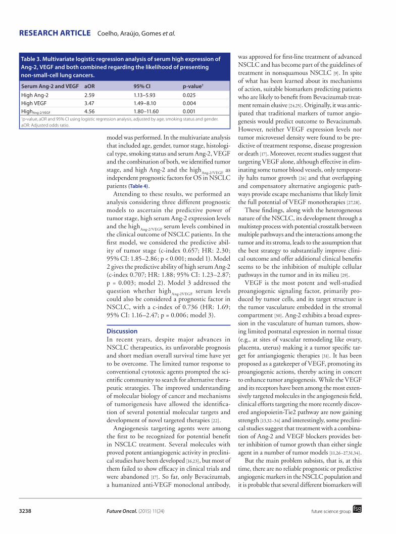
Future Oncol. (2015) 11(24)3238
RESEaRch aRticlE Coelho, Araújo, Gomes et al.
future science group
model was performed. In the multivariate analysis that included age, gender, tumor stage, histologi-cal type, smoking status and serum Ang-2, VEGF and the combination of both, we identified tumor stage, and high Ang-2 and the high
Ang-2/VEGF as
independent prognostic factors for OS in NSCLC patients (Table 4).
Attending to these results, we performed an analysis considering three different prognostic models to ascertain the predictive power of tumor stage, high serum Ang-2 expression levels and the high
Ang-2/VEGF serum levels combined in
the clinical outcome of NSCLC patients. In the first model, we considered the predictive abil-ity of tumor stage (c-index 0.657; HR: 2.30; 95% CI: 1.85–2.86; p < 0.001; model 1). Model 2 gives the predictive ability of high serum Ang-2 (c-index 0.707; HR: 1.88; 95% CI: 1.23–2.87; p = 0.003; model 2). Model 3 addressed the question whether high
Ang-2VEGF serum levels
could also be considered a prognostic factor in NSCLC, with a c-index of 0.736 (HR: 1.69; 95% CI: 1.16–2.47; p = 0.006; model 3).
DiscussionIn recent years, despite major advances in NSCLC therapeutics, its unfavorable prognosis and short median overall survival time have yet to be overcome. The limited tumor response to conventional cytotoxic agents prompted the sci-entific community to search for alternative thera-peutic strategies. The improved understanding of molecular biology of cancer and mechanisms of tumorigenesis have allowed the identifica-tion of several potential molecular targets and de velopment of novel targeted therapies [22].
Angiogenesis targeting agents were among the first to be recognized for potential benefit in NSCLC treatment. Several molecules with proved potent antiangiogenic activity in preclini-cal studies have been developed [16,23], but most of them failed to show efficacy in clinical trials and were abandoned [17]. So far, only Bevacizumab, a humanized anti-VEGF monoclonal antibody,
was approved for first-line treatment of advanced NSCLC and has become part of the guidelines of treatment in nonsquamous NSCLC [9]. In spite of what has been learned about its mechanisms of action, suitable biomarkers predicting patients who are likely to benefit from Bevacizumab treat-ment remain elusive [24,25]. Originally, it was antic-ipated that traditional markers of tumor angio-genesis would predict outcome to Bevacizumab. However, neither VEGF expression levels nor tumor microvessel density were found to be pre-dictive of treatment response, disease progression or death [17]. Moreover, recent studies suggest that targeting VEGF alone, although effective in elim-inating some tumor blood vessels, only temporar-ily halts tumor growth [26] and that overlapping and compensatory alternative angiogenic path-ways provide escape mechanisms that likely limit the full potential of VEGF monotherapies [27,28].
These findings, along with the heterogeneous nature of the NSCLC, its development through a multistep process with potential crosstalk between multiple pathways and the interactions among the tumor and its stroma, leads to the assumption that the best strategy to substantially improve clini-cal outcome and offer additional clinical benefits seems to be the inhibition of multiple cellular pathways in the tumor and in its milieu [29].
VEGF is the most potent and well-studied proangiogenic signaling factor, primarily pro-duced by tumor cells, and its target structure is the tumor vasculature embedded in the stromal compartment [30]. Ang-2 exhibits a broad expres-sion in the vasculature of human tumors, show-ing limited postnatal expression in normal tissue (e.g., at sites of vascular remodeling like ovary, placenta, uterus) making it a tumor specific tar-get for antiangiogenic therapies [31]. It has been proposed as a gatekeeper of VEGF, promoting its proangiogenic actions, thereby acting in concert to enhance tumor angiogenesis. While the VEGF and its receptors have been among the most exten-sively targeted molecules in the angiogenesis field, clinical efforts targeting the more recently discov-ered angiopoietin-Tie2 pathway are now gaining strength [13,32–34] and interestingly, some preclini-cal studies suggest that treatment with a combina-tion of Ang-2 and VEGF blockers provides bet-ter inhibition of tumor growth than either single agent in a number of tumor models [11,26–27,31,34].
But the main problem subsists, that is, at this time, there are no reliable prognostic or predictive angiogenic markers in the NSCLC population and it is probable that several different biomarkers will
Table 3. Multivariate logistic regression analysis of serum high expression of Ang-2, VEGF and both combined regarding the likelihood of presenting non-small-cell lung cancers.
Serum Ang-2 and VEGF aOR 95% CI p-value†
High Ang-2 2.59 1.13–5.93 0.025High VEGF 3.47 1.49–8.10 0.004HighAng-2/VEGF 4.56 1.80–11.60 0.001†p-value, aOR and 95% CI using logistic regression analysis, adjusted by age, smoking status and gender.aOR: Adjusted odds ratio.

3239
Figure 2. Association of serum levels of Ang-2 (A), VEGF (B) and both combined (C) with overall survival in non-small-cell lung cancers by Kaplan–Meier curves.
0.0
0.2
0.4
0.6
0.8
1.0
Cu
mu
lati
ve s
urv
ival
0 25 50 75 100 125
Overall survival (months)
High Ang-2expression
Low Ang-2 expression
Log-rank test, p = 0.001
++
++++
++
+
+
0.0
0.2
0.4
0.6
0.8
1.0
Cu
mu
lati
ve s
urv
ival
0 25 50 75 100 125Overall survival (months)
High VEGF expression
Low VEGF expression
Log-rank test, p = 0.619
+
++
+
+
0.0
0.2
0.4
0.6
0.8
1.0
Cu
mu
lati
ve s
urv
ival
0 25 50 75 100 125Overall survival (months)
High Ang-2 and VEGF expression
All cases
Log-rank test, p = 0.005
+
++++++
+++
++
+
Combined Ang-2 & VEGF serum levels: holding hands as a new integral biomarker in NSCLC RESEaRch aRticlE
future science group www.futuremedicine.com
be required to address the unique problems posed by antiangiogenic drugs [35]. Identifying biomarker candidates for prospective evaluation in rand-omized antiangiogenic trials, for efficacy, safety and cost considerations, remains an outstanding challenge in NSCLC and other cancers [36].
In the present study, we aimed to evaluate the prognostic significance of serum levels of Ang-2 and VEGF in patients with NSCLC prior to treat-ment. Moreover, due to the mutual influence that both molecules exert in tumor angiogenesis, we wanted to clarify if there is a co-dependent relation in both molecules serum levels and whether the combination of both factors could have a diagnos-tic and/or prognostic impact in NSCLC patients or in the s usceptibility for NSCLC development.
Our results demonstrate that, although isolated serum Ang-2 or VEGF levels are associated with susceptibility to NSCLC cancer, the combination of both yields a more pronounced effect in the risk for NSCLC development. That is, patients with high
Ang-2/VEGF present a higher risk for NCSLC
(OR: 4.66; p < 0.0001) than when considering Ang-2 or VEGF alone (OR: 2.63; p = 0.016; OR:
3.27; p = 0.003, respectively). Moreover, we have shown these two markers combined are valuable in discriminating between healthy subjects and NSCLC patients. Our results demonstrate that high
Ang-2/VEGF expression levels are associated with
a higher probability of NCSLC presence (AUC for high setting is 0.739; p < 0.0001), indicating the ability of these two markers in differentiating healthy individuals from patients with NSCLC.
We also evaluated the role of Ang-2 and VEGF expression levels as prognostic mark-ers in NSCLC and assessed their joint effect in OS of NSCLC patients. Although we stud-ied a Caucasian population, our results are in agreement with a previous study in an Asian population, of Park and co-workers [37], show-ing that high levels of Ang-2 are associated with a lower OS (21.0 vs 42.6 months, respectively; log rank test, p = 0.001) in a univariate analysis. Conversely of Park’s results, our study also proved that Ang-2 is a prognostic factor in a Cox multi-variate analysis. Moreover, when we consider the setting where both Ang-2 and VEGF expression levels are elevated simultaneously, a significant
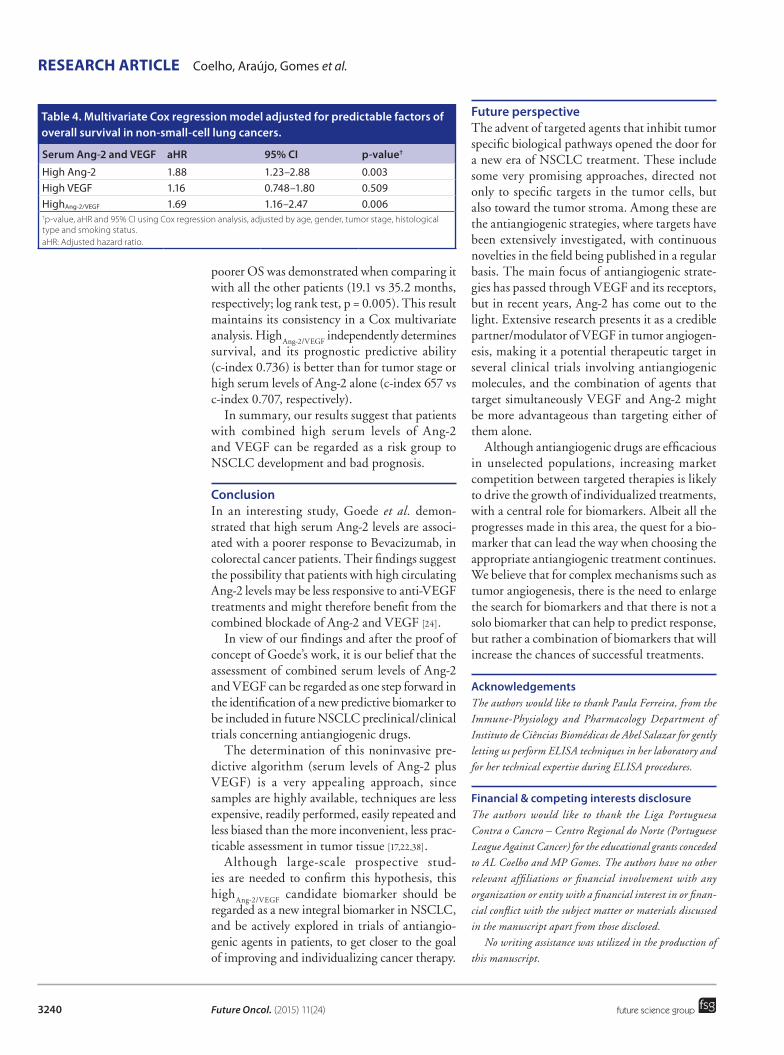
Future Oncol. (2015) 11(24)3240
RESEaRch aRticlE Coelho, Araújo, Gomes et al.
future science group
poorer OS was demonstrated when comparing it with all the other patients (19.1 vs 35.2 months, respectively; log rank test, p = 0.005). This result maintains its consistency in a Cox multivariate analysis. High
Ang-2/VEGF independently determines
survival, and its prognostic predictive ability (c-index 0.736) is better than for tumor stage or high serum levels of Ang-2 alone (c-index 657 vs c-index 0.707, respectively).
In summary, our results suggest that patients with combined high serum levels of Ang-2 and VEGF can be regarded as a risk group to NSCLC development and bad prognosis.
ConclusionIn an interesting study, Goede et al. demon-strated that high serum Ang-2 levels are associ-ated with a poorer response to Bevacizumab, in colorectal cancer patients. Their findings suggest the possibility that patients with high circulating Ang-2 levels may be less responsive to anti-VEGF treatments and might therefore benefit from the combined blockade of Ang-2 and VEGF [24].
In view of our findings and after the proof of concept of Goede’s work, it is our belief that the assessment of combined serum levels of Ang-2 and VEGF can be regarded as one step forward in the identification of a new predictive biomarker to be included in future NSCLC preclinical/clinical trials concerning antiangiogenic drugs.
The determination of this noninvasive pre-dictive algorithm (serum levels of Ang-2 plus VEGF) is a very appealing approach, since samples are highly available, techniques are less expensive, readily performed, easily repeated and less biased than the more inconvenient, less prac-ticable assessment in tumor tissue [17,22,38].
Although large-scale prospective stud-ies are needed to confirm this hypothesis, this high
Ang-2/VEGF candidate biomarker should be
regarded as a new integral biomarker in NSCLC, and be actively explored in trials of antiangio-genic agents in patients, to get closer to the goal of improving and individualizing cancer therapy.
Future perspectiveThe advent of targeted agents that inhibit tumor specific biological pathways opened the door for a new era of NSCLC treatment. These include some very promising approaches, directed not only to specific targets in the tumor cells, but also toward the tumor stroma. Among these are the antiangiogenic strategies, where targets have been extensively investigated, with continuous novelties in the field being published in a regular basis. The main focus of antiangiogenic strate-gies has passed through VEGF and its receptors, but in recent years, Ang-2 has come out to the light. Extensive research presents it as a credible partner/modulator of VEGF in tumor angiogen-esis, making it a potential therapeutic target in several clinical trials involving antiangiogenic molecules, and the combination of agents that target simultaneously VEGF and Ang-2 might be more advantageous than targeting either of them alone.
Although antiangiogenic drugs are efficacious in unselected populations, increasing market competition between targeted therapies is likely to drive the growth of individualized treatments, with a central role for biomarkers. Albeit all the progresses made in this area, the quest for a bio-marker that can lead the way when choosing the appropriate antiangiogenic treatment continues. We believe that for complex mechanisms such as tumor angiogenesis, there is the need to enlarge the search for biomarkers and that there is not a solo biomarker that can help to predict response, but rather a combination of biomarkers that will increase the chances of successful treatments.
AcknowledgementsThe authors would like to thank Paula Ferreira, from the Immune-Physiology and Pharmacology Department of Instituto de Ciências Biomédicas de Abel Salazar for gently letting us perform ELISA techniques in her laboratory and for her technical expertise during ELISA procedures.
Financial & competing interests disclosureThe authors would like to thank the Liga Portuguesa Contra o Cancro – Centro Regional do Norte (Portuguese League Against Cancer) for the educational grants conceded to AL Coelho and MP Gomes. The authors have no other relevant affiliations or financial involvement with any organization or entity with a financial interest in or finan-cial conflict with the subject matter or materials d iscussed in the manuscript apart from those disclosed.
No writing assistance was utilized in the production of this manuscript.
Table 4. Multivariate Cox regression model adjusted for predictable factors of overall survival in non-small-cell lung cancers.
Serum Ang-2 and VEGF aHR 95% CI p-value†
High Ang-2 1.88 1.23–2.88 0.003High VEGF 1.16 0.748–1.80 0.509HighAng-2/VEGF 1.69 1.16–2.47 0.006†p-value, aHR and 95% CI using Cox regression analysis, adjusted by age, gender, tumor stage, histological type and smoking status.aHR: Adjusted hazard ratio.

3241
Combined Ang-2 & VEGF serum levels: holding hands as a new integral biomarker in NSCLC RESEaRch aRticlE
future science group www.futuremedicine.com
EXEcUtiVE SUMMaRYBackground
● The purpose of this study was to evaluate the prognostic relevance of combined serum Ang-2 and VEGF levels in non-small-cell lung cancers (NSCLC) patients and based on the proposed interplay between VEGF and Ang-2, it was also examined if there is a co-dependent relation in serum levels of both molecules in patients with NSCLC and whether the combination of both had a diagnostic and/or prognostic impact in these patients or in the likelihood of developing NSCLC.
Patients & methods
● Study population:
ū The study included 145 newly diagnosed and untreated Caucasian patients with NSCLC, from the North region of Portugal, admitted to the Portuguese Institute of Oncology of Porto.
ū Inclusion criteria were histological or cytological confirmed diagnosis of NSCLC, no previous treatment, no prior oncologic disease, available clinical data and Eastern Cooperative Oncology Group performance status ≤2.
ū The control group consisted of 30 Caucasian healthy individuals from the same geographic area as the NSCLC patients, recruited from the blood donor bank of the Instituto Português de Oncologia–Porto, free of malign, inflammatory and connective tissue diseases.
● Serum Ang-2 and VEGF analysis:
ū Serum levels of VEGF and Ang-2 were measured by means of an ELISA using the Quantikine Human VEGF ELISA kit and the Human Angiopoietin-2 Quantikine ELISA kit, according to manufacturer’s instructions;
ū To evaluate its roles as diagnostic and prognostic markers in NSCLC, samples were grouped in high Ang-2 or high VEGF serum levels of expression and low Ang-2 or low VEGF levels. Moreover, patients with high serum levels of both molecules were grouped separately in a highAng-2/VEGF subgroup.
Results
● Serum Ang-2 and VEGF in NSCLC patients and healthy control individuals:
ū Serum levels of Ang-2 were significantly correlated with serum levels of VEGF.
● Serum Ang-2 and VEGF levels and NSCLC clinical outcome:
ū High Ang-2, but not high VEGF, is associated with worst disease outcome; when comparing the setting of highAng-2/VEGF phenotype against all other samples, there is a marked decrease in OS.
● Serum Ang-2 and VEGF levels and likelihood of presenting NSCLC:
ū Individuals with high Ang-2 or high VEGF serum levels have higher probability of presenting NSCLC than individuals with low serum levels; individuals with highAng-2/VEGF present an almost fivefold increased likelihood of having NSCLC, regardless of age, gender and smoking status.
Conclusion
● The assessment of combined serum levels of Ang-2 and VEGF can be regarded as one step forward in the identification of a new predictive biomarker to be included in future NSCLC preclinical/clinical trials concerning antiangiogenic drugs.
References1 Jemal A, Bray F, Center MM, Ferlay J, Ward
E, Forman D. Global cancer statistics. CA Cancer J. Clin. 61(2), 69–90 (2011).
2 Reck M, Heigener DF, Mok T, Soria JC, Rabe KF. Management of non-small-cell lung cancer: recent developments. Lancet 382(9893), 709–719 (2013).
3 Lammers PE, Horn L. Targeting angiogenesis in advanced non-small cell lung cancer. J. Natl Compr. Canc. Netw. 11(10), 1235–1247 (2013).
Ethical conduct of researchThe authors state that they have obtained appropriate institutional review board approval or have followed the principles outlined in the Declaration of Helsinki for all
human or animal experimental investigations. In addi-tion, for investigations involving human subjects, informed consent has been obtained from the participants involved.

Future Oncol. (2015) 11(24)3242
RESEaRch aRticlE Coelho, Araújo, Gomes et al.
future science group
4 Schettino C, Bareschino MA, Rossi A et al. The potential role of bevacizumab in early stages and locally advanced non-small cell lung cancer. Ther. Adv. Med. Oncol. 1(1), 5–13 (2009).
5 Grothey A, Galanis E. Targeting angiogenesis: progress with anti-vegf treatment with large molecules. Nat. Rev. Clin. Oncol. 6(9), 507–518 (2009).
6 Yancopoulos GD, Davis S, Gale NW, Rudge JS, Wiegand SJ, Holash J. Vascular-specific growth factors and blood vessel formation. Nature 407(6801), 242–248 (2000).
7 Scharpfenecker M, Fiedler U, Reiss Y, Augustin HG. The tie-2 ligand angiopoietin-2 destabilizes quiescent endothelium through an internal autocrine loop mechanism. J. Cell Sci. 118(Pt 4), 771–780 (2005).
8 Carmeliet P. Vegf as a key mediator of angiogenesis in cancer. Oncology 69(Suppl. 3), 4–10 (2005).
9 Schmid-Bindert G. Update on antiangiogenic treatment of advanced non-small cell lung cancer (NSCLC). Target Oncol. 8(1), 15–26 (2013).
10 Holash J, Maisonpierre PC, Compton D et al. Vessel cooption, regression, and growth in tumors mediated by angiopoietins and VEGF. Science 284(5422), 1994–1998 (1999).
11 Daly C, Eichten A, Castanaro C et al. Angiopoietin-2 functions as a Tie2 agonist in tumor models, where it limits the effects of VEGF inhibition. Cancer Res. 73(1), 108–118 (2013).
12 Augustin HG, Koh GY, Thurston G, Alitalo K. Control of vascular morphogenesis and homeostasis through the angiopoietin–Tie system. Nat. Rev. Mol. Cell. Biol. 10(3), 165–177 (2009).
13 Oliner J, Min H, Leal J et al. Suppression of angiogenesis and tumor growth by selective inhibition of angiopoietin-2. Cancer Cell 6(5), 507–516 (2004).
14 Cascone T, Heymach JV. Targeting the angiopoietin/Tie2 pathway: cutting tumor vessels with a double-edged sword? J. Clin. Oncol. 30(4), 441–444 (2012).
15 Lobov IB, Brooks PC, Lang RA. Angiopoietin-2 displays VEGF-dependent modulation of capillary structure and endothelial cell survival in vivo. Proc. Natl
Acad. Sci. USA 99(17), 11205–11210 (2002).
16 Wheatley-Price P, Shepherd FA. Targeting angiogenesis in the treatment of lung cancer. J. Thorac. Oncol. 3(10), 1173–1184 (2008).
17 Sessa C, Guibal A, Del Conte G, Ruegg C. Biomarkers of angiogenesis for the development of antiangiogenic therapies in oncology: tools or decorations? Nat. Clin. Pract. Oncol. 5(7), 378–391 (2008).
18 Eroglu Z, Stein CA, Pal SK. Targeting angiopoietin-2 signaling in cancer therapy. Expert Opin. Investig. Drugs 22(7), 813–825 (2013).
19 ClinicalTrials.gov. www.clinicaltrials.gov
20 Oken MM, Creech RH, Tormey DC et al. Toxicity and response criteria of the eastern cooperative oncology group. Am. J. Clin. Oncol. 5(6), 649–655 (1982).
21 Jelkmann W. Pitfalls in the measurement of circulating vascular endothelial growth factor. Clin. Chem. 47(4), 617–623 (2001).
22 Farhat FS, Tfayli A, Fakhruddin N et al. Expression, prognostic and predictive impact of VEGF and bFGF in non-small cell lung cancer. Crit. Rev. Oncol. Hematol. 84(2), 149–160 (2012).
23 Dempke WC. Targeted therapy for NSCLC – a double-edged sword? Anticancer Res. 35(5), 2503–2512 (2015).
24 Goede V, Coutelle O, Neuneier J et al. Identification of serum angiopoietin-2 as a biomarker for clinical outcome of colorectal cancer patients treated with bevacizumab-containing therapy. Br. J. Cancer 103(9), 1407–1414 (2010).
25 Murukesh N, Dive C, Jayson GC. Biomarkers of angiogenesis and their role in the development of VEGF inhibitors. Br. J. Cancer 102(1), 8–18 (2010).
26 Hashizume H, Falcon BL, Kuroda T et al. Complementary actions of inhibitors of angiopoietin-2 and VEGF on tumor angiogenesis and growth. Cancer Res. 70(6), 2213–2223 (2010).
27 Kienast Y, Klein C, Scheuer W et al. Ang-2-VEGF-a crossmab, a novel bispecific human IgG1 antibody blocking VEGF-A and ang-2 functions simultaneously, mediates potent anti-tumor, anti-angiogenic, and anti-metastatic efficacy. Clin. Cancer Res. 19(24), 6730–6740 (2013).
28 Crino L, Metro G. Therapeutic options targeting angiogenesis in nonsmall cell lung cancer. Eur. Respir. Rev. 23(131), 79–91 (2014).
29 Pallis AG, Syrigos KN. Targeting tumor neovasculature in non-small-cell lung cancer. Crit. Rev. Oncol. Hematol. 86(2), 130–142 (2013).
30 Willett CG, Boucher Y, Di Tomaso E et al. Direct evidence that the VEGF-specific antibody bevacizumab has antivascular effects in human rectal cancer. Nat. Med. 10(2), 145–147 (2004).
31 Thomas M, Kienast Y, Scheuer W et al. A novel angiopoietin-2 selective fully human antibody with potent anti-tumoral and anti-angiogenic efficacy and superior side effect profile compared with pan-angiopoietin-1/-2 inhibitors. PLoS ONE 8(2), e54923 (2013).
32 Gerald D, Chintharlapalli S, Augustin HG, Benjamin LE. Angiopoietin-2: an attractive target for improved antiangiogenic tumor therapy. Cancer Res. 73(6), 1649–1657 (2013).
33 Huang H, Lai JY, Do J et al. Specifically targeting angiopoietin-2 inhibits angiogenesis, Tie2-expressing monocyte infiltration, and tumor growth. Clin. Cancer Res. 17(5), 1001–1011 (2011).
34 Herbst RS, Hong D, Chap L et al. Safety, pharmacokinetics, and antitumor activity of AMG 386, a selective angiopoietin inhibitor, in adult patients with advanced solid tumors. J. Clin. Oncol. 27(21), 3557–3565 (2009).
35 Jubb AM, Oates AJ, Holden S, Koeppen H. Predicting benefit from anti-angiogenic agents in malignancy. Nat. Rev. Cancer 6(8), 626–635 (2006).
36 Duda DG, Ancukiewicz M, Jain RK. Biomarkers of antiangiogenic therapy: how do we move from candidate biomarkers to valid biomarkers? J. Clin. Oncol. 28(2), 183–185 (2010).
37 Park JH, Park KJ, Kim YS et al. Serum angiopoietin-2 as a clinical marker for lung cancer. Chest 132(1), 200–206 (2007).
38 Poste G, Carbone DP, Parkinson DR, Verweij J, Hewitt SM, Jessup JM. Leveling the playing field: Bringing development of biomarkers and molecular diagnostics up to the standards for drug development. Clin. Cancer Res. 18(6), 1515–1523 (2012).
Oncotarget1www.impactjournals.com/oncotarget
www.impactjournals.com/oncotarget/ Oncotarget, Advance Publications 2016
Angiogenesis in NSCLC: is vessel co-option the trunk that sustains the branches?
Ana Luísa Coelho1,2, Mónica Patrícia Gomes1,3, Raquel Jorge Catarino1,2, Christian Rolfo4,5, Agostinho Marques Lopes2,6, Rui Manuel Medeiros1,3,7 and António Manuel Araújo3,8
1 Instituto Português de Oncologia, Molecular Oncology Group, Porto, Portugal2 Faculdade de Medicina, University of Porto, Porto, Portugal3 Instituto de Ciências Biomédicas Abel Salazar, University of Porto, Porto, Portugal4 Phase I, Early Clinical Trials Unit, Antwerp University Hospital, Edegem, Belgium5 Centre of Oncological Research (CORE), Antwerp University, Edegem, Belgium6 Centro Hospitalar de S. João, Pulmonology Department, Porto, Portugal7 Liga Portuguesa Contra o Cancro (NRNorte), Research Department, Porto, Portugal8 Centro Hospitalar do Porto, Medical Oncology Department, Porto, Portugal
Correspondence to: Christian Rolfo, email: [email protected]: NSCLC, angiogenesis, anti-angiogenic strategies, vessel co-option, angiopoietin-2Received: January 25, 2016 Accepted: February 09, 2016 Published: February 29, 2016
ABSTRACTThe critical role of angiogenesis in tumor development makes its inhibition a
valuable new approach in therapy, rapidly making anti-angiogenesis a major focus in research. While the VEGF/VEGFR pathway is the main target of the approved anti-angiogenic molecules in NSCLC treatment, the results obtained are still modest, especially due to resistance mechanisms. Accumulating scientific data show that vessel co-option is an alternative mechanism to angiogenesis during tumor development in well-vascularized organs such as the lungs, where tumor cells highjack the existing vasculature to obtain its blood supply in a non-angiogenic fashion. This can explain the low/lack of response to current anti-angiogenic strategies. The same principle applies to lung metastases of other primary tumors. The exact mechanisms of vessel co-option need to be further elucidated, but it is known that the co-opted vessels regress by the action of Angiopoietin-2 (Ang-2), a vessel destabilizing cytokine expressed by the endothelial cells of the pre-existing mature vessels. In the absence of VEGF, vessel regression leads to tumor cell loss and hypoxia, with a subsequent switch to a neoangiogenic phenotype by the remaining tumor cells. Unravelling the vessel co-option mechanisms and involved players may be fruitful for numerous reasons, and the particularities of this form of vascularization should be carefully considered when planning anti-angiogenic interventions or designing clinical trials for this purpose. In view of the current knowledge, rationale for therapeutic approaches of dual inhibition of Ang-2 and VEGF are swiftly gaining strength and may serve as a launchpad to more successful NSCLC anti-vascular treatments.
INTRODUCTION
Cancer is a major health issue, constituting the second leading cause of death worldwide and expected to surpass heart diseases as the leading cause of death in the next few years [1, 2]. In 2013, the incidence of cancer cases worldwide was 14.9 million, with 8.2 million cancer-
related deaths. Lung cancer was the most common incident form of cancer, with an estimated 1.8 million new cases having deaths that exceeded those from any other type of malignancy worldwide, accounting for nearly one in five deaths (1.6 msillion deaths in total) [2]. Most lung cancers (~85%) are non-small cell lung cancers (NSCLC) which are divided according to two major histologic subtypes:
Oncotarget2www.impactjournals.com/oncotarget
the non-squamous carcinomas (mainly adenocarcinomas) and the squamous-cell carcinomas [3]. The parenchyma and the stroma are the two almost-indistinguishable compartments that compose the NSCLC and build up the tumor microenvironment [4]. The stromal cells contribute to the development and expression of certain cancer hallmark capabilities, defined by Hanahan and Weinberg in 2011 [5]. Among these, angiogenesis assumes major importance, since rate-limiting steps in tumor progressions include gaining access to the host vascular system and the generation of a tumor blood supply to obtain oxygen and nutrients, growth factors and hormones [6].
ANGIOGENESIS AND CANCER
While the identification of massive vascularization in tumors dates back to 1863 [7] and the importance of tumor angiogenesis has been recognised since 1908 [8], it was only through the work of Folkman in the early 1970s that the scientific community acknowledge angiogenesis as a potential target to inhibit cancer progression [9-12]. The therapeutic potential of anti-angiogenic strategies boosted this field of research, placing angiogenesis as one of the major hubs of current cancer research.
It is now widely accepted that most tumors and metastases originate as small avascular structures which must induce the development of new blood vessels from pre-existing ones in order to grow beyond a minimum size of 2-3 mm3 [6, 13]. To achieve this, tumors undergo an angiogenic switch, disrupting the equilibrium between pro and anti-angiogenic regulators and favouring pro-angiogenic mechanisms. Signalling molecules induce quiescent endothelial cells to continuously sprout from existing blood vessels, thereby forming new vessels that help to sustain expanding neoplastic growth [6, 14, 15], according to the conventional model of angiogenesis known as angiogenic sprout [16].
Decades of research investigating the molecular basis of angiogenesis led to the discovery of a number of angiogenic molecules that promote tumor angiogenesis [15]. Of all the identified angiogenic pathways, the most critical appears to be the one involving the VEGF family and their receptors (VEGFR1-2-3) [17-19], although a number of other important molecules and their receptors have also proven to work in combination with VEGF/VEGFR signalling in tumor angiogenesis [19]. These include the fibroblast growth factor receptors (FGFRs) family and their ligands, particularly FGF1 and FGF2, that induce the proliferation and migration of endothelial cells [20]; as well as the platelet-derived growth factor receptors (PDGFRs) and their ligands (PDGFs) that, either alone or in combination with FGF and VEGF, are associated with tumor vascularization in malignant disease, including NSCLC [21, 22] and the Ang-Tie-2 system [19, 22]. Ever since the identification of VEGF
as the first endothelium-acting specific cytokine in 1983 [13, 23, 24], its overexpression has been found in several human tumors, including NSCLC [25-29]. More recently, scientists are gaining a better understanding of the many functions of this molecule in the tumor angiogenic process [29]: it triggers multiple signalling networks that enhance endothelial cell proliferation and survival, increases migration and invasion of endothelial cells, increases vascular permeability of existing vessels, and enhances chemotaxis and mobilization of bone marrow derived endothelial progenitor cells (EPCs) into the peripheral circulation [30, 31].
The growing acknowledgment of VEGF’s key role in tumor angiogenesis has made it an attractive target for therapeutic intervention in cancer. The VEGF pathway is a promising avenue in research that aims to uncover more effective, targeted anti-angiogenic strategies [23, 32]. The extensive investigation in this field has led to the study of several anti-angiogenic agents, including monoclonal antibodies to block VEGF and its receptor VEGFR2 and VEGFR tyrosine kinase inhibitors (TKIs) [33].
ANTI-ANGIOGENIC THERAPY AND LUNG CANCER
From the multitude of potential therapeutic options that target angiogenesis in NSCLC, [34] (Table 1), there are currently three anti-angiogenic compounds approved by EMA for the treatment of NSCLC. Bevacizumab, an anti-VEGF monoclonal antibody that blocks the binding of VEGF to its high-affinity receptors, was the first angiogenic inhibitor to complete clinical development, showing clinical benefit in patients with metastatic colorectal cancer when combined with chemotherapy [31, 33]. It was approved in 2006 for the treatment of advanced non-squamous NSCLC in the first line setting in combination with chemotherapy [29]. In 2014, ramucirumab, a fully humanized monoclonal antibody that targets angiogenesis by specifically binding to VEGFR-2 with higher affinity than its natural ligand VEGF [35], was approved for the treatment of patients with metastatic NSCLC in second line setting, in combination with docetaxel [36]. In the same year, nintedanib, an oral medication that can simultaneously inhibit triple angiokinase, VEGFR, platelet-derived growth factor receptors (PDGFR), and fibroblast growth factor receptors (FGFR) signalling pathways, was approved to be used in combination with docetaxel in patients with locally advanced, metastatic, or locally recurrent NSCLC adenocarcinoma, after first-line chemotherapy [37]. There are also other potential agents that are under clinical evaluation, whether that be in the clinical trial stage or currently waiting for approval for treatment of metastatic or recurrent NSCLC [38, 39].
In spite of the impressive clinical efficacy of

Oncotarget3www.impactjournals.com/oncotarget
bevacizumab, ramucirumab, and nintedanib in various cancer treatment settings, the results were relatively modest and limited [40]. In addition, the clinical use of VEGF/VEGFR blockers as anti- angiogenic therapy for patients with advanced NSCLC has been more challenging than anticipated by the preclinical experiments in which long-term benefit of VEGF/VEGFR inhibition was achieved [41]. Anti-angiogenic agents are usually given to all patients for the approved indications; in a high fraction of these patients, however, the tumor is intrinsically refractory to the anti-angiogenic therapy and the disease progresses ceaselessly [42]. Moreover, when there is no intrinsic resistance, acquired resistance to therapy can rapidly occur and limit the efficacy of the anti-angiogenic treatments [41, 43], and the clinical benefit of prolonging cancer patients survival with advanced disease becomes limited, often in the order of weeks or months [16, 44].
Tumor resistance to the anti-angiogenic therapies (whether intrinsic or acquired), represents a significant problem faced in routine clinical practice. The mechanisms underlying the response to these therapies are far from being clearly understood, further fuelling this active field of research [43]. Preclinical investigations have shed some light on the subject, and although different authors propose escape ways from angiogenic inhibitors that are somewhat distinct, some key features appear to be consensual among most of them; these features are likely to be involved in primary and acquired resistance and deserve consideration [16, 18, 41-43, 45]. One of such features is invasive (or metastatic) co-option of normal quiescent vessels without requisite of angiogenesis.
VESSEL CO-OPTION AND LUNG CANCER GROWTH
It is widely accepted that tumor progression is heavily dependent on angiogenesis. Much less understood, is the concept that angiogenesis is necessary for a tumor to become larger than a few millimetres and become clinically detectable, as some research has shown that angiogenesis is not always a pre-requisite for tumor growth [46]. Hence, one possibility for anti-angiogenic therapy resistance is that some primary and metastatic tumors are non-angiogenic, meaning that these tumors do not need angiogenic sprout to obtain an efficient blood supply [47]. Rather, the tumors use alternative vascularization mechanisms. For example, in vessel-dense tissues, the most likely route is hijacking the pre-existing normal blood vessels [42, 44, 48], and more aggressive tumors can undergo vasculogenic mimicry, a process by which tumor cells dedifferentiate to an endothelial phenotype forming structures that provide tumour cells with a secondary circulation system independently of angiogenesis [49].
When tumors arise in well-vascularized organs, their growth will rely on the invasion of host tissue. Enhancement of invasion and metastasis facilitates access to normal tissue vasculature, and cancer cells stay in close contact with the surface of blood vessels [39, 50, 51]. This allows tumor cells to grow and migrate along quiescent normal vessels and take their oxygen and essential nutrients without obligate neovascularization, in
Table 1: Angiogenesis inhibitors in non-small cell lung cancer (NSCLC)ApprovedDrug Target Indication
Bevacizumab VEGF First-line treatment of nonsquamous NSCLC with CT
Nintedanib VEGFR 2, FGFR 1-3, PDGFRα and β TKI
Second-line treatment of adenocarcinoma NSCLC with CT
Ramucirumab VEGFR-2 Second-line treatment of NSCLC with CT
On clinical trials or not approvedDrug TargetVandetanib VEGFRs, EGFR, and RET Sunitinib VEGFRs, PDGFRs, KIT, FLT3, CSF-1R, and RETAflibercept VEGFSorafenib VEGFR, PDGFRs, FGFR, KIT, and RAF Motesanib VEGFRs, PDGFRs, and KIT Pazopanib VEGFRs, PDGFRs, FGFR, and KIT Cediranib VEGFRs Cabozantinib VEGFR, RET, and MET Axitinib VEGFRs, PDGFRs, and KIT
CT - chemotherapy; VEGF - vascular endothelial growth factor; VEGFR - vascular endothelial growth factor receptor; FGFR - fibroblast growth factor receptor; PDGFR - platelet-derived growth factor receptor; KIT - stem cell factor receptor; FLT3 - Fms-like tyrosine kinase-3; CSF-1R - colony stimulating factor receptor; RET - glial cell-line derived neurotrophic factor receptor; MET - met proto-oncogene;

Oncotarget4www.impactjournals.com/oncotarget
a process known as vessel (or vascular) co-option [42, 43, 49]. These non-angiogenic tumors are a separate group of fast-growing malignancies with little apoptosis and very efficient mitochondrial metabolism [52]. This seems to be the case of tumors arising in the lungs, liver, and brain, areas where this form of vascularization appears to assume a major role [31, 50, 51, 53, 54]. This is also true for tumor metastasis that occurs through lymph and blood vessels and outgrow mostly in these vessel-dense organs [55-58].
In recent years, research related to angiogenesis has been massive; but on the contrary, there is a scarcity of research focusing on tumors that escape pathways of classical angiogenesis and use vessel co-option as an alternative blood supply for tumor growth. This has led to a dearth in information regarding the mechanisms and players involved in that process.
The first insights into the relationship between vessel co-option and lung cancer were made by Pezzella and co-workers, who described NSCLC that grew without morphological evidence of neoangiogenesis but with signs of normal tissue vessel exploitation [59]. They characterized these tumors as having an alveolar pattern, with tumor cell nests filling the alveolar spaces without destruction of the lung parenchyma. The only vessels evident in these tumors appeared to belong to the trapped alveolar septa [59]. Moreover, patients with alveolar pattern tumors presented a worse survival rate than their angiogenic counterparts. Later, when investigating the possible role of microvessel count in NSCLC as a potential marker of disease prognosis, Offersen and colleagues [60] identified the same special vascular pattern in 17 out of 35 NSCLC samples, thus confirming the description of Pezzella’s group. Their observations led them to the hypothesis that these alveolar tumors are nonangiogenic and invasive and exploited the pre-existing
vascular beds. They also noted that some tumors exhibited only the alveolar pattern while other tumors presented a mixed alveolar pattern consisting of both alveolar and angiogenic features [60]. There was no correlation, however, between angiogenic or vessel co-option status and disease aggressiveness.
Taking into account the NSCLC growth patterns, Nia Sardari et al. suggested a modification of Pezzella’s classification according to morphological features, based on the biological properties of the tumor-lung interface, which is the region where the tumor expands and the tumor-stroma interactions are more active and homogeneous [61]. According to them, NSCLCs can be classified as having a destructive growth pattern (angiogenic growth pattern), papillary growth pattern (with preservation of the alveolar structure of the lung parenchyma at the interface with co-option of alveolar blood vessels with formation of stromal stalks and subsequent angiogenesis), and alveolar growth pattern (preservation of the alveolar structure of lung parenchyma with co-option of septal blood vessels and without evidence of new stroma formation at the interface). Moreover, they suggested that, in NSCLC, a low degree of ongoing angiogenesis is predictive of poorer prognosis [61, 62].
The hypothesis of co-option by lung metastases, which are often the main cause of death in many solid malignancies, was also proposed by Pezzella’s group back in the 1990’s. They observed that, regardless of the angiogenic status of the primary breast carcinomas, they could relapse as nonangiogenic tumors in the lungs. This was also true for lung metastases of human renal and colorectal carcinomas [56, 63, 64]. In a very recent study, Szabo and co-workers used cell lines from six different solid tumors, and showed that lung metastases
Figure 1: Vessel co-option and Ang-2 regulation in cancer development in vessel dense tissues. A. In well vascularized organs, such as the lung, tumor cells grow and migrate along quiescent normal vessels (vessel co-option). B. Over time, tumor cells induce extreme changes in the co-opted vessels and ECs start to express Ang-2, leading to vascular disruption and vessel regression. C. Regression of the co-opted vessel associated with regression of the ECs generates a hypoxic core in the tumor centre, with massive tumor cell loss. This triggers the angiogenic switch, with the remaining tumor cells expressing high amounts of VEGF. D. VEGF expression induces a robust angiogenic response that ultimately rescues the tumor and allows its growth and progression.
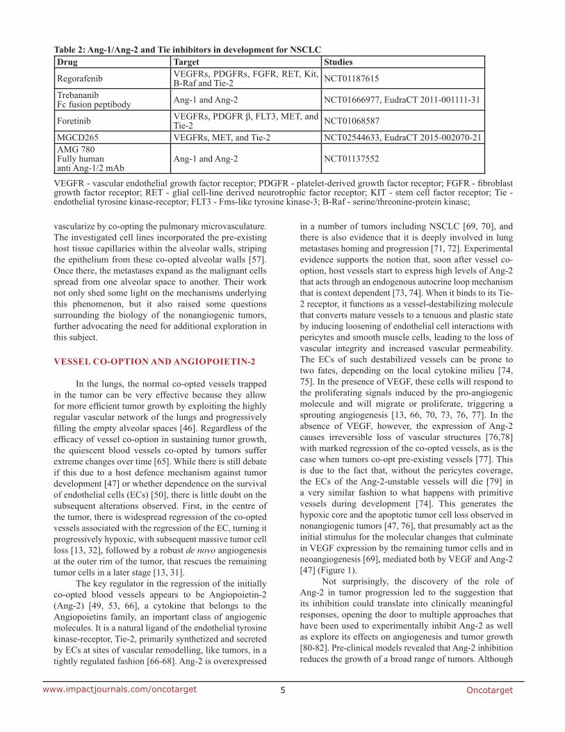
Oncotarget5www.impactjournals.com/oncotarget
vascularize by co-opting the pulmonary microvasculature. The investigated cell lines incorporated the pre-existing host tissue capillaries within the alveolar walls, striping the epithelium from these co-opted alveolar walls [57]. Once there, the metastases expand as the malignant cells spread from one alveolar space to another. Their work not only shed some light on the mechanisms underlying this phenomenon, but it also raised some questions surrounding the biology of the nonangiogenic tumors, further advocating the need for additional exploration in this subject.
VESSEL CO-OPTION AND ANGIOPOIETIN-2
In the lungs, the normal co-opted vessels trapped in the tumor can be very effective because they allow for more efficient tumor growth by exploiting the highly regular vascular network of the lungs and progressively filling the empty alveolar spaces [46]. Regardless of the efficacy of vessel co-option in sustaining tumor growth, the quiescent blood vessels co-opted by tumors suffer extreme changes over time [65]. While there is still debate if this due to a host defence mechanism against tumor development [47] or whether dependence on the survival of endothelial cells (ECs) [50], there is little doubt on the subsequent alterations observed. First, in the centre of the tumor, there is widespread regression of the co-opted vessels associated with the regression of the EC, turning it progressively hypoxic, with subsequent massive tumor cell loss [13, 32], followed by a robust de novo angiogenesis at the outer rim of the tumor, that rescues the remaining tumor cells in a later stage [13, 31].
The key regulator in the regression of the initially co-opted blood vessels appears to be Angiopoietin-2 (Ang-2) [49, 53, 66], a cytokine that belongs to the Angiopoietins family, an important class of angiogenic molecules. It is a natural ligand of the endothelial tyrosine kinase-receptor, Tie-2, primarily synthetized and secreted by ECs at sites of vascular remodelling, like tumors, in a tightly regulated fashion [66-68]. Ang-2 is overexpressed
in a number of tumors including NSCLC [69, 70], and there is also evidence that it is deeply involved in lung metastases homing and progression [71, 72]. Experimental evidence supports the notion that, soon after vessel co-option, host vessels start to express high levels of Ang-2 that acts through an endogenous autocrine loop mechanism that is context dependent [73, 74]. When it binds to its Tie-2 receptor, it functions as a vessel-destabilizing molecule that converts mature vessels to a tenuous and plastic state by inducing loosening of endothelial cell interactions with pericytes and smooth muscle cells, leading to the loss of vascular integrity and increased vascular permeability. The ECs of such destabilized vessels can be prone to two fates, depending on the local cytokine milieu [74, 75]. In the presence of VEGF, these cells will respond to the proliferating signals induced by the pro-angiogenic molecule and will migrate or proliferate, triggering a sprouting angiogenesis [13, 66, 70, 73, 76, 77]. In the absence of VEGF, however, the expression of Ang-2 causes irreversible loss of vascular structures [76,78] with marked regression of the co-opted vessels, as is the case when tumors co-opt pre-existing vessels [77]. This is due to the fact that, without the pericytes coverage, the ECs of the Ang-2-unstable vessels will die [79] in a very similar fashion to what happens with primitive vessels during development [74]. This generates the hypoxic core and the apoptotic tumor cell loss observed in nonangiogenic tumors [47, 76], that presumably act as the initial stimulus for the molecular changes that culminate in VEGF expression by the remaining tumor cells and in neoangiogenesis [69], mediated both by VEGF and Ang-2 [47] (Figure 1).
Not surprisingly, the discovery of the role of Ang-2 in tumor progression led to the suggestion that its inhibition could translate into clinically meaningful responses, opening the door to multiple approaches that have been used to experimentally inhibit Ang-2 as well as explore its effects on angiogenesis and tumor growth [80-82]. Pre-clinical models revealed that Ang-2 inhibition reduces the growth of a broad range of tumors. Although
Table 2: Ang-1/Ang-2 and Tie inhibitors in development for NSCLC Drug Target Studies
Regorafenib VEGFRs, PDGFRs, FGFR, RET, Kit, B-Raf and Tie-2 NCT01187615
TrebananibFc fusion peptibody Ang-1 and Ang-2 NCT01666977, EudraCT 2011-001111-31
Foretinib VEGFRs, PDGFR β, FLT3, MET, and Tie-2 NCT01068587
MGCD265 VEGFRs, MET, and Tie-2 NCT02544633, EudraCT 2015-002070-21AMG 780Fully humananti Ang-1/2 mAb
Ang-1 and Ang-2 NCT01137552
VEGFR - vascular endothelial growth factor receptor; PDGFR - platelet-derived growth factor receptor; FGFR - fibroblast growth factor receptor; RET - glial cell-line derived neurotrophic factor receptor; KIT - stem cell factor receptor; Tie - endothelial tyrosine kinase-receptor; FLT3 - Fms-like tyrosine kinase-3; B-Raf - serine/threonine-protein kinase;

Oncotarget6www.impactjournals.com/oncotarget
some of the results were modest, some revealed to be very promising and there is now a robust pipeline of drugs targeting the Ang/Tie-2 system in different clinical trials phases (Table 2) [67, 78, 83]. Furthermore, with Ang-2 being required to render endothelium responsive to VEGF and with both molecules contributing to tumor angiogenesis and metastases [84, 85], there seems to be a more encouraging response to the straightforward question of whether co-targeting of both ligands in a bispecific manner would improve the outcomes of current anti-angiogenic therapies [80, 83, 86-88].
VESSEL CO-OPTION AND CLINICAL IMPLICATIONS
The ability to identify tumors that make vascular co-option their primary source of blood supply does not envisage an easy task, hence why few strategies have been used to achieve this goal [48]. Research in the field has been scarce, especially when compared to the angiogenic field that has largely overshadowed alternative blood sources for tumor development. Moreover, much of the research has been performed in cell lines or murine models and only a few in human tissues [48]. While the findings are limited so far, what has been discovered highly advocates for unravelling the vessel co-option mechanisms and involved players. The precise identification of tumors that preferentially use this route to support growth and the factors driving them to switch from this to an angiogenic pattern may be crucial to delineate future cancer treatments for two main reasons. The first is that vascular co-option may represent a clever strategy by which tumors partly evade and resist conventional anti-angiogenic treatments [89]. Even if a treatment like bevacizumab is effective against one angiogenic factor such as VEGF, the therapy can still fail if this factor is not important for the endothelium in that given tumor, as appears to be the case in tumors that co-opt pre-existing vessels in NSCLC [45]. In these cases, vessel co-option may serve as a pathologic biomarker for selecting potentially nonresponsive patients [43]. There is also evidence that in some nonangiogenic tumors, cancer cells adapt by migrating more aggressively into normal tissue [42]; and when anti-angiogenic treatments are used indiscriminately, they may contribute to the selection of clones of nonangiogenic cells that will progress with a more aggressive behaviour [89, 90]. These features should be carefully considered when planning anti-angiogenic therapeutic interventions, suggesting the need for tailor-made treatments against such tumors.
Secondly, anti-angiogenic compounds do not affect incorporated pre-existent vasculature or matured tumor vasculature, making targeting existing vessels on which the tumor growth relies, an attractive approach to accomplish tumor regression [91]. This is also of primordial importance in cases of metastases that establish in well-vascularized organs, since vessel co-option may
constitute their primary feeding option [57]. Moreover, it can be speculated that in earlier stages of the tumor, the interval that mediates Ang-2 overexpression, co-opted vessels regression, and de novo angiogenesis seems to be the perfect therapeutic window for intervention using a dual-pronged approach with Ang-2 and VEGF blockers rather than in more advanced stages of the disease. This issue should be addressed by investigators developing pre-clinical/clinical trials of drugs that target angiogenesis or envisage tumor arrest by anti-angiogenic strategies.
CONCLUSIONS
Anti-angiogenic strategies focusing on VEGF/VEGFR in combination with chemotherapy marked a milestone in the field of cancer treatment, including NSCLC. However, a relevant number of patients are unresponsive or refractory to anti-angiogenic treatments. Some tumors obviate the need to generate angiogenesis by co-opting host mature vessels and growing along them, using them as blood sources. Vessel co-option is a mechanism that may help explain the limited success of anti-angiogenic therapy in these patients in an adjuvant setting.
Thus far, the only growth factors proven to be associated with vessel co-option are VEGF and Ang-2. This lack of information is likely due to the limited number of studies examining this subject. Ang-2 seems to have a particularly critical role in the process, but is also an extremely laborious study topic due to the complexity of its functions and regulation, which are both highly cell context dependent.
Tumors that grow in non-angiogenic fashions through exploitation of pre-existing vessels are non-responsive to anti-angiogenic molecules and raise a number of concerns in terms of treatment. First, little is known about the modifications a neoplastic cell must go through in order to co-opt a blood vessel, which is a huge obstacle for strategies that aim to interfere with this step in tumor progression. Second, once the tumor is committed to vascular co-option pathway, an effective way of blocking tumor progression would be to target existing tumor vasculature; this would require the availability of tumor-vessel specific targeting agents, however, and the few candidates that have been identified so far have failed to prove their clinical efficacy.
All of these concerns reinforce the need for better understanding of the mechanisms and molecular players underlying vessel co-option during tumor development within the proper biologic context. This would not only explore more assertive cancer treatments and help with the identification of tumors where vessel co-option is the growth support (instead of angiogenesis), but could also help identify patients who may be nonresponsive to current anti-angiogenic treatments. Additionally, it could open doors to novel areas of NSCLC research at both the
Oncotarget7www.impactjournals.com/oncotarget
molecular and microanatomical level.
ACKNOWLEDGMENTS
The authors would like to thank the Liga Portuguesa Contra o Cancro - Núcleo Regional do Norte, for the Educational Grant conceded to Ana Luísa Coelho.
The authors would like to thank Dr. Rachel Zsido for her valuable contribution in the critical review of redaction.
CONFLICTS OF INTEREST
The authors declare they have no potential conflict of interests with this research.
REFERENCES
1. Siegel RL, Miller KD, Jemal A. Cancer statistics, 2015. CA Cancer J Clin. 2015; 65: 5-29.
2. Fitzmaurice C, Dicker D, Pain A, Hamavid H, Moradi-Lakeh M, MacIntyre MF, Allen C, Hansen G, Woodbrook R, Wolfe C, Hamadeh RR, Moore A, Werdecker A, et al. The Global Burden of Cancer 2013. JAMA Oncol. 2015; 1: 505-27.
3. Reck M, Heigener DF, Mok T, Soria JC, Rabe KF. Management of non-small-cell lung cancer: recent developments. Lancet. 2013; 382: 709-19.
4. Schiavoni G, Gabriele L, Mattei F. The tumor microenvironment: a pitch for multiple players. Front Oncol. 2013; 3: 90.
5. Hanahan D, Weinberg RA. Hallmarks of cancer: the next generation. Cell. 2011; 144: 646-74.
6. Bergers G, Benjamin LE. Tumorigenesis and the angiogenic switch. Nat Rev Cancer. 2003; 3: 401-10.
7. Fan F, Schimming A, Jaeger D, Podar K. Targeting the tumor microenvironment: focus on angiogenesis. J Oncol. 2012; 2012: 281261.
8. Goldmann E. The Growth of Malignant Disease in Man and the Lower Animals, with special reference to the Vascular System. Proc R Soc Med. 1908; 1: 1-13.
9. Folkman J. Tumor angiogenesis: therapeutic implications. N Engl J Med. 1971; 285: 1182-6.
10. Folkman J, Merler E, Abernathy C, Williams G. Isolation of a tumor factor responsible for angiogenesis. J Exp Med. 1971; 133: 275-88.
11. Holleb AI, Folkman J. Tumor angiogenesis. CA Cancer J Clin. 1972; 22: 226-9.
12. Folkman J. Anti-angiogenesis: new concept for therapy of solid tumors. Ann Surg. 1972; 175: 409-16.
13. Holash J, Wiegand SJ, Yancopoulos GD. New model of tumor angiogenesis: dynamic balance between vessel regression and growth mediated by angiopoietins and VEGF. Oncogene. 1999; 18: 5356-62.
14. Hanahan D, Folkman J. Patterns and emerging mechanisms of the angiogenic switch during tumorigenesis. Cell. 1996; 86: 353-64.
15. Baeriswyl V, Christofori G. The angiogenic switch in carcinogenesis. Semin Cancer Biol. 2009; 19: 329-37.
16. Potente M, Gerhardt H, Carmeliet P. Basic and therapeutic aspects of angiogenesis. Cell. 2011; 146: 873-87.
17. Jain RK. Normalization of tumor vasculature: an emerging concept in antiangiogenic therapy. Science. 2005; 307: 58-62.
18. Dvorak HF. Vascular permeability factor/vascular endothelial growth factor: a critical cytokine in tumor angiogenesis and a potential target for diagnosis and therapy. J Clin Oncol. 2002; 20: 4368-80.
19. Zhao Y, Adjei AA. Targeting Angiogenesis in Cancer Therapy: Moving Beyond Vascular Endothelial Growth Factor. Oncologist. 2015; 20: 660-73.
20. Presta M, Dell’Era P, Mitola S, Moroni E, Ronca R, Rusnati M. Fibroblast growth factor/fibroblast growth factor receptor system in angiogenesis. Cytokine Growth Factor Rev. 2005; 16: 159-78.
21. Cao Y. Multifarious functions of PDGFs and PDGFRs in tumor growth and metastasis. Trends Mol Med. 2013; 19: 460-73.
22. Ferrara N, Kerbel RS. Angiogenesis as a therapeutic target. Nature. 2005; 438: 967-74.
23. Ferrara N. VEGF and the quest for tumour angiogenesis factors. Nat Rev Cancer. 2002; 2: 795-803.
24. Senger DR, Galli SJ, Dvorak AM, Perruzzi CA, Harvey VS, Dvorak HF. Tumor cells secrete a vascular permeability factor that promotes accumulation of ascites fluid. Science. 1983; 219: 983-5.
25. Yuan A, Yu CJ, Chen WJ, Lin FY, Kuo SH, Luh KT, Yang PC. Correlation of total VEGF mRNA and protein expression with histologic type, tumor angiogenesis, patient survival and timing of relapse in non-small-cell lung cancer. Int J Cancer. 2000; 89: 475-83.
26. Imoto H, Osaki T, Taga S, Ohgami A, Ichiyoshi Y, Yasumoto K. Vascular endothelial growth factor expression in non-small-cell lung cancer: prognostic significance in squamous cell carcinoma. J Thorac Cardiovasc Surg. 1998; 115: 1007-14.
27. Koukourakis MI, Giatromanolaki A, Thorpe PE, Brekken RA, Sivridis E, Kakolyris S, Georgoulias V, Gatter KC, Harris AL. Vascular endothelial growth factor/KDR activated microvessel density versus CD31 standard microvessel density in non-small cell lung cancer. Cancer Res. 2000; 60: 3088-95.
28. Stefanou D, Batistatou A, Arkoumani E, Ntzani E, Agnantis NJ. Expression of vascular endothelial growth factor (VEGF) and association with microvessel density in small-cell and non-small-cell lung carcinomas. Histol Histopathol. 2004; 19: 37-42.
29. Villaruz LC, Socinski MA. The role of anti-angiogenesis
Oncotarget8www.impactjournals.com/oncotarget
in non-small-cell lung cancer: an update. Curr Oncol Rep. 2015; 17: 26.
30. Hicklin DJ, Ellis LM. Role of the vascular endothelial growth factor pathway in tumor growth and angiogenesis. J Clin Oncol. 2005; 23: 1011-27.
31. Ellis LM, Hicklin DJ. VEGF-targeted therapy: mechanisms of anti-tumour activity. Nat Rev Cancer. 2008; 8: 579-91.
32. Korn C, Augustin HG. Mechanisms of Vessel Pruning and Regression. Dev Cell. 2015; 34: 5-17.
33. Ferrara N, Hillan KJ, Gerber HP, Novotny W. Discovery and development of bevacizumab, an anti-VEGF antibody for treating cancer. Nat Rev Drug Discov. 2004; 3: 391-400.
34. Crino L, Metro G. Therapeutic options targeting angiogenesis in nonsmall cell lung cancer. Eur Respir Rev. 2014; 23: 79-91.
35. Reinmuth N, Heigener D, Reck M. Novel angiogenesis inhibitors in nonsmall cell lung cancer. Curr Opin Oncol. 2015; 27: 79-86.
36. Garon EB, Ciuleanu TE, Arrieta O, Prabhash K, Syrigos KN, Goksel T, Park K, Gorbunova V, Kowalyszyn RD, Pikiel J, Czyzewicz G, Orlov SV, Lewanski CR, et al. Ramucirumab plus docetaxel versus placebo plus docetaxel for second-line treatment of stage IV non-small-cell lung cancer after disease progression on platinum-based therapy (REVEL): a multicentre, double-blind, randomised phase 3 trial. Lancet. 2014; 384: 665-73.
37. Reck M, Kaiser R, Mellemgaard A, Douillard JY, Orlov S, Krzakowski M, von Pawel J, Gottfried M, Bondarenko I, Liao M, Gann CN, Barrueco J, Gaschler-Markefski B, et al. Docetaxel plus nintedanib versus docetaxel plus placebo in patients with previously treated non-small-cell lung cancer (LUME-Lung 1): a phase 3, double-blind, randomised controlled trial. Lancet Oncol. 2014; 15: 143-55.
38. Aggarwal C, Somaiah N, Simon G. Antiangiogenic agents in the management of non-small cell lung cancer: where do we stand now and where are we headed? Cancer Biol Ther. 2012; 13: 247-63.
39. Pallis AG, Syrigos KN. Targeting tumor neovasculature in non-small-cell lung cancer. Crit Rev Oncol Hematol. 2013; 86: 130-42.
40. Jain RK. Normalizing tumor microenvironment to treat cancer: bench to bedside to biomarkers. J Clin Oncol. 2013; 31: 2205-18.
41. Carmeliet P, Jain RK. Molecular mechanisms and clinical applications of angiogenesis. Nature. 2011; 473: 298-307.
42. Bergers G, Hanahan D. Modes of resistance to anti-angiogenic therapy. Nat Rev Cancer. 2008; 8: 592-603.
43. Huang Y, Carbone DP. Mechanisms of and strategies for overcoming resistance to anti-vascular endothelial growth factor therapy in non-small cell lung cancer. Biochim Biophys Acta. 2015; 1855: 193-201.
44. Bertolini F, Marighetti P, Martin-Padura I, Mancuso P, Hu-Lowe DD, Shaked Y, D’Onofrio A. Anti-VEGF and beyond: shaping a new generation of anti-angiogenic
therapies for cancer. Drug Discov Today. 2011; 16: 1052-60.
45. Eikesdal HP, Kalluri R. Drug resistance associated with antiangiogenesis therapy. Semin Cancer Biol. 2009; 19: 310-7.
46. Passalidou E, Trivella M, Singh N, Ferguson M, Hu J, Cesario A, Granone P, Nicholson AG, Goldstraw P, Ratcliffe C, Tetlow M, Leigh I, Harris AL, et al. Vascular phenotype in angiogenic and non-angiogenic lung non-small cell carcinomas. Br J Cancer. 2002; 86: 244-9.
47. Ribatti D, Vacca A, Dammacco F. New non-angiogenesis dependent pathways for tumour growth. Eur J Cancer. 2003; 39: 1835-41.
48. Donnem T, Hu J, Ferguson M, Adighibe O, Snell C, Harris AL, Gatter KC, Pezzella F. Vessel co-option in primary human tumors and metastases: an obstacle to effective anti-angiogenic treatment? Cancer Med. 2013; 2: 427-36.
49. Hillen F, Griffioen AW. Tumour vascularization: sprouting angiogenesis and beyond. Cancer Metastasis Rev. 2007; 26: 489-502.
50. Dome B, Hendrix MJ, Paku S, Tovari J, Timar J. Alternative vascularization mechanisms in cancer: Pathology and therapeutic implications. Am J Pathol. 2007; 170: 1-15.
51. Carbonell WS, Ansorge O, Sibson N, Muschel R. The vascular basement membrane as “soil” in brain metastasis. PLoS One. 2009; 4: e5857.
52. Hu J, Bianchi F, Ferguson M, Cesario A, Margaritora S, Granone P, Goldstraw P, Tetlow M, Ratcliffe C, Nicholson AG, Harris A, Gatter K, Pezzella F. Gene expression signature for angiogenic and nonangiogenic non-small-cell lung cancer. Oncogene. 2005; 24: 1212-9.
53. Dvorak HF. Tumor Stroma, Tumor Blood Vessels, and Antiangiogenesis Therapy. Cancer J. 2015; 21: 237-43.
54. Adighibe O, Micklem K, Campo L, Ferguson M, Harris A, Pozos R, Gatter K, Pezzella F. Is nonangiogenesis a novel pathway for cancer progression? A study using 3-dimensional tumour reconstructions. Br J Cancer. 2006; 94: 1176-9.
55. Leenders WP, Kusters B, Verrijp K, Maass C, Wesseling P, Heerschap A, Ruiter D, Ryan A, de Waal R. Antiangiogenic therapy of cerebral melanoma metastases results in sustained tumor progression via vessel co-option. Clin Cancer Res. 2004; 10: 6222-30.
56. Evidence for novel non-angiogenic pathway in breast-cancer metastasis. Breast Cancer Progression Working Party. Lancet. 2000; 355: 1787-8.
57. Szabo V, Bugyik E, Dezso K, Ecker N, Nagy P, Timar J, Tovari J, Laszlo V, Bridgeman VL, Wan E, Frentzas S, Vermeulen PB, Reynolds AR, et al. Mechanism of tumour vascularization in experimental lung metastases. J Pathol. 2015; 235: 384-96.
58. Kusters B, Leenders WP, Wesseling P, Smits D, Verrijp K, Ruiter DJ, Peters JP, van Der Kogel AJ, de Waal RM. Vascular endothelial growth factor-A(165) induces

Oncotarget9www.impactjournals.com/oncotarget
progression of melanoma brain metastases without induction of sprouting angiogenesis. Cancer Res. 2002; 62: 341-5.
59. Pezzella F, Pastorino U, Tagliabue E, Andreola S, Sozzi G, Gasparini G, Menard S, Gatter KC, Harris AL, Fox S, Buyse M, Pilotti S, Pierotti M, et al. Non-small-cell lung carcinoma tumor growth without morphological evidence of neo-angiogenesis. Am J Pathol. 1997; 151: 1417-23.
60. Offersen BV, Pfeiffer P, Hamilton-Dutoit S, Overgaard J. Patterns of angiogenesis in nonsmall-cell lung carcinoma. Cancer. 2001; 91: 1500-9.
61. Sardari Nia P, Colpaert C, Blyweert B, Kui B, Vermeulen P, Ferguson M, Hendriks J, Weyler J, Pezzella F, Van Marck E, Van Schil P. Prognostic value of nonangiogenic and angiogenic growth patterns in non-small-cell lung cancer. Br J Cancer. 2004; 91: 1293-300.
62. Sardari Nia P, Colpaert C, Vermeulen P, Weyler J, Pezzella F, Van Schil P, Van Marck E. Different growth patterns of non-small cell lung cancer represent distinct biologic subtypes. Ann Thorac Surg. 2008; 85: 395-405.
63. Pezzella F, Di Bacco A, Andreola S, Nicholson AG, Pastorino U, Harris AL. Angiogenesis in primary lung cancer and lung secondaries. Eur J Cancer. 1996; 32A: 2494-500.
64. Sardari Nia P, Hendriks J, Friedel G, Van Schil P, Van Marck E. Distinct angiogenic and non-angiogenic growth patterns of lung metastases from renal cell carcinoma. Histopathology. 2007; 51: 354-61.
65. Ziyad S, Iruela-Arispe ML. Molecular mechanisms of tumor angiogenesis. Genes Cancer. 2011; 2: 1085-96. doi:10.1177/1947601911432334.
66. Mazzieri R, Pucci F, Moi D, Zonari E, Ranghetti A, Berti A, Politi LS, Gentner B, Brown JL, Naldini L, De Palma M. Targeting the ANG2/TIE2 axis inhibits tumor growth and metastasis by impairing angiogenesis and disabling rebounds of proangiogenic myeloid cells. Cancer Cell. 2011; 19: 512-26.
67. Cascone T, Heymach JV. Targeting the angiopoietin/Tie2 pathway: cutting tumor vessels with a double-edged sword? J Clin Oncol. 2012; 30: 441-4.
68. Augustin HG, Koh GY, Thurston G, Alitalo K. Control of vascular morphogenesis and homeostasis through the angiopoietin-Tie system. Nat Rev Mol Cell Biol. 2009; 10: 165-77.
69. Wong MP, Chan SY, Fu KH, Leung SY, Cheung N, Yuen ST, Chung LP. The angiopoietins, tie2 and vascular endothelial growth factor are differentially expressed in the transformation of normal lung to non-small cell lung carcinomas. Lung Cancer. 2000; 29: 11-22.
70. Daly C, Eichten A, Castanaro C, Pasnikowski E, Adler A, Lalani AS, Papadopoulos N, Kyle AH, Minchinton AI, Yancopoulos GD, Thurston G. Angiopoietin-2 functions as a Tie2 agonist in tumor models, where it limits the effects of VEGF inhibition. Cancer Res. 2013; 73: 108-18.
71. Holopainen T, Saharinen P, D’Amico G, Lampinen A, Eklund L, Sormunen R, Anisimov A, Zarkada G, Lohela M, Helotera H, Tammela T, Benjamin LE, Yla-Herttuala S, et al. Effects of angiopoietin-2-blocking antibody on endothelial cell-cell junctions and lung metastasis. J Natl Cancer Inst. 2012; 104: 461-75.
72. Minami T, Jiang S, Schadler K, Suehiro J, Osawa T, Oike Y, Miura M, Naito M, Kodama T, Ryeom S. The calcineurin-NFAT-angiopoietin-2 signaling axis in lung endothelium is critical for the establishment of lung metastases. Cell Rep. 2013; 4: 709-23.
73. Scharpfenecker M, Fiedler U, Reiss Y, Augustin HG. The Tie-2 ligand angiopoietin-2 destabilizes quiescent endothelium through an internal autocrine loop mechanism. J Cell Sci. 2005; 118: 771-80.
74. Yancopoulos GD, Davis S, Gale NW, Rudge JS, Wiegand SJ, Holash J. Vascular-specific growth factors and blood vessel formation. Nature. 2000; 407: 242-8.
75. Nasarre P, Thomas M, Kruse K, Helfrich I, Wolter V, Deppermann C, Schadendorf D, Thurston G, Fiedler U, Augustin HG. Host-derived angiopoietin-2 affects early stages of tumor development and vessel maturation but is dispensable for later stages of tumor growth. Cancer Res. 2009; 69: 1324-33.
76. Yu Q, Stamenkovic I. Angiopoietin-2 is implicated in the regulation of tumor angiogenesis. Am J Pathol. 2001; 158: 563-70.
77. Holash J, Maisonpierre PC, Compton D, Boland P, Alexander CR, Zagzag D, Yancopoulos GD, Wiegand SJ. Vessel cooption, regression, and growth in tumors mediated by angiopoietins and VEGF. Science. 1999; 284: 1994-8.
78. Huang H, Lai JY, Do J, Liu D, Li L, Del Rosario J, Doppalapudi VR, Pirie-Shepherd S, Levin N, Bradshaw C, Woodnutt G, Lappe R, Bhat A. Specifically targeting angiopoietin-2 inhibits angiogenesis, Tie2-expressing monocyte infiltration, and tumor growth. Clin Cancer Res. 2011; 17: 1001-11.
79. Huang H, Bhat A, Woodnutt G, Lappe R. Targeting the ANGPT-TIE2 pathway in malignancy. Nat Rev Cancer. 2010; 10: 575-85.
80. Hashizume H, Falcon BL, Kuroda T, Baluk P, Coxon A, Yu D, Bready JV, Oliner JD, McDonald DM. Complementary actions of inhibitors of angiopoietin-2 and VEGF on tumor angiogenesis and growth. Cancer Res. 2010; 70: 2213-23.
81. Brown JL, Cao ZA, Pinzon-Ortiz M, Kendrew J, Reimer C, Wen S, Zhou JQ, Tabrizi M, Emery S, McDermott B, Pablo L, McCoon P, Bedian V, et al. A human monoclonal anti-ANG2 antibody leads to broad antitumor activity in combination with VEGF inhibitors and chemotherapy agents in preclinical models. Mol Cancer Ther. 2010; 9: 145-56.
82. Oliner J, Min H, Leal J, Yu D, Rao S, You E, Tang X, Kim H, Meyer S, Han SJ, Hawkins N, Rosenfeld R, Davy E, et al. Suppression of angiogenesis and tumor growth by

Oncotarget10www.impactjournals.com/oncotarget
selective inhibition of angiopoietin-2. Cancer Cell. 2004; 6: 507-16.
83. Chae SS, Kamoun WS, Farrar CT, Kirkpatrick ND, Niemeyer E, de Graaf AM, Sorensen AG, Munn LL, Jain RK, Fukumura D. Angiopoietin-2 interferes with anti-VEGFR2-induced vessel normalization and survival benefit in mice bearing gliomas. Clin Cancer Res. 2010; 16: 3618-27.
84. Sheridan C. Amgen’s angiopoietin blocker fails in ovarian cancer. Nat Biotechnol. 2015; 33: 5-6.
85. Eklund L, Saharinen P. Angiopoietin signaling in the vasculature. Exp Cell Res. 2013; 319: 1271-80.
86. Bessho H, Wong B, Huang D, Tan J, Ong CK, Iwamura M, Hart S, Dangl M, Thomas M, Teh BT. Effect of Ang-2-VEGF-A Bispecific Antibody in Renal Cell Carcinoma. Cancer Invest. 2015; 33: 378-86.
87. Kienast Y, Klein C, Scheuer W, Raemsch R, Lorenzon E, Bernicke D, Herting F, Yu S, The HH, Martarello L, Gassner C, Stubenrauch KG, Munro K, et al. Ang-2-VEGF-A CrossMab, a novel bispecific human IgG1 antibody blocking VEGF-A and Ang-2 functions simultaneously, mediates potent antitumor, antiangiogenic, and antimetastatic efficacy. Clin Cancer Res. 2013; 19: 6730-40.
88. Koh YJ, Kim HZ, Hwang SI, Lee JE, Oh N, Jung K, Kim M, Kim KE, Kim H, Lim NK, Jeon CJ, Lee GM, Jeon BH, et al. Double antiangiogenic protein, DAAP, targeting VEGF-A and angiopoietins in tumor angiogenesis, metastasis, and vascular leakage. Cancer Cell. 2010; 18: 171-84.
89. Zhao C, Yang H, Shi H, Wang X, Chen X, Yuan Y, Lin S, Wei Y. Distinct contributions of angiogenesis and vascular co-option during the initiation of primary microtumors and micrometastases. Carcinogenesis. 2011; 32: 1143-50.
90. Paez-Ribes M, Allen E, Hudock J, Takeda T, Okuyama H, Vinals F, Inoue M, Bergers G, Hanahan D, Casanovas O. Antiangiogenic therapy elicits malignant progression of tumors to increased local invasion and distant metastasis. Cancer Cell. 2009; 15: 220-31.
91. Roodink I, Leenders WP. Targeted therapies of cancer: angiogenesis inhibition seems not enough. Cancer Lett. 2010; 299: 1-10.

41
4. GENERAL DISCUSSION AND CONCLUSIONS

42
Until the late 1990s, treatment of NSCLC followed the simple algorithm of
platinum based combination therapy, with or without surgery and/or radiation
therapy, depending upon tumor stage, irrespective of histological subtype and without
any option for further lines of treatment [115]. The limited tumor response to
conventional cytotoxic agents and the short median OS of NSCLC patients impelled the
scientific community to search for alternative therapeutic strategies that could have
better performances in limiting NSCLC disease progression. This goal was partially
achieved when increased knowledge on the molecular biology of cancer and on the
complex mechanisms that underlie NSCLC tumorigenesis revealed that lung cancer is
not a single entity, but rather a collection of diseases, identifiable by its molecular
abnormalities [116]. This constituted the launchpad to the era of personalized
medicine and promptly, several potential molecular targets were identified, and novel
targeted therapies were developed based on the inhibition of those targets [116].
Angiogenesis is a hallmark of cancer, and agents targeting key molecules in this
crucial process for tumor development were among the first to be recognized for
potential benefit in NSCLC treatment. The rationale behind the alleged success that
angiogenesis hindrance would bring into NSCLC treatment was very straightforward:
angiogenesis is stroma related, and therefore, transversal to all types of cancers, and
tumors cannot sustain growth without it [10,11,20,117].
The best studied pathway in the generation of a robust tumor vasculature is VEGF
and its receptors (VEGFRs) and not surprisingly, the first approved antiangiogenic
treatments were bevacizumab and ramucirumab, two compounds that block VEGF and
VEGFR2, respectively, administered in combination with cytotoxic chemotherapy
[35,39,41,42]. These agents primarily promote stable disease, targeting immature
blood vessels, and normalizing tumor vasculature, improving drug delivery to tumor
stroma [62,118]. But these treatments, although exhibiting a substantial improvement
in NSCLC median OS, felt short on the high expectations generated around them; there
is a considerable subset of patients who do not respond or even experience
progression under therapy [39].
Insights into the mechanisms that may be responsible for the weak responses to
anti-VEGF pathway inhibitors have shown that overlapping and compensatory
43
alternative angiogenic pathways provide escape routes that probably limit the full
potential of VEGF monotherapies [46], and revealed that the proangiogenic cytokine
Ang-2 is likely one of the most important regulators of these processes; Ang-2
overexpression is part of an ‘‘angiogenic rescue’’ when VEGF-VEGFR2 signalling is
blocked during tumor progression, being responsible for compensatory tumor
revascularization and exaggerating malignant tumor progression by increasing local
invasion and accelerating metastasis [119-123]. So, it was proposed that Ang-2
inhibitors, both as single agents or in combination with chemo- or anti-VEGF therapy,
could efficiently mediate anti-tumor effects, and several experimental models were
developed that confirm this theory [101,118].
After extensive bibliographic research, we pictured another important obstacle to
the success of anti-angiogenic monotherapies that target VEGF pathways. Several
studies suggest that some NSCLC, with more aggressive phenotypes, obviate the need
to generate angiogenesis by co-opting host mature vessels and growing along them, in
a process known as vessel co-option [51, 134]. Vessel co-option may be a potential
explanation as to why approved NSCLC antiangiogenic therapy in many cases does not
appear to be as beneficial as initially expected. Mature, non-angiogenic vessels are
intrinsically different from the immature vessels that originate from angiogenesis. They
are characteristically surrounded by tight pericyte coverage, showing low angiogenic
activity, if any. This constitutes a handicap to therapies aiming to halt tumor
development through the blockade of VEGF or its receptors, since there is no obvious
expression of these molecules in the tumor stroma during the first steps of tumor
progression. Intriguingly, it is observed that blocking VEGF signalling increases co-
option and growth of satellite tumors in some tumor models [135]. On the contrary,
correlative expression of Ang-2 by ECs in the context of tumor vessel co-option
supports the concept that this cytokine has a dominant biological role in this form of
tumor vascularization [62], and it has been proposed that it is the main responsible for
mature vessel destabilizing actions, which, ultimately, generates a hypoxic core in the
tumor, that is posteriorly rescued by an increased expression of VEGF, which induces a
robust angiogenic response [136].
Assuming the above scenario, it can be speculated that in earlier stages of the
tumor, the interval that mediates Ang-2 overexpression, co-opted vessels regression,
44
and angiogenesis seems to be the perfect therapeutic window for intervention using a
dual-pronged approach with Ang-2 and VEGF blockers rather than in more advanced
stages of the disease, serving as a launchpad to more successful NSCLC anti-vascular
treatments.
The improved efficacy when VEGF and Ang-2-targeting therapies are combined is
now solidly established in preclinical models, and several Ang2-targeting drugs are in
clinical trials [62], with very encouraging results, as previously exposed
[53,64,69,76,87,100-107].
But regardless of the promising success of the new anti-angiogenic approaches,
that inhibit both VEGF and Ang-2-associated pathways, an important issue subsists.
Considering the high cost of these therapies, specific biomarkers able to discriminate
patients for whom therapy with antiangiogenic inhibitors may be most beneficial and
the importance of these as prognostic factors in NSCLC are not only a clinical necessity,
but are also an economic requirement [124]. Extensive biomarker programs have been
built into numerous clinical studies, and originally, it was anticipated that traditional
markers of tumor angiogenesis would predict outcome to angiogenic inhibitors.
However, neither VEGF expression levels, nor tumour microvessel density (MVD) were
found to be predictive of treatment response, disease progression or death [125]. This
boosted a forefront of basic and applied vascular biology research, aiming to find
alternative biomarkers that may guide future pharmaceutical development to improve
antiangiogenic interventions.
The induction of Ang-2 mRNA in tumor endothelium has made Ang-2 a very
attractive circulating biomarker of angiogenic activation during tumorigenesis, and
some authors addressed the correlation between mRNA and Ang-2 expression in
tumor tissue, the protein circulating levels and cancer development and metastasis,
although few of them were able to document a correlation between this molecule and
disease clinical features or prognosis in lung cancer [126,127,128,129].
Taking into account the well-established correlation between Ang-2 mRNA and
Ang-2 expression in the context of tumorigenesis, the present study aimed to evaluate
the prognostic significance of Ang-2 mRNA detection in the cell fraction of peripheral
blood of patients with NSCLC, prior to treatment, using qRT-PCR. Moreover, it
intended to evaluate the possibility of using Ang-2 mRNA levels as an independent

45
prognostic factor for NSCLC. Our results demonstrate that high circulating Ang-2 mRNA
levels are a significantly unfavourable prognostic factor in NSCLC. When considering
all NSCLC stages, patients with high circulating Ang-2 mRNA levels present diminished
OS when compared to those with low mRNA expression, and this difference is even
more notorious when considering only patients with advanced stage disease (with
distant metastasis), the most suitable candidates to current antiangiogenic therapies.
Moreover, mRNA levels independently determine survival, and its prognostic
predictive ability increases when modelled in a simple and easy to apply nomogram
with NSCLC staging, patients’ smoking status and Ang-2 mRNA levels. Our findings are
in agreement with a similar study from Takanami et al, which measured Ang-2 mRNA
levels in NSCLC tissue, and observed that higher Ang-2 tissue mRNA levels also confer
worst disease prognosis [129].
Taken together, our observations prompted us to think that detection and
quantification of circulating Ang-2 mRNA in blood samples, along with proper NSCLC
staging, could serve as a biomarker of prognosis in NSCLC, as well as a determinant of
the angiogenic state of the tumor. This hypothesis is based on the fact that Ang-2
shows limited postnatal expression in normal tissues, but exhibits broad expression
and prominent upregulation in the tumor milieu [53]. This high Ang-2 expression is
strongly induced by ECs, and regulated at the transcriptional level, through the
upregulation of Ang-2 mRNA [62], and hence, circulating Ang-2 mRNA levels
theoretically reflect the overall angiogenic activity of the tumor. This biomarker would
offer major advantages over tissue based markers, since blood Ang-2 mRNA samples
are easily accessible, and allow the ability to carry out continuous, non-invasive
assessments over time and most important, do not rely on the availability of adequate
surgical or biopsy specimens, so difficult to obtain in NSCLC.
Keeping in mind that Ang-2 and VEGF are inseparable proteins in tumor angiogenic
settings, and the assumed relationship between both, the second part of this study
aimed to evaluate whether the serum expression levels of these proteins are
correlated in NSCLC patients, and if this has an impact in the disease OS. The
evaluation of the impact of combined serum Ang-2 and VEGF levels as a diagnostic tool
in NSCLC and its relation with the likelihood of an individual to present NSCLC were
secondary endpoints of this study.

46
Similarly to what was previously described by Park and co-workers [128], we
found that serum Ang-2 levels inversely correlate with OS, but unlike Park’s results,
this is also true after a multivariate analysis, adjusted by age, gender, tumor stage,
histological type and smoking status. Moreover, serum levels of VEGF alone did not
correlate with prognosis in the studied NSCLC population. An interesting finding of our
experiments was that grouping the samples with concomitant high levels of Ang-2 and
VEGF (HighAng-2/VEGF), seems to confer a cumulative decrease in OS, suggesting that
when both Ang-2 and VEGF are elevated in serum, the prognostic effect in the disease
is more pronounced than the elevation of each marker by itself. These results are not
surprising, given the recognized tight interplay of the VEGF and the Ang-Tie2 pathway
in vessel physiology and pathology. So, we propose that both high Ang-2 and HighAng-
2/VEGF serum levels could serve as non-invasive biomarkers of poor prognosis in NSCLC.
In the current era of antiangiogenic targeted therapies, the analysis of circulating
factors may not only help to determine prognosis but also direct the use of targeted
therapies [130], yielding personalized information on tumor biology and help to
predict tumor responses to angiogenic inhibitors. Herein, we propose two different
prognostic biomarkers for NSCLC overall survival, high circulating Ang-2 mRNA levels
and combined high serum levels of VEGF and Ang-2, which we believe that deserve
further exploitation as predictive biomarkers of NSCLC responses to combined
antiangiogenic targeted therapies. Circulating predictive biomarker assays would be of
great interest in the clinical practice, mainly because of the simplicity of sampling and
the future potential of automation of the technical methods for clinical applicability.
Moreover, this kind of tests would allow the tracking of tumor state in the course of
the disease.
Another interesting finding of our study was that individuals with isolated high
Ang-2 or high VEGF serum levels, have higher probability of presenting NSCLC than
individuals with low serum levels, and individuals with the HighAng-2/VEGF phenotype
present an almost 5 fold increased likelihood of presenting NSCLC. The conclusions are
valid regardless of age, gender and smoking status. In summary, our results suggest
that individuals with high serum levels of Ang-2 and VEGF (alone or in combination)
can be regarded as risk groups for having NSCLC.

47
Given the high prevalence of NSCLC and low survival rates, efforts have been
focused not only in the development of more efficacious targeted therapies, but also
on developing screening protocols for the high risk population likely to develop NSCLC,
since early detection of lung cancer is an important opportunity for decreasing lung
cancer mortality.
The National Comprehensive Cancer Network (NCCN) Clinical Practice Guidelines
in Oncology (NCCN Guidelines) for Lung Cancer Screening provides recommendations
for the selection of high risk groups of individuals, candidates for lung cancer
screening, performed with low-dose computed tomography (LDCT), based upon the
results of multiple randomized trials that assess whether LDCT screening decreases
disease-specific mortality [131]. According to these guidelines, LDCT screening should
be considered for high-risk individuals who are potential candidates for definitive
treatment. Current or past history of tobacco smoking has been firmly established as a
risk factor for lung cancer development with a relative risk for lung cancer being
approximately 20-fold higher for smokers than for nonsmokers. So, in the NCCN
Guidelines, current and former smokers aged 55 to 74 years with a 30 or more pack
year history of smoking tobacco are selected as the highest-risk group for lung cancer
and are recommended for screening. Former smokers with a 30 pack-year smoking
history who quit smoking less than 15 years ago are also included in this highest-risk
group [131]. Individuals aged aboveŽ50 years and with a history of 20 pack-year
smoking and one additional risk factor, such as radon exposure, occupational
exposure, cancer history, family history of lung cancer, disease history (COPD or
pulmonary fibrosis) with absence of symptoms or signs of lung cancer fall in the same
levels recommendation.
However, LDCT scans have low specificity, only 61%, when screening for lung
cancer. For approximately every one true positive scan there are 19 false positive
scans [132]. This configures a problem in the generalization of lung cancer LDCT
screening for psychological and economic reasons. On one hand, a false positive result
induces unnecessary anxiety and fear in the screened individual and his family, and on
the other hand, it can result in costly unnecessary follow-up interventions, including
invasive biopsies or surgical resection [133].

48
A way to improve the low specificity value that LDCT screening has on its own,
would be its association with easy-to-obtain molecular biomarkers of lung cancer risk
that could help to define a more refined subset of individuals at higher risk of
developing NSCLC. We believe that one of such risk biomarkers could be the presence
of the HighAng-2/VEGF phenotype in high risk individuals. It seems like a suitable candidate
because of its association with an almost 5-fold increased likelihood to present NSCLC
and considering this is a circulating biomarker, it would be easy to build minimally-
invasive, cost-effective tests aiming its detection in peripheral blood samples.

49
5. FUTURE PERSPECTIVES
50
Based on the assumption that angiogenesis is a common feature of all tumors and
that it is quite a homogeneous process, tumor angiogenic inhibitors have been
developed to apply to most solid tumor types, irrespective of the intrinsic biological
characteristics of each tumor. As a consequence of this simplified vision of
angiogenesis, clinical development of tumor antiangiogenics was not biomarker-based,
and the original promise of success of the current antiangiogenic treatments stumbled
across a great diversity of mechanisms of resistance in different patient settings.
The most obvious candidate to join VEGF pathway blockade in the quest for
improved antiangiogenic strategies is the inhibition Ang-2-Tie2 axis, which is involved
in the resistance to Anti-VEGF(R) agents. Based on current data, the combination of
Ang-2 and VEGF targeted modalities would yield more effective results than either
alone. Furthermore, targeting Ang-2 would add an extra layer of tumor
microenvironment effects by inhibiting the recruitment of TEMs that have been shown
to aid tumor and vascular growth. Clinical development and validation of these new
strategies would receive a great and decisive advantage from the availability of
biomarkers to select the patients who, in a given patient population, are more likely to
benefit from these new drugs and strategies.
Our results show that high circulating Ang-2 mRNA levels, high serum Ang-2 and
the combination of serum high levels of Ang-2 and VEGF, measured before treatment,
confer worst OS to NSCLC patients. So, we propose that they can serve as prognostic
biomarkers of survival in NSCLC, regardless of treatment. Large-scale prospective
studies need to be conducted to confirm our results.
We speculate that these prognostic biomarkers could as well be candidates to
predictive biomarkers of response to angiogenic inhibitors. To demonstrate that a
biomarker is predictive of treatment benefit, the study requires biomarker status on all
patients as well as patients who were treated with the agent of interest and patients
not so treated, preferably in the context of a randomized study. Therefore, we propose
the inclusion of the measurements of the combined serum levels of Ang-2 and VEGF
and of the circulating levels of Ang-2 mRNA in current and future randomized clinical
trials of antiangiogenic agents targeting VEGF and Ang-2 pathways, so the predictive
role of these biomarkers can be elucidated. To be confirmed, our hypothesis would
help to balance efficacy, toxicity and cost of these novel therapies.

51
The acknowledgement of vessel co-option as an alternative mechanism that solid
tumors use to avoid angiogenesis, opened doors to an entirely new and very
interesting field of research, that raises many questions and brings few answers. First,
little is known about the modifications a neoplastic cell suffers in order to be
committed to vessel co-option. Second, if the tumor is committed to the vascular co-
option pathway, an effective way of blocking tumor progression would be to target
existing tumor vasculature, but currently, there are no mature tumor-vessel specific
targeting agents. All of these concerns reinforce the need for a better understanding of
the mechanisms and molecular players underlying vessel co-option during tumor
development within the proper biologic context. This would not only explore more
assertive cancer treatments and help with the identification of tumors where vessel
co-option is the growth support (instead of angiogenesis), but could also help identify
patients who may be nonresponsive to current antiangiogenic treatments.
Additionally, it could unravel novel areas of NSCLC research at both the molecular and
microanatomical level.
Finally, we intend to delineate a population based study of lung cancer screening,
selecting a sample population with the high risk factors defined in the NCCN screening
guidelines and applying the LDCT scans to all of the individuals, and simultaneously
determining the combined serum levels of Ang-2 and VEGF, in order to assess the
value of HighAng-2/VEGF as a biomarker of lung cancer presence.

52
6. REFERENCES

53
1. Bray F, Ferlay J, Laversanne M, Brewster DH, Gombe Mbalawa C, et al. (2015) Cancer
Incidence in Five Continents: Inclusion criteria, highlights from Volume X and the
global status of cancer registration. International journal of cancer 137: 2060-
2071.
2. Siegel RL, Miller KD, Jemal A (2015) Cancer statistics, 2015. CA: a cancer journal for
clinicians 65: 5-29.
3. de Moor JS, Mariotto AB, Parry C, Alfano CM, Padgett L, et al. (2013) Cancer
survivors in the United States: prevalence across the survivorship trajectory and
implications for care. Cancer epidemiology, biomarkers & prevention : a
publication of the American Association for Cancer Research, cosponsored by the
American Society of Preventive Oncology 22: 561-570.
4. Ferlay J, Soerjomataram I, Dikshit R, Eser S, Mathers C, et al. (2015) Cancer incidence
and mortality worldwide: sources, methods and major patterns in GLOBOCAN
2012. International journal of cancer 136: E359-386.
5. Chen Z, Fillmore CM, Hammerman PS, Kim CF, Wong KK (2014) Non-small-cell lung
cancers: a heterogeneous set of diseases. Nature reviews Cancer 14: 535-546.
6. Santarpia M, Daffina MG, Karachaliou N, Gonzalez-Cao M, Lazzari C, et al. (2016)
Targeted drugs in small-cell lung cancer. Translational lung cancer research 5: 51-
70.
7. Davidson MR, Gazdar AF, Clarke BE (2013) The pivotal role of pathology in the
management of lung cancer. Journal of thoracic disease 5 Suppl 5: S463-478.
8. Righi L, Volante M, Rapa I, Vatrano S, Pelosi G, et al. (2014) Therapeutic biomarkers
in lung neuroendocrine neoplasia. Endocrine pathology 25: 371-377.
9. Ettinger DS, Akerley W, Borghaei H, Chang AC, Cheney RT, et al. (2013) Non-small
cell lung cancer, version 2.2013. Journal of the National Comprehensive Cancer
Network : JNCCN 11: 645-653; quiz 653.
10. Hanahan D, Weinberg RA (2011) Hallmarks of cancer: the next generation. Cell 144:
646-674.
11. Bergers G, Benjamin LE (2003) Tumorigenesis and the angiogenic switch. Nature
reviews Cancer 3: 401-410.
12. Fan F, Schimming A, Jaeger D, Podar K (2012) Targeting the tumor
microenvironment: focus on angiogenesis. Journal of oncology 2012: 281261.

54
13. Goldmann E (1908) The Growth of Malignant Disease in Man and the Lower
Animals, with special reference to the Vascular System. Proceedings of the Royal
Society of Medicine 1: 1-13.
14. Folkman J (1971) Tumor angiogenesis: therapeutic implications. The New England
journal of medicine 285: 1182-1186.
15. Folkman J, Merler E, Abernathy C, Williams G (1971) Isolation of a tumor factor
responsible for angiogenesis. The Journal of experimental medicine 133: 275-288.
16. Holleb AI, Folkman J (1972) Tumor angiogenesis. CA: a cancer journal for clinicians
22: 226-229.
17. Folkman J (1972) Anti-angiogenesis: new concept for therapy of solid tumors.
Annals of surgery 175: 409-416.
18. Holash J, Wiegand SJ, Yancopoulos GD (1999) New model of tumor angiogenesis:
dynamic balance between vessel regression and growth mediated by
angiopoietins and VEGF. Oncogene 18: 5356-5362.
19. Hanahan D, Folkman J (1996) Patterns and emerging mechanisms of the angiogenic
switch during tumorigenesis. Cell 86: 353-364.
20. Baeriswyl V, Christofori G (2009) The angiogenic switch in carcinogenesis. Seminars
in cancer biology 19: 329-337.
21. Potente M, Gerhardt H, Carmeliet P (2011) Basic and therapeutic aspects of
angiogenesis. Cell 146: 873-887.
22. Jain RK (2005) Normalization of tumor vasculature: an emerging concept in
antiangiogenic therapy. Science 307: 58-62.
23. Dvorak HF (2002) Vascular permeability factor/vascular endothelial growth factor:
a critical cytokine in tumor angiogenesis and a potential target for diagnosis and
therapy. Journal of clinical oncology : official journal of the American Society of
Clinical Oncology 20: 4368-4380.
24. Carmeliet P, Jain RK (2011) Molecular mechanisms and clinical applications of
angiogenesis. Nature 473: 298-307.
25. Ellis LM, Hicklin DJ (2008) VEGF-targeted therapy: mechanisms of anti-tumour
activity. Nature reviews Cancer 8: 579-591.
26. Goel HL, Mercurio AM (2013) VEGF targets the tumour cell. Nature reviews Cancer
13: 871-882.
55
27. Zhao Y, Adjei AA (2015) Targeting Angiogenesis in Cancer Therapy: Moving Beyond
Vascular Endothelial Growth Factor. The oncologist 20: 660-673.
28. Tammela T, Alitalo K (2010) Lymphangiogenesis: Molecular mechanisms and future
promise. Cell 140: 460-476.
29. Ferrara N (2002) VEGF and the quest for tumour angiogenesis factors. Nature
reviews Cancer 2: 795-803.
30. Senger DR, Galli SJ, Dvorak AM, Perruzzi CA, Harvey VS, et al. (1983) Tumor cells
secrete a vascular permeability factor that promotes accumulation of ascites fluid.
Science 219: 983-985.
31. Yuan A, Yu CJ, Chen WJ, Lin FY, Kuo SH, et al. (2000) Correlation of total VEGF
mRNA and protein expression with histologic type, tumor angiogenesis, patient
survival and timing of relapse in non-small-cell lung cancer. International journal
of cancer Journal international du cancer 89: 475-483.
32. Imoto H, Osaki T, Taga S, Ohgami A, Ichiyoshi Y, et al. (1998) Vascular endothelial
growth factor expression in non-small-cell lung cancer: prognostic significance in
squamous cell carcinoma. The Journal of thoracic and cardiovascular surgery 115:
1007-1014.
33. Koukourakis MI, Giatromanolaki A, Thorpe PE, Brekken RA, Sivridis E, et al. (2000)
Vascular endothelial growth factor/KDR activated microvessel density versus CD31
standard microvessel density in non-small cell lung cancer. Cancer research 60:
3088-3095.
34. Stefanou D, Batistatou A, Arkoumani E, Ntzani E, Agnantis NJ (2004) Expression of
vascular endothelial growth factor (VEGF) and association with microvessel
density in small-cell and non-small-cell lung carcinomas. Histology and
histopathology 19: 37-42.
35. Villaruz LC, Socinski MA (2015) The role of anti-angiogenesis in non-small-cell lung
cancer: an update. Current oncology reports 17: 26.
36. Hicklin DJ, Ellis LM (2005) Role of the vascular endothelial growth factor pathway in
tumor growth and angiogenesis. Journal of clinical oncology : official journal of the
American Society of Clinical Oncology 23: 1011-1027.
37. Korn C, Augustin HG (2015) Mechanisms of Vessel Pruning and Regression.
Developmental cell 34: 5-17.
56
38. Ferrara N, Hillan KJ, Gerber HP, Novotny W (2004) Discovery and development of
bevacizumab, an anti-VEGF antibody for treating cancer. Nature reviews Drug
discovery 3: 391-400.
39. Reinmuth N, Heigener D, Reck M (2015) Novel angiogenesis inhibitors in nonsmall
cell lung cancer. Current opinion in oncology 27: 79-86.
40. Garon EB, Ciuleanu TE, Arrieta O, Prabhash K, Syrigos KN, et al. (2014)
Ramucirumab plus docetaxel versus placebo plus docetaxel for second-line
treatment of stage IV non-small-cell lung cancer after disease progression on
platinum-based therapy (REVEL): a multicentre, double-blind, randomised phase 3
trial. Lancet 384: 665-673.
41. Aggarwal C, Somaiah N, Simon G (2012) Antiangiogenic agents in the management
of non-small cell lung cancer: where do we stand now and where are we headed?
Cancer biology & therapy 13: 247-263.
42. Pallis AG, Syrigos KN (2013) Targeting tumor neovasculature in non-small-cell lung
cancer. Critical reviews in oncology/hematology 86: 130-142.
43. Jain RK (2014) Antiangiogenesis strategies revisited: from starving tumors to
alleviating hypoxia. Cancer cell 26: 605-622.
44. Jain RK (2013) Normalizing tumor microenvironment to treat cancer: bench to
bedside to biomarkers. Journal of clinical oncology : official journal of the
American Society of Clinical Oncology 31: 2205-2218.
45. Bertolini F, Marighetti P, Martin-Padura I, Mancuso P, Hu-Lowe DD, et al. (2011)
Anti-VEGF and beyond: shaping a new generation of anti-angiogenic therapies for
cancer. Drug discovery today 16: 1052-1060.
46. Bergers G, Hanahan D (2008) Modes of resistance to anti-angiogenic therapy.
Nature reviews Cancer 8: 592-603.
47. Huang Y, Carbone DP (2015) Mechanisms of and strategies for overcoming
resistance to anti-vascular endothelial growth factor therapy in non-small cell lung
cancer. Biochimica et biophysica acta 1855: 193-201.
48. Welti J, Loges S, Dimmeler S, Carmeliet P (2013) Recent molecular discoveries in
angiogenesis and antiangiogenic therapies in cancer. The Journal of clinical
investigation 123: 3190-3200.

57
49. Lewis CE, Ferrara N (2011) Multiple effects of angiopoietin-2 blockade on tumors.
Cancer cell 19: 431-433.
50. Eikesdal HP, Kalluri R (2009) Drug resistance associated with antiangiogenesis
therapy. Seminars in cancer biology 19: 310-317.
51. Donnem T, Hu J, Ferguson M, Adighibe O, Snell C, et al. (2013) Vessel co-option in
primary human tumors and metastases: an obstacle to effective anti-angiogenic
treatment? Cancer medicine 2: 427-436.
52. Coelho AL, Gomes MP, Catarino RJ, Rolfo C, Lopes AM, et al. (2016) Angiogenesis in
NSCLC: is vessel co-option the trunk that sustains the branches? Oncotarget.
53. Huang H, Bhat A, Woodnutt G, Lappe R (2010) Targeting the ANGPT-TIE2 pathway
in malignancy. Nature reviews Cancer 10: 575-585.
54. Thomas M, Augustin HG (2009) The role of the Angiopoietins in vascular
morphogenesis. Angiogenesis 12: 125-137.
55. Fiedler U, Krissl T, Koidl S, Weiss C, Koblizek T, et al. (2003) Angiopoietin-1 and
angiopoietin-2 share the same binding domains in the Tie-2 receptor involving the
first Ig-like loop and the epidermal growth factor-like repeats. The Journal of
biological chemistry 278: 1721-1727.
56. Makinde T, Agrawal DK (2008) Intra and extravascular transmembrane signalling of
angiopoietin-1-Tie2 receptor in health and disease. Journal of cellular and
molecular medicine 12: 810-828.
57. Thurston G, Daly C (2012) The complex role of angiopoietin-2 in the angiopoietin-
tie signaling pathway. Cold Spring Harbor perspectives in medicine 2: a006550.
58. Kim KL, Shin IS, Kim JM, Choi JH, Byun J, et al. (2006) Interaction between Tie
receptors modulates angiogenic activity of angiopoietin2 in endothelial progenitor
cells. Cardiovascular research 72: 394-402.
59. Augustin HG, Koh GY, Thurston G, Alitalo K (2009) Control of vascular
morphogenesis and homeostasis through the angiopoietin-Tie system. Nature
reviews Molecular cell biology 10: 165-177.
60. Loughna S, Sato TN (2001) Angiopoietin and Tie signaling pathways in vascular
development. Matrix biology : journal of the International Society for Matrix
Biology 20: 319-325.

58
61. Fagiani E, Christofori G (2013) Angiopoietins in angiogenesis. Cancer letters 328:
18-26.
62. Gerald D, Chintharlapalli S, Augustin HG, Benjamin LE (2013) Angiopoietin-2: an
attractive target for improved antiangiogenic tumor therapy. Cancer research 73:
1649-1657.
63. Jeltsch M, Leppanen VM, Saharinen P, Alitalo K (2013) Receptor tyrosine kinase-
mediated angiogenesis. Cold Spring Harbor perspectives in biology 5.
64. Daly C, Eichten A, Castanaro C, Pasnikowski E, Adler A, et al. (2013) Angiopoietin-2
functions as a Tie2 agonist in tumor models, where it limits the effects of VEGF
inhibition. Cancer research 73: 108-118.
65. Scharpfenecker M, Fiedler U, Reiss Y, Augustin HG (2005) The Tie-2 ligand
angiopoietin-2 destabilizes quiescent endothelium through an internal autocrine
loop mechanism. Journal of cell science 118: 771-780.
66. Fiedler U, Scharpfenecker M, Koidl S, Hegen A, Grunow V, et al. (2004) The Tie-2
ligand angiopoietin-2 is stored in and rapidly released upon stimulation from
endothelial cell Weibel-Palade bodies. Blood 103: 4150-4156.
67. Gale NW, Thurston G, Hackett SF, Renard R, Wang Q, et al. (2002) Angiopoietin-2 is
required for postnatal angiogenesis and lymphatic patterning, and only the latter
role is rescued by Angiopoietin-1. Developmental cell 3: 411-423.
68. Oliner J, Min H, Leal J, Yu D, Rao S, et al. (2004) Suppression of angiogenesis and
tumor growth by selective inhibition of angiopoietin-2. Cancer cell 6: 507-516.
69. Cascone T, Heymach JV (2012) Targeting the angiopoietin/Tie2 pathway: cutting
tumor vessels with a double-edged sword? Journal of clinical oncology : official
journal of the American Society of Clinical Oncology 30: 441-444.
70. Venneri MA, De Palma M, Ponzoni M, Pucci F, Scielzo C, et al. (2007) Identification
of proangiogenic TIE2-expressing monocytes (TEMs) in human peripheral blood
and cancer. Blood 109: 5276-5285.
71. De Palma M, Venneri MA, Galli R, Sergi Sergi L, Politi LS, et al. (2005) Tie2 identifies
a hematopoietic lineage of proangiogenic monocytes required for tumor vessel
formation and a mesenchymal population of pericyte progenitors. Cancer cell 8:
211-226.

59
72. Lewis CE, De Palma M, Naldini L (2007) Tie2-expressing monocytes and tumor
angiogenesis: regulation by hypoxia and angiopoietin-2. Cancer research 67: 8429-
8432.
73. De Palma M, Naldini L (2011) Angiopoietin-2 TIEs up macrophages in tumor
angiogenesis. Clinical cancer research : an official journal of the American
Association for Cancer Research 17: 5226-5232.
74. Pucci F, Venneri MA, Biziato D, Nonis A, Moi D, et al. (2009) A distinguishing gene
signature shared by tumor-infiltrating Tie2-expressing monocytes, blood
"resident" monocytes, and embryonic macrophages suggests common functions
and developmental relationships. Blood 114: 901-914.
75. Coffelt SB, Tal AO, Scholz A, De Palma M, Patel S, et al. (2010) Angiopoietin-2
regulates gene expression in TIE2-expressing monocytes and augments their
inherent proangiogenic functions. Cancer research 70: 5270-5280.
76. Mazzieri R, Pucci F, Moi D, Zonari E, Ranghetti A, et al. (2011) Targeting the
ANG2/TIE2 axis inhibits tumor growth and metastasis by impairing angiogenesis
and disabling rebounds of proangiogenic myeloid cells. Cancer cell 19: 512-526.
77. Murdoch C, Tazzyman S, Webster S, Lewis CE (2007) Expression of Tie-2 by human
monocytes and their responses to angiopoietin-2. Journal of immunology 178:
7405-7411.
78. Scholz A, Plate KH, Reiss Y (2015) Angiopoietin-2: a multifaceted cytokine that
functions in both angiogenesis and inflammation. Annals of the New York
Academy of Sciences 1347: 45-51.
79. Cortes-Santiago N, Hossain MB, Gabrusiewicz K, Fan X, Gumin J, et al. (2016)
Soluble Tie2 overrides the heightened invasion induced by anti-angiogenesis
therapies in gliomas. Oncotarget.
80. Passalidou E, Trivella M, Singh N, Ferguson M, Hu J, et al. (2002) Vascular
phenotype in angiogenic and non-angiogenic lung non-small cell carcinomas.
British journal of cancer 86: 244-249.
81. Dome B, Hendrix MJ, Paku S, Tovari J, Timar J (2007) Alternative vascularization
mechanisms in cancer: Pathology and therapeutic implications. The American
journal of pathology 170: 1-15.

60
82. Carbonell WS, Ansorge O, Sibson N, Muschel R (2009) The vascular basement
membrane as "soil" in brain metastasis. PloS one 4: e5857.
83. Hillen F, Griffioen AW (2007) Tumour vascularization: sprouting angiogenesis and
beyond. Cancer metastasis reviews 26: 489-502.
84. Ribatti D, Vacca A, Dammacco F (2003) New non-angiogenesis dependent
pathways for tumour growth. European journal of cancer 39: 1835-1841.
85. Qian CN, Tan MH, Yang JP, Cao Y (2016) Revisiting tumor angiogenesis: vessel co-
option, vessel remodeling, and cancer cell-derived vasculature formation. Chinese
journal of cancer 35: 10.
86. Yancopoulos GD, Davis S, Gale NW, Rudge JS, Wiegand SJ, et al. (2000) Vascular-
specific growth factors and blood vessel formation. Nature 407: 242-248.
87. Yu Q, Stamenkovic I (2001) Angiopoietin-2 is implicated in the regulation of tumor
angiogenesis. The American journal of pathology 158: 563-570.
88. Holash J, Maisonpierre PC, Compton D, Boland P, Alexander CR, et al. (1999) Vessel
cooption, regression, and growth in tumors mediated by angiopoietins and VEGF.
Science 284: 1994-1998.
89. Huang H, Lai JY, Do J, Liu D, Li L, et al. (2011) Specifically targeting angiopoietin-2
inhibits angiogenesis, Tie2-expressing monocyte infiltration, and tumor growth.
Clinical cancer research : an official journal of the American Association for Cancer
Research 17: 1001-1011.
90. Wong MP, Chan SY, Fu KH, Leung SY, Cheung N, et al. (2000) The angiopoietins, tie2
and vascular endothelial growth factor are differentially expressed in the
transformation of normal lung to non-small cell lung carcinomas. Lung cancer 29:
11-22.
91. Zhang CL, Ge SL, Yang N, Zhang JR, Tian DD (2015) Elevated Serum Level of
Angiopoietin-2 as a Potential Marker for Poor Prognosis in Small Cell Lung Cancer.
The Tohoku journal of experimental medicine 236: 305-309.
92. Yamakawa M, Liu LX, Belanger AJ, Date T, Kuriyama T, et al. (2004) Expression of
angiopoietins in renal epithelial and clear cell carcinoma cells: regulation by
hypoxia and participation in angiogenesis. American journal of physiology Renal
physiology 287: F649-657.

61
93. Wang ZQ, Sun XL, Su HL, Liu XF, Xuan YJ, et al. (2015) Association between serum
angiopoietin-2 concentration and clinicopathological parameters in patients with
colorectal cancer. Genetics and molecular research : GMR 14: 15547-15552.
94. Sun XD, Liu XE, Wu JM, Cai XJ, Mou YP, et al. (2004) Expression and significance of
angiopoietin-2 in gastric cancer. World journal of gastroenterology 10: 1382-1385.
95. Lind AJ, Wikstrom P, Granfors T, Egevad L, Stattin P, et al. (2005) Angiopoietin 2
expression is related to histological grade, vascular density, metastases, and
outcome in prostate cancer. The Prostate 62: 394-399.
96. Li P, He Q, Luo C, Qian L (2015) Diagnostic and prognostic potential of serum
angiopoietin-2 expression in human breast cancer. International journal of clinical
and experimental pathology 8: 660-664.
97. Bupathi M, Kaseb A, Janku F (2014) Angiopoietin 2 as a therapeutic target in
hepatocellular carcinoma treatment: current perspectives. OncoTargets and
therapy 7: 1927-1932.
98. Belloni D, Marcatti M, Ponzoni M, Ciceri F, Veschini L, et al. (2015) Angiopoietin-2
in Bone Marrow milieu promotes Multiple Myeloma-associated angiogenesis.
Experimental cell research 330: 1-12.
99. Helfrich I, Edler L, Sucker A, Thomas M, Christian S, et al. (2009) Angiopoietin-2
levels are associated with disease progression in metastatic malignant melanoma.
Clinical cancer research : an official journal of the American Association for Cancer
Research 15: 1384-1392.
100. Kloepper J, Riedemann L, Amoozgar Z, Seano G, Susek K, et al. (2016) Ang-2/VEGF
bispecific antibody reprograms macrophages and resident microglia to anti-tumor
phenotype and prolongs glioblastoma survival. Proceedings of the National
Academy of Sciences of the United States of America.
101. Kienast Y, Klein C, Scheuer W, Raemsch R, Lorenzon E, et al. (2013) Ang-2-VEGF-A
CrossMab, a novel bispecific human IgG1 antibody blocking VEGF-A and Ang-2
functions simultaneously, mediates potent antitumor, antiangiogenic, and
antimetastatic efficacy. Clinical cancer research : an official journal of the
American Association for Cancer Research 19: 6730-6740.
62
102. Albini A, Noonan DM (2012) Angiopoietin2 and tie2: tied to lymphangiogenesis
and lung metastasis. New perspectives in antimetastatic antiangiogenic therapy.
Journal of the National Cancer Institute 104: 429-431.
103. Chae SS, Kamoun WS, Farrar CT, Kirkpatrick ND, Niemeyer E, et al. (2010)
Angiopoietin-2 interferes with anti-VEGFR2-induced vessel normalization and
survival benefit in mice bearing gliomas. Clinical cancer research : an official
journal of the American Association for Cancer Research 16: 3618-3627.
104. Bessho H, Wong B, Huang D, Tan J, Ong CK, et al. (2015) Effect of Ang-2-VEGF-A
Bispecific Antibody in Renal Cell Carcinoma. Cancer investigation 33: 378-386.
105. Holopainen T, Saharinen P, D'Amico G, Lampinen A, Eklund L, et al. (2012) Effects
of angiopoietin-2-blocking antibody on endothelial cell-cell junctions and lung
metastasis. Journal of the National Cancer Institute 104: 461-475.
106. Scholz A, Harter PN, Cremer S, Yalcin BH, Gurnik S, et al. (2015) Endothelial cell-
derived angiopoietin-2 is a therapeutic target in treatment-naive and
bevacizumab-resistant glioblastoma. EMBO molecular medicine 8: 39-57.
107. Brown JL, Cao ZA, Pinzon-Ortiz M, Kendrew J, Reimer C, et al. (2010) A human
monoclonal anti-ANG2 antibody leads to broad antitumor activity in combination
with VEGF inhibitors and chemotherapy agents in preclinical models. Molecular
cancer therapeutics 9: 145-156.
108. Marinis Fd, Bria E, Ciardiello F, Crinò L, Douillard JY, et al. (2016) International
Experts Panel Meeting of the Italian Association of Thoracic Oncology on Anti-
angiogenetic Drugs for Non-Small-Cell Lung Cancer: Realities and Hopes. Journal of
Thoracic Oncology e-pub ahead of print.
109. Dey N, De P, Brian LJ (2015) Evading anti-angiogenic therapy: resistance to anti-
angiogenic therapy in solid tumors. American journal of translational research 7:
1675-1698.
110. Fang B, Mehran RJ, Heymach JV, Swisher SG (2015) Predictive biomarkers in
precision medicine and drug development against lung cancer. Chinese journal of
cancer 34: 295-309.
111. Lammers PE, Horn L (2013) Targeting angiogenesis in advanced non-small cell
lung cancer. Journal of the National Comprehensive Cancer Network : JNCCN 11:
1235-1247.

63
112. Jain RK, Duda DG, Willett CG, Sahani DV, Zhu AX, et al. (2009) Biomarkers of
response and resistance to antiangiogenic therapy. Nature reviews Clinical
oncology 6: 327-338.
113. Schmid-Bindert G (2013) Update on antiangiogenic treatment of advanced non-
small cell lung cancer (NSCLC). Targeted oncology 8: 15-26.
114. Moreno L, Pearson AD (2013) How can attrition rates be reduced in cancer drug
discovery? Expert opinion on drug discovery 8: 363-368.
115. Reck M, Heigener DF, Mok T, Soria JC, Rabe KF (2013) Management of non-small-
cell lung cancer: recent developments. Lancet 382: 709-719.
116. Mok TS (2011) Personalized medicine in lung cancer: what we need to know.
Nature reviews Clinical oncology 8: 661-668.
117. Ferrara N, Kerbel RS (2005) Angiogenesis as a therapeutic target. Nature 438: 967-
974.
118. Hashizume H, Falcon BL, Kuroda T, Baluk P, Coxon A, et al. (2010) Complementary
actions of inhibitors of angiopoietin-2 and VEGF on tumor angiogenesis and
growth. Cancer research 70: 2213-2223.
119. Casanovas O, Hicklin DJ, Bergers G, Hanahan D (2005) Drug resistance by evasion
of antiangiogenic targeting of VEGF signaling in late-stage pancreatic islet tumors.
Cancer cell 8: 299-309.
120. Zaghloul N, Hernandez SL, Bae JO, Huang J, Fisher JC, et al. (2009) Vascular
endothelial growth factor blockade rapidly elicits alternative proangiogenic
pathways in neuroblastoma. International journal of oncology 34: 401-407.
121. Huang J, Bae JO, Tsai JP, Kadenhe-Chiweshe A, Papa J, et al. (2009) Angiopoietin-
1/Tie-2 activation contributes to vascular survival and tumor growth during VEGF
blockade. International journal of oncology 34: 79-87.
122. Ebos JM, Lee CR, Cruz-Munoz W, Bjarnason GA, Christensen JG, et al. (2009)
Accelerated metastasis after short-term treatment with a potent inhibitor of
tumor angiogenesis. Cancer cell 15: 232-239.
123. Paez-Ribes M, Allen E, Hudock J, Takeda T, Okuyama H, et al. (2009)
Antiangiogenic therapy elicits malignant progression of tumors to increased local
invasion and distant metastasis. Cancer cell 15: 220-231.
64
124. Lambrechts D, Lenz HJ, de Haas S, Carmeliet P, Scherer SJ (2013) Markers of
response for the antiangiogenic agent bevacizumab. Journal of clinical oncology :
official journal of the American Society of Clinical Oncology 31: 1219-1230.
125. Sessa C, Guibal A, Del Conte G, Ruegg C (2008) Biomarkers of angiogenesis for the
development of antiangiogenic therapies in oncology: tools or decorations?
Nature clinical practice Oncology 5: 378-391.
126. Naumnik W, Chyczewska E, Ossolinska M (2009) Serum levels of angiopoietin-1,
angiopoietin-2, and their receptor tie-2 in patients with nonsmall cell lung cancer
during chemotherapy. Cancer investigation 27: 741-746.
127. Naumnik W, Naumnik B, Niewiarowska K, Ossolinska M, Chyczewska E (2013)
Angiogenic axis angiopoietin-1 and angiopoietin-2/Tie-2 in non-small cell lung
cancer: a bronchoalveolar lavage and serum study. Advances in experimental
medicine and biology 788: 341-348.
128. Park JH, Park KJ, Kim YS, Sheen SS, Lee KS, et al. (2007) Serum angiopoietin-2 as a
clinical marker for lung cancer. Chest 132: 200-206.
129. Takanami I (2004) Overexpression of Ang-2 mRNA in non-small cell lung cancer:
association with angiogenesis and poor prognosis. Oncology reports 12: 849-853.
130. Park do J, Yoon C, Thomas N, Ku GY, Janjigian YY, et al. (2014) Prognostic
significance of targetable angiogenic and growth factors in patients undergoing
resection for gastric and gastroesophageal junction cancers. Annals of surgical
oncology 21: 1130-1137.
131. Wood DE, Kazerooni E, Baum SL, Dransfield MT, Eapen GA, et al. (2015) Lung
cancer screening, version 1.2015: featured updates to the NCCN guidelines.
Journal of the National Comprehensive Cancer Network : JNCCN 13: 23-34; quiz
34.
132. Bach PB, Mirkin JN, Oliver TK, Azzoli CG, Berry DA, et al. (2012) Benefits and harms
of CT screening for lung cancer: a systematic review. JAMA 307: 2418-2429.
133. Gyoba J, Shan S, Roa W, Bedard EL (2016) Diagnosing Lung Cancers through
Examination of Micro-RNA Biomarkers in Blood, Plasma, Serum and Sputum: A
Review and Summary of Current Literature. International journal of molecular
sciences 17.