ann oncol-2008-ocular adnexal malt limfoma
DESCRIPTION
Ocular Adnexal MALt LimfomaTRANSCRIPT
Annals of Oncology 19: 835–846, 2008
doi:10.1093/annonc/mdm513
Published online 6 November 2007review
Ocular adnexal MALT lymphoma: an intriguing modelfor antigen-driven lymphomagenesis andmicrobial-targeted therapy
A. J. M. Ferreri1,2*, R. Dolcetti3, M.-Q. Du4, C. Doglioni5, A. Giordano Resti6, L. S. Politi7,C. De Conciliis8, J. Radford9, F. Bertoni10, E. Zucca10, F. Cavalli10 & M. Ponzoni1,5
1Unit of Lymphoid Malignancies; 2Medical Oncology Unit, Department of Oncology, San Raffaele Scientific Institute, Milan; 3Cancer Bio-Immunotherapy Unit,
Department of Medical Oncology, Centro di Riferimento Oncologico, IRCCS National Cancer Institute, Aviano, Italy; 4Division of Molecular Histopathology, Department
of Pathology, University of Cambridge, Addenbrooke’s Hospital, Cambridge, UK; 5Pathology Unit; 6Ophthalmology Unit; 7Neuroradiology Unit, San Raffaele Scientific
Institute, Milan; 8Ophthalmology Unit, Ospedale San Giuseppe, Milan, Italy; 9Cancer Research UK, Department of Medical Oncology, Christie Hospital NHS Trust,
Manchester, UK; 10Oncology Institute of Southern Switzerland, Ospedale San Giovanni, Bellinzona, Switzerland
Received 11 July 2007; revised 26 September 2007; accepted 5 October 2007
Non-Hodgkin’s lymphomas constitute one half of malignancies arising in the orbit and the ocular adnexae.
Mucosa-associated lymphoid tissue (MALT)-type lymphoma is the most common histological category in this
anatomic region. The incidence of ocular adnexal lymphoma of mucosa-associated lymphoid tissue-type
(OAML) is increasing and recent studies offered new relevant insights in molecular, pathogenetic and
therapeutic issues on these neoplasms. A pathogenetic model of antigen-driven lymphoproliferation similar to
that reported for Helicobacter pylori-related gastric MALT lymphomas has been hypothesized for OAML. This
notion is supported by the association between OAML and Chlamydophila psittaci infection, an association that
is of likely pathogenetic relevance and may influence both the biological behavior and the therapeutic
management of these neoplasms. However, this association displays evident geographical variability indicating
that other etiopathogenic agents could be involved. These recent acquisitions coupled with the occurrence of
chromosomal translocations and other genetic alterations, as well as additional risk factors like autoimmune
disorders have contributed to render OAML an exciting challenge for a broad group of physicians and scientists.
OAML is an indolent and rarely lethal malignancy that, in selected patients, can be managed with observation
alone. Lymphomatous lesions are frequently responsible for symptoms affecting patient’s quality of life,
requiring, therefore, immediate treatment. Several therapeutic strategies are available, often associated with
relevant side-effects. However, the therapeutic choice in OAML is not supported by consolidated evidence due
to the lack of prospective trials. In this review, we analyze the most relevant biological, molecular, pathological
and clinical features of OAML and propose some therapeutic guidelines for patients affected by this malignancy.
Key words: chlamydia, extranodal lymphomas, interferon, MALT, ocular adnexae, rituximab
introduction
Non-Hodgkin’s lymphomas constitute one half of all orbitalmalignancies [1]. Five to fifteen per cent of all extranodallymphomas arise in the ocular adnexae, such as theconjunctiva, the lachrymal gland, the orbital fat, the eyelid andthe lachrymal sac [2]. Marginal zone B-cell lymphoma ofmucosa-associated lymphoid tissue (MALT)-type (OAML,ocular adnexal lymphoma of MALT-type) is the most commonlymphoma category arising in these anatomical structures,varying from 50–78% of all ocular adnexal lymphomas inWestern countries to 80–90% in Japan and Korea [3]. The
incidence of OAML is rapidly increasing, with annual rates>6%, and no evidence of peaking [4]. These epidemiologicfeatures are not correlated to changes in classification schemesconsidering that a comparable increase has not been observedat other extranodal organs displaying similar overallpercentages of MALT lymphomas [4].
The distinctive epidemiologic patterns of OAML call forfurther studies to identify environmental and genetic riskfactors and pathogenetic mechanisms, including the potentialrole of infectious agents [4]. The recently reported associationbetween chlamydial infection and OAML [5] offered newpathogenetic insights that have led to the development ofinnovative antimicrobial therapies. However, the optimaltreatment of OAML is closely related to several clinical andbiological variables, and the characterization of genetic
revie
w
*Correspondence to: A. J. M. Ferreri MD, Medical Oncology Unit, Department of
Oncology, San Raffaele Scientific Institute, Via Olgettina 60, 20132, Milan, Italy.
Tel: 0039-02-26437649; Fax: 0039-02-26437625; E-mail: [email protected]
ª The Author 2007. Published by Oxford University Press on behalf of the European Society for Medical Oncology.
All rights reserved. For permissions, please email: [email protected]
by guest on February 17, 2015http://annonc.oxfordjournals.org/
Dow
nloaded from
alterations may be potentially useful to predict therapeuticresponse and identify the best candidates for the differenttreatments.
In this review, we summarize current knowledge onpathogenesis, molecular, pathological, radiological and clinicalfeatures as well as available therapeutic strategies for OAML.Moreover, we discuss new therapeutic strategies that, byexploiting targets and mechanisms different from those ofconventional radiotherapy and chemotherapy, may avoid theundesirable side-effects frequently associated with theseapproaches.
pathological features
The orbital region lacks both resident lymphoid tissue andlymphatic drainage, and it is controversial whether MALT ispresent in normal conjunctiva. OAML may derive from theMALT tissue acquired following chronic inflammatory orautoimmune disorders [6]. Many ocular adnexal neoplasmspreviously classified as ‘pseudolymphomas’ or ‘benignlymphoid hyperplasia’ may actually contain clonal B-cellexpansions [7] and are, presumably, B-cell lymphomas. Indifferent studies, a variable number of cases were diagnosed as‘lymphoma, not further specified’, mostly because of thescarcity of diagnostic tissue; the majority of the entities herebydescribed, however, fall into the ‘low-grade lymphoma’category.
OAML displays the well-known classical histopathology andimmunophenotype profile of most MALT lymphomas.Classically, the histopathology of MALT lymphomasencompasses either neoplastic or non-neoplastic cells.Lymphomatous cells may be heterogeneous in appearance,since centrocytic-like cells, monocytoid cells or small-sizedlymphocytes may coexist in the same tissue, albeit withdifferent proportions varying from case to case. In some cases,a striking plasma cell differentiation is present. In those sitesprovided by epithelium, i.e. conjunctiva and lachrymal gland,tumor cells may infiltrate either glandular or superficialepithelium determining the formation of the so-called‘lymphoepithelial lesions’. Sometimes, neoplastic lymphocytesmay selectively grow within germinal centers (follicularcolonization), and a few scattered large cells (blasts) are usuallyencountered throughout the section. Tumor population isaccompanied by non-neoplastic cells, including reactivegerminal centers, a moderate to high amount of reactiveT cells and histiocytes.
The classical immunophenotype of MALT lymphomacomprises CD20+, CD79a+, usually IgM+ with light-chainrestriction, PAX5+, bcl-2+, TCL1+, CD11c+/2, CD43+/2,CD21+/2, CD35+/2, and IgD2, CD32, CD52, CD102,CD232, cyclin D12, bcl-62, MUM12 cells. OAML displayssome histopathologic and immunophenotypic peculiaritieswith respect to other MALT lymphomas, mostly regardingmarked plasmacellular differentiation and altered expressionof molecules regulating the cell cycle and apoptosis (Table 1).The main histopathological differential diagnoses of OAML aremantle cell lymphoma (CD5+, CD232, cyclin D1+), smalllymphocytic B-cell lymphoma/chronic lymphocytic leukemia(CD5+, CD23+) and follicular lymphoma (CD10+, bcl-6+).
A few cases of OAML display CD5 immunoreactivity (Tables 1and 2), increasing differential diagnostic difficulties; in thissetting, morphology coupled to cyclin D1 assessment as well asFISH analysis (i.e. absence of chromosome abnormalitiesfrequently observed either in chronic lymphocytic leukemiaor in mantle cell lymphoma) can be helpful for a correctdiagnosis. Some histopathological and immunophenotypicparameters useful as predictors of outcome have been reported,although mostly in small series (Table 2).
molecular features and cytogenetics
Similarly to other MALT lymphomas, PCR analysis ofimmunoglobulin heavy-chain gene rearrangement showeda clonal B-cell population in 55% of OAML [3] and somatichypermutations in two-thirds of these cases [16]. In particular,the VH3 family is expressed in nearly half of the cases, followedby VH4 in 23% of cases, showing thus a biased usage incomparison to adult peripheral blood B lymphocytes [16]. Themost frequently involved germline genes (DP-8, DP-10, DP-53,DP-63, DP-49, DP-54, DP-47) [3, 16] are those commonlyimplicated in the assembly of autoantibodies. Ongoingmutations have been described in OAML; their frequency is
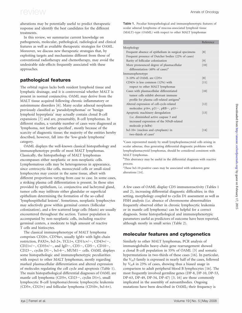
Table 1. Peculiar histopathological and immunophenotypic features of
ocular adnexal lymphoma of mucosa-associated lymphoid tissue
(MALT)-type (OAML) with respect to other MALT lymphomas
Reference
Morphology
Frequent absence of epithelium in surgical specimens [8]
Frequent presence of Dutcher bodies (22% of cases)
Rarity of follicular colonization [9]
More pronounced degree of plasmacellular
differentiation (40% of cases)a
[10]
Immunophenotype
3–10% of OAML are CD5+ [8]
CD43+ is less common (12%) with
respect to other MALT lymphomas
[11]
Cases with plasmacellular differentiated
tumor cells exhibit aberrant immune
profile for plasma cell-related antigensb
[10]
Altered expression of cell-cycle-related
molecules: p16+, p212, pRB2, p532
[12]
Apoptotic machinery deregulation
(i.e. diminished active caspase 3 and
increased expression of the NFjB-related
molecule p-IjBa)
[13]
bcl-10+ (nucleus and cytoplasm) in
two-thirds of casesc
[14]
aCases represented mainly by small lymphoplasmacytoid cells arising in
ocular adnexae, thus generating differential diagnostic problems with
lymphoplasmacytoid lymphomas, should be considered consistent with
MALT lymphomas.bThis aberrancy may be useful in the differential diagnosis with reactive
process.cThese bcl-10-positive cases may be associated with unknown gene
alterations [14].
review Annals of Oncology
836 | Ferreri et al. Volume 19 | No. 5 | May 2008
by guest on February 17, 2015http://annonc.oxfordjournals.org/
Dow
nloaded from
lower than that reported in gastrointestinal MALT lymphomas,but it is higher in cases with follicular dendritic cell networks,supporting a potential role of microenvironmental stimuli [9].Taken together, these features support the view that OAMLrepresent a clonal expansion of post-germinal-center memoryB cells, where, in two-thirds of the cases, antigen selection mayhave occurred [16].
Some chromosomal translocations, includingt(11;18)(q21;q21)/API2-MALT1, t(1;14)(p22;q32)/IGH-BCL10, t(14;18)(q32;q21)/IGH-MALT1 and t(3;14)(p14;q32)/IGH-FOXP1, are associated with MALT lymphomas, buttheir frequency markedly varies among different mucosal sites(Table 3). t(11;18), t(1;14) and t(14;18)/IGH-MALT1 are nearlyexclusively found in MALT lymphoma and their oncogenicproducts share the ability to enhance the activation of NFjB,a master transcriptional factor for a number of genes relevantfor lymphocyte activation, proliferation and survival [26]. Thet(3;14) has been detected both in MALT and in other B-celllymphomas [22, 25], although the molecular mechanism ofFOXP1-mediated lymphomagenesis remains to be investigated.The overall frequency of MALT-associated chromosomal
translocations in OAML is reported in Table 3; their clinicalsignificance in these malignancies remains to be defined.
There are only limited cytogenetic data in OAML (Table 3).Both conventional karyotyping and interphase FISH-basedcytogenetic analyses demonstrated that aneuploidy, particularlytrisomy 3 and 18, occurs frequently in t(11;18)-negative OAML[17–19, 23]. Trisomy 3 and 18 and t(14;18)(q32;q21) deserve tobe further investigated as possible predictors of multifocaldisease [27]. OAML with trisomy 18 seems to have distinctclinical features: it involves the conjunctiva, occurs in youngfemales and shows a high recurrence rate [19]. On the otherhand, trisomy 3 is significantly less common in conjunctivalMALT lymphomas with respect to orbit lymphomas (12%versus 81%) [11]. Comparison of European [18] and American[11] series seems to indicate the existence of geographicvariability in the incidence of recurring cytogeneticabnormalities in OAML. Variables influencing these featuresshould be further investigated.
Comparative genomic hybridization (CGH) carried out in10 OAML cases showed recurrent chromosomal gains at6p21 and 9q33-qter, in addition to trisomy 3, 12 and 18 [28].It will be noteworthy to survey the genomic gains and lossesof OAML using array CGH to explore whether these MALTlymphomas are also characterized by a conserved pattern ofchromosomal gains, as reported for other MALT lymphomas[29], and how these genomic alterations correlate withchlamydial infection and treatment response.
pathogenesis
OAML shares several clinicopathologic features with otherMALT lymphomas. In fact, OAML arises in tissues normallydevoid of innate immune system [6], often develops ona background of preexisting chronic inflammation (i.e.conjunctivitis) [9] and usually shows an indolent clinicalcourse. The presence of a preexisting inflammatory backgroundseems to be of pathogenetic relevance for MALT lymphomas,underlying the possible role of exogenous triggers (infections)and autoimmune reactions. Somatic immunoglobulinongoing mutations detected in OAML (see above) areconsistent with a process driven by chronic antigenicstimulation. Moreover, the biased usage of VH genes, frequentlyrearranged in autoantibodies production [30] and oftenoverrepresented in B-cell malignancies [31], further supports
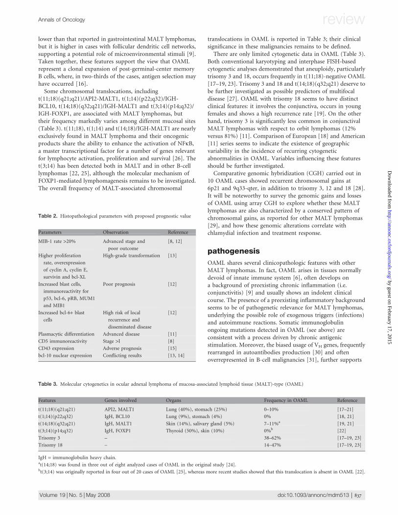
Table 2. Histopathological parameters with proposed prognostic value
Parameters Observation Reference
MIB-1 rate >20% Advanced stage and
poor outcome
[8, 12]
Higher proliferation
rate, overexpression
of cyclin A, cyclin E,
survivin and bcl-XL
High-grade transformation [13]
Increased blast cells,
immunoreactivity for
p53, bcl-6, pRB, MUM1
and MIB1
Poor prognosis [12]
Increased bcl-6+ blast
cells
High risk of local
recurrence and
disseminated disease
[12]
Plasmacytic differentiation Advanced disease [11]
CD5 immunoreactivity Stage >I [8]
CD43 expression Adverse prognosis [15]
bcl-10 nuclear expression Conflicting results [13, 14]
Table 3. Molecular cytogenetics in ocular adnexal lymphoma of mucosa-associated lymphoid tissue (MALT)-type (OAML)
Features Genes involved Organs Frequency in OAML Reference
t(11;18)(q21;q21) API2, MALT1 Lung (40%), stomach (25%) 0–10% [17–21]
t(1;14)(p22;q32) IgH, BCL10 Lung (9%), stomach (4%) 0% [18, 21]
t(14;18)(q32;q21) IgH, MALT1 Skin (14%), salivary gland (5%) 7–11%a [19, 21]
t(3;14)(p14;q32) IgH, FOXP1 Thyroid (50%), skin (10%) 0%b [22]
Trisomy 3 – 38–62% [17–19, 23]
Trisomy 18 – 14–47% [17–19, 23]
IgH = immunoglobulin heavy chain.at(14;18) was found in three out of eight analyzed cases of OAML in the original study [24].bt(3;14) was originally reported in four out of 20 cases of OAML [25], whereas more recent studies showed that this translocation is absent in OAML [22].
Annals of Oncology review
Volume 19 | No. 5 | May 2008 doi:10.1093/annonc/mdm513 | 837
by guest on February 17, 2015http://annonc.oxfordjournals.org/
Dow
nloaded from
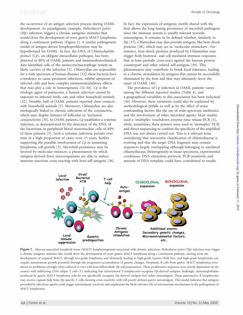
the occurrence of an antigen selection process during OAMLdevelopment. As paradigmatic example, Helicobacter pylori(Hp) infection triggers a chronic antigenic stimulus thatwould drive the development of overt gastric MALT lymphomaalong a continuum pathway (Figure 1). A similar pathogeneticmodel of antigen-driven lymphoproliferation may behypothesized for OAML. In fact, the DNA of Chlamydophilapsittaci (Cp), an obligate intracellular pathogen, has beendetected in 80% of OAML patients and immunohistochemicaldata identified cells of the monocyte/macrophage system aslikely carriers of the infection [5]. Chlamydiae are responsiblefor a wide spectrum of human diseases [32]; these bacteria havea tendency to cause persistent infections, inhibit apoptosis ofinfected cells and have complex immunomodulatory effectsthat may play a role in tumorigenesis [32–34]. Cp is theetiologic agent of psittacosis, a human infection caused byexposure to infected birds, cats and other household animals[32]. Notably, half of OAML patients reported close contactswith household animals [5]. Moreover, Chlamydiae are alsoetiologically linked to chronic infections of the conjunctiva,which may display features of follicular or ‘inclusion’conjunctivitis [35]. In OAML patients, Cp establishes a systemicinfection, as demonstrated by the detection of the DNA ofthe bacterium in peripheral blood mononuclear cells of 40%of these patients [5]. Such a systemic infection persists overtime in a high proportion of cases, even >5 years, furthersupporting the possible involvement of Cp in sustaininglymphoma cell growth [5]. Microbial persistence may befavored by molecular mimicry, a phenomenon by whichantigens derived from microorganisms are able to induceimmune reactions cross-reacting with host self-antigens [36].
In fact, the expression of antigenic motifs shared with thehost allows the long-lasting persistence of microbial pathogenssince the immune system is usually tolerant towardsautoantigens. It remains to be defined whether, similarly toHp [37], Chlamydiae may also provide antigens, like heat-shockproteins [38], which may act as ‘molecular mimickers’. Forinstance, heat-shock proteins produced by Chlamydiae maytrigger both humoral- and cell-mediated immune responsesthat at least partially cross-react against the human proteincounterpart and other related self-antigens [39]. Thisphenomenon may contribute to break local tolerance, leadingto a chronic stimulation by antigens that cannot be successfullyeliminated by the host and that may ultimately favor theonset of OAML [40].
The prevalence of Cp infection in OAML patients variesamong the different reported studies (Table 4), anda geographical variability in this association has been indicated[44]. However, these variations could also be explained bymethodological pitfalls as well as by the effect of someconfounding factors like the use of wide-spectrum antibioticsand the involvement of other microbial agents. Most studiesused a ‘multiplex’ touchdown enzyme time release-PCR [5],while, sometimes, these primers were used in ‘monoplex’ PCR,and direct sequencing to confirm the specificity of the amplifiedDNA was not always carried out. This is a relevant issueconsidering that taxonomic classification of chlamydiaceae isevolving and that the target DNA fragment may containsequences largely overlapping although belonging to unrelatedchlamydiaceae. Heterogeneity in tissue specimens, experimentalconditions, DNA extraction protocol, PCR sensitivity andamount of DNA template could have contributed to results
Figure 1. Mucosa-associated lymphoid tissue (MALT) lymphomagenesis associated with chronic infections: Helicobacter pylori (Hp) infection may trigger
a chronic antigenic stimulus that would drive the development of overt gastric MALT lymphoma along a continuum pathway, starting from the
development of acquired MALT, through low-grade lymphoma and ultimately leading to high-grade tumors. Both low- and high-grade lymphomas can
acquire autonomous growth potential through the progressive accumulation of genetic changes. Neoplastic B cells from gastric MALT lymphomas were
shown to proliferate strongly when cultured in vitro with heat-killed whole Hp cell preparations. These proliferative responses were strictly dependent on the
contact with infiltrating CD4+ helper T cells (T), indicating that intratumoral T lymphocytes recognize Hp-derived antigens. Strikingly, immunoglobulins
produced by gastric MALT lymphoma cells do not specifically recognize Hp-derived antigens but rather autoantigens. These autoreactive B lymphocytes
may receive cognate help from Hp-specific T cells showing cross-reactivity with still poorly defined gastric autoantigens. This model indicates that antigens
provided by infectious agents could trigger autoimmune reactivity and emphasizes the likely relevant role of autoimmune mechanisms in the pathogenesis of
MALT lymphomas.
review Annals of Oncology
838 | Ferreri et al. Volume 19 | No. 5 | May 2008
by guest on February 17, 2015http://annonc.oxfordjournals.org/
Dow
nloaded from
variability. From a clinical point of view, most patients withconjunctival or orbital lesions are firstly considered as affectedby inflammatory or infectious processes instead of lymphomas,and are usually treated with topic or systemic wide-spectrumantibiotics, a common practice in OAML patients that couldfurther reduce the local chlamydial population, resulting inPCR false negatives. Finally, the potential involvement of othermicrobial agents in the development and maintaining ofOAML should be taken into account. This is indicated by theintriguingly tumor regression observed in one-third ofChlamydia-negative OAML after doxycycline treatment [46](see ‘Treatment’). Available evidence seems to rule out the
possible involvement of other infectious agents commonlyassociated with chronic eye diseases, such as Chlamydiatrachomatis, herpes simplex virus 1 and 2 and adenovirus 8 and19 [5, 55], whereas Chlamydia pneumoniae DNA was detectedsporadically in a few cases of OAML [56, 57] (R. Dolcetti,unpublished data). Like for other B-cell lymphomas, hepatitis Cvirus (HCV) could play a role in the development of OAML.HCV seropositivity has been detected in 13% of OAMLpatients, and seems to be associated with more disseminatedand aggressive lymphomas [58].
The study of putative mechanisms regulating lymphocyteshoming to the ocular adnexa constitutes an interesting issue inthe genesis and development of these neoplasms. Available dataare preliminary and limited to the reported absence ofexpression of the a4b7 integrin, a crucial regulator oflymphocyte trafficking, and its ligand MAdCAM-1 [59], and tothe expression of the chemoattractant cytokine CXCL13 onneoplastic lymphocytes [55].
clinical features
clinical presentation
Lymphomas can infiltrate any orbital and ocular adnexal tissue.The clinical picture of OAML depends greatly on the structurescompromised. Twenty-five percent of OAML displaysconjunctival involvement, while intraorbital masses are presentin 75% of cases and bilateral involvement is observed in 10–15% of cases, mostly in conjunctival forms [51,60–63]. It isdifficult to differentiate clinically OAML from other orbitaldiseases due to the lack of pathognomonic signs or symptoms.Every lymphoma histotype can arise in the ocular adnexae,although with similar presenting symptoms and requiringsurgical biopsy for histopathological diagnosis considering thattreatment and prognosis remarkably vary among differentlymphoma categories.
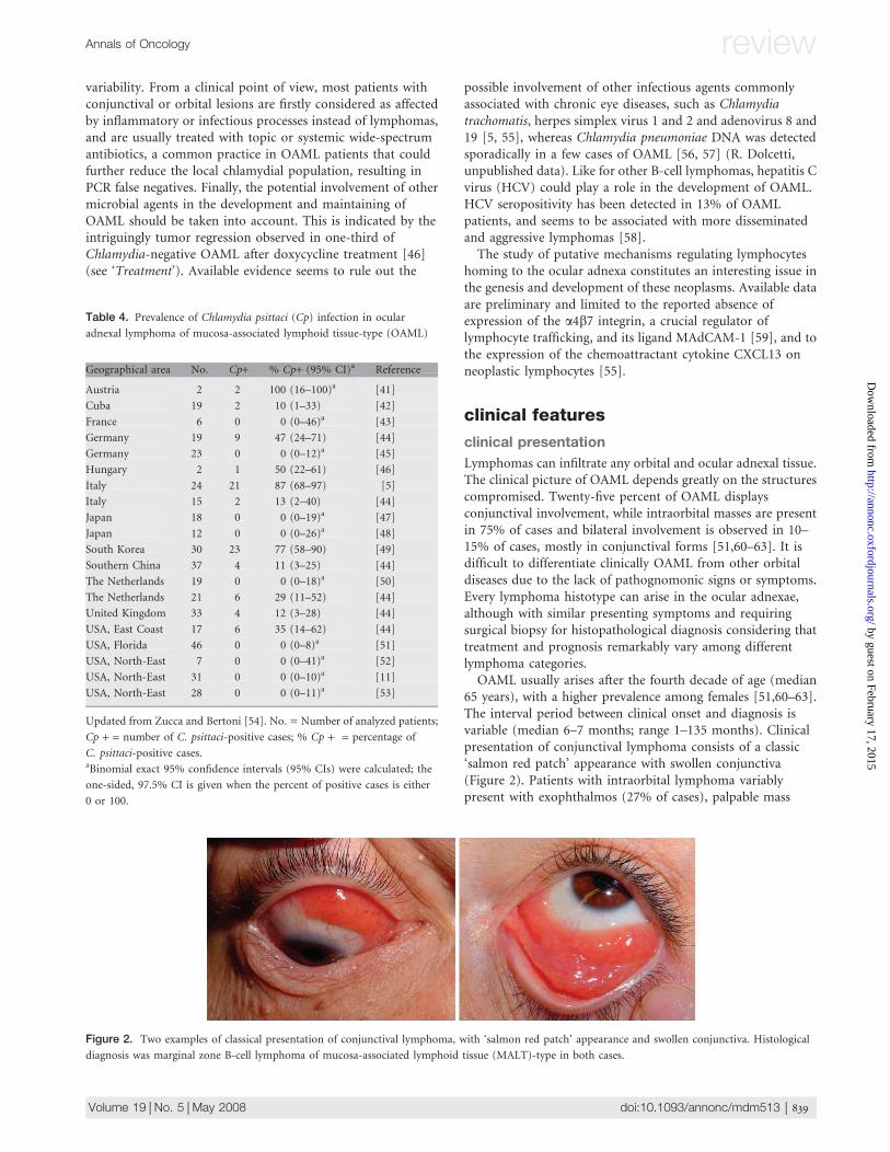
OAML usually arises after the fourth decade of age (median65 years), with a higher prevalence among females [51,60–63].The interval period between clinical onset and diagnosis isvariable (median 6–7 months; range 1–135 months). Clinicalpresentation of conjunctival lymphoma consists of a classic‘salmon red patch’ appearance with swollen conjunctiva(Figure 2). Patients with intraorbital lymphoma variablypresent with exophthalmos (27% of cases), palpable mass
Table 4. Prevalence of Chlamydia psittaci (Cp) infection in ocular
adnexal lymphoma of mucosa-associated lymphoid tissue-type (OAML)
Geographical area No. Cp+ % Cp+ (95% CI)a Reference
Austria 2 2 100 (16–100)a [41]
Cuba 19 2 10 (1–33) [42]
France 6 0 0 (0–46)a [43]
Germany 19 9 47 (24–71) [44]
Germany 23 0 0 (0–12)a [45]
Hungary 2 1 50 (22–61) [46]
Italy 24 21 87 (68–97) [5]
Italy 15 2 13 (2–40) [44]
Japan 18 0 0 (0–19)a [47]
Japan 12 0 0 (0–26)a [48]
South Korea 30 23 77 (58–90) [49]
Southern China 37 4 11 (3–25) [44]
The Netherlands 19 0 0 (0–18)a [50]
The Netherlands 21 6 29 (11–52) [44]
United Kingdom 33 4 12 (3–28) [44]
USA, East Coast 17 6 35 (14–62) [44]
USA, Florida 46 0 0 (0–8)a [51]
USA, North-East 7 0 0 (0–41)a [52]
USA, North-East 31 0 0 (0–10)a [11]
USA, North-East 28 0 0 (0–11)a [53]
Updated from Zucca and Bertoni [54]. No. = Number of analyzed patients;
Cp + = number of C. psittaci-positive cases; % Cp + = percentage of
C. psittaci-positive cases.aBinomial exact 95% confidence intervals (95% CIs) were calculated; the
one-sided, 97.5% CI is given when the percent of positive cases is either
0 or 100.
Figure 2. Two examples of classical presentation of conjunctival lymphoma, with ‘salmon red patch’ appearance and swollen conjunctiva. Histological
diagnosis was marginal zone B-cell lymphoma of mucosa-associated lymphoid tissue (MALT)-type in both cases.
Annals of Oncology review
Volume 19 | No. 5 | May 2008 doi:10.1093/annonc/mdm513 | 839
by guest on February 17, 2015http://annonc.oxfordjournals.org/
Dow
nloaded from
(19%), eyelid ptosis (6%), diplopia (2%), eyelid nodule, orbitaledema, epiphora and a variable degree of impaired ocularmotility [51,60–63]. Extraocular muscle imbalance andlimitation of the excursion of the eye are usually indicators ofexpansive effect of the lesion rather than of muscle damage.Clinical manifestations usually consist of a slowly growing,painless mass that displaces the normal structures, butsometimes are acute, with inflammatory-like signs andsymptoms. Only in rare cases of rapidly growing tumors, visualacuity and field defects or choroidal folds are observed, anda few cases of OAML infiltrating the eye with devastatingconsequences have been reported [64].
staging procedures
More than 75% of OAML presents with a single lesion (stageIE). With conventional lymphoma staging procedures, regionallymphadenopathies are detected in <5% of cases (stage IIE),and extraorbital disease, mostly in extranodal organs, isobserved in 10–15% of cases (stage IVE), rarely in patients withconjunctival lymphoma [5, 51,61–63]. Conversely, the use ofmore extensive and invasive staging showed that 38% of OAMLpatients have at least one concomitant, extraorbital site ofdisease at diagnosis [27]. The usefulness of extended staging inOAML remains a matter of debate. On one hand, the definitionof stage I disease is important because these patients are usuallytreated with radiotherapy alone. On the other hand, even ifpatients with stage I disease have significantly better relapse-freesurvival in comparison to those with advanced disease, nodifference in cause-specific survival between these subgroupshas been reported [63]. Comprehensively, OAML patientsshould be assessed with conventional lymphoma stagingprocedures, whereas an extensive gastrointestinal workup in theabsence of clinical symptoms suggestive of lymphoma does notseem necessary [27].
Some OAML patients have a history of autoimmunedisorders, mostly thyrotoxicosis (5% of cases) or Sjogrensyndrome [65], the concomitance of which should be assessedat diagnosis since their negative impact on therapeutic outcomein extraorbital MALT lymphomas [66].
neuroimaging
Neuroimaging techniques are fundamental for distinguishingOAML from other orbital masses and for accurate staging andtherapeutic response definition since they allow precisevolumetric measurements. At neuroimaging examination,OAML usually present as well-defined lesions, mostly placed inthe superior-lateral quadrant of the orbit, often surroundingand displacing extraocular muscles, without signs of ocularinfiltration. On basal computed tomography images, OAMLappears homogeneously iso- or slightly hyperdense comparedwith extraocular muscles. OAML contrast enhancement ishomogeneous and its intensity is comparable to that oflachrymal glands and extraocular muscles. Magnetic resonanceimaging (MRI) shows a great potential in differentiating OAMLfrom other orbital expansive lesions. Location, margins and thedistinctive T2 and diffusion-weighted imaging (DWI) signalintensities allow OAML identification and characterization(Figure 3). Similarly to what was observed in lymphomas of
other districts, OAML presents high DWI signal and lowapparent diffusion coefficient (ADC) values due to the highcellularity and high nucleus-to-cytoplasm ratio. Preliminarydata indicate that ADC of OAML is lower than that of allorbital normal structures and expansive lesions, being thususeful for differential diagnosis. Furthermore, DWI is helpful inestablishing involvement or persistence of disease in thelachrymal gland, where both signal intensity and contrastenhancement do not allow unambiguous differentiationbetween normal and pathologic tissue. A- and B-scan orbitalultrasonography provides additional information to MRI fordistinguishing OAML from other orbital masses (Figure 4).
clinical behavior and prognosis
OAML shows a better prognosis in comparison to otherlymphoma categories arising in the ocular adnexae [63]. MostOAML patients display good prognostic indicators like limiteddisease, good performance status and absence of systemicsymptoms, and, if adequately treated, these patients exhibita favorable outcome [51, 67]. Some anecdotal cases ofspontaneous tumor remission in OAML patients have beenreported, mostly in Japanese patients with conjunctival MALTlymphoma [68]. However, the real rate of this phenomenonwarrants further investigation since some of these patients havebeen treated with topical steroids or antibiotics, which couldhave affected results interpretation. Presenting symptoms aresometimes severe, requiring a proper and timely treatment.Local control rates vary according to the used therapy, with a5-year relapse-free survival of �65%. Some patients experiencemultiple relapses, which usually involve the contralateral orbitand distant extranodal organs, particularly in patients treatedwith radiotherapy. Systemic dissemination occurs in 5–10% ofcases, being rare in patients with conjunctival lymphoma. Lessthan 5% of OAML patients die of lymphoma, with a 5-yearcause-specific and overall survival of 100% and >90%,respectively [62, 63]. Reliable prognostic factors remain to bedefined. A few studies conducting multivariate analysisindicated that nodal involvement (<5% of cases), systemicsymptoms (1%), increased lactate dehydrogenase serum levels(1%) and non-conjunctival sites are negative predictors ofoutcome [51, 61, 62]. Some of these aggressiveness parameterspredict high-grade transformation, which has been reported in1–3% of cases [67, 69].
treatment
Current therapeutic knowledge in OAML results froma limited number of small, and variably treated, retrospectiveseries, which included different lymphoma categories,diagnosed before the World Health Organizationclassification era and a single prospective trial [46]. Thus,universally accepted therapeutic guidelines for OAML do notexist. Therapeutic decision is usually driven from appraisalof several variables related to the patient (age, performancestatus, co-morbidity—i.e. autoimmune disorders—concomitant infections useful as therapeutic targets), tothe lymphoma (stage, site of disease—i.e. surgicalaccessibility—symptoms due to infiltration, histological
review Annals of Oncology
840 | Ferreri et al. Volume 19 | No. 5 | May 2008
by guest on February 17, 2015http://annonc.oxfordjournals.org/
Dow
nloaded from

and molecular indicators of aggressiveness and response)and to the risk of severe treatment-related toxicity andsequelae. Efficacy and kinetic of response are two importantparameters for therapeutic choice, mostly in ‘less-indolent’lymphomas that could determine a fast impairment of ocularfunction.
surgical resection
Surgical resection is a necessary diagnostic step and, in selectedcases, a part of therapeutic approach to OAML (Figure 5).Complete excision can be carried out in many conjunctival andlachrymal gland MALT lymphomas, especially inpseudoencapsulated lesions. However, additional efforts tocompletely resect lymphomatous lesions should be avoided in
OAML patients since an aggressive approach could beassociated with a high risk of complications, especially in thearea of the lachrymal gland and in the deeper orbit, andconsidering that the extent of surgical resection does notinfluence survival [67]. ‘Wait and watch’ strategy after surgicalresection or biopsy in patients with stage I disease producessimilar results, in terms of time to progression, systemicdissemination, high-grade transformation and lymphoma-related deceases, to those reported with immediateradiotherapy, with a 10-year overall survival of 94% [67]. Thisstrategy could be safely proposed to selected OAML patients,like elderly patients or patients with severe co-morbidity,completely resected lesions and/or indolent and asymptomaticdisease.
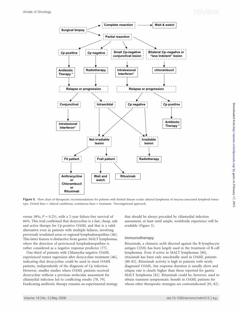
Figure 3. Magnetic resonance imaging (MRI) of ocular adnexal lymphoma of mucosa-associated lymphoid tissue-type (OAML). Coronal basal T1-weighted
(w) (A), coronal T2-w (B), coronal and axial post-gadolinium T1-w (C, E), coronal and axial diffusion-weighted imaging (DWI) (D, F) image, obtained
with a b value of 700 mm2/s. The OAML (large arrow) is located in superior-lateral quadrant of the left orbit and involves both intra- and extraconal
structures. It surrounds the optic nerve (small arrow) and the superior pole of the left ocular globe (§), the lateral rectus and the superior extraocular
muscles (EOM). The left lachrymal gland is infiltrated. On T1-w image (A), the OAML signal is comparable to that of EOM. On the T2-w image (B), OAML
presents the same signal intensity of cerebral gray matter and is slightly hyperintense to EOM. Usually, T1 and T2 signal intensities within the lesion are
homogeneous. OAML contrast enhancement is uniform and its conspicuity is comparable to that observed in lachrymal glands and EOM. Using parallel
imaging technique is now possible to obtain DWI images of the orbit with reasonable scan times and without occurrence of significant susceptibility artifacts
(D, F). DWI is an MRI-based technique that evaluates the rate of microscopic water diffusion in tissues and represents a useful technique for characterizing
lymphomas of the central nervous system, the neck and the orbits. On DWI images, OAML appears hyperintense compared with all other orbital structures.
DWI is also helpful in establishing involvement of the lachrymal gland, where both signal intensity and contrast enhancement do not allow unambiguous
differentiation between normal and pathologic tissue. Please compare the contrast enhancement and DWI signal intensity of affected left lachrymal gland
and normal right one (*).
Annals of Oncology review
Volume 19 | No. 5 | May 2008 doi:10.1093/annonc/mdm513 | 841
by guest on February 17, 2015http://annonc.oxfordjournals.org/
Dow
nloaded from
radiotherapy
Radiotherapy is the most extensively studied treatment inOAML patients. However, only a few series have been focusedon OAML treated exclusively with radiotherapy [62, 63, 70, 71].An universally accepted radiation schedule for OAML patientsdoes not exist. Recent studies indicate a radiation fieldincluding a gross tumor volume with 0.5- to 1-cm margin fora planning target volume and a dose of 25–30 Gy in 10–15fractions (minimal target dose >25 Gy) [62, 70, 71]. Electronbeams (4–12 MeV) and 4–9 MV photon beams are advisable inconjunctival and intraorbital lymphomas, respectively. A singleanterior field or a wedge pair of anterior fields has been used inmost series. In conjunctival lymphomas, the entire conjunctivaand eyelid should be irradiated, while preliminary data seem toindicate that the entire orbit should be irradiated in patientswith intraorbital lymphoma [62]. Brachytherapy can providelocal control in conjunctival lymphomas, but the risk ofcomplications and marginal relapses is unacceptably high [72].
Most irradiated patients with stage I OAML achieve anobjective response, which is slow and gradual [62, 70]. In-fieldrelapses are rare and seem to be related to low-radiation doses[71] or to the use of lens shielding [62]. Relapse rate at 4 yearsis 20–25%; most relapses involve the contralateral orbit (half ofrelapses) and distant extranodal organs [63, 70, 71].
With the above-indicated schedule, radiotherapy is usuallywell tolerated [62, 70]. The most common toxic effects of grade‡2 are cataract (38% of cases), retinal disorders (17%),xerophthalmia (17%) and glaucoma (2%) [70]. Toxicity ismore common with doses >36 Gy [70]. Presently, the role ofupfront radiotherapy is being reviewed due to its relatedtoxicity, the development of new therapeutic strategies and therecent insights into the biology of OAML.
chemotherapy
Prospective trials assessing chemotherapy efficacy in OAML donot exist; in most retrospective OAML series, only a smallproportion of patients has been treated with chemotherapy
alone. The largest experience regards chlorambucil, analkylating agent largely used in indolent lymphomas. This drugis an active and well-tolerated therapy for stage I OAML, witha 100% overall response rate, a 79% complete remission rateand a 5-year relapse-free survival of 60% [69]. Relapses afterchlorambucil mostly involve extraorbital tissues, with rare cases(3%) of high-grade transformation [69]. Chlorambucil couldbe proposed especially to OAML patients who experiencerelapse in previously irradiated areas or with disseminated orbilateral disease and in the case of radiotherapy inaccessibility(Figure 5). The use of other drugs, like fludarabine [73],cladribine [74] and oxaliplatin [75], deserves more caution.Tolerability of these drugs is sometimes unsatisfactory [74],and evidence of their efficacy is limited to a few prospectivetrials on unselected MALT lymphomas including a smallnumber of OAML patients. The use of upfront anthracycline-based chemotherapy did not show any clinical advantage incomparison with chlorambucil alone [2].
bacteria-eradicating therapy
In gastric MALT lymphoma, antibiotic therapy, aimed toeradicate the Hp infection, is followed by lymphoma regressionin 60–70% of stage IE cases [26]. Although a role for Hp insustaining the growth also of non-gastric MALT lymphomaswas hypothesized, reported evidence shows that gastric Hpinfection in OAML patients does not influence clinicalpresentation and course and that Hp-eradicating antibiotictherapy is not active against OAML [76]. Conversely, theeradication of Cp infection with doxycycline, a tetracyclinederivative largely used in the treatment of psittacosis, has beenproposed as a valid alternative for OAML patients. Ina multicenter phase II trial [46], 11 patients with Cp-positiveOAML and 16 with Cp-negative OAML have been treated withdoxycycline, obtaining, after a median follow-up of 14 months,an overall response rate of 48%. Lymphoma regression wasusually slow and gradual and has been observed in bothCp-positive and -negative patients (overall response rate = 64%
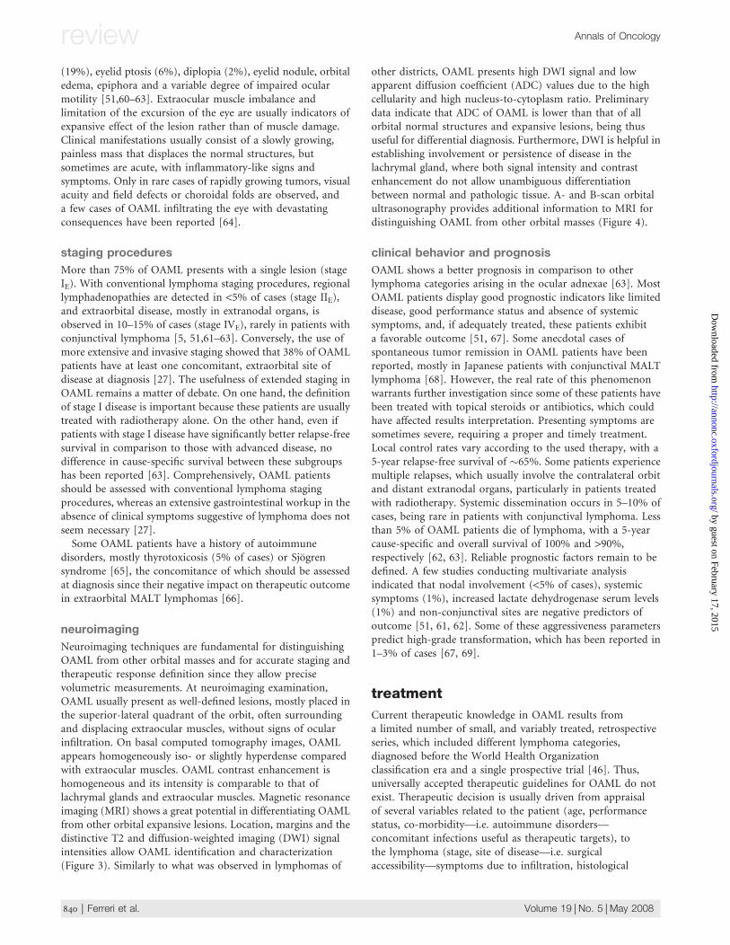
Figure 4. A- and B-scan orbital ultrasonography plays an important role in differential diagnosis of orbital masses. B- (left side) and A-scan (right side)
orbital ultrasonography of the eye and the orbit in a patient with a retrobulbar ocular adnexal lymphoma of mucosa-associated lymphoid tissue-type
(OAML). C, cornea; L, lens; V, vitreous humor; R, retina. B-scan orbital ultrasonography (left side) shows a well-delimited, hyporeflective retroocular solid
mass (Ly; surrounded by arrows). A-scan orbital ultrasonography (right side) confirms the presence of a retroocular, homogeneously hyporeflective lesion
(Ly) delimited by two peaks. Histological diagnosis was OAML. Orbital ultrasonography provides useful information on the site, morphology and structure
of the lesion (B-scan) and on the acoustic structure, internal reflectivity, vascularization and margins of the lesion (A-scan). Low reflectivity is characteristic
in OAML and other lymphomas of the orbit, which is due to the high cellular density distinctive for these disorders. Kindly provided by Dr Luisa Pierro,
Ophthalmology Unit, San Raffaele Scientific Institute, Milan, Italy.
review Annals of Oncology
842 | Ferreri et al. Volume 19 | No. 5 | May 2008
by guest on February 17, 2015http://annonc.oxfordjournals.org/
Dow
nloaded from
versus 38%; P = 0.25), with a 2-year failure-free survival of66%. This trial confirmed that doxycycline is a fast, cheap, safeand active therapy for Cp-positive OAML and that is a validalternative even in patients with multiple failures, involvingpreviously irradiated areas or regional lymphadenopathies [46].This latter feature is distinctive from gastric MALT lymphomas,where the detection of perivisceral lymphadenopathies israther considered as a negative response predictor [77].
One-third of patients with Chlamydia-negative OAMLexperienced tumor regression after doxycycline treatment [46],indicating that doxycycline could be used in most OAMLpatients, independently of the diagnosis of Cp infection.However, smaller studies where OAML patients receiveddoxycycline without a previous molecular assessment forchlamydial infection led to conflicting results [78, 79].Eradicating antibiotic therapy remains an experimental strategy
that should be always preceded by chlamydial infectionassessment, at least until ample, worldwide experience will beavailable (Figure 5).
immunotherapy
Rituximab, a chimeric mAb directed against the B-lymphocyteantigen CD20, has been largely used in the treatment of B-celllymphomas. Even if active in MALT lymphomas [80],rituximab has been only anecdotally used in OAML patients[80–82]. Rituximab activity is high in patients with newlydiagnosed OAML, but response duration is usually short andrelapse rate is clearly higher than those reported for gastricMALT lymphoma [82]. Rituximab could be, however, used toobtain transient symptomatic benefit in OAML patients forwhom other therapeutic strategies are contraindicated [81, 82].
Surgical biopsy
Complete resection
Partial resection
Cp positive Cp negative
AntibioticTherapy *
Radiotherapy
Relapse or progression
IntraorbitalConjunctival
Frail patientFit patient
Wait andwatch
IntralesionalInterferon*
Anthracyclineor
Chlorambucilor
Rituximab
Bilateral Cp–negative or“less Indolent” lesion
chlorambucilIntralesionalInterferon*
Wait & watch
Relapse or progression
Not irradiablelesion
Cp positiveCp negative
AntibioticTherapy *
Irradiablelesion
Radiotherapy
Rituximab
Small Cp-negativeconjunctival lesion
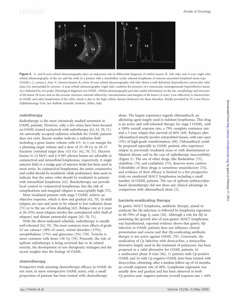
Figure 5. Flow chart of therapeutic recommendations for patients with limited disease ocular adnexal lymphoma of mucosa-associated lymphoid tissue-
type. Dotted lines = clinical conditions; continuous lines = treatment. *Investigational approach.
Annals of Oncology review
Volume 19 | No. 5 | May 2008 doi:10.1093/annonc/mdm513 | 843
by guest on February 17, 2015http://annonc.oxfordjournals.org/
Dow
nloaded from
Intralesional injection of interferon a is a relatively simpleand quick procedure that has been successfully used forconjunctival lymphomas [83, 84] (Figure 5). Side-effects consistof local hemorrhage, chemosis and minor systemic effects [83,84]. The efficacy of this approach remains, however, to bedefined considering that follow-up of reported cases is shortand that unsuccessfully treated patients may have not beenreported.
future perspectives
Future studies will investigate whether Cp infection isresponsible for the rapidly increasing incidence of OAML.The role of putative Cp-derived antigens in lymphomagenesis,the involvement of distinct Cp strains as well as themechanisms of tolerance and antigenic chronic stimulationinduced by this microorganism, that may ultimately favorthe onset of OAML, should be addressed in well-designedstudies. From a therapeutic point of view, and following theexample of gastric MALT lymphoma [85], the analysis ofpathologic and molecular predictors of response will play animportant role in selecting the best candidates forCp-eradicating antibiotic therapy. Well-designed prospectivetrials will lead to establish new therapeutic strategies that,exploiting novel mechanisms, could contribute to furtherimprove the outcome of these patients. For instance, theefficacy of antiviral therapy with interferon and ribavirin [86]in HCV-positive patients or new antibiotic combinationsshould be investigated. In the years ahead, it is hoped thatinternational, multidisciplinary efforts could address mostof the fundamental, clinical and biological research questionsfor OAML.
references
1. Margo CE, Mulla ZD. Malignant tumors of the orbit. Analysis of the Florida cancer
registry. Ophthalmology 1998; 19(5): 835–846.
2. Sasai K, Yamabe H, Dodo Y et al. Non-Hodgkin’s lymphoma of the ocular
adnexa. Acta Oncol 2001; 40(4): 485–490.
3. Mannami T, Yoshino T, Oshima K et al. Clinical, histopathological, and
immunogenetic analysis of ocular adnexal lymphoproliferative disorders:
characterization of malt lymphoma and reactive lymphoid hyperplasia. Mod
Pathol 2001; 14(7): 641–649.
4. Moslehi R, Devesa SS, Schairer C et al. Rapidly increasing incidence of ocular
non-Hodgkin lymphoma. J Natl Cancer Inst 2006; 98(13): 936–939.
5. Ferreri AJ, Guidoboni M, Ponzoni M et al. Evidence for an association between
Chlamydia psittaci and ocular adnexal lymphomas. J Natl Cancer Inst 2004;
96(8): 586–594.
6. Du MQ, Isaccson PG. Gastric MALT lymphoma: from aetiology to treatment.
Lancet Oncol 2002; 3(2): 97–104.
7. Neri A, Jakobiec FA, Pelicci PG et al. Immunoglobulin and T cell receptor beta
chain gene rearrangement analysis of ocular adnexal lymphoid neoplasms:
clinical and biologic implications. Blood 1987; 70(5): 1519–1529.
8. Coupland SE, Krause L, Delecluse HJ et al. Lymphoproliferative lesions of the
ocular adnexa. Analysis of 112 cases. Ophthalmology 1998; 105(8):
1430–1441.
9. Hara Y, Nakamura N, Kuze T et al. Immunoglobulin heavy chain gene analysis of
ocular adnexal extranodal marginal zone B-cell lymphoma. Invest Ophthalmol Vis
Sci 2001; 42(11): 2450–2457.
10. Coupland SE, Damato B. Lymphomas involving the eye and the ocular adnexa.
Curr Opin Ophthalmol 2006; 17(6): 523–531.
11. Ruiz A, Reischl U, Swerdlow SH et al. Extranodal marginal zone B-cell
lymphomas of the ocular adnexa: multiparameter analysis of 34 cases including
interphase molecular cytogenetics and PCR for Chlamydia psittaci. Am J Surg
Pathol 2007; 31(5): 792–802.
12. Coupland SE, Hellmich M, Auw-Haedrich C et al. Prognostic value of cell-cycle
markers in ocular adnexal lymphoma: an assessment of 230 cases. Graefes Arch
Clin Exp Ophthalmol 2004; 242(2): 130–145.
13. Franco R, Camacho FI, Caleo A et al. Nuclear bcl10 expression characterizes
a group of ocular adnexa MALT lymphomas with shorter failure-free survival.
Mod Pathol 2006; 19(8): 1055–1067.
14. Adachi A, Tamaru JI, Kaneko K et al. No evidence of a correlation between
BCL10 expression and API2-MALT1 gene rearrangement in ocular adnexal MALT
lymphoma. Pathol Int 2004; 54(1): 16–25.
15. Nola M, Lukenda A, Bollmann M et al. Outcome and prognostic factors in ocular
adnexal lymphoma. Croat Med J 2004; 45(3): 328–332.
16. Coupland SE, Foss HD, Anagnostopoulos I et al. Immunoglobulin VH gene
expression among extranodal marginal zone B-cell lymphomas of the ocular
adnexa. Invest Ophthalmol Vis Sci 1999; 40(3): 555–562.
17. Remstein ED, Kurtin PJ, James CD et al. Mucosa-associated lymphoid tissue
lymphomas with t(11;18)(q21;q21) and mucosa-associated lymphoid tissue
lymphomas with aneuploidy develop along different pathogenetic pathways. Am
J Pathol 2002; 161(1): 63–71.
18. Streubel B, Simonitsch-Klupp I, Mullauer L et al. Variable frequencies of MALT
lymphoma-associated genetic aberrations in MALT lymphomas of different sites.
Leukemia 2004; 18(10): 1722–1726.
19. Tanimoto K, Sekiguchi N, Yokota Y et al. Fluorescence in situ hybridization
(FISH) analysis of primary ocular adnexal MALT lymphoma. BMC Cancer 2006;
6: 249.
20. Murga Penas EM, Hinz K, Roser K et al. Translocations t(11;18)(q21;q21)
and t(14;18)(q32;q21) are the main chromosomal abnormalities involving
MLT/MALT1 in MALT lymphomas. Leukemia 2003; 17(11): 2225–2229.
21. Ye H, Gong L, Liu H et al. MALT lymphoma with t(14;18)(q32;q21)/IGH-MALT1 is
characterized by strong cytoplasmic MALT1 and BCL10 expression. J Pathol
2005; 205(3): 293–301.
22. Haralambieva E, Adam P, Ventura R et al. Genetic rearrangement of FOXP1 is
predominantly detected in a subset of diffuse large B-cell lymphomas with
extranodal presentation. Leukemia 2006; 20(7): 1300–1303.
23. Ott G, Katzenberger T, Greiner A et al. The t(11;18)(q21;q21) chromosome
translocation is a frequent and specific aberration in low-grade but not high-
grade malignant non-Hodgkin’s lymphomas of the mucosa-associated lymphoid
tissue (MALT-) type. Cancer Res 1997; 57(18): 3944–3948.
24. Streubel B, Lamprecht A, Dierlamm J et al. T(14;18)(q32;q21) involving IGH and
MALT1 is a frequent chromosomal aberration in MALT lymphoma. Blood 2003;
101(6): 2335–2339.
25. Streubel B, Vinatzer U, Lamprecht A et al. T(3;14)(p14.1;q32) involving IGH and
FOXP1 is a novel recurrent chromosomal aberration in MALT lymphoma.
Leukemia 2005; 19(4): 652–658.
26. Isaacson PG, Du MQ. MALT lymphoma: from morphology to molecules. Nat Rev
Cancer 2004; 4(8): 644–653.
27. Raderer M, Wohrer S, Streubel B et al. Assessment of disease dissemination in
gastric compared with extragastric mucosa-associated lymphoid tissue
lymphoma using extensive staging: a single-center experience. J Clin Oncol
2006; 24(19): 3136–3141.
28. Matteucci C, Galieni P, Leoncini L et al. Typical genomic imbalances in primary
MALT lymphoma of the orbit. J Pathol 2003; 200(5): 656–660.
29. Zhou Y, Ye H, Martin-Subero JI et al. Distinct comparative genomic
hybridisation profiles in gastric mucosa-associated lymphoid tissue
lymphomas with and without t(11;18)(q21;q21). Br J Haematol 2006; 133(1):
35–42.
30. Pascual V, Capra JD. VH4-21, a human VH gene segment overrepresented in the
autoimmune repertoire. Arthritis Rheum 1992; 35(1): 11–18.
31. Bahler DW, Campbell MJ, Hart S et al. Ig VH gene expression among human
follicular lymphomas. Blood 1991; 78(6): 1561–1568.
32. Byrne GI, Ojcius DM. Chlamydia and apoptosis: life and death decisions of an
intracellular pathogen. Nat Rev Microbiol 2004; 2(10): 802–808.
review Annals of Oncology
844 | Ferreri et al. Volume 19 | No. 5 | May 2008
by guest on February 17, 2015http://annonc.oxfordjournals.org/
Dow
nloaded from
33. Smith JS, Munoz N, Herrero R et al. Evidence for Chlamydia trachomatis as
a human papillomavirus cofactor in the etiology of invasive cervical cancer in
Brazil and the Philippines. J Infect Dis 2002; 185(3): 324–331.
34. Miyairi I, Byrne GI. Chlamydia and programmed cell death. Curr Opin Microbiol
2006; 9(1): 102–108.
35. Yeung L, Tsao YP, Chen PY et al. Combination of adult inclusion conjunctivitis
and mucosa-associated lymphoid tissue (MALT) lymphoma in a young adult.
Cornea 2004; 23(1): 71–75.
36. Oldstone MB. Molecular mimicry and immune-mediated diseases. FASEB J
1998; 12(13): 1255–1265.
37. Negrini R, Savio A, Poiesi C et al. Antigenic mimicry between helicobacter pylori
and gastric mucosa in the pathogenesis of body atrophic gastritis.
Gastroenterology 1996; 111(3): 655–665.
38. Lamb DJ, El-Sankary W, Ferns GA. Molecular mimicry in atherosclerosis: a role for
heat shock proteins in immunisation. Atherosclerosis 2003; 167(2): 177–185.
39. Pockley AG. Heat shock proteins as regulators of the immune response. Lancet
2003; 362(9382): 469–476.
40. Ishii E, Yokota K, Sugiyama T et al. Immunoglobulin G1 antibody response to
helicobacter pylori heat shock protein 60 is closely associated with low-grade
gastric mucosa-associated lymphoid tissue lymphoma. Clin Diagn Lab Immunol
2001; 8(6): 1056–1059.
41. Aigelsreiter A, Stelzl E, Deutsch A et al. Association between Chlamydia psittaci
infection and extranodal marginal zone B-cell lymphoma of mucosa associated
lymphoid tissue (MALT)-lymphomas. J Clin Oncol 2006; 24 (18 Suppl): (Abstr
7568).
42. Gracia E, Froesch P, Mazzucchelli L et al. Low prevalence of Chlamydia psittaci
in ocular adnexal lymphomas from Cuban patients. Leuk Lymphoma 2007;
48(1): 104–108.
43. De Cremoux P, Subtil A, Ferreri AJ et al. Low prevalence of Chlamydia psittaci
infection in french patients with ocular adnexal lymphomas. J Natl Cancer Inst
2006; 98: 365–366.
44. Chanudet E, Zhou Y, Bacon CM et al. Chlamydia psittaci is variably associated
with ocular adnexal MALT lymphoma in different geographical regions. J Pathol
2006; 209(3): 344–351.
45. Goebel N, Serr A, Mittelviefhaus H et al. Chlamydia psittaci, Helicobacter pylori
and ocular adnexal lymphoma-is there an association? The German experience.
Leuk Res 2007; 31: 1450–1452.
46. Ferreri AJ, Ponzoni M, Guidoboni M et al. Bacteria-eradicating therapy with
doxycycline in ocular adnexal MALT lymphoma: a multicenter prospective trial.
J Natl Cancer Inst 2006; 98(19): 1375–1382.
47. Daibata M, Nemoto Y, Togitani K et al. Absence of Chlamydia psittaci in ocular
adnexal lymphoma from Japanese patients. Br J Haematol 2006; 132(5):
651–652.
48. Liu YC, Ohyashiki JH, Ito Y et al. Chlamydia psittaci in ocular adnexal lymphoma:
Japanese experience. Leuk Res 2006; 30(12): 1587–1589.
49. Yoo C, Ryu MH, Huh J et al. Chlamydia psittaci infection and clinico-pathologic
analysis of ocular adnexal lymphomas in Korea. Am J Hematol 2007; 82:
821–823.
50. Mulder MM, Heddema ER, Pannekoek Y et al. No evidence for an association of
ocular adnexal lymphoma with Chlamydia psittaci in a cohort of patients from the
Netherlands. Leuk Res 2006; 30(10): 1305–1307.
51. Rosado MF, Byrne JG, Ding F et al. Ocular adnexal lymphoma:
a clinicopathological study of a large cohort of patients with no evidence
for an association with Chlamydia psittaci. Blood 2005; 107(2): 467–472.
52. Vargas RL, Fallone E, Felgar RE et al. Is there an association between ocular
adnexal lymphoma and infection with Chlamydia psittaci? The University of
Rochester experience. Leuk Res 2005; 33: 547–551.
53. Zhang GS, Winter JN, Variakojis D et al. Lack of an association between Chlamydia
psittaci and ocular adnexal lymphoma. Leuk Lymphoma 2007; 48(3): 577–583.
54. Zucca E, Bertoni F. Chlamydia or not Chlamydia, that is the question: which is
the microorganism associated with MALT lymphomas of the ocular adnexa?
J Natl Cancer Inst 2006; 98(19): 1348–1349.
55. Falkenhagen KM, Braziel RM, Fraunfelder FW et al. B-cells in ocular adnexal
lymphoproliferative lesions express B-cell attracting chemokine 1 (CXCL13). Am
J Ophthalmol 2005; 140(2): 335–337.
56. Shen D, Yuen HK, Galita DA et al. Detection of Chlamydia pneumoniae in
a bilateral orbital mucosa-associated lymphoid tissue lymphoma. Am J
Ophthalmol 2006; 141(6): 1162–1163.
57. Chan CC, Shen D, Mochizuki M et al. Detection of Helicobacter pylori and
Chlamydia pneumoniae genes in primary orbital lymphoma. Trans Am
Ophthalmol Soc 2006; 104: 62–70.
58. Ferreri AJ, Viale E, Guidoboni M et al. Clinical implications of hepatitis C virus
infection in MALT-type lymphoma of the ocular adnexa. Ann Oncol 2006; 17(5):
769–772.
59. Liu YX, Yoshino T, Ohara N et al. Loss of expression of alpha4beta7 integrin and
L-selectin is associated with high-grade progression of low-grade MALT
lymphoma. Mod Pathol 2001; 14(8): 798–805.
60. Bhatia S, Paulino AC, Buatti JM et al. Curative radiotherapy for primary orbital
lymphoma. Int J Radiat Oncol Biol Phys 2002; 54(3): 818–823.
61. Martinet S, Ozsahin M, Belkacemi Y et al. Outcome and prognostic factors in
orbital lymphoma: a rare cancer network study on 90 consecutive patients
treated with radiotherapy. Int J Radiat Oncol Biol Phys 2003; 55(4): 892–898.
62. Uno T, Isobe K, Shikama N et al. Radiotherapy for extranodal, marginal zone,
B-cell lymphoma of mucosa-associated lymphoid tissue originating in the ocular
adnexa: a multiinstitutional, retrospective review of 50 patients. Cancer 2003;
98(4): 865–871.
63. Fung CY, Tarbell NJ, Lucarelli MJ et al. Ocular adnexal lymphoma: clinical
behavior of distinct world health organization classification subtypes. Int J Radiat
Oncol Biol Phys 2003; 57(5): 1382–1391.
64. Sarraf D, Jain A, Dubovy S et al. Mucosa-associated lymphoid tissue lymphoma
with intraocular involvement. Retina 2005; 25(1): 94–98.
65. Nutting CM, Shah-Desai S, Rose GE et al. Thyroid orbitopathy possibly
predisposes to late-onset of periocular lymphoma. Eye 2006; 20(6): 645–648.
66. Raderer M, Osterreicher C, Machold K et al. Impaired response of gastric MALT-
lymphoma to helicobacter pylori eradication in patients with autoimmune
disease. Ann Oncol 2001; 12(7): 937–939.
67. Tanimoto K, Kaneko A, Suzuki S et al. Long-term follow-up results of no initial
therapy for ocular adnexal MALT lymphoma. Ann Oncol 2006; 17(1): 135–140.
68. Matsuo T, Yoshino T. Long-term follow-up results of observation or radiation for
conjunctival malignant lymphoma. Ophthalmology 2004; 111(6): 1233–1237.
69. Ben Simon GJ, Cheung N, McKelvie P et al. Oral chlorambucil for extranodal,
marginal zone, B-cell lymphoma of mucosa-associated lymphoid tissue of the
orbit. Ophthalmology 2006; 113(7): 1209–1213.
70. Ejima Y, Sasaki R, Okamoto Y et al. Ocular adnexal mucosa-associated lymphoid
tissue lymphoma treated with radiotherapy. Radiother Oncol 2006; 78(1): 6–9.
71. Tsang RW, Gospodarowicz MK, Pintilie M et al. Localized mucosa-associated
lymphoid tissue lymphoma treated with radiation therapy has excellent clinical
outcome. J Clin Oncol 2003; 21(22): 4157–4164.
72. Regueiro CA, Valcarcel FJ, Romero J et al. Treatment of conjunctival lymphomas
by beta-ray brachytherapy using a strontium-90-yttrium-90 applicator. Clin
Oncol (R Coll Radiol) 2002; 14(6): 459–463.
73. Zinzani PL, Stefoni V, Musuraca G et al. Fludarabine-containing chemotherapy as
frontline treatment of nongastrointestinal mucosa-associated lymphoid tissue
lymphoma. Cancer 2004; 100(10): 2190–2194.
74. Jager G, Neumeister P, Quehenberger F et al. Prolonged clinical remission in
patients with extranodal marginal zone B-cell lymphoma of the mucosa-
associated lymphoid tissue type treated with cladribine: 6 year follow-up of
a phase II trial. Ann Oncol 2006; 17(11): 1722–1723.
75. Raderer M, Wohrer S, Bartsch R et al. Phase II study of oxaliplatin for treatment
of patients with mucosa-associated lymphoid tissue lymphoma. J Clin Oncol
2005; 23(33): 8442–8446.
76. Ferreri AJ, Ponzoni M, Viale E et al. Association between helicobacter pylori
infection and MALT-type lymphoma of the ocular adnexa: clinical and therapeutic
implications. Hematol Oncol 2006; 24(1): 33–37.
77. Ruskone-Fourmestraux A, Lavergne A, Aegerter PH et al. Predictive factors for
regression of gastric MALT lymphoma after anti-helicobacter pylori treatment.
Gut 2001; 48(3): 297–303.
78. Abramson DH, Rollins I, Coleman M. Periocular mucosa-associated lymphoid/low
grade lymphomas: treatment with antibiotics. Am J Ophthalmol 2005; 140(4):
729–730.
Annals of Oncology review
Volume 19 | No. 5 | May 2008 doi:10.1093/annonc/mdm513 | 845
by guest on February 17, 2015http://annonc.oxfordjournals.org/
Dow
nloaded from
79. Grunberger B, Hauff W, Lukas J et al. ‘Blind’ antibiotic treatment targeting
Chlamydia is not effective in patients with MALT lymphoma of the ocular adnexa.
Ann Oncol 2006; 17(3): 484–487.
80. Conconi A, Martinelli G, Thieblemont C et al. Clinical activity of rituximab in
extranodal marginal zone B-cell lymphoma of MALT type. Blood 2003; 102(8):
2741–2745.
81. Nuckel H, Meller D, Steuhl KP et al. Anti-CD20 monoclonal antibody therapy in
relapsed MALT lymphoma of the conjunctiva. Eur J Haematol 2004; 73(4):
258–262.
82. Ferreri AJ, Ponzoni M, Martinelli G et al. Rituximab in patients with mucosal-
associated lymphoid tissue-type lymphoma of the ocular adnexa. Haematologica
2005; 90(11): 1578–1579.
83. Lachapelle KR, Rathee R, Kratky V et al. Treatment of conjunctival mucosa-
associated lymphoid tissue lymphoma with intralesional injection of interferon
alfa-2b. Arch Ophthalmol 2000; 118(2): 284–285.
84. Blasi MA, Gherlinzoni F, Calvisi G et al. Local chemotherapy with interferon-alpha
for conjunctival mucosa-associated lymphoid tissue lymphoma: a preliminary
report. Ophthalmology 2001; 108(3): 559–562.
85. Liu H, Ye H, Ruskone-Fourmestraux A et al. T(11;18) is a marker for all stage
gastric MALT lymphomas that will not respond to H. pylori eradication.
Gastroenterology 2002; 122(5): 1286–1294.
86. Vallisa D, Bernuzzi P, Arcaini L et al. Role of anti-hepatitis C virus (HCV)
treatment in HCV-related, low-grade, B-cell, non-Hodgkin’s lymphoma:
a multicenter Italian experience. J Clin Oncol 2005; 23(3): 468–473.
review Annals of Oncology
846 | Ferreri et al. Volume 19 | No. 5 | May 2008
by guest on February 17, 2015http://annonc.oxfordjournals.org/
Dow
nloaded from