antibody supervised training of a deep learning based
TRANSCRIPT
422 IEEE JOURNAL OF BIOMEDICAL AND HEALTH INFORMATICS, VOL. 25, NO. 2, FEBRUARY 2021
Antibody Supervised Training of a DeepLearning Based Algorithm for Leukocyte
Segmentation in Papillary Thyroid CarcinomaSebastian Stenman , Dmitrii Bychkov , Hakan Kücükel, Nina Linder, Caj Haglund,
Johanna Arola , and Johan Lundin
Abstract—The quantity of leukocytes in papillary thyroidcarcinoma (PTC) potentially have prognostic and treatmentpredictive value. Here, we propose a novel method fortraining a convolutional neural network (CNN) algorithm forsegmenting leukocytes in PTCs. Tissue samples from tworetrospective PTC cohort were obtained and representativetissue slides from twelve patients were stained with hema-toxylin and eosin (HE) and digitized. Then, the HE slideswere destained and restained immunohistochemically (IHC)with antibodies to the pan-leukocyte anti CD45 antigen andscanned again. The two stain-pairs of all representative tis-sue slides were registered, and image tiles of regions of in-terests were exported. The image tiles were processed andthe 3,3′-diaminobenzidine (DAB) stained areas representinganti CD45 expression were turned into binary masks. Thesebinary masks were applied as annotations on the HE imagetiles and used in the training of a CNN algorithm. Ten wholeslide images (WSIs) were used for training using a five-foldcross-validation and the remaining two slides were used asan independent test set for the trained model. For visual
Manuscript received December 6, 2019; revised March 26, 2020 andMay 8, 2020; accepted May 10, 2020. Date of publication May 29, 2020;date of current version February 4, 2021. This work was supported inpart by Sigrid Jusélius Foundation (C.H., J.L.), in part by Medicinska Un-derstödsföreningen Liv och Hälsa (J.A., C.H., N.L., J.L.), and in part byFInska Läkaresällskapet. (Corresponding author: Sebastian Stenman.)
Sebastian Stenman is with the Institute for Molecular Medicine Fin-land – FIMM, University of Helsinki, 00290 Helsinki, Finland, andwith the HUSLAB Pathology Department, Helsinki University Hos-pital, 00290 Helsinki, Finland, and also with the Department ofSurgery, Helsinki University Hospital, 00290 Helsinki, Finland (e-mail:[email protected]).
Dmitrii Bychkov and Hakan Kücükel are with the Institute for MolecularMedicine Finland – FIMM, University of Helsinki, 00290 Helsinki, Finland(e-mail: [email protected]; [email protected]).
Nina Linder is with the Institute for Molecular Medicine Finland –FIMM, University of Helsinki, 00290 Helsinki, Finland, and also with theDepartment of Women’s and Children’s Health, International Maternaland Child Health, Uppsala University, 75310 Uppsala, Sweden (e-mail:[email protected]).
Caj Haglund is with the Department of Surgery, Helsinki UniversityHospital, 00290 Helsinki, Finland, and also with the Research ProgramsUnit in Translational Cancer Medicine, University of Helsinki, 00290Helsinki, Finland (e-mail: [email protected]).
Johanna Arola is with the HUSLAB Pathology Department, Universityof Helsinki, 00290 Helsinki, Finland (e-mail: [email protected]).
Johan Lundin is with the Institute for Molecular Medicine Finland –FIMM, University of Helsinki, 00290 Helsinki, Finland, and also with theDepartment of Global Public Health, Karolinska Insitutet, 171 77 Solna,Sweden (e-mail: [email protected]).
This article has supplementary downloadable material available athttps://ieeexplore.ieee.org, provided by the authors.
Digital Object Identifier 10.1109/JBHI.2020.2994970
evaluation, the algorithm was run on all twelve WSIs, and intotal 238,144 tiles sized 500 × 500 pixels were analyzed. Thetrained CNN algorithm had an intersection over union of0.82 for detection of leukocytes in the HE image tiles whencomparing the prediction masks to the ground truth antiCD45 mask. We conclude that this method for generatingantibody supervised annotations using the destain-restainIHC guided annotations resulted in high accuracy segmen-tations of leukocytes in HE tissue images.
Index Terms—Digital pathology, artificial neural network,antibody-supervised learning, papillary thyroid carcinoma,tumor-infiltrating lymphocytes.
I. INTRODUCTION
PAPILLARY thyroid carcinoma (PTC), the most commonvariant of thyroid cancer, shows an increase in incidence
and is about three times more common in women [1]–[3]. Inthe US, about 52,000 new cases of PTC are diagnosed annually.However, treated with surgery and radioiodine ablation therapy,the vast majority of patients are cured, and the 5-year survivalrate for PTC is over 98% [1]–[4].
The immune response plays a crucial role in the defenseagainst the development of cancer. However, there is also ev-idence that inflammatory cells can be actively tumor promoting[5]. The inflammatory milieu of PTC plays a crucial role in tumorprogression, metastasis and recurrence of thyroid cancer [6], [7].The presence of immune cells has been shown to correlate with afavorable outcome of PTC [8], [9]. The prognostic significanceof specific immune cells in PTC has also been studied byanalyzing immunological parameters specific to certain cells[10]. Several specific immunological markers have been shownto be prognostically significant, including CD8 and PD-L1 [11].
Tumor-infiltrating lymphocytes (TILs) predict a more favor-able survival in numerous types of cancers; e.g. breast cancer[12], colon cancer [13], and melanoma [14]. Immune cells arecurrently to the largest extent quantified by pathologists throughmicroscopy of tissue sections [15]. However, this method istime consuming, has high inter- and intraobserver variability,and consequently a poor reproducibility. Thus, new and moreobjective methods for immune cell quantification are needed.
A class of artificial intelligence methods showing great perfor-mance in various image recognition tasks in digital pathologyis deep learning-based algorithms [16]. Convolutional neural
This work is licensed under a Creative Commons Attribution 4.0 License. For more information, see https://creativecommons.org/licenses/by/4.0/

STENMAN et al.: ANTIBODY SUPERVISED TRAINING OF A DEEP LEARNING 423
networks (CNNs) have been applied to many tasks in pathol-ogy, including cell detection [17], outcome prediction [18],as well as analyzing complex spatial patterns within tumors[19]. Also, deep learning algorithms have been applied to awide range of tasks in image cytometry [20]. Indeed, CNNshave already shown promising results in quantifying TILs inhematoxylin and eosin (HE) stained tissue samples [21]. Fur-thermore, reproducibility can even further be improved whenusing leukocyte-specific immunohistochemical (IHC) stains asa reference when annotating leukocytes in HE stained samples[22], [23]. However, the tissue morphology might significantlychange in consecutive tissue sections, particularly on cell level.This can prove to be problematic when using one section as anannotation reference for another. Therefore, sequential stainingand digitization of the same tissue section, as has been proposedin previous works, would be preferred for referencing purposes[24], [25].
In the present proof-of-concept study, we propose a methodfor generating antibody-supervised annotations for training ofCNN algorithms. Our aim was to assess the feasibility of amachine-learning based method for segmenting leukocytes. Inthe present study, we trained the model on HE stained tissue sec-tions since it is the most widely used stain in routine diagnostics.The annotations were generated using a novel destain-restainprotocol where the pan-leukocyte anti CD45 antibody stainingformed the ground truth for the HE stained samples.
II. MATERIAL AND METHODS
A. Patient Cohort
The twelve patient cases used in the present study derivedfrom two different patient cohorts. Five patients were originallyincluded in a cohort consisting of 65 PTC patients treatedbetween 1973 and 1996 [26], [27]. The remaining seven casesderived from a newer series of PTC patients treated between2003 and 2013. All patients were treated at the Helsinki Uni-versity Hospital. These representative cases visually containedvarying amounts of leukocyte infiltration were selected to beused in training and testing of the CNN algorithm. As no clinicalrecords were retrieved for this study, and the study contained nopersonal identifiers, no written consent was required accordingto the Ministry of Social Affairs and Health, Finland Act on theMedical Use of Human Organs, Tissues and Cells (Amendmentsincluding and up to 227/2013)
B. Staining Protocol and Digitization of Tissue Samples
Two researchers (S.S., J.A.) reviewed all available originaltissue glass slides of the twelve patients included in the trainingand the independent test set. One formalin-fixed and paraffin-embedded (FFPE) tissue block containing the most representa-tive tumor material was selected for each of the twelve patients.The selected FFPE blocks were retrieved from the archives ofHelsinki University Hospital Laboratory (HUSLAB, Helsinki,Finland). Sections (0.3 µm) were freshly cut and fixed on glassslides. The tissue slides were then stained with HE accordingto standard procedures. The HE stained samples were digitized
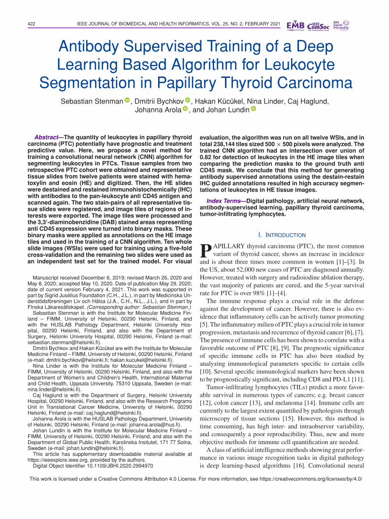
with a whole-slide image scanner (Pannoramic 250, 3DHistech,Hungary). The HE procedure as well as the scanner specificsare described in detail in the appendix. The scanned images(0.24 µm/pixel) were then imported to an image managementplatform (WebMicroscope, Aiforia Technologies Oy, Helsinki,Finland). After digitization of the HE slides, the coverslips weresoaked off in xylene and the sections were rehydrated. Then theHE was boiled off during antigen retrieval for 20 min in 99 °C10 mM Tris/ 1mM EDTA pH9 solution. For antigen retrieval,slides were pre-treated with Cell Conditioning 1 buffer (VentanaMedical Systems, Inc., Arizona, USA) for 20 min. The tissuesections were incubated with the primary anti CD45 antibody(RTU CD45, clone 2B11PD7/26, Ventana) for 44 min at roomtemperature and the tissue sections were then processed usingan automated staining system (BenchMark ULTRA system,Ventana). A 3,3′-diaminobenzidine (DAB) kit (UltraVIEW, Ven-tana) was used as the detection. Finally, the anti CD45 stainedtissue sections were digitized into whole-slide images (WSIs)using the same scanner as with the HE stained samples (Fig. 1).
C. Creation of Binary Masks
For creating binary masks, we first created a custom soft-ware using C# in Dotnet (Microsoft, Redmond, WA) and Win-dows Presentation Foundation (WPF, Microsoft, Redmond, WA)frameworks. The five WSI pairs of HE and anti CD45 DABantibody stained tissue sections were imported to the softwareand registered. Using the abovementioned custom software, wemanually registered the WSIs using the HE as base layer and theground truth anti CD45 DAB immunostaining as the top layer.First, the slides were roughly registered using morphologicallandmarks that could be seen in the tissue slides. Following this,regions of interest (ROIs) were exported as 5000 × 5000-pixelsized tiles. These tiles were then re-layered and re-registered,now on a cell level, and further tiled into smaller 500× 500-pixelsized image tiles. This two-step matching and tiling procedurewas performed to limit the impact of potential stitching shiftsthat occur in the digitization of the slides (Fig. 2). During tilingand exporting of the 500 × 500 sized images, the ground truthmask, the anti CD45 DAB stained sample, was turned into abinary mask in multiple steps. First, the image tiles were splitinto red, green and blue color channels. Since the blue colorchannel had the best contrast between DAB stained anti CD45positive regions and the background, the red and green colorchannels were discarded. The blue color channel image wasthen converted into a binary mask by manually selecting thethreshold matrix prior to export. After this, the binary mask wasfurther processed by blurring. Finally, noise was filtered out bydiscarding areas smaller than a total area of 350 pixels (Fig. 3).
D. Image Datasets
A total of 1,738 500 × 500-pixel image tiles of the twelvedestained-restained WSIs (range: 48–321 tiles per tissue slide)were selected and used in the training and testing of the CNNalgorithm. The tiles of ten of these slides (n = 1,387) wereused for training and validating the CNN algorithm. The trainedmodel was then tested on an independent test set comprising 351
424 IEEE JOURNAL OF BIOMEDICAL AND HEALTH INFORMATICS, VOL. 25, NO. 2, FEBRUARY 2021
Fig. 1. Flowchart of protocol used in the study. First, tissue sections were stained with hematoxylin and eosin. Then, the sections were digitizedinto whole-slide images (WSIs) before being destained. The sections were then restained with anti CD45 antibody immunostaining and againscanned into WSIs. This staining protocol yielded HE and anti CD45 antibody stains of the same tissue section.
500 × 500-pixel tiles exported from the remaining two destain-restained tissue slides (Fig. 4). The entire WSIs in the trainingset were analyzed for internal validation outside the tiled regionsused for training.
The WSIs were analyzed in 500 × 500-pixel image tiles.Before performing the analysis, we discarded the all-white tileswhich did not include any tissue. Excluding the training andtesting tiles, 236,377 tiles of 500 × 500 pixels (mean 19,698tiles per slide, range: 11,464–30,476 tiles) were analyzed withthe CNN algorithm. Of these, 197,071 were from the ten WSIsincluded in the cross-validation training. The 39,306 remainingtiles were from the WSIs in the independent test set.
E. Deep Convolutional Neural Network Image Analysis
The U-Net architecture [28] is a CNN tailored to solve variousimage segmentation tasks in the medical domain. Specifically,the U-Net performs dense semantic segmentation, where eachpixel of the input image is assigned a corresponding class label.In our study, we adapted the U-Net architecture with ImageNet[29] pre-trained weights and ResNet-18 [30] backbone to per-form binary segmentation, i.e. separation of leukocytes fromthe rest of the tissue. The upward path was left identical to theoriginal U-Net architecture. We used the default learning rateof 0.001. Batch normalization was not used, nor was dropout orL1/L2 regularization. Also, both encoder and decoder weightswere optimized at training phase. We utilized on the fly dataaugmentation by applying random horizontal and vertical flipsto the image-mask pairs as well as random shear up to 15 percent.Training was done by presenting augmented HE tissue samples
as input and corresponding anti CD45 DAB binary masks as out-put. A five-fold cross-validation method was implemented. Thefolds were made by preserving the percentage of tiles from eachof the WSIs in both training and validation splits. In each fold,an average of 1,110 tiles were used for training and an average277 tiles were used for validation (Fig. 4). After the networkmodels reached their best performance on the validation set, itwas evaluated on held-out image-mask pairs to validate that thetrained model generalizes and performed well on unseen data.Each of the five models trained in cross-validation were appliedto the held-out set and the results were averaged. Evaluation ofthe results was done both quantitatively, by calculating segmen-tation accuracy metrics, as well as qualitatively, by visual ex-amination and comparison of ground truth labels with predictedsegmentation masks. For quantitative assessment of algorithmperformance, the prediction map created by the algorithm wasturned into binary masks which were compared to the anti CD45DAB ground truth binary masks. For visual assessment, theprobability score for each pixel generated by the algorithm wasturned into a color intensity score which resulted in a heatmapdirectly based on the probability map. The generated heatmapwas then registered with the HE stain for each correspondingWSI for visual assessment. At training an adaptive learning rateoptimization algorithm [31] was minimizing the Jaccard indexin mini batches of size 16 over 45 epochs.
F. Ethical Statement
The Ethics Committee at the University of Helsinki approvedthe study protocol (226/E6/2006, extension 17.4.2013). The

STENMAN et al.: ANTIBODY SUPERVISED TRAINING OF A DEEP LEARNING 425
Fig. 2. The hematoxylin and eosin (HE) and pan-leukocyte anti CD45antibody stained tissue slides were layered and manually registeredusing a custom software. Larger, 5000 × 5000-pixel image tiles of re-gions of interest were then exported. Following this, the tiles were againlayered and matched and further tiled into 500 × 500-pixel image tiles.During the last export, the registered positively DAB stained anti CD45regions were processed in multiple steps and turned into binary masks.The binary masks were used as annotations for the HE stained imagesand a convolutional neural network (CNN) algorithm was trained basedon the patterns of these masks. Results of the algorithm is illustrated asheatmaps.
National Authority of Welfare and Health approved the retro-spective study (Valvira Dnro 10041/06.01.03.01/2012).
III. RESULTS
For performance evaluation, the trained CNN algorithm wastested on 351 image tiles sized 500 × 500 pixels. For eachof these tiles, the probability masks generated by the CNNalgorithm were turned into binary masks and compared to theground truth mask based on the anti CD45 DAB staining. Thus,in pixel-wise comparisons of the algorithm result mask and theground truth mask, a total of 87.8 million pixels were compared.Based on this independent test set, we observed an intersectionover union (IoU) of 0.82. By averaging results from the fivemodels trained in cross-validation, we observed a receiver oper-ating characteristics area under the curve (ROC AUC) of 0.96 onthe held-out data when comparing the anti CD45 DAB groundtruth mask to the algorithm result mask on pixel level (Fig. 5).
For visual performance assessment, the HE WSIs of all twelvedestain-restained tissue sections were analyzed by the trainedalgorithm. For convenient visual evaluation, we registered thealgorithm result heatmap and the HE stain of the ten WSIsincluded in the training set. For the test WSIs, we registeredthe HE stain, the algorithm result heatmap, as well as the antiCD45 DAB ground truth stain. The algorithm results could bevisually compared to the anti CD45 DAB antibody stain bymoving through the layers (Fig. 6). The WSIs subject to thevisual performance assessment can be explored via the followingURL: https://tinyurl.com/qorlnlg
IV. DISCUSSION
In this proof-of-concept study, we used a novel method forantibody-supervised training of a CNN algorithm. The trainedalgorithm was highly accurate both measured on a pixel-levelin the test set image tiles and through visual examination of theanalyzed WSIs, which indicates that the proposed method ofgenerating training sets is feasible.
Machine learning-based tools have previously been used toquantify leukocytes within tumors [21], [22]. However, thesemethods use supervised learning and rely on manual annota-tions. This method is both laborious and subjective and thus haspoor reproducibility. We propose a method that require no man-ual annotations and is more objective. To the best of our knowl-edge, the proposed method for applying antibody supervisedannotations derived from binary masks using a destain-restainprotocol of the same tissue section has not been described.
Previously, a method for transferring annotations from an IHCstain to HE has been proposed [25]. However, this method differsfrom the present method in a few ways. First, in our method, wesuggest using binary masks when transferring the annotationsin between stains which allows for pixel-wise training of thealgorithm. In contrast, in the paper by Tellez et al, they trainthe first CNN patch-wise using 100 × 100-pixel tiles. Secondly,in the method proposed in this paper, no manual annotationsare required, compared to an average of 2 hours per observer.However, we manually reviewed and selected the exported tileswhich introduced a manual element in our method as well.
In order to train an algorithm for segmentation tasks, the anno-tated or labeled training material has to be as precise as possible.Thus, creating high quality training material is a time-consumingprocess. Our proposed method replaces the manual annotationtask with the IHC staining mask that is directly applied as anno-tations to the HE-stained training material. This trains the CNNmodel to output a virtual IHC stain e.g. a “digital biomarker”that mimics the performance of the particular antibody used asthe mask.
The proposed method for generating training data is fast;matched and marked areas are tiled, processed, and exported in amatter of minutes and can easily be extended to whole WSIs thatare hard to fully label by a human annotator. Also, as compared tomanual annotation, the antibody-supervised annotation methodis likely to be more reproducible.
We explored several different thresholding methods for theimage processing, including automatic methods such as Otsu’s

426 IEEE JOURNAL OF BIOMEDICAL AND HEALTH INFORMATICS, VOL. 25, NO. 2, FEBRUARY 2021
Fig. 3. Image processing protocol. First, the images were split into red, green and blue color channels, and the red and green channels werediscarded (a). Since the blue color channel had the best contrast between anti CD45 DAB positive and background regions, we used this for furtherprocessing. We then turned the blue channel image into a binary mask (b). This image was then blurred and noise smaller than a total area of 350pixels were filtered out (c).
Fig. 4. A consort diagram showing the datasets used in the study. Atotal of twelve destained-restained tissue slides were used. Ten WSIswere used in a five-fold cross validation training protocol. The tiles fromall ten WSIs were pooled and divided up in five batches so that eachfold had an equal number of tiles from all ten WSIs averaging 1,110tiles for training and 277 for validation. The performance of the fivemodels generated were averaged and run on unseen image tiles fromthe two WSIs in the test set and the performance was evaluated bothquantitatively and visually.
method. The masks created by various thresholding methodswere visually reviewed and we decided on a manual thresh-olding method since it gave superior results compared to theother methods. This, in turn, means that the threshold has tobe manually selected and thus introduces some subjectivity.However, compared to a fully manual way of creating trainingannotations, this method of antibody-supervised training is stilla more objective way of creating annotations. In our proposedmethod we selected the blue color channel for further imageprocessing. This color channel was selected because it offeredgood contrast with the positively DAB stained areas and the restof the tissue and thus could easily be converted to a binary mask.A method for color deconvolution has previously been proposed[32]. We also explored this method which resulted in similarbinary masks as with our proposed method (supplementaryFig. 1). However, using an image with only one-color channel,as we propose, decreases the size of the files being processed to
Fig. 5. Average receiver operating characteristics (ROC) curve of thefive-fold cross validation algorithm. The pixel-level predictions scores ofthe five models were averaged and turned into binary masks which werethen compared with the anti CD45 DAB ground truth masks of the imagetiles in the independent test set. The independent test set comprised of351 tiles of 500 × 500 pixels. Thus, 87.8 million pixels were evaluated.The area under the curve was 0.96.
one third compared to using RGB images. This is a significantdecrease in data being processed and using color deconvolutioncould drastically slow down the image processing step in biggerdatasets.
Since the annotations of the training material is directly basedon the IHC stains, the staining quality, in turn, directly affectsalgorithm performance. In the present study, we limited thisby manually reviewing all exported image tiles and selectingthe training data. Another issue that needs to be taken intoconsideration is the scanner stitching artifacts. When digitizinga tissue slide, the slide is first being scanned in tiles and thenstitched together. This might cause imperfect alignment andthus some shift between the scanned tiles. This effect is furtheramplified when layering multiple WSIs and had to be considered

STENMAN et al.: ANTIBODY SUPERVISED TRAINING OF A DEEP LEARNING 427
Fig. 6. Analysis of one of the whole slide images in the independent test set. Two separate regions of interest zoomed-in from the hematoxylinand eosin slide (a and b) and the corresponding areas in the immunohistochemically stained slide (e and f). The images in between these (c andd) represents heatmaps of the analysis result of the trained deep convolutional neural network algorithm. The rest of the WSIs can be explored indetail via the following URL: https://tinyurl.com/qorlnlg.
in the layering and tiling process. Therefore, we used a two-stepmatching and tiling protocol previously described in the methodssection to limit the impact of the stitching shifts (Fig. 2). Also,training images with misalignments due to stitching shift werediscarded in the manual review process.
We trained the algorithm using a five-fold cross validation on1,387 image tiles cropped from ten WSIs and annotated usingthe proposed antibody-supervised method. In the quantitativeperformance evaluation, the algorithm accurately segmentedleukocytes in the HE stained WSIs when comparing algorithmprediction masks to the ground truth masks. For visual per-formance evaluation, we then ran the algorithm on all twelveWSIs. The material in the training and testing comprised ofonly 0.73% of the entire WSI tissue area analyzed by thealgorithm. Even though the algorithm was trained on a relativelysmall training set and few individual tissue slides, the algorithmaccurately segmented leukocytes in the test WSIs (URL: https://tinyurl.com/qorlnlg). Here, the algorithm can also be seenworking well on slides containing few leukocytes (supplemen-tary Fig. 2). However, due to several factors, the accuracy tends todrop when testing algorithms on samples coming from differentcenters [33]. Thus, including more samples representing a largervariety of fixation, staining and scanning protocols from multiplecenters in the training set needs to be evaluated in further studies.
The prognostic and predictive value of the quantity of per-itumoral and tumor infiltrating immune cells in PTC is stilllargely unclear. The HE stain is widely used for identification ofleukocytes in tumor tissue in addition to identification of specificantibodies [34]. Therefore, we used HE as the selected stainingfor training of the CNN algorithm. Our aim was to study whetheran algorithm can be trained to detect leukocytes in HE stained
samples guided by a leukocyte-specific antibody. The anti CD45pan-leukocyte marker was used as the immune cell marker in thepresent study, but the method needs to be evaluated using otherimmunohistochemistry stainings in future studies.
In conclusion, we show that a destain-restain protocol andtransfer of the anti CD45 DAB stain to be used as a mask onHE stained samples is a feasible method for quickly generatingannotations for training accurate machine learning models.
APPENDIX
The freshly cut tissue glass slides were stained with hema-toxylin & eosin according to standard procedure. First, the tissuesections were deparaffinized and rehydrated after which theywere incubated for 10 min at room temperature in Mayer’shemalum solution (Merck 1.09249, Merck Life Science, Darm-stadt, Germany). The slides were then rinsed under tap waterfor 5 min and the staining was differentiated by dipping theslides twice in 70% EtOH/1% HCl solution. The slides werethen rinsed with tap water for 5 min more after which they wereincubated in aqueous 1% eosin solution (Sigma E4382, SigmaCorporation, USA) for 2 min. After dehydration in alcohol andxylene, the slides were mounted in DPX mounting medium(Sigma, USA).
Both the HE and the immunohistochemistry stained sam-ples were digitized with a scanner (Pannoramic 250 FLASH3DHISTECH Ltd., Budapest, Hungary) equipped with a plan-apochromat 20x objective (NA 0.8), a VCC-F52U24CL camera(CIS, Tokyo, Japan) with three image sensors (1,224 × 1,624;4.4 × 4,4 µm/pixels), and a 1.0 adapter. After digitization,the scanned images were compressed into a wavelet format
428 IEEE JOURNAL OF BIOMEDICAL AND HEALTH INFORMATICS, VOL. 25, NO. 2, FEBRUARY 2021
(Enhanced Compressed Wavelet, ECW, ER Mapper, Intergraph,Atlanta, GA) with a compression ratio of 1:9 and importedto an image management platform (WebMicroscope, AiforiaTechnologies Oy, Helsinki, Finland).
ACKNOWLEDGMENT
The author would like to thank the FIMM Digital Microscopyand Molecular Pathology Unit supported by Helsinki Universityand Biocenter Finland for outstanding assistance.
DISCLOSURE STATEMENT
Johan Lundin is a Founder and Chief Scientific Officer atAiforia Oy, Helsinki, Finland.
REFERENCES
[1] A. M. Noone et al., “SEER Cancer Statistics Review 1975-2016. NationalCancer Insitute,” Bethesda, MD, USA, based on Nov. 2017 SEER datasubmission, posted to the SEER web site, Apr. 2018. [Online]. Available:https://seer.cancer.gov/csr/1975_2015/
[2] L. Davies and H. G. Welch, “Current thyroid cancer trends in theUnited States,” Otolaryngology—Head Neck Surgery, vol. 140, no. 4,pp. 317–322, Apr. 2014, doi: 10.1001/jamaoto.2014.1.
[3] R. V. Lloyd, R. Y. Osamura, and G. Kloppel, WHO Classification ofTumours: Pathology and Genetics of Tumours of Endocrine Organs, 4thed. Lyon, France: IARC, 2017.
[4] D. S. McLeod, A. M. Sawka, and D. S. Cooper, “Controversies in primarytreatment of low-risk papillary thyroid cancer,” Lancet, vol. 381, no. 9871,pp. 1046–1057, Mar. 23, 2013, doi: 10.1016/S0140-6736(12)62205-3.
[5] D. Hanahan and R. A. Weinberg, “The hallmarks of cancer,” Cell, vol. 100,no. 1, pp. 57–70, Jan. 2000, doi: 10.1016/s0092-8674(00)81683-9.
[6] J. Modi, A. Patel, R. Terrell, R. M. Tuttle, and G. L. Francis, “Papil-lary thyroid carcinomas from young adults and children contain a mix-ture of lymphocytes,” J. Clin. Endocrinol. Metabolism, vol. 88, no. 9,pp. 4418–4425, Sep. 2003, doi: 10.1210/jc.2003-030342.
[7] J. S. Jeong et al., “Coexistence of chronic lymphocytic thyroiditis withpapillary thyroid carcinoma: Clinical manifestation and prognostic out-come,” J. Korean Med. Sci., vol. 27, no. 8, pp. 883–889, Aug. 2012, doi:10.3346/jkms.2012.27.8.883.
[8] S. K. Kim et al., “Chronic lymphocytic thyroiditis and BRAF V600E inpapillary thyroid carcinoma,” Endocrine-Related Cancer, vol. 23, no. 1,pp. 27–34, Jan. 2016, doi: 10.1530/ERC-15-0408.
[9] L. L. Cunha and L. S. Ward, “Concurrent lymphocytic thyroiditis is asso-ciated to less aggressive papillary thyroid carcinomas,” Eur. Arch. Otorhi-nolaryngol., vol. 269, no. 2, pp. 699–700, Feb. 2012, doi: 10.1007/s00405-011-1764-y.
[10] L. L. Cunha et al., “CD8+ tumour-infiltrating lymphocytes and COX2expression may predict relapse in differentiated thyroid cancer,” Clin.Endocrinology (Oxf), vol. 83, no. 2, pp. 246–253, Aug. 2015, doi:10.1111/cen.12586.
[11] M. J. Aghajani, T. Yang, C. E. McCafferty, S. Graham, X. Wu, andN. Niles, “Predictive relevance of programmed cell death protein1 and tumor-infiltrating lymphocyte expression in papillary thyroidcancer,” Surgery, vol. 163, no. 1, pp. 130–136, Jan. 2018, doi:10.1016/j.surg.2017.04.033.
[12] P. Savas et al., “Clinical relevance of host immunity in breast cancer: FromTILs to the clinic,” Nature Rev. Clin. Oncol., vol. 13, no. 4, pp. 228–241,Apr. 2016, doi: 10.1038/nrclinonc.2015.215.
[13] J. Galon et al., “Towards the introduction of the ‘Immunoscore’ inthe classification of malignant tumours,” J. Pathol., vol. 232, no. 2,pp. 199–209, Jan. 2014, doi: 10.1002/path.428.
[14] S. M. Ferrari et al., “Thyroid autoimmune disorders and can-cer,” Semin. Cancer Biol., vol. 64, pp. 135–146, Aug. 2020, doi:10.1016/j.semcancer.2019.05.019.
[15] R. Salgado et al., “The evaluation of tumor-infiltrating lymphocytes (TILs)in breast cancer: Recommendations by an International TILs WorkingGroup 2014,” Ann. Oncology, vol. 26, no. 2, pp. 259–271, Feb. 2015, doi:10.1093/annonc/mdu450.
[16] D. Shen, G. Wu, and H. I. Suk, “Deep learning in medical image anal-ysis,” Annu. Rev. Biomed. Eng., vol. 19, pp. 221–248, Jun. 2017, doi:10.1146/annurev-bioeng-071516-044442.
[17] F. Xing and L. Yang, “Robust nucleus/cell detection and segmenta-tion in digital pathology and microscopy images: A comprehensivereview,” IEEE Rev. Biomed. Eng., vol. 9, pp. 234–263, 2016, doi:10.1109/RBME.2016.2515127.
[18] D. Bychkov et al., “Deep learning based tissue analysis predicts outcomein colorectal cancer,” Sci. Rep., vol. 8, no. 1, pp. 3395-018–21758-3,Feb. 2018, doi: 10.1038/s41598-018-21758-3.
[19] G. Corredor et al., “Spatial architecture and arrangement of tumor-infiltrating lymphocytes for predicting likelihood of recurrence in early-stage non-small cell lung cancer,” Clin. Cancer Res., vol. 25, vol. 5,pp. 1526–1534, Mar. 2019, doi: 10.1158/1078-0432.CCR-18-201.
[20] A. Gupta et al., “Deep learning in image cytometry: A review,” CytometryA., vol. 95, no. 4, pp. 366–380, Apr. 2019, doi: 10.1002/cyto.a.23701.
[21] N. Linder et al., “Deep learning for detecting tumour-infiltrating lym-phocytes in testicular germ cell tumours,” J. Clin. Pathol., vol. 72, no. 2,pp. 157–164, Feb. 2019, doi: 10.1136/jclinpath-2018-205328.
[22] R. Turkki, N. Linder, P. E. Kovanen, T. Pellinen and J. Lundin, “Antibody-supervised deep learning for quantification of tumor-infiltrating immunecells in hematoxylin and eosin stained breast cancer samples ” J. Pathol.Informat., vol. 7, Sep. 2016, Art. no. 38, doi: 10.4103/2153-3539.189703.
[23] M. Valkonen et al., “Cytokeratin-supervised deep learning for automaticrecognition of epithelial cells in breast cancers stained for ER, PR, andKi-67,” IEEE Trans. Med. Imaging, vol. 39, no. 2, pp. 534–542, Feb. 2020,doi: 10.1109/TMI.2019.2933656.
[24] H. O. Helin, M. E. Lundin, M. Laakso, J. Lundin, H. J. Helin, and J. Isola,“Virtual microscopy in prostate histopathology: simultaneous viewingof biopsies stained sequentially with hematoxylin and eosin, and alpha-methylacyl-coenzyme A racemase/p63 immunohistochemistry,” J. Urol-ogy, vol. 175, no. 2, pp. 495–499, Feb. 2006, doi: S0022-5347(05)00164-3.
[25] D. Tellez et al., “Whole-slide mitosis detection in H&E breast histologyusing PHH3 as a reference to train distilled stain-invariant convolutionalnetworks,” IEEE Trans. Med. Imaging, vol. 37, no. 9, pp. 2126–2136,Sep. 2018, doi: 10.1109/TMI.2018.2820199.
[26] P. Siironen et al., “Prognostic factors in papillary thyroid cancer: an evalu-ation of 601 consecutive patients,” Tumour Biol., vol. 26, no. 2, pp. 57–64,Mar./Apr. 2005, [Online]. Available: https://doi.org/10.1159/000085586.
[27] S. Stenman, P. Siironen, H. Mustonen, J. Lundin, C. Haglund, and J. Arola,“The prognostic significance of tall cells in papillary thyroid carcinoma:A case-control study,” Tumour Biol., vol. 40, no. 7, Jul. 2018, doi:10.1177/1010428318787720.
[28] O. Ronneberger, P. Fischer and T. Brox, “U-Net: Convolutional net-works for biomedical image segmentation,” in Medical Image Comput-ing Computer-Assisted Intervention. Lecture Notes in Computer Science,Springer, Cham, vol 9351, 2015.
[29] J. Deng, W. Dong, R. Socher, L. Li, K. Li, and L. Fei-Fei, “Imagenet:A large-scale hierarchical image database,” in Proc. IEEE Conf. Comput.Vision Pattern Recogn., 2009, pp. 248–255.
[30] K. He, X. Zhang, S. Ren, and J. Sun, “Deep residual learning for imagerecognition,” in Proc. IEEE Conf. Comput. Vision Pattern Recognition,2016, pp. 770–778.
[31] D. P. Kingma and J. Ba, “Adam: A method for stochastic optimization,” inProc. 3rd Int. Conf. Learn. Representations, San Diego, CA, USA, 2015,arXiv:1412.6980.
[32] A. C. Ruifrok and D. A. Johnston, “Quantification of histochemicalstaining by color deconvolution,” Anal. Quantitative Cytology Histology,vol. 23, no. 4, pp. 291–299, Aug. 2001.
[33] F. Wang, L. P. Casalino and D. Khullar, “Deep learning in medicine—Promise, progress, and challenges,” Intemed, vol. 179, no. 3, pp. 293–294,2019, doi: 10.1001/jamainternmed.2018.7117.
[34] R. Salgado et al., “The evaluation of tumor-infiltrating lymphocytes (TILs)in breast cancer: Recommendations by an International TILs workinggroup 2014,” Ann. Oncology, vol. 26, no. 2, pp. 259–271, Feb. 2015, doi:10.1093/annonc/mdu450.