appendices appendix a morphometric analysis …studentsrepo.um.edu.my/6085/7/appendix3.pdf146...
TRANSCRIPT

146
APPENDICES
Appendix A – Morphometric analysis guideline of frog and its spinal cord
Dorsal view of Fejervarya limnocharis
Ventral view of frog spinal cord (C: length of cervical level, T: length of thoracic level,
L: length of lumbar level, S: length of sacral level)
Snout-to-vent length
Total length
C T L S
147
Appendix B – Preparation of chemical solutions for animal perfusion and histology
General
1) Tricaine methanesulfonate (MS-222)
Tricaine methanesulfonate 350mg
Distilled water 1000ml
2) 0.6% frog physiological saline
Sodium chloride (NaCl) 0.6g
Distilled water 100ml
3) 10% neutral buffered formalin
Formaldehyde 100ml
Distilled water 900ml
Sodium phosphate, monobasic, monohydrate (NaH2PO4) 4g
Sodium phosphate, dibasic, anyhydrous (Na2HPO4) 6.5g
4) 70% alcohol solution
95% alcohol solution 700ml
Distilled water 250ml
5) 85% alcohol solution
95% alcohol solution 850ml
Distilled water 100ml
148
6) Mayer’s albumin
Egg white 5g
Acetone 2ml
Distilled water 2ml
Hematoxylin and Eosin (H&E) Staining Method
1) Eosin stain
Eosin Y solution (water-soluble) 0.2ml
95% alcohol 99.8ml
2) Harris’ alum hematoxylin stain
Hematoxylin crystals 1g
Absolute alcohol (99.5%) 10ml
Aqueous aluminum ammonium sulphate 1ml
Note: Hematoxylin crystals are dissolved in alcohol and then, aluminum ammonium
sulphate is added and brought to boil. 0.5 gram of mercuric oxide is slowly added to
oxidize the staining solution. The solution is left to cool and then filtered.
149
Nissl Staining Method
1) Buffer solution pH 3.5
0.1M acetic acid (6ml/1,000ml water) 94ml
0.1M sodium acetate (13.6g/1,000ml water) 6ml
2) Cresyl violet (can only be used twice)
Cresyl violet acetate 0.2g
Distilled water 150ml
3) Working solution
Buffer solution 100ml
Cresyl violet 10ml
Lillie’s Variant of the Weil-Weigert Method (Lillie, 1954)
1) 1% acetic acid
Acetic acid 1ml
Distilled water 100ml
2) 1% alcoholic hematoxylin
Hematoxylin crystals 1g
Absolute alcohol 100ml
3) 10% alcoholic hematoxylin
Hematoxylin crystals 10g
Absolute alcohol 100ml
150
4) 0.5% iron alum
Iron alum 0.5g
Distilled water 100ml
5) 4% iron alum
Ferric ammonium sulphate 4g
Distilled water 100ml
6) 2.5% potassium ferricyanide solution
Potassium ferricyanide 2.5g
Distilled water 100ml
7) Safranin O
Safranin 0.1g
1% acetic acid 100ml

151
Modification of Golgi Method
1) 3% potassium bichromate
Potassium bichromate 3g
Distilled water 100ml
2) 2% silver nitrate
Silver nitrate 2g
Distilled water 100ml
Thionin Staining Method (Gurr, 1956)
1) 0.5% thionin in 10% formalin
Thionin 0.5g
10% neutral buffered formalin 100ml
152
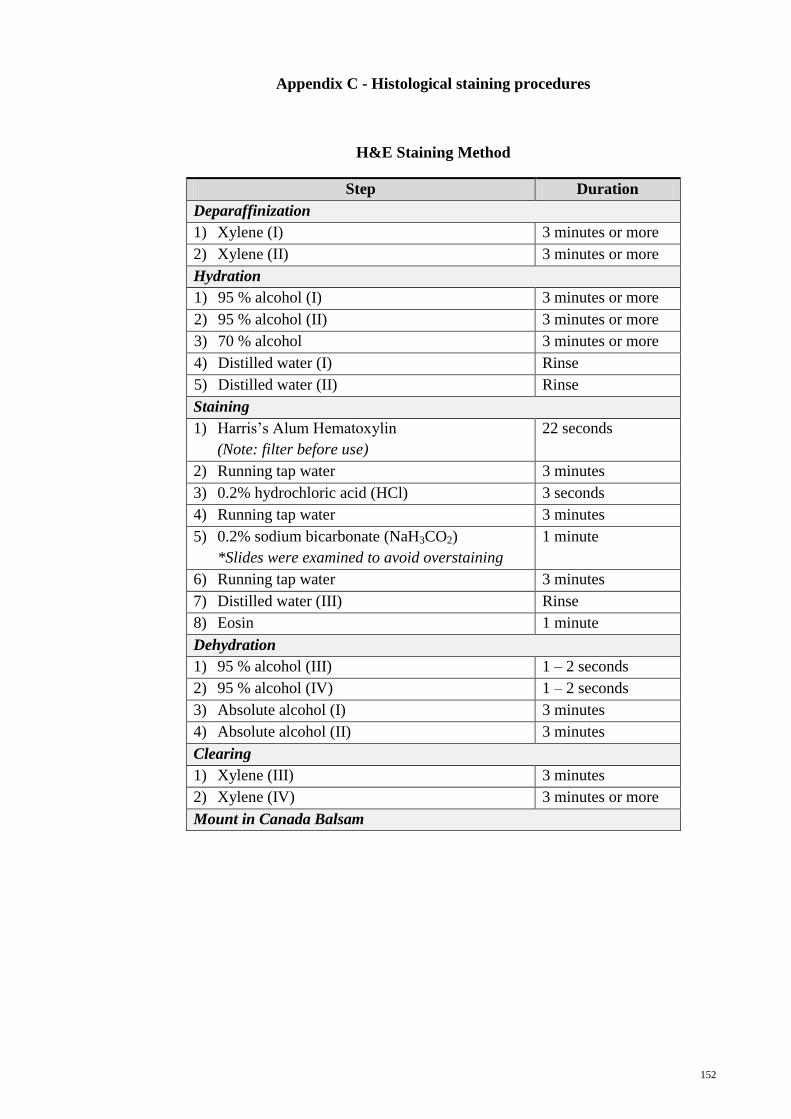
Appendix C - Histological staining procedures
H&E Staining Method
Step Duration
Deparaffinization
1) Xylene (I) 3 minutes or more
2) Xylene (II) 3 minutes or more
Hydration
1) 95 % alcohol (I) 3 minutes or more
2) 95 % alcohol (II) 3 minutes or more
3) 70 % alcohol 3 minutes or more
4) Distilled water (I) Rinse
5) Distilled water (II) Rinse
Staining
1) Harris’s Alum Hematoxylin
(Note: filter before use)
22 seconds
2) Running tap water 3 minutes
3) 0.2% hydrochloric acid (HCl) 3 seconds
4) Running tap water 3 minutes
5) 0.2% sodium bicarbonate (NaH3CO2)
*Slides were examined to avoid overstaining
1 minute
6) Running tap water 3 minutes
7) Distilled water (III) Rinse
8) Eosin 1 minute
Dehydration
1) 95 % alcohol (III) 1 – 2 seconds
2) 95 % alcohol (IV) 1 – 2 seconds
3) Absolute alcohol (I) 3 minutes
4) Absolute alcohol (II) 3 minutes
Clearing
1) Xylene (III) 3 minutes
2) Xylene (IV) 3 minutes or more
Mount in Canada Balsam
153
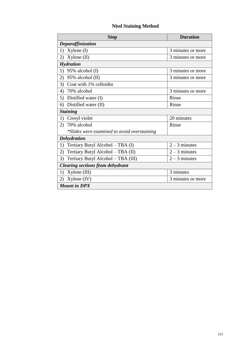
Nissl Staining Method
Step Duration
Deparaffinization
1) Xylene (I) 3 minutes or more
2) Xylene (II) 3 minutes or more
Hydration
1) 95% alcohol (I) 3 minutes or more
2) 95% alcohol (II) 3 minutes or more
3) Coat with 1% celloidin
4) 70% alcohol 3 minutes or more
5) Distilled water (I) Rinse
6) Distilled water (II) Rinse
Staining
1) Cresyl violet 20 minutes
2) 70% alcohol
*Slides were examined to avoid overstaining
Rinse
Dehydration
1) Tertiary Butyl Alcohol – TBA (I) 2 – 3 minutes
2) Tertiary Butyl Alcohol – TBA (II) 2 – 3 minutes
3) Tertiary Butyl Alcohol – TBA (III) 2 – 3 minutes
Clearing sections from dehydrant
1) Xylene (III) 3 minutes
2) Xylene (IV) 3 minutes or more
Mount in DPX
154
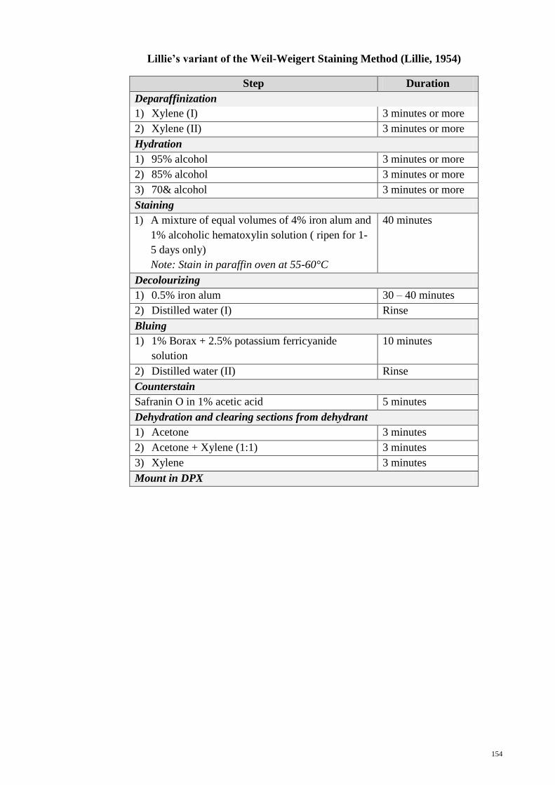
Lillie’s variant of the Weil-Weigert Staining Method (Lillie, 1954)
Step Duration
Deparaffinization
1) Xylene (I) 3 minutes or more
2) Xylene (II) 3 minutes or more
Hydration
1) 95% alcohol 3 minutes or more
2) 85% alcohol 3 minutes or more
3) 70& alcohol 3 minutes or more
Staining
1) A mixture of equal volumes of 4% iron alum and
1% alcoholic hematoxylin solution ( ripen for 1-
5 days only)
Note: Stain in paraffin oven at 55-60°C
40 minutes
Decolourizing
1) 0.5% iron alum 30 – 40 minutes
2) Distilled water (I) Rinse
Bluing
1) 1% Borax + 2.5% potassium ferricyanide
solution
10 minutes
2) Distilled water (II) Rinse
Counterstain
Safranin O in 1% acetic acid 5 minutes
Dehydration and clearing sections from dehydrant
1) Acetone 3 minutes
2) Acetone + Xylene (1:1) 3 minutes
3) Xylene 3 minutes
Mount in DPX
155
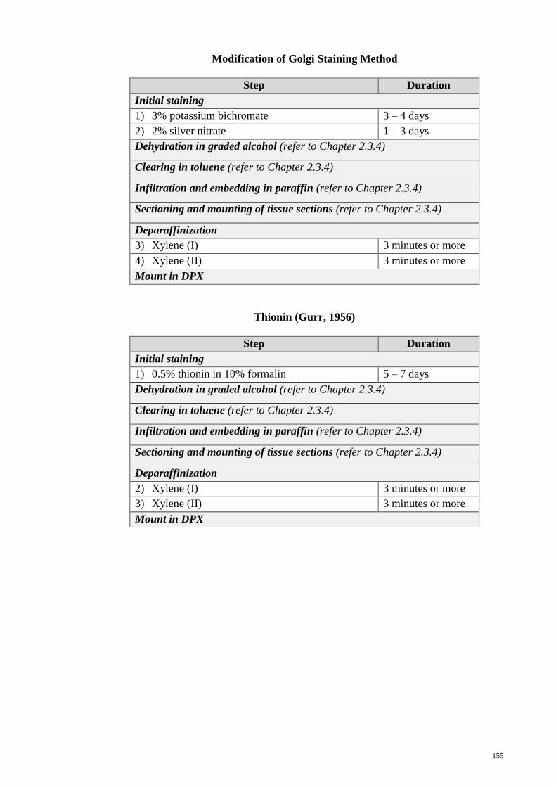
Modification of Golgi Staining Method
Step Duration
Initial staining
1) 3% potassium bichromate 3 – 4 days
2) 2% silver nitrate 1 – 3 days
Dehydration in graded alcohol (refer to Chapter 2.3.4)
Clearing in toluene (refer to Chapter 2.3.4)
Infiltration and embedding in paraffin (refer to Chapter 2.3.4)
Sectioning and mounting of tissue sections (refer to Chapter 2.3.4)
Deparaffinization
3) Xylene (I) 3 minutes or more
4) Xylene (II) 3 minutes or more
Mount in DPX
Thionin (Gurr, 1956)
Step Duration
Initial staining
1) 0.5% thionin in 10% formalin 5 – 7 days
Dehydration in graded alcohol (refer to Chapter 2.3.4)
Clearing in toluene (refer to Chapter 2.3.4)
Infiltration and embedding in paraffin (refer to Chapter 2.3.4)
Sectioning and mounting of tissue sections (refer to Chapter 2.3.4)
Deparaffinization
2) Xylene (I) 3 minutes or more
3) Xylene (II) 3 minutes or more
Mount in DPX

156
Appendix D – Preparation of chemical solutions for immunohistochemistry
1) 2% silane adhesive solution in acetone
Silane 10ml
Acetone 500ml
2) TBS-Tween 20 wash buffer
Tris-buffered saline (TBS) 1000 ml
Tween-20 0.5 ml
3) 10% normal goat serum
Normal goat serum 1000 µl
TBS 100 µl
4) 0.2% ammonia water solution (Bluing)
Ammonium hydroxide (concentrated) 2 ml
Distilled water 1000 ml

157
Appendix E – Immunohistochemistry procedure
Step Duration
Pre-warm slides at 50°C 20 minutes
Deparaffinization
1) Xylene 20 minutes
Hydration
1) Absolute alcohol 10 minutes
2) 95% alcohol 3 minutes
3) 70% alcohol 3 minutes
4) 50% alcohol 3 minutes
5) Distilled water 2 minutes
Epitope retrieval
1) Tris/EDTA pH 9.0 buffer solution 20 – 30 minutes
2) Leave to cool at room temperature 20 minutes
3) Wash buffer (TBS-Tween 20) Rinse
Endogenous peroxidase blocking
1) Peroxidase-blocking solution (H2O2) 30 minutes
2) Wash in TBS-Tween 20 5 minutes
Normal serum blocking
1) 10% normal goat serum 30 minutes
Primary antibody incubation (either one)
1) Enkephalin (1:500), or 30 minutes
Substance P (1:500), or 30 minutes
Serotonin (1:200) 40 minutes
2) Wash in TBS-Tween 20 (Repeat twice) 5 minutes
Secondary antibody incubation
1) Secondary antibody (Dako REAL™ Envision™
Detection System)
60 minutes
2) Wash in TBS-Tween 20 (Repeat thrice) 5 minutes
Incubation with chromogen
1) DAB (Dako REAL™ Envision™ Detection
System)
5 minutes
2) Distilled water Rinse
Counterstaining
1) Mayer’s Hematoxylin 30 – 60 seconds
2) Distilled water Rinse
3) 0.2% ammonia (NH4OH) for bluing 10 seconds
4) Distilled water Rinse
Mount in Glycergel

158
Appendix F – Statistical analysis on neuronal somas occurrence using SPSS
Test 1
Objective : One-way ANOVA was conducted to compare the differences in the
number of recorded somas among five somatal shapes (i.e. spindle,
triangular, polygonal, tear and others).
DESCRIPTIVES
ANOVA
MEAN PLOT
Conclusion
: It was indicated that there was a significant difference in the numbers
of recorded somas among the five somatal shapes [F(4, 25) = 37.046,
p<0.05].
159
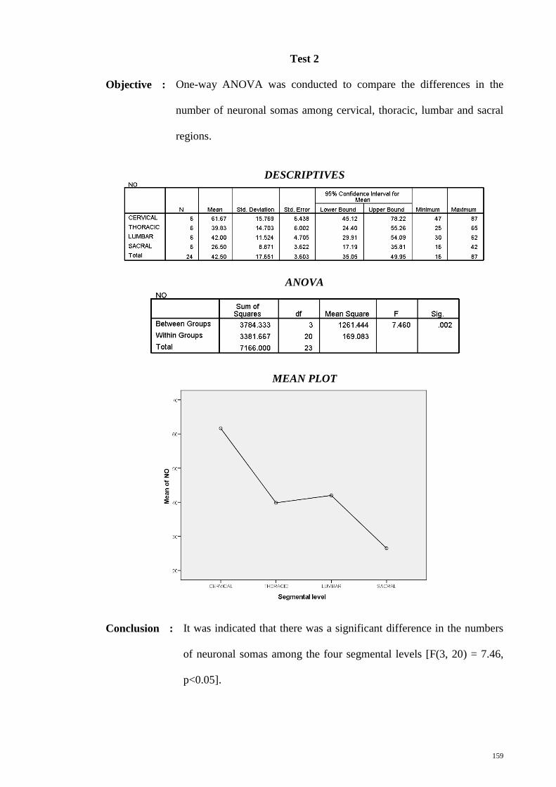
Test 2
Objective : One-way ANOVA was conducted to compare the differences in the
number of neuronal somas among cervical, thoracic, lumbar and sacral
regions.
DESCRIPTIVES
ANOVA
MEAN PLOT
Conclusion : It was indicated that there was a significant difference in the numbers
of neuronal somas among the four segmental levels [F(3, 20) = 7.46,
p<0.05].

160
Test 3
Objective : One-way between subjects ANOVA was conducted to compare the
differences in the number of recorded somas among seven fields of the
spinal grey, i.e. dorsal field (DF), lateral field (LF), central field (CF),
lateral motor field (LMF), medial motor field (MMF), ventrolateral field
(VLF) and ventromedial field (VMF).
DESCRIPTIVES
ANOVA
MEAN PLOT
Conclusion
: It was indicated that there was a significant difference in the numbers
of recorded somas among the seven fields of the spinal grey [F(6, 35)
= 47.258, p<0.05].
161
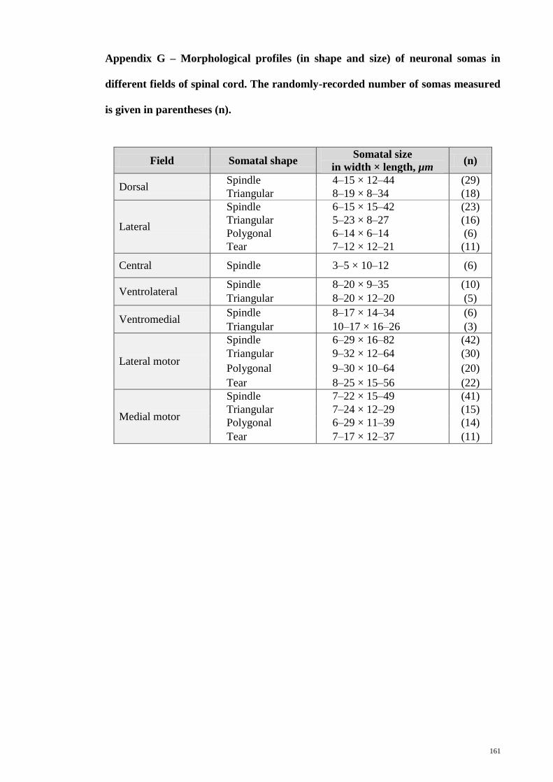
Appendix G – Morphological profiles (in shape and size) of neuronal somas in
different fields of spinal cord. The randomly-recorded number of somas measured
is given in parentheses (n).
Field Somatal shape Somatal size
in width × length, µm (n)
Dorsal Spindle 4–15 × 12–44 (29)
Triangular 8–19 × 8–34 (18)
Lateral
Spindle 6–15 × 15–42 (23)
Triangular 5–23 × 8–27 (16)
Polygonal 6–14 × 6–14 (6)
Tear 7–12 × 12–21 (11)
Central Spindle 3–5 × 10–12 (6)
Ventrolateral Spindle 8–20 × 9–35 (10)
Triangular 8–20 × 12–20 (5)
Ventromedial Spindle 8–17 × 14–34 (6)
Triangular 10–17 × 16–26 (3)
Lateral motor
Spindle 6–29 × 16–82 (42)
Triangular 9–32 × 12–64 (30)
Polygonal 9–30 × 10–64 (20)
Tear 8–25 × 15–56 (22)
Medial motor
Spindle 7–22 × 15–49 (41)
Triangular 7–24 × 12–29 (15)
Polygonal 6–29 × 11–39 (14)
Tear 7–17 × 12–37 (11)
162
Appendix H – List of publications and presentations
Published Journal
a) ISI-Cited Publication
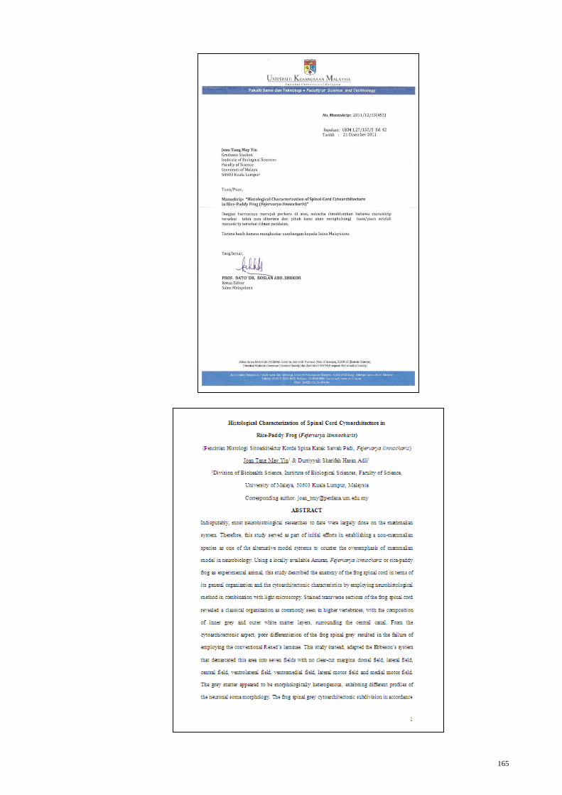
Tang, J.M.Y., and Durriyyah S. H. A. (2012). Histological characterization of spinal
cord cytoarchitecture in Rice-Paddy Frog (Fejervarya limnocharis). (Submitted
to Sains Malaysiana)
b) SCOPUS-Cited Publication
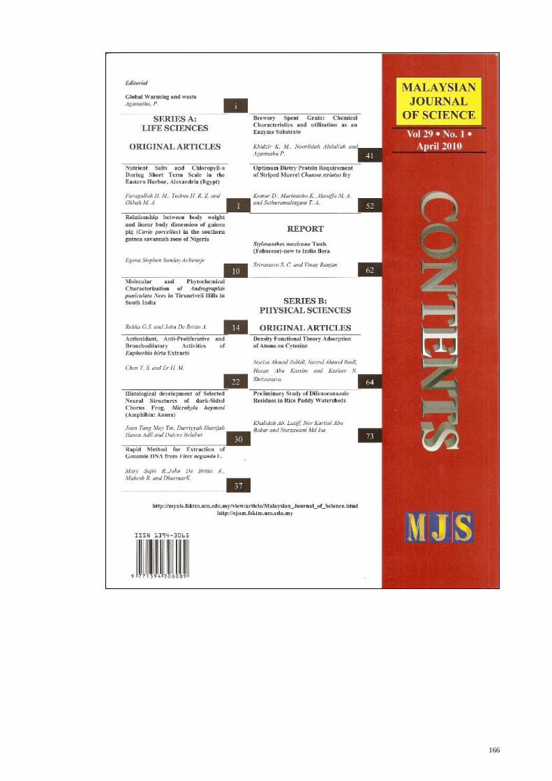
Tang, J.M.Y., Durriyyah S. H. A. and Belabut, D. (2010). Histological development of
selected neural structures of Dark-sided Chorus Frog, Microhyla heymonsi
(Amphibia: Anura). Malaysian Journal of Science, 29(1): 30-36.
Conference Proceeding
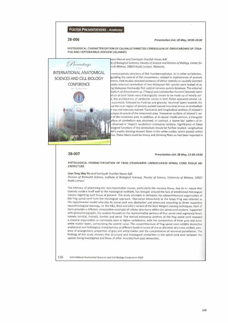
Tang, J. M. Y., and Durriyyah S. H. A. (2010). Histological Characterization of Frog
(Fejervarya limnocharis) Spinal Cord Tissue Architecture. In Proceedings of the
International Anatomical Sciences and Cell Biology Conference held on 26 May
– 29 May 2011 at the Department of Anatomy, National University of Singapore,
Singapore (pp. 116). Singapore: National University of Singapore
Chapter in Book
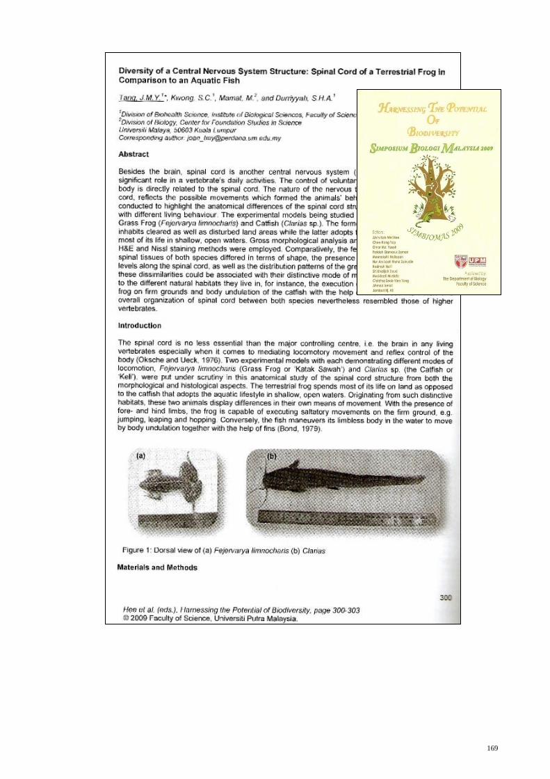
Tang, J. M. Y., Kwong, S. C., Mamat, M., and Durriyyah S. H. A. (2010). Diversity of a
Central Nervous System Structure: Spinal Cord of a Terrestrial Frog in
Comparison to an Aquatic Fish. In Hee et al. (Eds.). Harnessing the Potential of
Biodiversity (pp. 300-303). Kuala Lumpur: Universiti Putra Malaysia. (ISBN
978-983-2519-03-4)
PUBLICATIONS
163
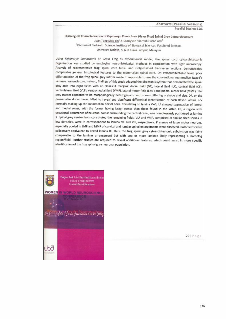
10 – 11 Oct 2011 Oral Presentation in Women in World Neurosciences Conference
2011, Universiti Brunei Darussalam, Brunei
Presentation Title: Histological Characterization of Fejervarya
limnocharis (Grass Frog)Spinal Grey Cytoarchitecture]
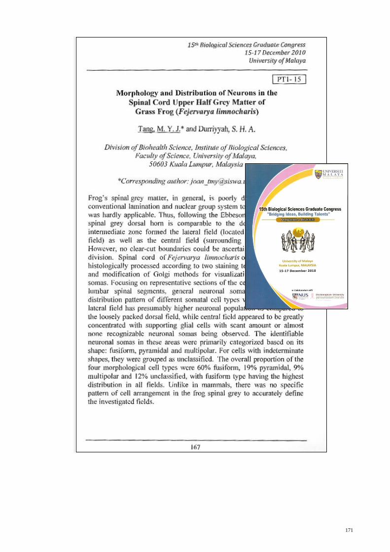
15 – 17 Dec 2010 : Poster Presentation in the 15th
Biological Sciences Graduate
Congress (BSGC) 2010, University of Malaya
Poster Title: Morphology and Distribution of Neurons in the
Spinal Cord Upper Half Grey Matter of Grass Frog (Fejervarya
limnocharis)
26 – 29 May 2010 : Poster Presentation in International Anatomical Sciences and Cell
Biology Conference (IASCBC) 2010, National University of
Singapore
Poster Title: Histological Characterization of Frog (Fejervarya
limnocharis) Spinal Cord Tissue Architecture.
14 April 2010 : Candidature Defence in Institute of Biological Sciences, Faculty
of Science, University of Malaya
Presentation Title: Spinal Cord Cytoarchitectonic Organization of
Fejerverya limnocharis in Relation to Nociceptive System.
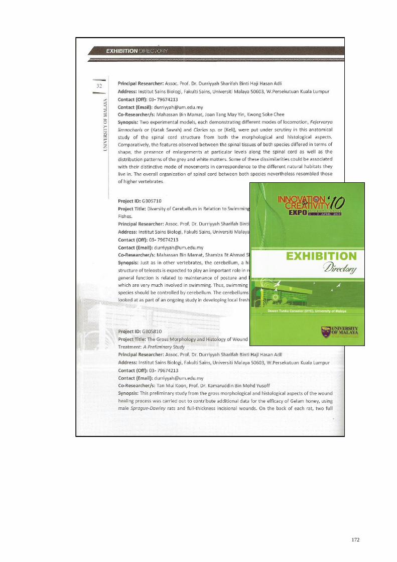
1 – 3 April 2010 : Poster Presentation in Innovation and Creativity Expo University
of Malaya 2010, University of Malaya
Poster Title: Diversity of a Central Nervous System Structure:
Spinal Cord of a Terrestrial Frog in Comparison to an Aquatic
Fish.
PRESENTATIONS
164
17 – 18 Nov 2009 : Poster Presentation in Simposium Biologi Malaysia 2009,
Universiti Putra Malaysia.
Poster Title: Diversity of a Central Nervous System Structure:
Spinal Cord of a Terrestrial Frog in Comparison to an Aquatic
Fish
1 – 2 Jul 2009
:
Oral Presentation in Postgraduate Research Seminar Biohealth
Science Programme 2009, Institute of Biological Sciences,
Faculty of Science, University of Malaya.
Presentation Title: Spinal Cord Cytoarchitectonic Organization of
Fejerverya limnocharis in Relation to Nociceptive System.
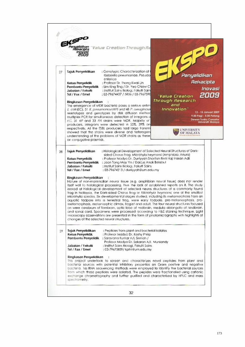
13 – 15 Jan 2009 : Poster Presentation in Eskpo Penyelidikan, Rekacipta & Inovasi
2009, University of Malaya.
Poster Title: Histological Development of Selected Neural
Structures of Dark-sided Chorus Frog, Microhyla heymonsi
(Amphibia: Anura)
165
166

167
168
169
170
171
172
173