author’s choice © 2018 by the american society for ... · spectrome´trie de masse (prism) ......
TRANSCRIPT
Spatially-Resolved Top-down ProteomicsBridged to MALDI MS Imaging Reveals theMolecular Physiome of Brain Regions*□S
Vivian Delcourt‡§**, Julien Franck‡**, Jusal Quanico‡, Jean-Pascal Gimeno‡,Maxence Wisztorski‡, Antonella Raffo-Romero‡, Firas Kobeissy¶, Xavier Roucou§,Michel Salzet‡�, and Isabelle Fournier‡�
Tissue spatially-resolved proteomics was performed on 3brain regions, leading to the characterization of 123 ref-erence proteins. Moreover, 8 alternative proteins fromalternative open reading frames (AltORF) were identified.Some proteins display specific post-translational modifi-cation profiles or truncation linked to the brain regionsand their functions. Systems biology analysis performedon the proteome identified in each region allowed to as-sociate sub-networks with the functional physiology ofeach brain region. Back correlation of the proteins iden-tified by spatially-resolved proteomics at a given tissuelocalization with the MALDI MS imaging data, was thenperformed. As an example, mapping of the distributionof the matrix metallopeptidase 3-cleaved C-terminalfragment of �-synuclein (aa 95–140) identified its spe-cific distribution along the hippocampal dentate gyrus.Taken together, we established the molecular physiomeof 3 rat brain regions through reference and hiddenproteome characterization. Molecular & Cellular Pro-teomics 17: 10.1074/mcp.M116.065755, 357–372, 2018.
On-tissue spatially-resolved proteomics provides a directmeans to examine proteomic fluctuations at the cellular level
in response to changes in the tissue microenvironment (1). Itsimportance is evident in physiopathological diseases such ascancer, where proteomic analysis of the complete tissue doesnot take into account tumor heterogeneity and thus the cel-lular cross-talks occurring in different regions of the tumor(2–8). Combined with MALDI mass spectrometry imaging(MSI)1, which can map the distribution of molecules (9, 10),on-tissue spatially-resolved proteomics can provide details ofthe molecular events occurring at cellular level in such dis-crete regions. In this context, our team made an ongoingeffort to develop microscale techniques that can achieve re-liable identification by shot-gun proteomics and quantificationof proteins within an area of the most limited size, and corre-late these expression changes with alterations in cell pheno-types and/or biological state (1, 11, 12).
Liquid microjunction (LMJ) microextraction was the firsttechnique developed for this purpose (11, 13–24). LMJ is theapplication of a droplet (1–2 �l) of solvent on top of a locallydigested area, in order to extract peptides after on-tissuetrypsin digestion. About 1500 protein groups from a tissuearea of about 650 �m in diameter corresponding to less than1900 cells can be identified (1). A method providing automaticmicroextraction and injection into the nanoLC-MS instrumentfrom a tissue surface for shotgun microproteomics was alsoimplemented. Thus an online LMJ coupling to on-tissue di-gestion using automatic microspotting of the digestion en-zyme allows the analysis of a very limited area of the tissuesection down to 250 �m spot size (corresponding to an equiv-alent average number of 300 cells) (25). Application to ovariancancer resulted in the identification of 1148 protein groups(12).
Parafilm-Assisted Microdissection (PAM) consists ofmounting the tissue on a glass slide covered with a stretchedlayer of Parafilm M™ (17, 26, 27). Regions of interest previ-ously highlighted by MALDI-MSI are then manually microdis-sected. The microdissected areas are then submitted to
From the ‡Laboratoire Proteomique, Reponse Inflammatoire etSpectrometrie de Masse (PRISM) - INSERM U1192, Universite Lille 1,Bat SN3, 1er etage, Cite Scientifique, F-59655 Villeneuve d’AscqCedex, France; §Departement de Biochimie Lab. Z8–2001, Facultede Medecine et des Sciences de la Sante, Universite de Sherbrooke,Sherbrooke, Canada; ¶Department of Biochemistry and MolecularGenetics, Faculty of Medicine, American University of Beirut, Beirut,Lebanon
Author’s Choice—Final version free via Creative CommonsCC-BY license.
Received November 23, 2016, and in revised form, October 11,2017
Published, MCP Papers in Press, November 9, 2017, DOI 10.1074/mcp.M116.065755
Author contributions: I.F., J.F., and M.S. conceived the study. V.D.,J.F., J.Q., J.P.G., M.W., A.R.R., and F.K. performed the experiments.I.F., J.F., and M.S. supervised the project, and participated in exper-imental design, data analyses and writing of the manuscript withcontributions coming from all co-authors. M.S., I.F., X.R., and J.F.also obtained funds for the project.
1 The abbreviations used are: MSI, mass spectrometry imaging;LMJ, liquid microjunction; PAM, paraffin-assisted microdissection;PTM, post-translational modification; CDS, coding DNA sequence;altORF, alternative open reading frame; ROI, regions of interest.
Research
Author’s Choice © 2018 by The American Society for Biochemistry and Molecular Biology, Inc.This paper is available on line at http://www.mcponline.org
los
Molecular & Cellular Proteomics 17.2 357

in-solution digestion and nanoLC-MS/MS, allowing the identi-fication and relative quantification of many proteins (17). Ap-plication to prostate cancer biomarker discovery led to theidentification of 1251 proteins, 485 of which fit the Fisher’stest criterion. 135 were upregulated and 73 downregulated in8 prostate cancer biopsies (27).
All these strategies based on bottom-up proteomics remainlimited as it is difficult to determine whether the protein is in itsnative or truncated form. Also, there is no direct informationabout post-translational modifications (PTMs), which oftenrequire specific enrichment steps. The top-down proteomicsapproach gives a unique solution for intact protein character-ization with applications to monoclonal antibody character-ization; de novo sequencing and PTM elucidation without anyconventional PTM-specific enrichment usually applied forbottom-up strategies and has already proven disease-moni-toring capabilities for various pathologies (28–33). However,this approach usually needs large amounts of protein samplesand extensive fractionation techniques to be competitive withconventional bottom-up strategies in terms of unique proteinIDs, mostly because of the need for accumulation of moremicroscans required for intact protein MS and MS/MS togenerate spectra suitable for analysis. The molecular weightdistribution tends to be restricted to lower molecular weightproducts as it remains challenging for the mass analyzer tomeasure the exact mass of high molecular weight com-pounds. Currently, top-down proteomics gives great oppor-tunities for the better understanding of biological mechanismsand has been used complementary to bottom-up proteomicsto gain information about PTMs, intact molecular weight andtruncated forms of proteins, all of which can be critical forbiomarker hunting. However, its association with tissueMALDI-MSI and clinical investigations remains rare but prom-ising (34, 35). Notably, one study involving on-tissue extrac-tion and direct infusion of protein extracts permitted thedetection of a specific proteoform in nonalcoholic steato-hepatitis patient tissues that could not be reliably identified bythe bottom-up approach, showing great promises for diseasecharacterization (34, 35).
Recently, it has been shown that the proteome of highermammals might has been under evaluated. We recently dem-onstrated the presence of several proteins issued from amature mRNA that is normally assumed to contain a singlecoding DNA sequence (CDS). These proteins, so-called alter-native proteins (also known as microproteins, micropeptidesand SEPs), are issued from alternative open reading frames(altORFs) (also known as smORFs and sORFs) and corre-spond to the hidden proteome (36). AltORFs are defined aspotential protein-coding ORFs exterior to, or in different read-ing frames from, annotated CDSs in mRNAs and ncRNAs.Indeed, proteins translated from nonannotated altORFs weredetected in several studies by MS (36, 37). AltORFs are pres-ent in untranslated mRNA regions (UTRs) or overlap canonicalor reference ORFs (refORFs) in a different reading frame.
Thus, alternative proteins are not identical to reference pro-teins (36, 37). For example, AltMRVI1, an alternative protein ofthe MRVI1 gene present in the 3�UTR region of the MRVI1mRNA, has been shown to interact with BRCA1 (36). Trans-lation of altORFs in human mRNAs in addition to refORFsprovides access to a large set of novel proteins whose func-tions have not been characterized, and that cannot be de-tected using conventional protein databases. Moreover, con-ventional bottom-up proteomics is not well suited for theiranalysis because these proteins are relatively small (between2 and 20 kDa) and more often do not contain enzyme-cleav-able sites. Thus, the number of enzymatically cleaved pep-tides generated is too small compared with those of referenceproteins. Consequently, the probability of peptide and proteinidentification is poor, in the absence of low-mass proteinenrichment steps. In this context, top-down proteomics offersbetter capabilities to detect alternative proteins, consideringthat no enzymatic digestion steps are used and this strategyis well suited to low-mass proteins.
In this article, further investigation of the hidden proteomeon biological tissues was done. For this purpose, we devel-oped a novel strategy based on MALDI-MSI coupled to on-tissue spatially-resolved top-down proteomics to identify low-mass proteins and to localize them. We performed ouranalyses on rat brain to compare the reference proteome andthe hidden proteome in different regions. Differential distri-butions of unique and common biological and functionalpathways among the three different regions were then deter-mined. A direct link can be drawn between the classes ofproteins identified and the biological functions associatedwith each specific brain region. Interestingly, we identifieddifferent large peptide fragments from either neuropeptideprecursors or from constitutive synapse proteins. These largepeptides are different in each brain region and are in line withthe presence of specific endocrine processing enzymes likeprohormone convertases (38), neutral endopeptidases (39), orangiotensin converting enzymes (40, 41).
We also showed the presence of specific PTMs associatedto each brain region and in relation with their local function.Moreover, we demonstrated the presence of novel proteinsissued from alternative ORFs and specific for each brainregion. Finally, we performed back correlation between theidentified proteins and their relative quantification at a givencellular localization with MALDI-MSI. Taken together, wecould depict a molecular proteomic pattern in three differentrat brain regions in relation with the biological and physiolog-ical functions of each specific brain area.
EXPERIMENTAL PROCEDURES
Experimental Design and Statistical Rationale—We first acquiredMS images of lipids. These images were subjected to spatial seg-mentation to identify regions of interest (ROIs) that can be subjectedto LMJ or PAM spatially-resolved proteomics. For this purpose, sev-eral tissue sections were obtained from rat brain. LMJ and PAM werefollowed by top-down proteomics for protein identification from 3
On-tissue Spatially-Resolved Top-down Proteomics Bridged to MALDI-MSI
358 Molecular & Cellular Proteomics 17.2

different brain regions. Back correlation by MALDI-MSI was thenperformed (n � 3). Reference and alternative proteins were thusidentified and localized in the 3 rat brain regions.
Chemicals—MS grade water (H2O), acetonitrile (ACN), methanol(MeOH), ethanol (EtOH) and chloroform were purchased from Bio-solve (Dieuze, France). The cleavable detergent ProteaseMAX waspurchased from Promega (Charbonnieres, France). Parafilm M, 2,5-dihydroxybenzoic acid (DHB), sinapinic acid (SA), �-cyano-4-hy-droxycinnamic acid (HCCA), aniline, sodium dodecyl sulfate (SDS),DL-dithiothreitol (DTT), trifluoroacetic acid (TFA) and formic acid (FA)were purchased from Sigma (Saint-Quentin Fallavier, France).
Tissues—Male Wistar rats of adult age were sacrificed by CO2
asphyxiation and dissected. Brain tissues were frozen in isopentaneat �50 °C and stored at �80 °C until use.
Tissue Section Preparation—For MALDI-MSI experiments, tissueswere cut in 10 �m slices using a cryostat (Leica Microsystems,Nanterre, France) and were mounted on Indium Tin Oxide (ITO)coated glass slides (LaserBio Labs, Sophia-Antipolis, France) by fin-ger-thawing. For LMJ and PAM, MSI-adjacent tissue slices were cutat 30 �m thickness. For LMJ, the tissues were mounted on polylysineglass slides (Thermo Fisher Scientific, Courtaboeuf, France) whereasfor PAM, the tissues were mounted on Parafilm M-covered polylysineglass slides (17). After tissue section preparation, the slides wereimmediately dehydrated under vacuum at room temperature for 20min. The slides were then scanned and stored at - 80 °C until use.
Intact Protein Extraction Buffer—To ensure little-to-no protein hy-drolysis by endogenous proteases, every step from buffer preparationto nanoLC-MS/MS analysis were carried out within the same day withon-ice conservation in between sample processing steps. A 1%solution of temperature- and acid-cleavable commercial detergent(ProteaseMAX) was prepared in 50 �M DTT and was aliquoted andimmediately stored at �20 °C until use according to manufacturer’srecommendations. The aliquots were processed the same day ofsample extraction to ensure minimal degradation of the detergentover time. An aliquot was further diluted in ice-cold 50 �M DTT toobtain a final detergent concentration of 0.1% and stored on ice untiluse. Each aliquot was used within the day without conservation of theremaining solution.
LMJ Experiments—To ensure optimal protein extraction, lipidswere depleted from the tissue section by immersing the glass slidesin consecutive solvent baths consisting of 70 and 95% EtOH (1 mineach time) and chloroform (30 s) with complete solvent evaporationunder reduced pressure at room temperature between each washingstep. The slides were then re-scanned to obtain better optical imageswith better contrast as the washing steps improve the visibility of thestructures on the tissue section. The tissue slide for LMJ extractionwas placed on a TriVersa NanoMate instrument (Advion, Ithaca, NY).Proteins were then extracted from every ROI by completing six cyclesof extraction consisting of pipetting up 1.5 �l of detergent solution,dispensing 0.8 �l of extraction buffer on the surface of the selectedROI with 10 iterations of up-and-down pipetting, aspiration of 2.5 �lby the device and expulsion of 4 �l from the pipette tip into a cleantube to ensure complete retrieval of the initial 1.5 �l volume for eachcycle. Per ROI, the final collected volume was 9 �l; the extracts wereimmediately placed on ice until further processing.
PAM Experiments—10 �l of extraction buffer was transferred into atube. Selected ROIs were manually dissected using a clean scalpelblade and transferred into the protein extraction buffer. Excision ofthe ROIs was performed with the aid of a microscope. The sampleswere placed on ice until further processing.
nanoLC-MS/MS—The extracts obtained using either the LMJ orPAM approaches were sonicated for 5 min and incubated at 55 °C for15 min to ensure reduction of disulfide bonds. These were thenquickly centrifuged to rally condensation droplets at the bottom of the
tube. The parafilm pieces were then carefully removed from the tubesusing a pipette tip and the tubes were then heated at 95 °C for 10 minto ensure complete detergent dissociation. The tubes were thenquickly centrifuged and placed on ice. 11 �l of 10% ACN in 0.4% FAin water were added to each tube to obtain a final ACN concentrationlike initial LC gradient conditions and the samples were stored at 4 °Cuntil nanoLC-MS/MS analysis on the same day.
5 �l of each sample was loaded onto a 2 cm X 150 �m internaldiameter (i.d.) PLRP-S (Varian, Palo Alto, CA) IntegraFrit sample trap-column (New Objective, Woburn, MA) at a maximum pressure of 280bar using a Proxeon EASY nLC-II chromatographic system (Proxeon,Thermo Scientific, Bremen, Germany). Proteins were separated on a15 cm X 100 �m diameter i.d. PLRP-S column with a linear gradientof ACN from 5 to 100% and a flow rate of 300 nL/min. 10 �l of thesamples were also injected and separated using a 3-h gradient.
Data were acquired on a Q-Exactive mass spectrometer (ThermoFisher Scientific, Bremen, Germany) equipped with a nanoESI source(Proxeon, Thermo Fisher Scientific, Bremen, Germany). 1.6 kV wasapplied on the PicoTip nanospray emitter (New Objective) and thespectra were acquired in data-dependent mode using a top 3 strat-egy. Full scans were acquired by averaging 4 microscans at 70,000resolution (at m/z 400) within a m/z range of 800–2000 with an AGCtarget of 1 � 10 6 and a maximum accumulation time of 200 ms. Thethree most abundant ions with charge states superior than �3 orunassigned were selected for fragmentation. Precursors were se-lected within an m/z selection window of 15 by the quadrupole andfragmented by averaging two microscans at a resolution of 70,000with a Normalized Collision Energy (NCE) of 25. The AGC target wasset to 1 � 10 6 with a maximum accumulation time of 500 ms.Dynamic exclusion was set to 20 s.
Data Analysis—RAW files were processed with ProSight PC 3.0 or4.0 (Thermo Fisher Scientifique, Bremen Germany). Spectral datawere deisotoped using the cRAWler algorithm and searched againstthe complex Rattus norvegicus ProSightPC database version2014_07. Using a similar approach, a second search was performedto detect alternative protein products, by interrogating RAW files witha concatenated custom database containing every reference proteinsand their isoforms. These were generated from an in-silico transcrip-tome-wide translated database that contains every possible refer-ence and alternative protein products from the Ensembl Rnor 6.0transcripts sequence database with at least 30 amino acids (36). Foralternative protein identification, it was verified that the ID was comingfrom a specific precursor that was not identified during the referenceprotein search. Files were searched using a two-step search treecontaining a 1-kDa precursor tolerant search (“Absolute”) and a “Bio-marker” search and MS/MS spectra were matched with sequenceswithin a 15-ppm mass tolerance. Proteins were considered identifiedwhen one of the two steps gave expected values (E-value) inferior to1 � 10�4.
Likewise, data from PMID 27512083 (42) were interrogated usingthe same search strategy with the concatenated database to iden-tify alternative proteins that were not interrogated in the originalpublication.
As ProsightPC’s “Absolute” search mode adds multiple identifica-tions for a single spectrum, output files were filtered using a customR script. For each identified spectrum, 1) the one with the bestE-Value and (2) identification that had the closest experimental masscompared with ProsightPC database was selected, which were con-catenated in a single table. In this table, the ProsightPC PTMs wereconsidered true if this PTM matches both its theoretical and experi-mental masses. On the other hand, mass shifts that matched knownshifts were annotated accordingly (e.g. �80 for phosphorylation, �42for acetylation) whereas undescribed shifts were automaticallymarked as unmodified (supplemental Data S1). Finally, a nonredun-
On-tissue Spatially-Resolved Top-down Proteomics Bridged to MALDI-MSI
Molecular & Cellular Proteomics 17.2 359

dant identification file was generated (supplemental Data S2) contain-ing information about identifications, methods, ROIs, found modifica-tions, E-values, best P-score, and spectral-count.
The mass spectrometry proteomics data have been deposited tothe ProteomeXchange Consortium via the PRIDE (43) partnerrepository with the data set identifier PXD005424.
Subnetwork Enrichment Pathway Analyses and Statistical Test-ing—Elsevier’s Pathway Studio version 10.0 (Ariadne Genomics/Elsevier) was used to deduce relationships among differentially ex-pressed proteomics protein candidates using the Ariadne ResNetdatabase (44, 45). “Subnetwork Enrichment Analysis” (SNEA) algo-rithm was selected to extract statistically significant altered biologicaland functional pathways pertaining to each identified set of proteinhits among the different groups. SNEA utilizes Fisher’s statistical testset to determine if there are nonrandom associations between twocategorical variables organized by specific relationships. IntegratedVenn diagram analysis was performed using “the InteractiVenn”: aweb-based tool for the analysis of complex data sets (46). See sup-plemental Data S3 and S4 for the listed differential pathways.
MALDI-MSI—DHB matrix (50 mg/ml in 6:4 v/v MeOH/0.1% TFA inwater) was manually sprayed using a syringe pump connected to anelectrospray nebulizer at a flow rate of 300 �l/h under nitrogen gasflow. The nebulizer was moved uniformly across the entire tissue untilcrystallization was sufficient to ensure optimal lipid detection. Thetissue was then analyzed using an UltraFlex II MALDI-TOF/TOF massspectrometer equipped with a Smartbeam Nd-YAG 355 nm laser andcontrolled by FlexControl software (Bruker Daltonics, Bremen, Ger-many). Acquisition was performed in positive reflector mode with anm/z range of 50 to 900 and a spatial resolution of 300 �m. Each imagepixel was obtained by averaging 300 laser shots at a rate of 200 Hz.External calibration was performed using the Peptide calibrationstandard mix 6 (LaserBio Labs). Lipid ion distributions were generatedusing FlexImaging software version 3.0 (Bruker Daltonics).
For intact protein imaging, SA and HCCA liquid ionic matrices wereused. These were prepared by dissolving the matrices in 7:3 v/vACN/0.1% TFA in water containing 7.2 �l of aniline at a concentrationof 15 and 10 mg/ml, respectively. The matrices were deposited on thetissue sections using ImagePrep (Bruker Daltonics). Images wereacquired using the UltraFlex II instrument in positive linear mode withan m/z range of 3000–25000 and 2000–25000, respectively, at 50 �mresolution with the laser size set using “Medium” setting. Each imagepixel was obtained by accumulating 500 laser shots at a rate of 200Hz. External calibration was performed using the Protein Calibrationstandard I (Bruker Daltonics).
Image files were processed using SCiLS Software (version 2015b,SCiLS GmbH, Bremen, Germany). Baseline removal was performedby applying the tophat filter, and normalization was done based ontotal ion count (TIC). Peak detection was performed by orthogonalmatching pursuit, and the peaks were aligned to the mean spectrumby centroid matching. The m/z intervals were set to � 5 Da. Spatialsegmentation was made using the bisecting k-means algorithm usingManhattan distance calculation. After analysis, the ROIs were deter-mined by selecting regions where the correlation distances weresignificantly distant from one another. The ion images of the individualpeaks were plotted following medium denoising and automatichotspot removal.
For back-correlation between protein MALDI-MS and top-downproteomics identification, spectra underwent realignment after m/zintervals were defined at � 5Da for both HCCA and SA images usingSCiLS. The maxima of the m/z intervals obtained after peak detection(Observed Mavg) were individually matched with the average masses(Mavgs) of top-down-identified proteins derived from their measuredmonoisotopic masses (Mmono.). Matching was performed with �Mavgs
� 6 Da all throughout the measured mass range and by considering
that MALDI MS mass deviations tends to increase with high molecularweight. When available, tissue brain in situ hybridization images fromAllen Brain Atlas (47) were added to analysis (supplemental Data S7).
Tissue Immunofluorescence—Immunofluorescence was per-formed on 10-�m sagittal rat brain sections (supplemental Data S9).The sections were immersed in blocking buffer (PBS 1� containing1% bovine serum albumin, 1% ovalbumin, 2% Triton, 1% NDS, and0.1 M Glycine) for 1 h. The primary antibodies monoclonal mouseAnti-GFAP (1:500, Millipore, Molsheim, France), Anti-Stathmin (1:100,Abcam, Cambridge, UK), Anti-�-synuclein C-terminal (20 �g/ml, Ab-cam) and Anti-BASP1 (1:100, Abcam) were diluted with the blockingbuffer and applied to the sections except for the negative controlwhere only the blocking buffer was applied. The sections were thenincubated overnight at 4 °C. The following day, the sections werewashed three times with PBS 1x, and incubated for 1h at 37 °C withthe secondary antibody Alexa fluor donkey anti-mouse (1:1000, LifeTechnologies, ThermoFisher Scientific, Courtaboeuf, France) for Anti-GFAP and Alexa fluor rabbit anti-mouse (1:2000, Life Technologies)diluted in blocking buffer without 0.1 M glycine. Afterward, the sec-tions were further washed with several changes of PBS 1x, stainedwith Sudan black 0.3% for 10 min to decrease the backgroundgenerated by lipids, and were eventually counterstained with Hoechstsolution (1: 10,000). The slides were then washed with PBS 1�, andDako fluorescent mounting medium was applied on the sectionsbefore putting cover slips. Confocal images were obtained using aconfocal microscope (Leica Biosystems, Nussloch, Germany). Proc-essing of the images was performed using Zen version and applied onthe entire images as well as on controls.
RESULTS
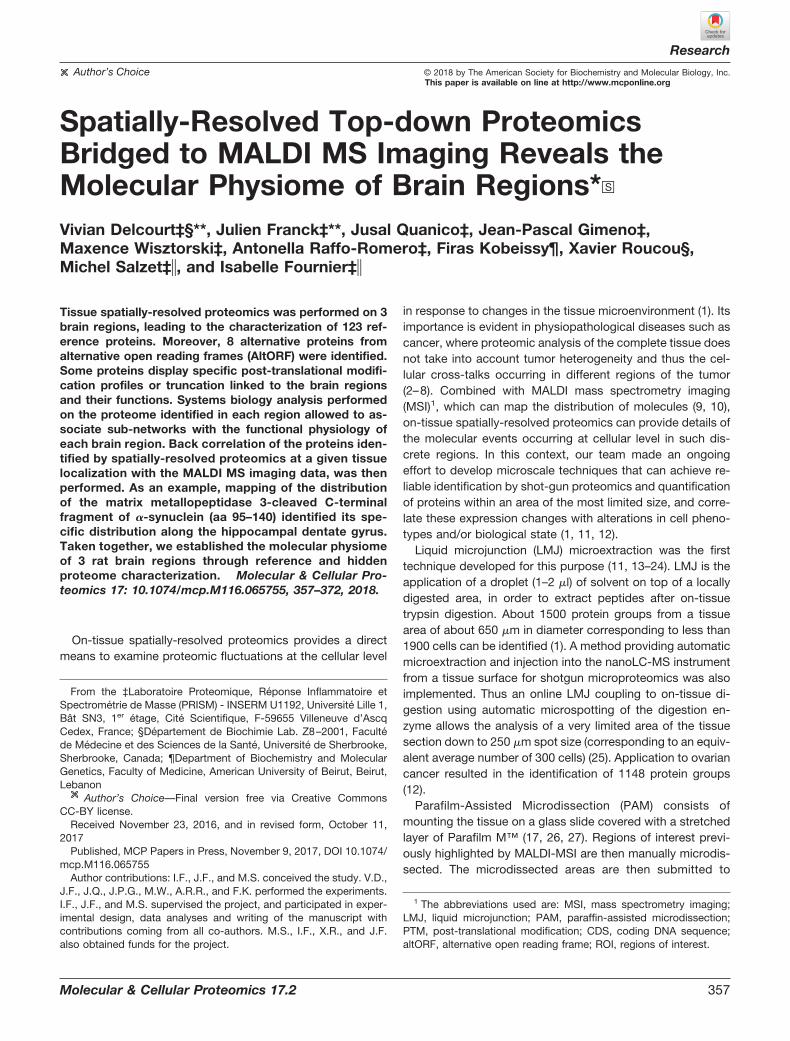
Spatially-Resolved Top-Down Proteomics and MALDI-MSI—Different types of molecules can be used in MALDI-MSIto determine ROIs from biological tissues such as lipids,endogenous or tryptic peptides and proteins. However, lipidMALDI-MSI is the most convenient to our approach as it givesgood spatial resolution and does not need extensive samplepreparation steps. Our first developments were performed onrat brain tissue sections (Fig. 1). Different ROIs can be re-trieved after lipid MALDI-MSI (Fig. 1A) followed by nonsuper-vised spatial segmentation analysis (Fig. 1B, bottom) com-pared with the optical image (Fig. 1B, top). Three ROIs in thehippocampus, corpus callosum, and medulla oblongata(Bregma Index lateral 1.90 mm) were selected for furtherprocessing as their segmentation profiles were sufficientlydistinct.
Based on these selected ROIs, the two main strategies toperform spatially-resolved proteomics studies were then re-alized i.e. PAM (Fig. 1C) or LMJ (Fig. 1D). Based on theidentified proteins, our approach mostly enables identificationof low molecular weight (from 1.6 to 21.9 kDa) and mostabundant proteins. These two strategies allowed the identifi-cation of proteins that were common within the three regionsas well as specific ones. Analyses of the three ROIs gave atotal of 123 proteins identified (Fig. 1E and 1F, supplementalData S1 and S2). One hundred eleven proteins have beenidentified in PAM and 45 in LMJ. The number of specificproteins identified is higher with PAM than with LMJ, whichmight be related to tissue washing steps prior to protein
On-tissue Spatially-Resolved Top-down Proteomics Bridged to MALDI-MSI
360 Molecular & Cellular Proteomics 17.2

extraction and smaller area of extraction. By combining thetwo approaches, 15 specific nonredundant proteins wereidentified from the corpus callosum, 17 from medulla oblon-gata, and 24 from hippocampus (Fig. 1E and 1F, supplemen-tal Data S1 and S2). Thirty-five are common to the 3 brainregions; 16 are shared between corpus callosum and medullaoblongata, 8 between corpus callosum and hippocampus,and 8 between medulla oblongata and hippocampus. Mostidentified spectra exhibited a mass shift close to 0 Da (Fig. 1G,inset). The mass tolerant identification approach allowedcharacterization of modified forms of proteins, which caneither be truncated compared with database prediction ormodified (Fig. 1G) in a similar fashion to what is described byChick et al. (48).
Systems Biology Analyses of the Identified Proteins—Func-tional enrichment analysis using Search Tool for RecurringInstances of Neighboring Genes (STRING, (49)) identified 4GO terms associated with Molecular function: Hydrogen ionstransmembrane transport (GO 0015078), Cytochrome-c oxi-dase activity (GO: 0004129), Ion transmembrane transporteractivity (GO: 0015075), and Oxidoreductase activity (GO:0016491). Systems biology analysis was then performed on
the over-expressed proteins of each group for LMJ (Fig. 2A)and for PAM (Fig. 2C). Differential distributions of unique andcommon statistically significant biological and functionalpathways among the three different regions are depicted inFig. 2A for LMJ and 2C for PAM, including 39 versus 18pathways for corpus callosum, 91 versus 34 pathways formedulla oblongata and 31 versus 82 pathways for hippocam-pus (Please refer to supplemental Data S3 for the identity ofeach of the unique pathways). Combined differential path-ways were analyzed across the three regions. Three pathwaysin LMJ versus 2 in PAM were shared between corpus callo-sum and medulla oblongata, 6 versus 15 pathways betweenhippocampus and medulla oblongata, and 5 versus 3 path-ways between corpus callosum and hippocampus. IntegratedVenn diagram analysis was performed using “the Interac-tiVenn”: a web-based tool for the analysis of complex datasets (Figs. 3A–3B) (46). See supplemental Data S3 for thelisted differential pathways. Overexpressed proteins commonto medulla oblongata and hippocampus (Fig. 3A) are involvedin learning, epilepsy, neuronal activity and plasticity, neu-rotransmission and ischemia. For hippocampus and corpuscallosum (Fig. 3A), the identified proteins are mainly involved
m/z 820.6 mz 772.5
m/z 848.6 m/z 866.8 Segmentation PAM LMJ
DCBA
E F G
2415
17
Corpus callosum
Medulla oblongata
Hippocampus
3516 8
8
78
12
LMJ
PAM
33
38(K+)
22(Na+)
54(Fe)
98(H3PO4)
80(Phos)
1
−114 (loss of Asn or Gly-Gly)
−261(loss of Glu-Ser) 16
(Ox)
−1
42(Ac)
359
0
25
50
75
100
−400 −200 0 200 400Mass shift (Da)
Num
ber o
f ide
ntifi
ed s
pect
ra
Truncation
−114(loss of Asn or Gly-Gly)
−261(loss of Glu-Ser) 16
(Ox)
−138
(K+)
22(Na+)
54(Fe)
98(H3PO4)
80(Phos)
1 42(Ac)
359
Modification
0
400
800
0 1000Mass shift (Da)
Iden
tifie
d sp
ect ra
5001000 500
Overall
FIG. 1. A, Molecular images after median normalization of spectra followed by medium denoizing and automatic hotspot removal.B, Optical image with highlighted regions of interest corpus callosum (yellow), hippocampus (blue) and medulla oblongata (brown) (top) andspatial segmentation analysis using the Bisecting k-Means approach using Correlation as the distance metric (bottom). C–D, Optical imagesof PAM and LMJ tissue sections with the top and bottom panels showing the tissue sections before and after ROI processing, respectively.E, Venn Diagram of the extracted proteins per technique (LMJ or PAM) and (F) global unique identifications using both strategies. G, Overallmass shifts of observed proteins precursors versus their theoretical masses (G, inset) and most abundant observed mass shifts within a � 400Da tolerance window (G) with annotation of known mass shifts. �114 Da corresponds to loss of “Asn” at N-term of ATP synthase-couplingfactor 6, mitochondrial or loss of “Gly-Gly” at C-term of Ubiquitin monomer and �261 corresponds to loss of Glu-Ser at C-term of Thymosinbeta-4.
On-tissue Spatially-Resolved Top-down Proteomics Bridged to MALDI-MSI
Molecular & Cellular Proteomics 17.2 361

in neurogenesis, cell proliferation and oxidative stress. Formedulla oblongata and corpus callosum (Fig. 3A), the patternis more related to cell damage and life span. The same anal-ysis for unique pathways in hippocampus clearly showedprotein patterns involved in neurogenesis, synaptogenesis,neurite outgrowth, neuroprotection, and axogenesis (Fig. 3B,supplemental Data S4). For medulla oblongata the proteinsare mainly involved in pathways related to memory consoli-dation, epilepsy, cognition disorders, oligodendrocytes differ-entiation, amyotrophic lateral sclerosis, and spinocerebralataxia type 1 (Fig. 3B). For corpus callosum, the proteins are
mainly implicated in beta thalassemia, anemia and relatedhemoglobinopathies (Fig. 3B). All the results are in line withbiological and physiological functions of these 3 brainregions.
PTM Analysis of Identified Proteins—PTM analysis of pro-teins from the 3 regions revealed the presence of 91 proteinsthat were identified with PTMs, of which, 29 were detectedin the hippocampus, 40 in the corpus callosum and 37 inthe medulla oblongata (supplemental Data S2). Interestingly,some proteins show region-specific PTMs (Table I, supple-mental Data S2). As an illustration, the most abundant PTM of
A B
C D
39(22.3%)
91(52%)
3(1.7%)
6(3.4%)
05(2.9%)
31(17.7%)
Corpus callosum Medulla oblongata
Hippocampus
18(11.7%)
34(22.1%)
2(1.3%)
15(9.7%)
03(1.9%)
82(53.2%)
Corpus callosum Medulla oblongata
Hippocampus
57
11 166
00 4 0
48200
41 4
4 5
0
19
21
20
00
00
0 0
043
21
3
00
0
010
0
1
0
0
000
2
010
0
0
16
200
0
00
0
100
39
0 003
0215 8
19 10
6 2
0 2
0
87
20
00
01
00
0 0
001
00
0
00
0
000
0
1
3
0
000
0
030
0
0
16
000
0
10
0
000
FIG. 2. Differential distribution of unique and common/intersected biological and functional pathways among the three brain regions(corpus callosum, hippocampus and medulla oblongata) obtained with LMJ (A) or PAM (C) extraction methods. Each brain region wasanalyzed across the three regions using a comprehensive Venn analysis representation extracted from Subnetwork Enrichment Analysis (B withLMJ and D with PAM).
On-tissue Spatially-Resolved Top-down Proteomics Bridged to MALDI-MSI
362 Molecular & Cellular Proteomics 17.2

A
Common Corpus Callosumand Hippocampus
Common Corpus Collosumand Medulla Oblongata
Common Hippocampusand Medulla Oblongata
Hippocampus
Medulla Oblongata
B
Corpus Callosum
FIG. 3. A, Global pathway analyses of the over-expressed proteins common to two different regions i. e. hippocampus and medullaoblongata, hippocampus and corpus callosum and medulla oblongata and corpus callosum. B, Over-expressed proteins in the hippocampus,medulla oblongata or corpus callosum were involved in globally altered molecular pathways.
On-tissue Spatially-Resolved Top-down Proteomics Bridged to MALDI-MSI
Molecular & Cellular Proteomics 17.2 363

TAB
LEI
Reg
ion
spec
ific
pos
t-tr
ansl
atio
nally
mod
ified
pro
tein
s.P
TMs
from
Pro
sigh
tPC
wer
eco
ncat
enat
edw
ithim
put
edP
TMs
from
mas
ssh
ifts
(i.e.
Ace
tyla
tion
(�42
);P
hosp
hory
latio
n(�
80))
Reg
ion
Acc
essi
onnu
mb
erP
TM(s
)P
rote
inna
me
Theo
.m
ass
(Da)
Ob
s.m
ass
(Da)
Shi
ft(D
a)
Cor
pus
callo
sum
P13
668
N-a
cety
l-L-
alan
ine,
O-p
hosp
ho-L
-ser
ine
Sta
thm
in17
268.
917
269.
00.
094
P31
399
N-a
cety
l-L-
alan
ine
ATP
synt
hase
sub
unit
d,
mito
chon
dria
l18
662.
618
662.
60.
082
G3V
9C0
N-a
cety
l-L-
serin
eH
isto
neH
2A14
037.
914
038.
00.
05Q
5U31
8N
-ace
tyl-
L-al
anin
e,O
-pho
spho
-L-s
erin
eA
stro
cytic
pho
spho
pro
tein
PE
A-1
515
021.
715
021.
80.
05D
3ZH
W9
N-a
cety
l-L-
serin
eP
rote
inS
hfm
181
83.5
381
83.6
0.04
4B
2RZ
27N
-ace
tyl-
L-se
rine
Pro
tein
Sh3
bgr
l310
381.
210
381.
30.
033
D3Z
ZW
2N
-ace
tyl-
L-se
rine
Pro
tein
LOC
1009
1067
869
72.9
6972
.9�
0.00
8D
3ZTB
5N
-ace
tyl-
L-al
anin
eP
rote
inS
100a
1311
101.
911
101.
0�
0.90
9H
ipp
ocam
pus
Q04
940
O-p
hosp
ho-L
-ser
ine
�P
hosp
hory
latio
n(�
80)
Neu
rogr
anin
7440
.43
7520
.580
.041
M0R
5I3
Pho
spho
ryla
tion
(�80
)H
igh
mob
ility
grou
pnu
cleo
som
alb
ind
ing
dom
ain
3,is
ofor
mC
RA
_a10
236.
410
316.
479
.973
Q5U
1W8
Pho
spho
ryla
tion
(�80
)H
igh-
mob
ility
grou
pnu
cleo
som
eb
ind
ing
dom
ain
199
87.3
1006
7.3
79.9
64P
0455
0N
-ace
tyl-
L-se
rine
�P
hosp
hory
latio
n(�
80)
Par
athy
mos
in11
463.
211
543.
179
.942
P62
329
N-a
cety
l-L-
serin
e�
Ace
tyla
tion
(�42
)Th
ymos
inb
eta-
449
60.4
950
02.5
42.0
29M
edul
laob
long
ata
P06
302
N-a
cety
l-L-
serin
e�
Ace
tyla
tion
(�42
)P
roth
ymos
inal
pha
1228
6.1
1232
8.1
41.9
93P
0463
1N
-ace
tyl-
L-se
rine
Pro
tein
S10
0-B
1064
810
648.
10.
05B
2RY
S2
N-a
cety
l-L-
alan
ine
Cyt
ochr
ome
b-c
1co
mp
lex
sub
unit
713
460.
913
460.
90.
047
P63
041
N-a
cety
l-L-
met
hion
ine
Com
ple
xin-
115
154.
515
154.
50.
047
P0C
C09
N-a
cety
l-L-
serin
eH
isto
neH
2Aty
pe
2-A
1399
7.9
1399
7.9
0.04
2P
0262
5N
-ace
tyl-
L-se
rine
Par
valb
umin
alp
ha11
829
1182
9.0
0.03
2P
1195
1N
-ace
tyl-
L-se
rine
Cyt
ochr
ome
cox
idas
esu
bun
it6C
-283
60.4
283
60.4
0.02
2Q
5U31
8N
-ace
tyl-
L-al
anin
eA
stro
cytic
pho
spho
pro
tein
PE
A-1
514
941.
814
940.
8�
0.91
8P
3104
4N
-ace
tyl-
L-al
anin
eP
hosp
hatid
ylet
hano
lam
ine-
bin
din
gp
rote
in1
2069
9.4
2069
8.4
�0.
935
On-tissue Spatially-Resolved Top-down Proteomics Bridged to MALDI-MSI
364 Molecular & Cellular Proteomics 17.2

stathmin in the corpus callosum (identified) and the hip-pocampus (detected but not identified) was the Nter-Acetyl �
1 Phosphorylation, whereas in the medulla oblongata (identi-fied) it was the Nter-Acetylation (Fig. 4). Similarly, neurograninwas specifically phosphorylated in the hippocampus. Anotherexample is the Astrocytic phosphoprotein (PEA-15), whichwas observed with a phosphorylated residue in the corpuscallosum but not in the medulla oblongata (Table I and sup-plemental Data S2). Similarly, Parathymosin was identifiedwith a mass shift of �79.94 Da in hippocampus by twospectra and with 5.89 and 5.38 ppm mass errors comparedwith theoretical mass plus a phosphorylation, thus implying aphosphorylated residue (Table I, Fig. 1G and supplementalData S1 and 2). These data clearly revealed that the PTM stateof proteins is linked to the brain regions where they arelocalized, and consequently with the biological function of theprotein in relation to the physiological function of the consid-ered brain region.
Protein Fragments Linked to Brain Region Localization—Data analyses revealed the presence of protein fragments inthe three brain regions (Table II and supplemental Data S8).
These fragments are derived from large proteins such asneuropeptide precursors (somatostatin, proenkephalin, se-cretogranin 1 and 2), Synuclein (alpha, beta and gamma),Synaptosomal associated protein 25, DNA-(apurinic orapyrimidinic) protein (APEX), Hematological and neurologicalexpressed 1 protein (HN1), Myelin basic protein (MBP) andThymosin beta 4. The generated fragments are linked tothe presence of processing enzymes e.g. pro-protein con-vertases, neutral endopeptidases, angiotensin-converting en-zymes and aminopeptidases, which are differentially ex-pressed in the brain region (38–41, 50, 51). Neuropeptidefragment precursors, neuromodulin and secretogranin 1 areprincipally detected in hippocampus whereas fragments ofMBP and somatostatin are detected in majority in medullaoblongata. HN1 fragments are detected in hippocampus,whereas Secretogranin 2 is present in both hippocampus andmedulla oblongata.
Alternative Protein Identification—Three alternative proteinswere detected in spatially-resolved top-down proteomics ex-periments. AltCd3e and AltMyo1f were detected in hippocam-pus using LMJ and PAM, respectively, and AltGrb10 was
Stathmin
Mw : 17268.9 DaError : 4.32 ppmE-Value : 1.77 E-17
m/z
25
50
75
100
Relat
ive A
bund
ance
(%)
956 957 958 959 960 961 962 963 964 965
m/z
Cor
pus
callo
sum
Hip
poca
mpu
sM
edul
la o
blon
gata
b5+
y5+
b7+
y8+
b10+
b11+
b172+
y172+
b182+
b192+
b202+
b212+
b222+
y222+
b232+
b183+
b193+
b203+
b213+
b223+
y223+
b233+
b313+
b333+ b34
3+
b314+
b334+
b344+
y395+
y465+
y576+ y116
11+
y11712+
y11113+
y11613+
y11713+
y11714+
[M+17H]17+
y464+
A S S D I Q V K E L E K R A S G Q A F E L I L S P R S K E S
V P E F P L S P P K K K D L S L E E I Q K K L E A A E E R R
K S H E A E V L K Q L A E K R E H E K E V L Q K A I E E N N
N F S K M A E E K L T H K M E A N K E N R E A Q M A A K L E
R L R E K D K H V E E V R K N K E S K D P A D E T E A D
Ac
P
Stathmin (P13668)
1017.0 1017.4 1017.8
m/z
25
50
75
100
Relat
ive A
bund
ance
(%)
956 957 958 959 960 961 962 963 964 965
25
50
75
100
Relat
ive A
bund
ance
(%)
956 957 958 959 960 961 962 963 964 965
12500
10000
7500
5000
2500
0400 600 800 1000 1200 1400
ΔM = 80 Da ΔM = 80 Da
Inte
nsity
A B
Cm/z
m/z
FIG. 4. ROI-specific PTM profile of Stathmin (A), which was identified in corpus callosum and medulla oblongata but not inhippocampus. N-terminal acetylation is marked red, whereas the phosphorylations are marked blue. Precursor (z � 17) and assignedfragmentation spectrum of Nter-Acetylated and phosphorylated (Ser-38) Stathmin (B) and fragmentation map (C).
On-tissue Spatially-Resolved Top-down Proteomics Bridged to MALDI-MSI
Molecular & Cellular Proteomics 17.2 365

TAB
LEII
Mos
td
etec
ted
trun
cate
dp
rote
in.
M.O
:m
edul
laob
long
ata;
C.C
:co
rpus
callo
sum
;H
i:hi
pp
ocam
pus
;A
A:
amin
oac
ids
Acc
essi
onP
rote
ind
escr
iptio
nM
.OC
.CH
iD
etec
ted
leng
th(A
A)
Full
leng
th(A
A)
Frag
men
t(A
A)
Frag
men
tp
ositi
on
P21
571
Cha
in3
3–10
8in
ATP
synt
hase
-cou
plin
gfa
ctor
6,m
itoch
ond
rial
��
�76
108
33–1
08C
-ter
min
alfr
agm
ent
P10
818
Cha
in2
7–11
1in
Cyt
ochr
ome
cox
idas
esu
bun
it6A
1,m
itoch
ond
rial
��
�85
111
27–1
11C
-ter
min
alfr
agm
ent
P11
240
Cha
in3
8–14
6in
Cyt
ochr
ome
cox
idas
esu
bun
it5A
,m
itoch
ond
rial
��
�10
914
638
–146
C-t
erm
inal
frag
men
tQ
71U
E8
Cha
in1
–76
inN
ED
D8
��
�76
811–
76N
-ter
min
alfr
agm
ent
P35
171
Cha
in2
4–83
in
Cyt
ochr
ome
cox
idas
esu
bun
it7A
2,m
itoch
ond
rial
��
�60
8324
–83
C-t
erm
inal
frag
men
tP
2157
1A
TPsy
ntha
se-c
oup
ling
fact
or6,
mito
chon
dria
l�
��
5310
856
–108
C-t
erm
inal
frag
men
tP
4794
2D
ihyd
rop
yrim
idin
ase-
rela
ted
pro
tein
2�
��
5557
251
8–57
2C
-ter
min
alfr
agm
ent
Q63
429
Pol
yub
iqui
tin-C
��
7481
01–
74N
-ter
min
alfr
agm
ent
P28
073
Pro
teas
ome
sub
unit
bet
aty
pe-
6�
�17
238
78–9
4In
tern
alfr
agm
ent
P80
432
Cha
in1
7–63
in
Cyt
ochr
ome
cox
idas
esu
bun
it7C
,m
itoch
ond
rial
��
�47
6317
–63
C-t
erm
inal
frag
men
tD
4A5W
9S
ynap
toso
mal
-ass
ocia
ted
pro
tein
��
�45
206
162–
206
C-t
erm
inal
frag
men
tP
1366
8S
tath
min
�11
214
938
–149
C-t
erm
inal
frag
men
tP
2157
1A
TPsy
ntha
se-c
oup
ling
fact
or6,
mito
chon
dria
l�
��
7510
834
–108
C-t
erm
inal
frag
men
tF1
LQ96
Gam
ma-
synu
clei
n�
�30
122
93–1
22C
-ter
min
alfr
agm
ent
F1LU
V9
Neu
ralc
ella
dhe
sion
mol
ecul
e1
�61
833
773–
833
C-t
erm
inal
frag
men
tP
3737
7A
lpha
-syn
ucle
in�
7314
068
–140
C-t
erm
inal
frag
men
tO
3531
4S
ecre
togr
anin
-1�
3067
529
2–32
1In
tern
alfr
agm
ent
P19
527
Neu
rofil
amen
tlig
htp
olyp
eptid
e�
7454
246
9–54
2C
-ter
min
alfr
agm
ent
Q5M
7W5
Mic
rotu
bul
e-as
soci
ated
pro
tein
4�
1610
5731
–46
Inte
rnal
frag
men
tP
2677
210
kDa
heat
shoc
kp
rote
in,
mito
chon
dria
l�
�37
102
66–1
02C
-ter
min
alfr
agm
ent
Q8R
1R5
CD
99an
tigen
-lik
ep
rote
in2
��
�24
246
223–
246
C-t
erm
inal
frag
men
tQ
6PC
U8
Cha
in3
6–10
8in
NA
DH
deh
ydro
gena
seu
biq
uino
ne
flavo
pro
tein
3,m
itoch
ond
rial
��
�73
108
36–1
08C
-ter
min
alfr
agm
ent
F1LQ
96G
amm
a-sy
nucl
ein
��
4812
275
–122
C-t
erm
inal
frag
men
t
On-tissue Spatially-Resolved Top-down Proteomics Bridged to MALDI-MSI
366 Molecular & Cellular Proteomics 17.2

detected in the medulla oblongata using PAM (Table III).These results suggest that the spatially-resolved proteomicsstrategy was suitable for studying the reference and hiddenproteomes. We then enlarged this study by re-analyzing pre-vious data obtained using whole rat brain sections (PMID:27512083) (42). Reanalysis of this dataset allowed the identi-fication of 5 more alternative proteins (Table III, supplementalData S6). These alternative proteins are translated from se-quences located in mRNAs 3�UTR (AltSstr3, AltKcnq5, Al-tLdlr), 5�UTR regions (AltZbtb8a) of mRNAs and from a puta-tive noncoding RNA (AltRn50_X_0580.1).
Back Correlation to Localization by MALDI-MSI—Intact pro-tein MSI experiments were performed to show ion distribu-tions of the proteins identified by top-down MS. To this end,two images were acquired; the first section was prepared withHCCA/aniline matrix and the second one with SA/aniline. Theimages were acquired only on the three ROIs specified in theprevious imaging experiment. Peaks obtained from these im-ages were then matched with the Mavg derived from thetop-down MS analysis performed on the entire rat brain tissuesection. Thirty-five protein IDs obtained from the referenceproteome were assigned to peaks obtained from both imageswith a �Mavgs cutoff � 6 Da (Fig. 5A–5D and supplementalData S7). This includes five proteins previously matchedalso with top-down MS data, namely PEP-19 (Pcp4, Fig. 5D),ubiquitin (Ubc), thymosin �-4 (Tmsb4x), thymosin �-10(Tmsb10), and calmodulin (Calm1) (34).
Fig. 6A shows the ion image of m/z 4966 assigned as theintact form (as hematopoietic system regulatory peptide) ofthymosin �-4 (monoisotopic theoretical mass � 4960.49). Thespecific localization of m/z 4966 in the hippocampus can beclearly observed. Topdown data indicate that this isoform,detected as the [M�5H]5� charge state, is the N-acetylatedisoform after methionine excision (Fig. 6B). Its distribution inthe hippocampus in MSI correlates well with the top-downdata where this form was detected using PAM. Furthermore,its detection by MSI and assignment of N-acetylation bytop-down is in accord with the MSI database reported byMaier et al. (52).
Fig. 6C shows the mapping of m/z 5180 assigned asthe C-terminal fragment of �-synuclein (observed as the
[M�4H]4� charge state in topdown, Fig. 6C), showing itsparticularly intense distribution along the hippocampal den-tate gyrus. Its distribution in the cerebral cortex observed inthe ROI that includes the corpus callosum, was also detectedin both MSI and spatially-resolved top-down proteomics. Toverify the specific formation of this fragment, the putativeprotease cleavage sites found in the full amino acid sequenceof �-synuclein was mapped using the PROtease SpecificityPrediction servER (PROSPER, (53), https://prosper.erc.monash.edu.au), where it can be observed that cleavage bymatrix metallopeptidase 3 (MMP3) can induce the generationof the C-terminal fragment (Fig. 6D). In situ hybridization of thegenes that code for �-synuclein (Snca) and MMP3 in mousebrain obtained from the Allen Mouse Brain Atlas (http://mouse.brain-map.org/) (47) confirms the distribution of �-sy-nuclein (strong) and MMP3 (weak) along the mouse hip-pocampal dentate gyrus (Fig. 6E). Localization of the�-synuclein was validated by tissue immunofluorescenceshowing strong signal in the hippocampus and corpus callo-sum and weak signal in the medulla oblongata (Fig. 6F).
In addition to the �-synuclein, the distribution of proteinsGFAP, BASP1 and stathmin were further validated by IF ex-periments. supplemental Data S9 shows the confocal imagesafter immunostaining against GFAP (red), showing highly pos-itive astrocytes (GFAP���) localized between the dentategyrus and CA3 of Ammon’s horn of the hippocampal forma-tion. The signal is markedly absent in the corpus callosum andsurrounding region (GFAP�), and is slightly present in themedulla oblongata region (GFAP�/�) as evidenced by immu-noreactivity of several astrocyte processes projecting in dif-ferent directions. The specific localization of GFAP-positiveastrocytes in the hippocampus was confirmed after gatheringz-series of images at varying focal planes throughout theentire tissue thickness of 10 �m. Likewise, GFAP was de-tected only in the hippocampus in MALDI-MSI and spatially-resolved top-down proteomics experiments. Results for theother proteins tested are shown in supplemental Data S9.
DISCUSSION
We developed a novel strategy combining MALDI MS Im-aging and spatially-resolved top-down proteomics to deter-
TABLE IIIAlternative protein products identified by tissue top-down proteomics
Region E-Value (P-score)Observed
MassProtein Gene
AltORF localizationon RNA
Transcript
Hippocampus 2.14 E-05 (3.80E-11) 4642.28 AltCd3e Cd3e 3�UTR ENSRNOT000000472917.70E-05 (1.37E-10) 8154.94 AltMyo1f Myo1f CDS ENSRNOT00000011513
Medulla Oblongata 1.06E-05 (1.89E-11) 15025.79 AltGrb10 Grb10 CDS ENSRNOT00000085175Whole brain section
PMID 275120833.15E-05 (2.24E-10) 4760.46 AltRn50_X_0580.1 Rn50_X_0580.1 ncRNA ENSRNOT00000066392
3.42E-05 (2.43E-10) 5000.62 AltSstr3 Sstr3 3�UTR ENSRNOT000000096124.23E-07 (7.52E-13) 3344.66 AltZbtb8a Zbtb8a 5�UTR ENSRNOT000000109832.48E-09 (1.77E-14) 2825.44 AltKcnq5 Kcnq5 3�UTR ENSRNOT000000400341.36E-05 (9.68E-11) 4440.29 AltLdlr Ldlr 3�UTR ENSRNOT00000013496
On-tissue Spatially-Resolved Top-down Proteomics Bridged to MALDI-MSI
Molecular & Cellular Proteomics 17.2 367

mine localized proteoforms, including truncated forms, frag-ments, and possibly altprots. First, molecular histology wasperformed using MALDI-MSI and spatial segmentation todistinguish ROIs within a tissue. These ROIs were then sub-jected to protein microextraction with ProteaseMAX ratherthan SDS or organic solvents. Protein microextraction effi-ciency was confirmed by nanoLC high resolution MS/MSanalysis of rat brain tissue because we identified many pro-teins (123) compared with the 36 previously identified from awhole tissue proteomics study that performed extraction us-ing acidified MeOH (34). Only 19 proteins were in commonwith those identified from this study. The 17 proteoformsabsent in our study are small peptides less than 4500 Da andare more related to the neuropeptide family, e.g. chromogra-nin-A, cholecystokinin, proneuropeptide Y, secretogranin-2,proSAAS, cocaine- and amphetamine-regulated transcriptprotein, and oxysterol-binding protein, consistent with the
brain regions selected in our study. Nevertheless, the com-mon proteoforms identified are the same with the same PTMs.
It is interesting to note that LMJ and PAM do not identify thesame proteins and are thus complementary, giving a total of123 protein IDs overall. For example, somatostatin and pep-tide 143–185 of proenkephalin-A were specifically identified inLMJ samples whereas �-synuclein and neuromodulin wherespecifically identified in PAM experiments. Considering thatthe average size of brain cells is 15 �m and that we havemicroextracted 0.8 mm2 with LMJ and 1 mm2 with PAM, weestimate that we identified proteins from 4444 cells for LMJand 5662 cells for PAM. By combining the two approaches,15 specific and nonredundant proteins were identified fromthe corpus callosum, 17 from the medulla oblongata and 24from the hippocampus (Tables I and II). 35 are common to the3 brain regions, 16 between corpus callosum and medullaoblongata, 8 between corpus callosum and hippocampus and
Ncam1 (F1LUV9), Neural cell adhesion molecule 1Chain 773-833 (C-terminal fragment)Mmono th. Mavg th. Mavg obs. ΔMavg
3453.67 3455.687 3455.687 (LC) 0.0013453 (MSI) 3
Ncam1 ISH*
Scg1 (O35314), Secretogranin-1Chain 277-321 (internal fragment)
Mmono th. Mavg th. Mavg obs. ΔMavg
5145.56 5148.567 5148.572 (LC) 0.0055148 (MSI) 1 Scg1 ISH*
Cox7a2 (P35171), cytochrome c oxidase subunit 7A2Chain 24-83 (C-terminal fragment)Mmono th. Mavg th. Mavg obs. ΔMavg
6644.41 6648.772 6648.797 (LC) 0.0256649 (MSI) 0
Cox7a2 ISH*
Pcp4 (P63055), Purkinje cell protein 4Nter-Ac after Met excision (intact protein)
Mmono th. Mavg th. Mavg obs. ΔMavg
6714.26 6718.2536719.269 (LC) 1.016
6721 (MSI, HCCA) 36724 (MSI, SA) 6 Pcp4 ISH*
A
B
C
D
m/z 3453
m/z 5148
m/z 6649
m/z 6721
m/z 6724
FIG. 5. MALDI-MS images of top-down identified proteoforms of Ncam1 C-terminal fragment (A), Scg1 internal fragment (B), Cox7a2C-terminal fragment (C) and intact Pcp4 N-terminally acetylated after initiator methionine excision (D) with their correspondingtop-down (LC) and MSI m/z and their in situ hybridization images (ISH*). All values in tables are given in a.m.u. See also supplemental DataS7. *Image credit: Allen Institute (47).
On-tissue Spatially-Resolved Top-down Proteomics Bridged to MALDI-MSI
368 Molecular & Cellular Proteomics 17.2

8 between medulla oblongata and hippocampus. Proteinsidentified with PAM are mainly present in the cytoplasm(62%), mitochondrial membrane (9.3%) or organelles andplasma membranes (28.7%). With LMJ, the proteins identifiedare from organelles (51.5%) and the cytoplasm (47.7%).
These studies performed by spatially-resolved top-downproteomics are in line and complementary to our previousstudies based on spatially-resolved bottom-up proteomics (1,17, 27) as it gives information about the precursor mass andPTMs detectable by measuring the �M(s) between the intactprecursor within a close retention time window. Indeed, ourapproach successfully discriminate stathmin PTMs betweendifferent regions of rat brain tissue (Fig. 4). We showed thatstathmin is more abundant in corpus callosum and medulla
oblongata and its PTM pattern is specific for each of thesetwo regions. The ratio phospho-stathmin/Nter-Ac was signif-icantly higher in the corpus callosum, suggesting a differentbiological activity in these two regions of the brain (Fig. 4).Similarly, out of the 41 unique proteins that were identifiedwith PTMs (supplemental Data S1 and 2), 22 had regionspecific PTMs (Table I). The most prevalent PTMs are theN-acetylation of proteins and phosphorylation. For example,we found that �-synuclein presents one PTM, i.e. N-acetyl-L-methionine in medulla oblongata, hippocampus and corpuscallosum. In literature it has been shown that �-synucleinacetylation at Met in position 1 seems to be important for itsproper folding (54)(55). Similarly, the Astrocytic phosphopro-tein (PEA-15) possesses N-acetyl-L-alanine in medulla oblon-
m/z 49660
200 000
993.0 993.9 994.8m/z
Inte
nsity
Thymosin beta-4(Nter-Ac after Met excision)
S D K P D M A E I E K F D
K S K L K K T E T Q E K N P
L P S K E T I E Q E K Q A G
E S
Ac
LMJ - HippocampusMw : 4960.526 DaError : 8.01 ppm
E-value : 1.17 E-108
[M+5H]5+
A
m/z 51800
75 000
1296.0 1297.0 1298.0m/z
Inte
nsity
Alpha-synuclein 95-140(C-terminal fragment)
PAM - Corpus callosumPAM - Hippocampus
[M+4H]4+
B
�Mmp3*
AAATGF VKKDQM
C
Snca**
Mmp3**
D E
95927920 1401
1
2
3
1. Medulla oblongata 2. Hippocampus 3. Corpus callosum
F
FIG. 6. A, MALDI-MSI of m/z 4966 attributed to intact N-terminally acetylated form of Thymosin beta-4 with correspondingidentification by spatially-resolved top-down proteomics (B). C, MALDI-MSI of m/z 5180 attributed to C-terminal fragment of �-synucleinidentified in hippocampus and corpus callosum. D, Schematic representation of protein fragment with predicted Stromelysin (Mmp3) cleavagesites (arrows and amino acid numbers) by PROSPER* (53) and identified form (red arrows). E, In situ- hybridization of �-synuclein (top) andStromelysin (bottom). F, Tissue immunofluorescence image of �-synuclein using an antibody targeting the C-terminal of the protein. Whitescale bars � 50 �m. **Image credit: Allen Institute (47).
On-tissue Spatially-Resolved Top-down Proteomics Bridged to MALDI-MSI
Molecular & Cellular Proteomics 17.2 369

gata and N-acetyl-L-alanine plus O-phospho-L-serine in cor-pus callosum. None of them have been previously identified(56).
In the same way, we identified protein fragments from pro-teins with distribution and presented a specific cleavage formacross each brain region. Majority of the identified fragmentsare large neuropeptides like synenkephalin and secretogra-nins 1 and 2. These fragments are produced by enzymaticcleavage of the pro-protein convertase family like PC1/3, PC2or PC5, PACE4 (38). We previously demonstrated the role ofthese enzymes in proenkephalin maturation (51, 57) andfound some of these neuropeptide fragments in temporallobe epilepsy (58) and Alzheimer’s disease (59), such assecretogranins for example. Synenkephalin is implicated incircadian rhythm in the hippocampus (60), Snap25 is impli-cated in synaptogenesis and memory consolidation (61–63). As previously demonstrated, we confirmed that thesomatostatin is present in medulla oblongata (64) whereaswe showed for the first time the presence of the hemato-logical and neurological expressed 1 protein in the hip-pocampus (fragment) and corpus callosum (full length aftermethionine excision).
Besides these novel protein fragments, another small familyof proteins has been identified from the hidden proteome. Infact, more and more evidence suggests that mRNAs containmore than one coding sequence and could be translated intoan annotated or reference protein and at least one alternativeprotein (36, 37). We tested if our strategy was able to detectintact alternative proteins. We identified 3 alternative proteins(Table III) by the spatially-resolved top-down proteomics ap-proach that share no sequence similarity with annotated rat-tus norvegicus proteins. Of the 5 novel altprots identified byreanalysis of the study on whole tissue sections (Alt-Kcnq5,Alt-Zbtb8a, Alt-Sstr3, Alt-Ldlr and a noncoding RNA Alt-Rn50_X_0580.1), 3 of them are receptors as reference pro-teins i.e. somatostatin 3 receptor, potassium voltage-gatedchannel subfamily Q member 5, and low-density lipoproteinreceptor. It is interesting to note that these 3 receptors areknown to be expressed in hippocampus specifically(65–67).
Back correlation of spatially-resolved top-down proteomicsprotein IDs with MALDI MS images allowed to localize 35identified proteins (Fig. 5 and supplemental Data S7). Thecorrelation included proteins with PTMs or enzymatic cleav-age whose distribution varies differently in the 3 regions in linewith identified biological processes taking place in each indi-vidual region. As an example, the truncated, N-acetylatedform of thymosin �-4 was mapped in MSI and its distributionwas compared with the result of the top-down data, showinggood correlation of the results from the two approaches (Fig.6A and 6B). The C-terminal fragment of �-synuclein likewiseshowed very good correlation of results (Fig. 6C). More im-portantly, the distribution of this fragment in the hippocampaldentate gyrus in MSI can be correlated with the abundance of
�-synuclein and MMP3 in the same region in ISH experimentson mouse brain. MMP3 can cleave �-synuclein at F94, yieldingthe natively unstructured C-terminal fragment aa 95–140 (5.74kDa) (Fig. 6D and 6E). Tissue immunofluorescence validated�-synuclein’s localization showing strong signal in hippocam-pus and corpus callosum indicating the presence of the pro-tein in these regions (Fig. 6F). However, MALDI-MSI revealedthat the C-terminal fragment has a strong and precise tissuelocalization in the hippocampal dentate gyrus and moderatelyaround the corpus callosum, matching the MMP3 in situ hy-bridization (47). This result exposes the great capabilities ofspatially-resolved top-down proteomics associated toMALDI-MSI to detect and localize truncated proteoforms thatcan be challenging using antibody-based tissue characteriza-tion methods. Other MMP3-produced C-terminally truncatedpeptides of �-synuclein (aa 1–78, 1–91 and 1–93) have beenreported under stress conditions, with aa 1–93 being impli-cated in dopamine neuronal loss in substantia nigra, suggest-ing that overexpression of the fragments could have a signif-icant impact in Parkinson’s disease (68). What role aa 95–140has in this regard thus needs to be further investigated.
Taken together, our results show that spatially-resolvedtop-down proteomics linked to MALDI-MSI can be used tosearch for biomarkers, PTM detection and to identify novelproteins expressed from altORFs.
DATA AVAILABILITY
The mass spectrometry proteomics data have been depos-ited to the ProteomeXchange Consortium via the PRIDE (43)partner repository with the data set identifier PXD005424.
* This work was supported by grants from Region Hauts de France(CM/YB N°2015.2097/12) and PROTEO (FRQNT-RS-188158) (V. Del-court), University Lille 1 (BQR to Dr. Julien Franck), University Lille 1and Region Hauts de France (NEU/2015058) (A. Raffo-Romero), Ca-nadian Institutes for Health Research (MOP-136962) and CanadaResearch Chairs in Functional Proteomics and Discovery of NewProtein (Prof. X. Roucou), PRISM (Prof. M. Salzet), Ministere del’Enseignement Superieur et de la Recherche via Institut Universitairede France (Prof. I. Fournier), SIRIC ONCOLille (Prof. I. Fournier), andGrant INCa-DGOS-Inserm 6041.
□S This article contains supplemental material.� To whom correspondence should be addressed: Reponse In-
flammatoire et Spectrometrie de Masse (PRISM) - INSERM U1192 -Universite Lille 1, Bat SN3, 1er etage, Cite Scientifique, F-59655Villeneuve d’Ascq Cedex. Tel.: �33-(0)3-20-43-41-94; Fax: �33-(0)3-20-43-40-54; E-mail: [email protected] or LaboratoireProteomique, Reponse Inflammatoire et Spectrometrie de Masse(PRISM) - INSERM U1192 - Universite Lille 1, Bat SN3, 1er etage,Cite Scientifique, F-59655 Villeneuve d’Ascq Cedex. Phone: 33-(0)3-20-43-41-94; Fax: 33-(0)3-20-43-40-54; E-mail: [email protected].
** These authors contributed equally to this work.
REFERENCES
1. Quanico, J., Franck, J., Dauly, C., et al. (2013) Development of liquidmicrojunction extraction strategy for improving protein identificationfrom tissue sections. J. Proteomics 79, 200–218 doi: 10.1016/j.jprot.2012.11.025
On-tissue Spatially-Resolved Top-down Proteomics Bridged to MALDI-MSI
370 Molecular & Cellular Proteomics 17.2

2. Viale, G., Slaets, L., de Snoo, F. A., et al. (2016) Discordant assessment oftumor biomarkers by histopathological and molecular assays in theEORTC randomized controlled 10041/BIG 03–04 MINDACT trial breastcancer : Intratumoral heterogeneity and DCIS or normal tissue compo-nents are unlikely to be the cau. Breast Cancer Res. Treat 155, 463–469doi: 10.1007/s10549–016-3690–6
3. Massard, C., Oulhen, M., Le Moulec, S., et al. (2016) Phenotypic andgenetic heterogeneity of tumor tissue and circulating tumor cells inpatients with metastatic castration-resistant prostate cancer: A reportfrom the PETRUS prospective study. Oncotarget 7, 55069–55082 doi:10.18632/oncotarget.10396
4. Kronig, M., Walter, M., Drendel, V., et al. (2015) Cell type specific geneexpression analysis of prostate needle biopsies resolves tumor tissueheterogeneity. Oncotarget 6, 1302–1314 doi: 10.18632/oncotarget.2744
5. Johann, D. J., Rodriguez-Canales, J., Mukherjee, S., et al. (2009) Ap-proaching solid tumor heterogeneity on a cellular basis by tissueproteomics using laser capture microdissection and biological massspectrometry. J. Proteome Res. 8, 2310–2318 doi: 10.1021/pr8009403
6. Johann, D. J., Mukherjee, S., Prieto, D. A., et al. (2011) Profiling solid tumorheterogeneity by LCM and biological MS of fresh-frozen tissue sections.Methods Mol. Biol. 755, 95–106 doi: 10.1007/978–1-61779–163-5_8
7. Sugihara, Y., Taniguchi, H., Kushima, R., et al. (2013) Laser microdis-section and two-dimensional difference gel electrophoresis reveal pro-teomic intra-tumor heterogeneity in colorectal cancer. J. Proteomics78, 134–147. doi: 10.1016/j.jprot.2012.11.009
8. Celis, J. E., Celis, P., Palsdottir, H., et al. (2002) Proteomic strategies toreveal tumor heterogeneity among urothelial papillomas. Mol. Cell. Pro-teomics 1, 269–279
9. Bonnel, D., Longuespee, R., Franck, J., et al. (2011) Multivariate analysesfor biomarkers hunting and validation through on-tissue bottom-up orin-source decay in MALDI-MSI: application to prostate cancer. Anal.Bioanal. Chem. 401, 149–165
10. Bruand, J., Alexandrov, T., Sistla, S., et al. (2011) AMASS: Algorithm forMSI analysis by semi-supervised segmentation. J. Proteome Res. 10,4734–4743 doi: 10.1021/pr2005378
11. Wisztorski, M., Desmons, A., Quanico, J., et al. (2016) Spatially-resolvedprotein surface microsampling from tissue sections using liquid ex-traction surface analysis. Proteomics 16, 1622–1632 doi: 10.1002/pmic.201500508
12. Wisztorski, M., Fatou, B., Franck, J., et al. (2013) Microproteomics by liquidextraction surface analysis: application to FFPE tissue to study thefimbria region of tubo-ovarian cancer. Proteomics Clin. Appl. 7, 234–240doi: 10.1002/prca.201200070
13. Walworth, M. J., Stankovich, J. J., Van Berkel, G. J., et al. (2011) Hydro-phobic treatment enabling analysis of wettable surfaces using a liquidmicrojunction surface sampling probe/electrospray ionization-massspectrometry system. Anal. Chem. 83, 591–597 doi: 10.1021/ac102634e
14. Walworth, M. J., ElNaggar, M. S., Stankovich, J. J., et al. (2011) Directsampling and analysis from solid-phase extraction cards using an auto-mated liquid extraction surface analysis nanoelectrospray mass spec-trometry system. Rapid Commun. Mass Spectrom. 25, 2389–2396 doi:10.1002/rcm.5132
15. Van Berkel, G. J., and Kertesz, V. (2013) Continuous-flow liquid microjunc-tion surface sampling probe connected on-line with high-performanceliquid chromatography/mass spectrometry for spatially resolved analysisof small molecules and proteins. Rapid Commun. Mass Spectrom. 27,1329–1334 doi: 10.1002/rcm.6580
16. Van Berkel, G. J., and Kertesz, V. (2009) Application of a liquid extractionbased sealing surface sampling probe for mass spectrometric analysis ofdried blood spots and mouse whole-body thin tissue sections. Anal.Chem. 81, 9146–9152 doi: 10.1021/ac901712b
17. Franck, J., Quanico, J., Wisztorski, M., et al. (2013) Quantification-basedmass spectrometry imaging of proteins by parafilm assisted microdis-section. Anal. Chem. 85, 8127–8134 doi: 10.1021/ac4009397
18. Kertesz, V., Weiskittel, T. M., and Van Berkel, G. J. (2015) An enhanceddroplet-based liquid microjunction surface sampling system coupledwith HPLC-ESI-MS/MS for spatially resolved analysis. Anal. Bioanal.Chem. 407, 2117–2125. doi: 10.1007/s00216–014-8287–5
19. Kertesz, V., and Van Berkel, G. J. (2014) Sampling reliability, spatial reso-lution, spatial precision, and extraction efficiency in droplet-based liquid
microjunction surface sampling. Rapid Commun. Mass Spectrom. 28,1553–1560 doi: 10.1002/rcm.6931
20. Kertesz, V., and Van Berkel, G. J. (2013) Automated liquid microjunctionsurface sampling-HPLC-MS/MS analysis of drugs and metabolites inwhole-body thin tissue sections. Bioanalysis 5, 819–826 doi:10.4155/bio.13.42
21. Kertesz, V., and Van Berkel, G. J. (2010) Fully automated liquid extraction-based surface sampling and ionization using a chip-based robotic na-noelectrospray platform. J. Mass Spectrom. 45, 252–260 doi:10.1002/jms.1709
22. Kertesz, V., and Van Berkel, G. J. (2010) Liquid microjunction surfacesampling coupled with high-pressure liquid chromatography-electro-spray ionization-mass spectrometry for analysis of drugs and metabo-lites in whole-body thin tissue sections. Anal. Chem. 82, 5917–5921 doi:10.1021/ac100954p
23. Emory, J. F., Walworth, M. J., Van Berkel, G. J., et al. (2010) Direct analysisof reversed-phase high-performance thin layer chromatography sepa-rated tryptic protein digests using a liquid microjunction surface sam-pling probe/electrospray ionization mass spectrometry system. Eur. J.Mass Spectrom. 16, 21–33 doi: 10.1255/ejms.1041
24. Elnaggar, M. S., Barbier, C., and Van Berkel, G. J. (2011) Liquid micro-junction surface sampling probe fluid dynamics: computational andexperimental analysis of coaxial intercapillary positioning effects onsample manipulation. J. Am. Soc. Mass Spectrom. 22, 1157–1166 doi:10.1007/s13361–011-0145–5
25. Quanico, J., Franck, J., Cardon, T., et al. (2016) NanoLC-MS coupling ofliquid microjunction microextraction for on-tissue proteomic analysis.Biochim. Biophys. Acta - Proteins Proteomics doi: 10.1016/j.bbapap.2016.11.002
26. Zimmerman, T. A., Rubakhin, S. S., Sweedler J V. (2011) MALDI massspectrometry imaging of neuronal cell cultures. J. Am. Soc. Mass Spec-trom. 22, 828–836 doi: 10.1007/s13361–011-0111–2
27. Quanico, J., Franck, J., Gimeno, J. P., et al. (2015) Parafilm-assistedmicrodissection: a sampling method for mass spectrometry-based iden-tification of differentially expressed prostate cancer protein biomarkers.Chem. Commun. 51, 4564–4567 doi: 10.1039/C4CC08331H
28. Nicolardi, S., Switzar, L., Deelder, A. M., et al. (2015) Top-down MALDI-in-source decay-FTICR mass spectrometry of isotopically resolved pro-teins. Anal. Chem. 87, 3429–3437 doi: 10.1021/ac504708y
29. Kou, Q., Zhu, B., Wu, S., et al. (2016) Characterization of proteoforms withunknown post-translational modifications using the MIScore. J. Pro-teome Res. 15, 2422–2432 doi: 10.1021/acs.jproteome.5b01098
30. Fellers, R. T., Greer, J. B., Early, B. P., et al. (2015) ProSight Lite: graphicalsoftware to analyze top-down mass spectrometry data. Proteomics 15,1235–1238 doi: 10.1002/pmic.201570050
31. Birner-Gruenberger, R., and Breinbauer, R. (2015) Weighing the protea-some for covalent modifications. Chem. Biol. 22, 315–316 doi: 10.1016/j.chembiol.2015.03.003
32. Tran, J. C., Zamdborg, L., Ahlf, D. R., et al. (2011) Mapping intact proteinisoforms in discovery mode using top-down proteomics. Nature 480,254–258 doi: 10.1038/nature10575
33. Liu, X., Hengel, S., Wu, S., et al. (2013) Identification of ultramodifiedproteins using top-down tandem mass spectra.
34. Ye, H., Mandal, R., Catherman, A., et al. (2014) Top-down proteomicswith mass spectrometry imaging: a pilot study towards discovery ofbiomarkers for neurodevelopmental disorders. PLoS ONE 9, e92831doi: 10.1371/journal.pone.0092831
35. Laouirem, S., Le Faouder, J., Alexandrov, T., et al. (2014) Progressionfrom cirrhosis to cancer is associated with early ubiquitin post-trans-lational modifications: identification of new biomarkers of cirrhosis atrisk of malignancy. J. Pathol. 234, 452–463 doi: 10.1002/path.4398
36. Vanderperre, B., Lucier, J.-F., Bissonnette, C., et al. (2013) Direct detec-tion of alternative open reading frames translation products in humansignificantly expands the proteome. PLoS ONE 8, e70698 doi:10.1371/journal.pone.0070698
37. Mouilleron, H., Delcourt, V., and Roucou, X. (2016) Death of a dogma:eukaryotic mRNAs can code for more than one protein. Nucleic AcidsRes. 44:14–23 doi: 10.1093/nar/gkv1218
38. Zheng, M., Seidah, N. G., and Pintar, J. E. (1997) The developmentalexpression in the rat CNS and peripheral tissues of proteases PC5 andPACE4 mRNAs: comparison with other proprotein processing enzymes.
On-tissue Spatially-Resolved Top-down Proteomics Bridged to MALDI-MSI
Molecular & Cellular Proteomics 17.2 371

Dev. Biol. 181, 268–283 doi: 10.1006/dbio.1996.840239. Walther, T., Albrecht, D., Becker, M., et al. (2009) Improved learning and
memory in aged mice deficient in amyloid beta-degrading neutral endo-peptidase. PLoS ONE 4, e4590 doi: 10.1371/journal.pone.0004590
40. Saavedra, J. M., Fernandez-Pardal, J., and Chevillard, C. (1982) Angioten-sin-converting enzyme in discrete areas of the rat forebrain and pituitarygland. Brain Res 245, 317–325
41. Harmer, D., Gilbert, M., Borman, R., and Clark, K. L. (2002) QuantitativemRNA expression profiling of ACE 2, a novel homologue of angiotensinconverting enzyme. FEBS Lett. 532, 107–110
42. Quanico, J., Franck, J., Salzet, M., and Fournier, I. (2016) On-tissue directmonitoring of global hydrogen/deuterium exchange by MALDI massspectrometry: TDXMS. Mol. Cell. Proteomics 33, 0–3 doi: 10.1074/mcp.O116.059832
43. Vizcaino, J. A., Csordas, A., Del-Toro, N., et al. (2016) 2016 update of thePRIDE database and its related tools. Nucleic Acids Res. 44, D447–D456doi: 10.1093/nar/gkv1145
44. Bonnet, A., Lagarrigue, S., Liaubet, L., et al. (2009) Pathway results from thechicken data set using GOTM, Pathway Studio and Ingenuity softwares.BMC Proc. 3, S11. doi: 10.1186/1753–6561-3-S4-S11
45. Yuryev, A., Kotelnikova, E., and Daraselia, N. (2009) Ariadne’s ChemEffectand pathway studio knowledge base. Expert Opin Drug Discov 4,1307–1318 doi: 10.1517/17460440903413488
46. Heberle, H., Meirelles, G. V., da Silva, F. R., et al. (2015) InteractiVenn: aweb-based tool for the analysis of sets through Venn diagrams. BMCBioinformatics 16, 169 doi: 10.1186/s12859–015-0611–3
47. Lein, E. S., Hawrylycz, M. J., Ao, N., et al. (2007) Genome-wide atlas ofgene expression in the adult mouse brain. Nature 445, 168–176 doi:10.1038/nature05453
48. Chick, J. M., Kolippakkam, D., Nusinow, D. P., et al. (2015) A mass-tolerant database search identifies a large proportion of unassignedspectra in shotgun proteomics as modified peptides. Nat. Biotechnol.doi: 10.1038/nbt.3267
49. Szklarczyk, D., Franceschini, A., Wyder, S., et al. (2015) STRING v10:protein-protein interaction networks, integrated over the tree of life.Nucleic Acids Res. 43, D447–D452 doi: 10.1093/nar/gku1003
50. Salzet, M., Vieau, D., and Day, R. (2000) Crosstalk between nervous andimmune systems through the animal kingdom: focus on opioids. TrendsNeurosci. 23, 550–555
51. Day R, Salzet M The neuroendocrine phenotype, cellular plasticity, and thesearch for genetic switches: redefining the diffuse neuroendocrine sys-tem. Neuro. Endocrinol. Lett. 23, 447–451
52. Maier, S. K., Hahne, H., Gholami, A. M., et al. (2013) Comprehensiveidentification of proteins from MALDI imaging. Mol. Cell. Proteomics 12,2901–2910 doi: 10.1074/mcp.M113.027599
53. Song, J., Tan, H., Perry, A. J., et al. (2012) PROSPER: an integratedfeature-based tool for predicting protease substrate cleavage sites.PLoS ONE doi: 10.1371/journal.pone.0050300
54. Trexler, A. J., and Rhoades, E. (2012) N-Terminal acetylation is critical forforming �-helical oligomer of �-synuclein. Protein Sci. 21, 601–605 doi:10.1002/pro.2056
55. Sarafian, T. A., Ryan, C. M., Souda, P., et al. (2013) Impairment ofmitochondria in adult mouse brain overexpressing predominantly full-length, N-terminally acetylated human ??-synuclein. PLoS ONE doi:10.1371/journal.pone.0063557
56. Lundby, A., Secher, A., Lage, K., et al. (2012) Quantitative maps of proteinphosphorylation sites across 14 different rat organs and tissues. Nat.Commun. 3, 876 doi: 10.1038/ncomms1871
57. Salzet M. (2001) Neuroimmunology of opioids from invertebrates to human.Neuro. Endocrinol. Lett. 22, 467–474
58. Meriaux, C., Franck, J., Park, D. B., et al. (2014) Human temporal lobeepilepsy analyses by tissue proteomics. Hippocampus 24, 628–642 doi:10.1002/hipo.22246
59. Ho Kim, J., Franck, J., Kang, T., et al. (2015) Proteome-wide character-ization of signalling interactions in the hippocampal CA4/DG subfieldof patients with Alzheimer’s disease. Sci. Rep. 5, 11138 doi:10.1038/srep11138
60. Miguel Asai, M. A., Lilian Mayagoitia, L. M., David García, D. G., et al. (2007)Rat brain opioid peptides-circadian rhythm is under control of melatonin.Neuropeptides 41, 389–397 doi: 10.1016/j.npep.2007.09.001
61. Hou, Q., Gao, X., Zhang, X., et al. (2004) SNAP-25 in hippocampal CA1region is involved in memory consolidation. Eur. J. Neurosci. 20,1593–1603 doi: 10.1111/j.1460–9568.2004.03600.x
62. Song, H., Stevens, C. F., and Gage, F. H. (2002) Neural stem cells fromadult hippocampus develop essential properties of functional CNS neu-rons. Nat. Neurosci. 5, 438–445. doi: 10.1038/nn844
63. Aigner, L., Arber, S., Kapfhammer, J. P., et al. (1995) Overexpression of theneural growth-associated protein GAP-43 induces nerve sprouting in theadult nervous system of transgenic mice. Cell 83, 269–278
64. Johansson, O., Hokfelt, T., and Elde, R. P. (1984) Immunohistochemicaldistribution of somatostatin-like immunoreactivity in the central nervoussystem of the adult rat. Neuroscience 13, 265–339
65. Dournaud, P., Jazat-Poindessous, F., Slama, A., et al. (1996) Correlationsbetween water maze performance and cortical somatostatin mRNA andhigh-affinity binding sites during ageing in rats. Eur. J. Neurosci. 8,476–485
66. Yus-Najera, E., Munoz, A., Salvador, N., et al. (2003) Localization of KCNQ5in the normal and epileptic human temporal neocortex and hippocampalformation. Neuroscience 120, 353–364
67. Poirier, J., Baccichet, A., Dea, D., and Gauthier, S. (1993) Cholesterolsynthesis and lipoprotein reuptake during synaptic remodelling in hip-pocampus in adult rats. Neuroscience 55, 81–90
68. Choi, D. H., Kim, Y. J., Kim, Y. G., et al. (2011) Role of matrix metallopro-teinase 3-mediated �-synuclein cleavage in dopaminergic cell death.J. Biol. Chem. 286, 14168–14177 doi: 10.1074/jbc.M111.222430
On-tissue Spatially-Resolved Top-down Proteomics Bridged to MALDI-MSI
372 Molecular & Cellular Proteomics 17.2