b0 magnetic field homogeneity and shimming for in vivo magnetic...
TRANSCRIPT
lable at ScienceDirect
Analytical Biochemistry xxx (2016) 1e13
Contents lists avai
Analytical Biochemistry
journal homepage: www.elsevier .com/locate/yabio
B0 magnetic field homogeneity and shimming for in vivo magneticresonance spectroscopy
Christoph Juchem a, b, *, Robin A. de Graaf a, c
a Department of Radiology and Biomedical Imaging, Yale University School of Medicine, New Haven, CT 06520, USAb Department of Neurology, Yale University School of Medicine, New Haven, CT 06520, USAc Department of Biomedical Engineering, Yale University, New Haven, CT 06520, USA
a r t i c l e i n f o
Article history:Received 26 April 2016Received in revised form26 May 2016Accepted 1 June 2016Available online xxx
Keywords:B0 homogeneityB0 field modelingB-0 shimmingSpherical harmonic functionsFASTMAPDYNAMITE
Abbreviations used: MRS, magnetic resonance spedimensional; ROI, region of interest; ppm, parts per mmixing time; FWHM, full width at half maximum; CRecho planar imaging; SH, spherical harmonic; SD, stanspecific absorption rate; MRSI, multi-slice MR spectro* Corresponding author. Department of Radiology a
E-mail address: [email protected] (C. Juc
http://dx.doi.org/10.1016/j.ab.2016.06.0030003-2697/© 2016 Elsevier Inc. All rights reserved.
Please cite this article in press as: C. Juchespectroscopy, Analytical Biochemistry (2016
a b s t r a c t
The homogenization of B0 conditions is necessary for every magnetic resonance spectroscopy (MRS)investigation. Its direct consequence is narrow spectral lines, on which reliable separation and quanti-fication of biochemicals, and thus experimentally obtainable metabolic information, fundamentally re-lies. Besides spectral linewidth, unwanted B0 inhomogeneity also impairs other aspects of the MRSexperiment, such as water suppression and editing efficiency, that rely on exact frequency definition.Therefore, experimental B0 homogenization, called B0 shimming, is mandatory for meaningful MRS, andhigh-level B0 shimming is arguably one of the most important ingredients for successful MRS in-vestigations. In this review, we describe the relevance of B0 homogeneity for in vivo MRS and summarizecommon concepts and specific solutions for its experimental optimization.
© 2016 Elsevier Inc. All rights reserved.
Contents
B0 homogeneity in the human brain . . . . . . . . . . . . . . . . . . . . . . . . . . . . . . . . . . . . . . . . . . . . . . . . . . . . . . . . . . . . . . . . . . . . . . . . . . . . . . . . . . . . . . . . . . . . . . . . . . 00B0 inhomogeneity and spectral linewidth . . . . . . . . . . . . . . . . . . . . . . . . . . . . . . . . . . . . . . . . . . . . . . . . . . . . . . . . . . . . . . . . . . . . . . . . . . . . . . . . . . . . . . . . . . . . . 00Other impacts of B0 inhomogeneity on MRS . . . . . . . . . . . . . . . . . . . . . . . . . . . . . . . . . . . . . . . . . . . . . . . . . . . . . . . . . . . . . . . . . . . . . . . . . . . . . . . . . . . . . . . . . . 00Basics of B0 shimming . . . . . . . . . . . . . . . . . . . . . . . . . . . . . . . . . . . . . . . . . . . . . . . . . . . . . . . . . . . . . . . . . . . . . . . . . . . . . . . . . . . . . . . . . . . . . . . . . . . . . . . . . . . . . . 00Passive shimming . . . . . . . . . . . . . . . . . . . . . . . . . . . . . . . . . . . . . . . . . . . . . . . . . . . . . . . . . . . . . . . . . . . . . . . . . . . . . . . . . . . . . . . . . . . . . . . . . . . . . . . . . . . . . . . . . . 00Active shimming with spherical harmonic shapes . . . . . . . . . . . . . . . . . . . . . . . . . . . . . . . . . . . . . . . . . . . . . . . . . . . . . . . . . . . . . . . . . . . . . . . . . . . . . . . . . . . . . . 00Fast automatic shimming technique by mapping along projections . . . . . . . . . . . . . . . . . . . . . . . . . . . . . . . . . . . . . . . . . . . . . . . . . . . . . . . . . . . . . . . . . . . . . . . 00Dynamic shimming with spherical harmonic shapes . . . . . . . . . . . . . . . . . . . . . . . . . . . . . . . . . . . . . . . . . . . . . . . . . . . . . . . . . . . . . . . . . . . . . . . . . . . . . . . . . . . 00Real-time shimming with spherical harmonic shapes . . . . . . . . . . . . . . . . . . . . . . . . . . . . . . . . . . . . . . . . . . . . . . . . . . . . . . . . . . . . . . . . . . . . . . . . . . . . . . . . . . . 00Static and dynamic multi-coil shimming . . . . . . . . . . . . . . . . . . . . . . . . . . . . . . . . . . . . . . . . . . . . . . . . . . . . . . . . . . . . . . . . . . . . . . . . . . . . . . . . . . . . . . . . . . . . . . 00Summary and outlook: which shimming method is best for me? . . . . . . . . . . . . . . . . . . . . . . . . . . . . . . . . . . . . . . . . . . . . . . . . . . . . . . . . . . . . . . . . . . . . . . . 00Acknowledgments . . . . . . . . . . . . . . . . . . . . . . . . . . . . . . . . . . . . . . . . . . . . . . . . . . . . . . . . . . . . . . . . . . . . . . . . . . . . . . . . . . . . . . . . . . . . . . . . . . . . . . . . . . . . . . . . . 00References . . . . . . . . . . . . . . . . . . . . . . . . . . . . . . . . . . . . . . . . . . . . . . . . . . . . . . . . . . . . . . . . . . . . . . . . . . . . . . . . . . . . . . . . . . . . . . . . . . . . . . . . . . . . . . . . . . . . . . . . 00
ctroscopy; NMR, nuclear magnetic resonance; MRI, magnetic resonance imaging; MR, magnetic resonance; 3D, three-illion; PFC, prefrontal cortex; RF, radiofrequency; GABA, g-aminobutyric acid; GSH, glutathione; TE, echo time; TM,
LB, CramereRao lower bound; NAA, N-acetyl aspartate; SNR, signal-to-noise ratio; CHESS, chemical shift selective; EPI,dard deviation; tCr, total creatine; FASTMAP, fast automatic shimming technique by mapping along projections; SAR,scopic imaging; MC, multi-coil; DYNAMITE, dynamic multi-coil technique.nd Biomedical Imaging, Yale University School of Medicine, New Haven, CT 06520, USA.hem).
m, R.A. de Graaf, B0 magnetic field homogeneity and shimming for in vivo magnetic resonance), http://dx.doi.org/10.1016/j.ab.2016.06.003
C. Juchem, R.A. de Graaf / Analytical Biochemistry xxx (2016) 1e132
Magnetic resonance spectroscopy (MRS) provides a wealth ofintramolecular microscopic information from relatively simple“macroscopic” experiments. Chemical shift and J-coupling enablethe acquisition of compound-specific information, thereby layingthe foundation for the key role of MRS in structural and analyticalchemistry. By contrast, the spectroscopic characteristics of theneurochemicals detectable by 1H MRS in the human brain in vivoare well known [1]. Therefore, the goal of in vivo MRS is no longerto study the physicochemical properties of unknown substancesbut rather to exploit existing knowledge on substance-specificpatterns (i.e., their spectroscopic fingerprint) to separate andquantify them in intact biological samples. Based on this importantparadigm shift, in vivo MRS faces a different set of challengescompared with its counterpart in analytical chemistry. Althoughnear-optimal experimental conditions can be obtained in high-resolution nuclear magnetic resonance (NMR) systems, this is notalways possible under in vivo conditions. As such, limited acquisi-tion sensitivity, spectral overlap, and insufficient B0 homogeneityare further pronounced in vivo.
In particular, imperfections in B0 field conditions and methodsfor their mitigation are the focus of this review. We describe therelevance of B0 homogeneity for in vivo MRS and summarizecommon concepts and specific solutions for its experimentaloptimization in a process called B0 shimming. B0 shimming is anauxiliary process for the MRS application at hand and does not byitself qualify to take center stage. The homogenization of B0 con-ditions, however, is necessary for every MRS investigation becausethe information content of an experiment relies on it. Althoughsome magnetic resonance imaging (MRI) artifacts induced bylimited B0 homogeneity can be ameliorated by post-processing,similar corrections are inherently impossible for MRS. Therefore,experimental optimization of B0 homogeneity is mandatory forMRS, and high-level B0 shimming is arguably one of the mostimportant ingredients for successful metabolic profiling with MRS.
B0 homogeneity in the human brain
Magnetic fields are vector fields and, as such, are characterizedby both an amplitude and a direction at every spatial point. Mostmagnetic resonance (MR) scanners apply a solenoid B0 coil toproduce a dominant magnetic field along the direction of the borecommonly defined as the scanner's z-axis. Components perpen-dicular to the z-axis, so-called Maxwell terms, are typically smallcompared with the B0 field and negligible with respect to the MRexperiment. Although the B0 field of an MR scanner has vectorcharacter like any other magnetic field, it is largely described by theMR-relevant z-component alone, namely, a three-dimensional (3D)amplitude distribution. A perfectly homogeneous B0 magnetic fieldexhibits no spatial variance in that it has the same amplitudethroughout the considered region of interest (ROI).
Although manufacturing imperfections such as minute varia-tions in magnet coil windings exist, the majority of magnetic fieldimperfections encountered in vivo are induced by the sample itself.Materials differ in their permeability to magnetic fields, an effectgoverned by the material's magnetic susceptibility [2]. Whenplaced in a magnetic field, therefore, materials of different mag-netic susceptibility exhibit different internal magnetic fields as wellas transition zones at their boundaries. Human tissues and bone, forinstance, are diamagnetic, reducing the scanner B0 field on theorder of a few parts per million (ppm) relative to the surroundingparamagnetic air. Most magnetic field alterations induced in thehuman brain are simple in nature because deviations between thehuman head geometry and a sphere are small. Some specific de-viations, however, can cause complex and high-amplitude fieldterms. The largest differences in magnetic susceptibility occur
Please cite this article in press as: C. Juchem, R.A. de Graaf, B0 magnetispectroscopy, Analytical Biochemistry (2016), http://dx.doi.org/10.1016/j.
between brain tissue and air from the nasal and auditory passages.The specifics of their geometry lead to highly localized magneticfield distortions in the prefrontal cortex (PFC) and the temporallobes, respectively (Fig. 1).
Unwanted B0 inhomogeneity throughout the subject of studycan affect MRS experiments in a variety of ways, including spectralline broadening, impairment of MRS functions such as water sup-pression that rely on exact frequency definition, and erroneousMRS voxel localization. Their link to B0 inhomogeneity and theexperimental consequences they entail are described as follows.
B0 inhomogeneity and spectral linewidth
A series of powerful in vivo MRS methods have been developedto noninvasively retrieve biochemical information from intact tis-sue. Quantification of in vivoMRS, however, commonly suffers fromlimited acquisition sensitivity and severe spectral overlap (e.g.,most 1H brain metabolites resonate in the range of 1e4 ppm). Acontinuous effort in the MRS community seeks to achieve everhigher B0 magnetic field strength in both humans and animals toimprove the attainable acquisition sensitivity and spectral disper-sion. However, such benefits of higher B0 field are achieved only ifcertain associated challenges can be overcome. At ultra-high B0field (�7 T), the concomitant difficulties are primarily related toaspects of radiofrequency (RF) behavior and B0 inhomogeneity. Theshape of B0 inhomogeneity induced by varying magnetic suscep-tibility conditions is independent of the B0 field strength. However,its magnitude is proportional to B0, and if not corrected properly,the resultant detrimental consequences for MRS are magnifiedaccordingly. In one of the first 1HMRS studies of the human brain at7 T, Tkac and coworkers discussed the fundamental importance ofB0 shimming for obtaining a net benefit at ultra-high B0 field and,more specifically, the spectroscopic separation of glutamate andglutamine [3]. They expressed the spectral linewidth obtainedin vivo as a combination of various factors with contributions from(i) natural linewidth, based on the spinespin relaxation time T2,plus additional broadening due to (ii) microscopic or (iii) macro-scopic B0 fluctuations throughout the ROI:
Dv1=2 ¼ 1p$T2
þ Dvmicroscopic þ Dvmacroscopic:
The latter B0 variations lead to a spread of Larmor frequenciesand a broadening of the resultant peak beyond its natural line-width. Microscopic line broadening is attributed to B0 effects on avery small spatial scale, as from microscopic heterogeneity of thetissue and/or from accumulation of magnetic iron ions in the sub-stantia nigra in Parkinson's disease, and by definition cannot becorrected for. Note that microscopic B0 fluctuations also involvesignal loss due to random, and thus irrecoverable, diffusion inmicroscopic field gradients [4]. By contrast, line broadening due tomacroscopic B0 variations can, in principle, be addressed by B0correction fields, that is, B0 shimming. The optimal linewidth underperfect macroscopic B0 shim conditions, however, remains a func-tion of B0 field strength and brain region, that is, tissue composition[5].
The accurate quantification of 1H MRS brain metabolites de-pends on the reliable separation of their respective spectroscopicsignals. 1H MRS of the human brain, however, inherently suffersfrom severe spectral overlap of many metabolites, includingglutamate and glutamine around 2.4 ppm; creatine, phosphocrea-tine, g-aminobutyric acid (GABA), and glutathione (GSH) atapproximately 3 ppm; and choline, taurine, phosphoethanolamine,and alanine at approximately 3.2 ppm. Spectral overlap cannot befully avoided in 1H MRS of the human brain at 7 T even at near-
c field homogeneity and shimming for in vivo magnetic resonanceab.2016.06.003

Fig.1. Example B0 distributions in the human brain at 4 T. First row: Axial anatomical images of five subjects of mixed age, sex, and ethnicity. The extracted brain areas are overlaidin old rose. Second row: Strong and highly localized distortions are primarily observed above the sinuses and the ear cavities (red/yellow foci). Although these B0 distortions showsimilar patterns in all subjects, considerable inter-subject variability is observed. (For interpretation of the references to color in this figure legend, the reader is referred to the Webversion of this article.)Adapted from Juchem et al., Magn. Res. Med. 63 (2010)
C. Juchem, R.A. de Graaf / Analytical Biochemistry xxx (2016) 1e13 3
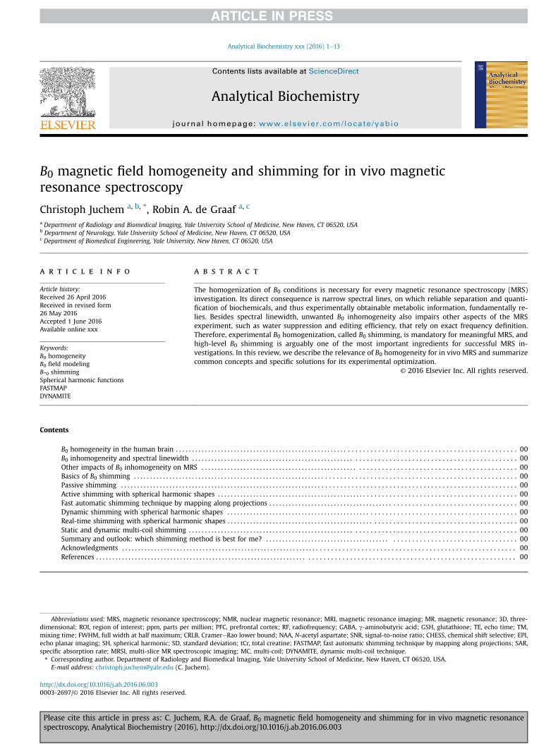
optimal B0 homogeneity and corresponding linewidths of 10 Hz(Fig. 2, simulated STEAM MRS, echo time (TE) 10 ms, mixing time(TM) 50 ms, metabolite concentrations from Refs. [1,6], no macro-molecules, relaxation not considered). The minimization of suchoverlap and the preservation of as much spectroscopic detail aspossible are of paramount importance to allow the numericaldisentanglement of individual metabolite signals and to maximizethe attainable information content (Fig. 2A, full width at halfmaximum (FWHM) 10 Hz). However, suboptimal B0 homogeneitybroadens spectral lines, reduces MRS signal amplitudes, and cor-rupts otherwise attainable metabolic information (Fig. 2B, FWHM15 Hz). At poor B0 conditions, spectral peaks progressively mergetogether and eventually become inseparable (Fig. 2C, FWHM20 Hz). Although quantification accuracy, for which CramereRaolower bound (CRLB) often serves as the maximum confidencemeasure, might remain sufficient for metabolites containing strongsinglets such as N-acetyl aspartate (NAA) and creatine, the quan-tification of low-amplitude and/or strongly overlapping signalssuch as GABA, GSH, and glutamate versus glutamine becomesprogressively impossible. The sensitivity of in vivo MRS is inher-ently limited, and a significant noise floor is the rule rather than theexception. As such, suboptimal B0 conditions directly translate toreduced signal-to-noise ratios (SNRs). For simplicity, pure Lor-entzian line broadening representing a shortened mono-exponential transverse decay time T2 has been applied in thisexample simulation to illustrate the effects of linewidth on spectraloverlap and attainable SNRs. In reality, such line broadening reflectsthe underlying Larmor frequencies and, therefore, the backgroundB0 distribution. As such, B0 inhomogeneity not only broadens MRSsignals but also affects their line shape in a complex fashion. Notethat the above simulations also neglected the influence of macro-molecules. At short TE, macromolecules contribute a significantfraction of the observed signals [7,8]. In addition to potentialconfusion among overlapping (short T2) metabolites, then, thespectroscopic quantification is further impaired at poor B0 condi-tions when peaks or multiplet structures exhibit linewidths similarto those of macromolecular signals.
Other impacts of B0 inhomogeneity on MRS
Chemical shift selective (CHESS) water suppression employsfrequency-selective RF pulses to selectively excite and subsequentlydephase the water magnetization throughout a chosen volume,
Please cite this article in press as: C. Juchem, R.A. de Graaf, B0 magnetispectroscopy, Analytical Biochemistry (2016), http://dx.doi.org/10.1016/j.
thereby minimizing the spectral contribution of water [9]. Inho-mogeneous B0 conditions result in a distribution of water fre-quencies instead of a single frequency. This reduces the suppressionefficiency if the frequency variation is significant compared withthe bandwidth and profile of the selective RF pulses. Note that thesidebands of the RF pulses applied for MRS localization can extendsignificantly beyond the MRS voxel volume itself. If B0 conditionssurrounding the MRS voxel are inhomogeneous, as in the humanPFC, reduced suppression efficiency in these areas can manifestitself as residual water signal from outside the voxel.
Frequency-selective RF pulses are also commonly employed forspectral editing techniques such as J-difference editing for GABAquantification [10]. Here, frequency variations lead to alteredediting efficiency and erroneous metabolite quantification. Editingschemes relying on a specific symmetry, such as those applied tominimize macromolecule contributions [11], are potentiallyaffected in a similar fashion.
B0 field inhomogeneity during the application of magnetic fieldgradients inevitably leads to erroneous spatial assignments thatcan become a limiting factor for low-bandwidth MRI applicationssuch as echo planar imaging (EPI). The impact of B0 inhomogeneityon localization accuracy, although less critical for MRS, is describedhere in the context of this review because the origin of thisimportant B0-induced artifact is universal (Fig. 3).
Linear field gradients are used to encode spatial position infrequency for slice selection and MRS voxel localization by atailored combination of a linear field gradient (Fig. 3, y-gradientdBz/dy, black line) and an RF pulse (upper gray horizontal bar). Sliceposition and width are determined by the combination of gradientstrength and RF pulse offset/bandwidth, respectively (right verticalgray bar, Dy1). Spatial localization with linear magnetic field gra-dients is based on the assumption that the field distributionthroughout the subject is based solely on the applied field gradi-ents. In reality, however, this implicit assumption is regularlyviolated by apparent background B0 field inhomogeneity (dashedblue line). Such variations in the PFC locally alter the effectivegradient field, thereby leading to an erroneous slice profile andposition (left vertical gray bar). Thus, an RF pulse of unchangedbandwidth (lower horizontal gray bar) results in a slice of reducedthickness Dy2. Note that these artifacts are given by the shape andstrength of the B0 distortion relative to the applied gradientamplitude and polarity. Therefore, the effect is highly positiondependent and the opposite artifact, a stretch instead of a squeeze,
c field homogeneity and shimming for in vivo magnetic resonanceab.2016.06.003
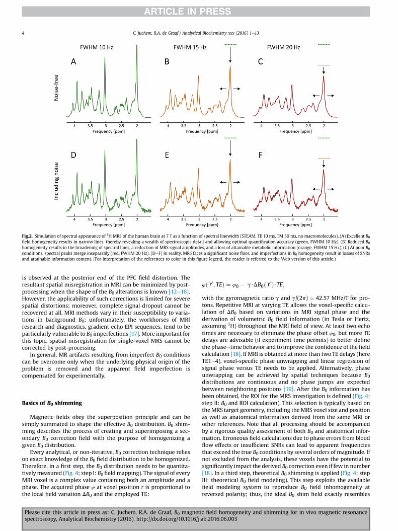
Fig.2. Simulation of spectral appearance of 1H MRS of the human brain at 7 T as a function of spectral linewidth (STEAM, TE 10 ms, TM 50 ms, no macromolecules). (A) Excellent B0field homogeneity results in narrow lines, thereby revealing a wealth of spectroscopic detail and allowing optimal quantification accuracy (green, FWHM 10 Hz). (B) Reduced B0homogeneity results in the broadening of spectral lines, a reduction of MRS signal amplitudes, and a loss of attainable metabolic information (orange, FWHM 15 Hz). (C) At poor B0conditions, spectral peaks merge inseparably (red, FWHM 20 Hz). (DeF) In reality, MRS faces a significant noise floor, and imperfections in B0 homogeneity result in losses of SNRsand attainable information content. (For interpretation of the references to color in this figure legend, the reader is referred to the Web version of this article.)
C. Juchem, R.A. de Graaf / Analytical Biochemistry xxx (2016) 1e134
is observed at the posterior end of the PFC field distortion. Theresultant spatial misregistration in MRI can be minimized by post-processing when the shape of the B0 alterations is known [12e16].However, the applicability of such corrections is limited for severespatial distortions; moreover, complete signal dropout cannot berecovered at all. MRI methods vary in their susceptibility to varia-tions in background B0; unfortunately, the workhorses of MRIresearch and diagnostics, gradient echo EPI sequences, tend to beparticularly vulnerable to B0 imperfections [17]. More important forthis topic, spatial misregistration for single-voxel MRS cannot becorrected by post-processing.
In general, MR artifacts resulting from imperfect B0 conditionscan be overcome only when the underlying physical origin of theproblem is removed and the apparent field imperfection iscompensated for experimentally.
Basics of B0 shimming
Magnetic fields obey the superposition principle and can besimply summated to shape the effective B0 distribution. B0 shim-ming describes the process of creating and superimposing a sec-ondary B0 correction field with the purpose of homogenizing agiven B0 distribution.
Every analytical, or non-iterative, B0 correction technique relieson exact knowledge of the B0 field distribution to be homogenized.Therefore, in a first step, the B0 distribution needs to be quantita-tivelymeasured (Fig. 4; step I: B0 fieldmapping). The signal of everyMRI voxel is a complex value containing both an amplitude and aphase. The acquired phase 4 at voxel position r is proportional tothe local field variation DB0 and the employed TE:
Please cite this article in press as: C. Juchem, R.A. de Graaf, B0 magnetispectroscopy, Analytical Biochemistry (2016), http://dx.doi.org/10.1016/j.
4ð r!; TEÞ ¼ 40 � g$DB0ð r!Þ$TE;
with the gyromagnetic ratio g and g/(2p) ¼ 42.57 MHz/T for pro-tons. Repetitive MRI at varying TE allows the voxel-specific calcu-lation of DB0 based on variations in MRI signal phase and thederivation of volumetric B0 field information (in Tesla or Hertz,assuming 1H) throughout the MRI field of view. At least two echotimes are necessary to eliminate the phase offset 40, but more TEdelays are advisable (if experiment time permits) to better definethe phaseetime behavior and to improve the confidence of the fieldcalculation [18]. If MRI is obtained at more than twoTE delays (hereTE1e4), voxel-specific phase unwrapping and linear regression ofsignal phase versus TE needs to be applied. Alternatively, phaseunwrapping can be achieved by spatial techniques because B0distributions are continuous and no phase jumps are expectedbetween neighboring positions [19]. After the B0 information hasbeen obtained, the ROI for the MRS investigation is defined (Fig. 4;step II: B0 and ROI calculation). This selection is typically based ontheMRS target geometry, including theMRS voxel size and positionas well as anatomical information derived from the same MRI orother references. Note that all processing should be accompaniedby a rigorous quality assessment of both B0 and anatomical infor-mation. Erroneous field calculations due to phase errors from bloodflow effects or insufficient SNRs can lead to apparent frequenciesthat exceed the true B0 conditions by several orders of magnitude. Ifnot excluded from the analysis, these voxels have the potential tosignificantly impact the derived B0 correction even if few in number[18]. In a third step, theoretical B0 shimming is applied (Fig. 4; stepIII: theoretical B0 field modeling). This step exploits the availablefield modeling system to reproduce B0 field inhomogeneity atreversed polarity; thus, the ideal B0 shim field exactly resembles
c field homogeneity and shimming for in vivo magnetic resonanceab.2016.06.003
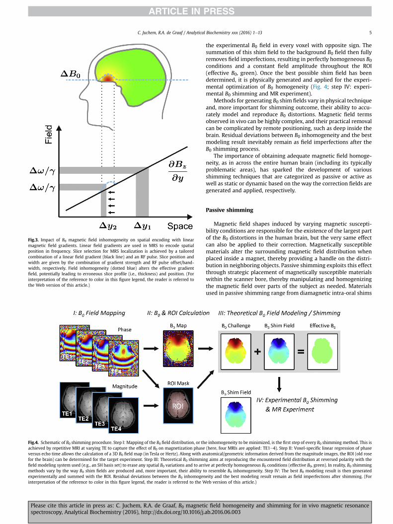
Fig.3. Impact of B0 magnetic field inhomogeneity on spatial encoding with linearmagnetic field gradients. Linear field gradients are used in MRS to encode spatialposition in frequency. Slice selection for MRS localization is achieved by a tailoredcombination of a linear field gradient (black line) and an RF pulse. Slice position andwidth are given by the combination of gradient strength and RF pulse offset/band-width, respectively. Field inhomogeneity (dotted blue) alters the effective gradientfield, potentially leading to erroneous slice profile (i.e., thickness) and position. (Forinterpretation of the reference to color in this figure legend, the reader is referred tothe Web version of this article.)
Fig.4. Schematic of B0 shimming procedure. Step I: Mapping of the B0 field distribution, or thachieved by repetitive MRI at varying TE to capture the effect of B0 on magnetization phaseversus echo time allows the calculation of a 3D B0 field map (in Tesla or Hertz). Along with anfor the brain) can be determined for the target experiment. Step III: Theoretical B0 shimminfield modeling system used (e.g., an SH basis set) to erase any spatial B0 variations and to arrmethods vary by the way B0 shim fields are produced and, more important, their ability texperimentally and summed with the ROI. Residual deviations between the B0 inhomogeninterpretation of the reference to color in this figure legend, the reader is referred to the W
C. Juchem, R.A. de Graaf / Analytical Biochemistry xxx (2016) 1e13 5
Please cite this article in press as: C. Juchem, R.A. de Graaf, B0 magnetispectroscopy, Analytical Biochemistry (2016), http://dx.doi.org/10.1016/j.
the experimental B0 field in every voxel with opposite sign. Thesummation of this shim field to the background B0 field then fullyremoves field imperfections, resulting in perfectly homogeneous B0conditions and a constant field amplitude throughout the ROI(effective B0, green). Once the best possible shim field has beendetermined, it is physically generated and applied for the experi-mental optimization of B0 homogeneity (Fig. 4; step IV: experi-mental B0 shimming and MR experiment).
Methods for generating B0 shim fields vary in physical techniqueand, more important for shimming outcome, their ability to accu-rately model and reproduce B0 distortions. Magnetic field termsobserved in vivo can be highly complex, and their practical removalcan be complicated by remote positioning, such as deep inside thebrain. Residual deviations between B0 inhomogeneity and the bestmodeling result inevitably remain as field imperfections after theB0 shimming process.
The importance of obtaining adequate magnetic field homoge-neity, as in across the entire human brain (including its typicallyproblematic areas), has sparked the development of variousshimming techniques that are categorized as passive or active aswell as static or dynamic based on the way the correction fields aregenerated and applied, respectively.
Passive shimming
Magnetic field shapes induced by varying magnetic suscepti-bility conditions are responsible for the existence of the largest partof the B0 distortions in the human brain, but the very same effectcan also be applied to their correction. Magnetically susceptiblematerials alter the surrounding magnetic field distribution whenplaced inside a magnet, thereby providing a handle on the distri-bution in neighboring objects. Passive shimming exploits this effectthrough strategic placement of magnetically susceptible materialswithin the scanner bore, thereby manipulating and homogenizingthe magnetic field over parts of the subject as needed. Materialsused in passive shimming range from diamagnetic intra-oral shims
e inhomogeneity to be minimized, is the first step of every B0 shimming method. This is(here, four MRIs are applied: TE1e4). Step II: Voxel-specific linear regression of phaseatomical/geometric information derived from the magnitude images, the ROI (old roseg aims at reproducing the encountered field distribution at reversed polarity with theive at perfectly homogeneous B0 conditions (effective B0, green). In reality, B0 shimmingo resemble B0 inhomogeneity. Step IV: The best B0 modeling result is then generatedeity and the best modeling result remain as field imperfections after shimming. (Foreb version of this article.)
c field homogeneity and shimming for in vivo magnetic resonanceab.2016.06.003
C. Juchem, R.A. de Graaf / Analytical Biochemistry xxx (2016) 1e136
for the minimization of field artifacts in the human PFC [20e22] toexternal dia- and paramagnetic passive shims for whole-brainshimming in the mouse [23,24]. Matching the magnetic proper-ties of a human head holder (with pyrolytic graphite foam) to thediamagnetic properties of the head/neck that it hosts can also beunderstood as a form of passive shimming [25]. Passive shimmingwith ferromagnetic substances provides an inexpensive means ofefficiently producing very strong magnetic fields. Ferromagneticmaterials have also been applied in a subject-specific fashion[26,27].
Since the early days of MR, the B0 distribution of the MRscanner itself is improved by placement of small pieces of iron orsteel (“shims”) to strategic positions; as such, the term “shim-ming,” now applied to describe any type B0 homogenizationtechnique, is rooted in the passive variant. Notably, optimizednumerical methods have been recently proposed to efficientlycalculate the B0 fields induced by arbitrary magnetic susceptibilitydistributions, thereby providing a versatile tool for understandingthe B0 effects induced in the human body and their possiblecorrection with passive shimming [28,29]. Passive shimming ismarked by ease of field generation via simple placement of theassemblies to be polarized inside the scanner field. However, thecreation and adjustment of such passive shim assemblies iscumbersome. More important, passive shimming generally lacksthe flexibility to accommodate experiment-specific conditions andvarying shim requirements due to differences in subject anatomy(compare, e.g., the five cases in Fig. 1), subject placement, oraltered susceptibility distributions (e.g., due to the nasal conges-tion of a subject with a cold). Although the benefits of passiveshimming for specific MRS applications have been demonstrated,its practical shortcomings have prevented it from becoming thecommon method of choice.
Active shimming with spherical harmonic shapes
Active shimming refers to B0 homogenization with correctionfields produced by electrical coils. The standard approach tominimizing magnetic field variations for MRS with active shim-ming is to superimpose magnetic fields with a spatial variationgoverned by spherical harmonic (SH) functions [30,31]. Themagnetic field in free space obeys the Laplace equation, and so-lutions thereof can be expressed as SH expansions. Although thespace is arguably no longer empty when objects are studied withMRS, the SH framework has prevailed likely due to its experi-mental practicality and ubiquity in the fields of chemistry andphysics. SH functions are orthogonal and organized in orders Nwith 2Nþ 1 terms per order (Fig. 5). The single zero-order shape isa general offset (not shown), and the three first-order termsdescribe linear field gradients identical to those applied for spatialencoding. Higher orders contribute shapes with higher-ordersymmetry and multiplicity (Fig. 5; cf. first-order X, second-orderX2eY2 and XY, third-order X3, and fourth-order X4) and, there-fore, enable the modeling of increasingly complex target fieldshapes. In general, the higher the maximum SH order applied forB0 shimming, the more likely it is that it will resemble the spatialfeatures of given B0 distortions and thereby allow their subse-quent compensation by B0 shimming. In other words, the numberof basis functions available for modeling the field distortion de-termines the flexibility of magnetic field shaping; thus, the qualityof the expected shim outcome is improved if higher orders areincluded [32].
The B0 field generated by electrical charges flowing through aconducting wire is described by BioteSavart's law. Shaping thewirepattern allows a handle on the current distribution and the mag-netic field shape that is created. This principle is exploited to
Please cite this article in press as: C. Juchem, R.A. de Graaf, B0 magnetispectroscopy, Analytical Biochemistry (2016), http://dx.doi.org/10.1016/j.
produce individual SH shapes by dedicated current-driven wirepatterns, one for each term, and a set of SH coils is nested inside thescanner bore to form a shim system that generates B0 correctionfields over the subject. In practice, SH shim systems are limited tolow-order terms due to space and cost restrictions. Most humanMR systems are equipped with shim coils capable of generating SHfields up to the second or third order. SH shimming is a robust andflexible method that can be fully automated to provide objectiveuser-independent magnetic field homogeneity [33e36].
The orthogonality of the SH basis functions allows the individualadjustment of SH shim terms under the assumption that the fieldsgenerated by the SH wire patterns are indeed pure and truly in-dependent. In reality, however, most SH coils also produce otherunwanted terms along with the primary shape that determines thecoil's name. These cross-terms are not necessarily a problem as longas they can be modeled with B0 shapes from other available shimcoils and subsequently considered in the computation of theapplicable set of B0 shim currents [37].
Fig. 6 shows an example analysis of SH field modeling andtheoretical B0 shimming for single voxel MRS in the human PFC at4 T (ROI size 4 � 4 � 4 cm3). Magnetic susceptibility boundariesnear the frontal bone result in strong and complex B0 in-homogeneities that can severely disrupt and potentially ruin MRSinvestigations in the PFC. The inclusion of higher-order SH func-tions and shapes facilitates progressively better resemblance of B0distortions and improved B0 shim outcome, reflected in reducedstandard deviation (SD) of the residual field distribution.
B0 shimming aims to improve the overall B0 homogeneity of theROI. Numerically, this is typically achieved by least-squares mini-mization of the sum of the original B0 challenge and the B0 shimfield to be applied. With this approach, the residual field deviationsof every voxel are combined quadratically and, therefore, irre-spectively of sign. The synthesized field with the smallest overalldeviation in terms of this single metric is considered the best fit. Ifthe B0 challenge can be perfectly resembled by the shim system,this quality measure becomes zero and perfect B0 homogeneity isachieved. In most cases, the applied basis set is not capable ofperfectly resembling all field distortion. Although there is only oneperfectly homogeneous B0 scenario, imperfect solutions are notunique but to some degree rely on the chosen B0 homogeneitymetric. Note that the residual field imperfection depends on theemployed set of basis functions and, thus, can vary in both shapeand polarity with the inclusion of additional SH orders (Fig. 6, ar-rows). Although the overall B0 homogeneity is improved, regionalB0 homogeneity might be compromised and residual distortionscan potentially even reverse polarity. This might seem counterin-tuitive at first but can be well understood given a particular patternof B0 inhomogeneity, the available B0 shim system, and the char-acteristics of the numerical optimization routine (i.e., the costfunction).
Tkac and coworkers estimated the resolution limit for 7-T 1HMRS of adult human brain to correspond with a linewidth of 9 Hzfor total creatine or the combination of creatine and phosphocre-atine [3]. This finding is consistent with our experience and caneven be obtained in the PFC when B0 shimming is optimal (Fig. 7A;7 T, STEAM, TE 10 ms, TM 50 ms, 8 cc, FWHM of total creatine (tCr)8.8 ± 0.2 Hz, FWHM of NAA 7.3 ± 0.2 Hz). Excellent B0 homogeneityis the basis for reliable quantification of the overall neurochemicalprofile [38] and, moreover, the separation of glutamate and gluta-mine at 7 T. At this spectral quality, even the multiplet structure ofthe glutamate C4 protons at 2.35 ppm becomes clearly visible inboth the experimental spectrum and the scaled glutamate basisfunction.
c field homogeneity and shimming for in vivo magnetic resonanceab.2016.06.003

Fig.5. Selection of first- to fourth-order SH shapes. SH functions are organized in orders N with 2N þ 1 terms per order. The single zero-order shape represents a global offset (notshown), and the three first-order terms describe linear field gradients identical to those applied for spatial encoding. Higher orders contribute shapes with higher-order symmetryand multiplicity (compare figure third-order X3 and fourth-order X4 with first-order X and second-order X2eY2 and XY) and, therefore, can model progressively advanced targetfield shapes. In general, the higher the maximum SH order applied in B0 shimming and the larger the number of shapes included, the more likely the successful modeling of B0distortions and their subsequent compensation by B0 shimming.
Fig.6. SH field modeling and B0 shimming for single-voxel MRS in the human PFC at 4 T as a function of SH order (cube size 4 � 4 � 4 cm3). Magnetic susceptibility transitions in thefrontal head result in strong and complex-shaped B0 inhomogeneities that have the potential to render MRS investigations in the PFC useless. The inclusion of higher-order SHfunctions and shapes allows progressively better resemblance of the B0 distortions and improved B0 shim outcomes, corresponding to reduced SD of the residual field distribution.Note that the residual field imperfection is a function of the employed set of basis functions and can vary regionally in both shape and polarity with the inclusion of additional SHorders (arrows: first order, strongly positive; second order, intermediate negative; third order, slightly positive).
C. Juchem, R.A. de Graaf / Analytical Biochemistry xxx (2016) 1e13 7
Fast automatic shimming technique by mapping alongprojections
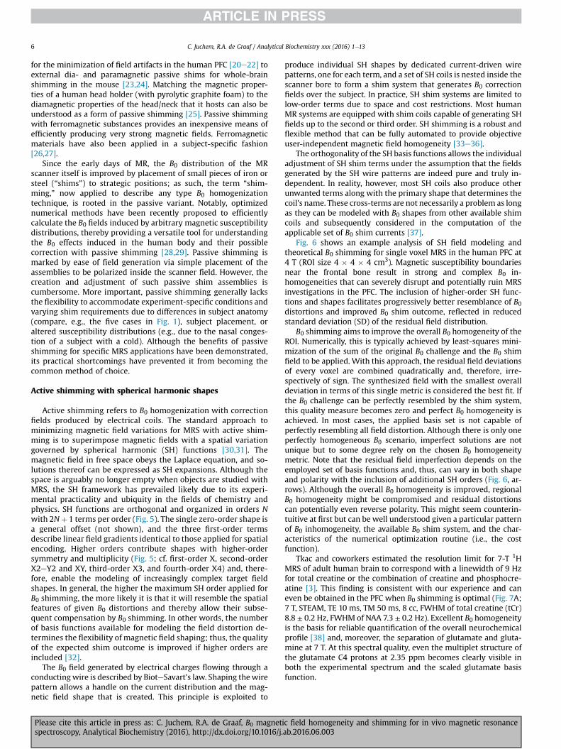
Full 3D mapping of B0 field conditions can be time-consuming.Instead, the fast automatic shimming technique by mappingalong projections (FASTMAP) samples the B0 field distributions tobe homogenized along six exemplary orthogonal column
Please cite this article in press as: C. Juchem, R.A. de Graaf, B0 magnetispectroscopy, Analytical Biochemistry (2016), http://dx.doi.org/10.1016/j.
projections only (Fig. 8; cube size 14 mm, projection length19.8 mm). Basic linear algebra is applied to unambiguously derivethe unique solutions for the apparent first- and second-order SHfield imperfections from this small set of measured polynomial B0terms. Given the available B0 shim system, the apparent B0 in-homogeneity is then converted to the corresponding correctionfield. Note that the key of the FASTMAP approach lies in the efficient
c field homogeneity and shimming for in vivo magnetic resonanceab.2016.06.003

Fig.7. (A) In vivo single-voxel 1H MRS of the human PFC at 7 T (STEAM, TE 10 ms, TM50 ms). Third-order SH B0 shimming allows excellent line width (FWHM of NAA7.3 ± 0.2 Hz, Cr/PCr 8.8 ± 0.2 Hz). (B,C) Clear separation of glutamate and glutamineand resultant high-accuracy LCModel decomposition of metabolic component. At thisspectral quality, even the multiplet structure of the glutamate C4 protons at 2.35 ppmcan be appreciated in both the experimental spectrum and the glutamate basis func-tion. Ins, myo-inositol; PE, phosphorylethanolamine; Cr, creatine; PCr, phosphocrea-tine; Glu, glutamate; Gln, glutamine; Tau, taurine; Cho, choline; GSH, glutathione;GABA, g-aminobutyric acid; Asp, aspartate; NAA, N-acetyl aspartate.
C. Juchem, R.A. de Graaf / Analytical Biochemistry xxx (2016) 1e138
determination of 3D B0 conditions with a sparse samplingapproach. The subsequent determination of the B0 shim field isstandard.
The sampling of selected column projections assumes smoothand well-behaved B0 field conditions. Obviously, very localizeddistortions that are not captured by one of the column projectionsare not considered. If applied to reasonably small volumes forsingle-voxel MRS, however, such cases are rare. Full 3D field map-ping does provide a more detailed and complete picture of theapparent B0 conditions of such a specialized scenario. In practicalreality, the limited capability of second-order SH field modeling toresemble such extreme B0 scenarios is more likely to be the limitingfactor than the sparsity of the employed column projections.
FASTMAP is an elegant analytical method that provides a ver-satile tool for second-order SH B0 homogenization in cubic volumestypical for single-voxel MRS investigations. As such, FASTMAP (andits derivatives [39,40]) has become the method of choice in many
Please cite this article in press as: C. Juchem, R.A. de Graaf, B0 magnetispectroscopy, Analytical Biochemistry (2016), http://dx.doi.org/10.1016/j.
laboratories due to significantly reduced acquisition timescompared with B0 shimming methods that are based on full 3DMRI. An optimized FASTMAP implementation at 7 T was recentlyshown to allow localized B0 shimming at low specific absorptionrate (SAR; i.e., RF power deposition) in less than 1 min [41].
The B0 shim ROI typically resembles the ROI of the MR investi-gation but can differ as long as the characteristics of the B0 distri-bution to be homogenized are properly captured. FASTMAPoperates on cubic ROIs and is typically chosen to provide B0shimming for MRS in single voxels. A moderate extension of the B0shim ROI beyond the geometry targeted by the MR investigation issometimes useful if very small ROIs are considered to improve therobustness of the B0 shim computation. This approach should beused with care, however, because it rests on the assumption thatthe B0 distribution in the vicinity of the original ROI resembles theshapes encountered within the original ROI. Therefore, a moderateROI extension can be applied in parts of the brain where B0 con-ditions can be expected to change slowly, but it must be avoided inareas of severe local B0 gradients or at the brain surface if no furtherquality and selection measures are applied. The homogenization ofB0 conditions in slabs or complex-shaped volumes such as theentire human brain requires 3D B0 mapping methods along withalgorithms to select the desired ROI based on geometric conditions(e.g., slab position and thickness) and/or anatomical aspects (e.g.,brain segmentation).
Dynamic shimming with spherical harmonic shapes
High-quality B0 homogeneity can be readily achieved with low-order SH B0 shimming in small areas considered for single-voxelMRS. B0 shimming with SH shapes is also capable of removing alarge part of the B0 imperfections encountered throughout theentire human brain (Fig. 9). As expected, the inclusion of higher-order shapes provides progressively better results and a nearlyperfect elimination of the widespread shallow terms [32]. Complexand localized magnetic field distortions, such as those observed inthe human PFC or the temporal lobes, are beyond the modelingcapability of current static SH shimming. They can be somewhatreduced by the low-order SH fields to which current technology islimited, but they cannot be removed. As such, limited B0 homoge-neity throughout the human brain has been a long-standingproblem.
Global B0 shimming aims to optimize the B0 conditions over theentire object under investigation at once (Fig.10A,1D example, graydotted range). The best B0 shim field is then applied experimentallyand remains constant, or static, throughout the MR investigation. Ifthe MR sequence employs a serial scheme in which different sub-volumes of the considered object are sampled sequentially, as ininterleaved multi-voxel MRS or multi-slice MRI, a series of B0 shimconditions can be optimized for every such subvolume separately(Fig. 10B). The B0 shim condition tailored to an individual sub-volume is then applied immediately before the MR signal ismeasured and updated to the next B0 shim setting and subvolumeimmediately after. The adjustment of subunit-specific shim settingswith this dynamic shim approach allows the improved optimiza-tion of magnetic field homogeneity over the original larger volumebased on the individual improvements in the constituentsubvolumes.
After the introduction of dynamic shimming for multi-slice MRIwith linear gradients [42,43], the benefits of including second-order SH terms [44,45] and third-order SH terms [37] have beendemonstrated. Besides the application of dynamic shimming toMRI, multi-voxel MRS with dynamically updated, voxel-specificshim settings has been shown to allow multi-fold efficiency gains[46,47]. In the same vein, multi-slice MR spectroscopic imaging
c field homogeneity and shimming for in vivo magnetic resonanceab.2016.06.003
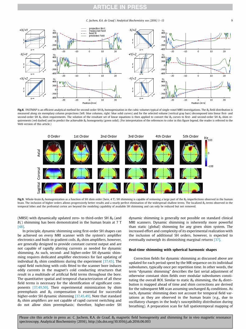
Fig.8. FASTMAP is an efficient analytical method for second-order SH B0 homogenization in the cubic volumes typical of single-voxel MRS investigations. The B0 field distribution ismeasured along six exemplary column projections (left: blue columns, right: blue solid curves) and for the selected volume (vertical gray bars) decomposed into linear first- andsecond-order SH B0 shim requirements. The solution of the resultant set of linear equations is then applied to convert the B0 curves to first- and second-order SH B0 shim re-quirements (red dashed) and to predict the achievable B0 homogeneity (green solid). (For interpretation of the references to color in this figure legend, the reader is referred to theWeb version of this article.)
Fig.9. Whole-brain B0 homogenization as a function of SH shim order (here, 4 T). SH shimming is capable of removing a large part of the B0 imperfections observed in the humanbrain. The inclusion of higher orders allows progressively better results and a nearly perfect elimination of the widespread shallow terms. The localized B0 terms observed in thetemporal lobes and the prefrontal cortex are beyond the modeling capability of available SH shimming and can only be reduced but not removed.
C. Juchem, R.A. de Graaf / Analytical Biochemistry xxx (2016) 1e13 9
(MRSI) with dynamically updated zero- to third-order SH B0 (andB1þ) shimming has been demonstrated in the human brain at 7 T
[48].In principle, dynamic shimming using first-order SH shapes can
be achieved on every MRI scanner with the system's amplifierelectronics and built-in gradient coils. B0 shim amplifiers, however,are generally designed to provide constant current output and arenot capable of rapidly altering currents as needed for dynamicshimming. As such, second- and higher-order SH dynamic shim-ming requires dedicated amplifier electronics for fast updating ofindividual B0 shim conditions during the experiment [37,45]. Therapid field switching with coils fitted to the scanner bore induceseddy currents in the magnet's cold conducting structures thatresult in a multitude of artificial field terms throughout the bore.The quantitative spatial and temporal characterization of all thesefield terms is necessary for the identification of significant com-ponents [37,49,50]. Their experimental minimization by shimpreemphasis and B0 compensation is essential for successfulhigher-order SH dynamic shimming [37,45,49]. Note that standardB0 shim amplifiers are not capable of rapid current switching anddo not allow shim preemphasis; therefore, higher-order SH
Please cite this article in press as: C. Juchem, R.A. de Graaf, B0 magnetispectroscopy, Analytical Biochemistry (2016), http://dx.doi.org/10.1016/j.
dynamic shimming is generally not possible on standard clinicalMRI scanners. Dynamic shimming is inherently more powerfulthan static (global) shimming for any given shim system. Theincreased effort and complexity of its experimental realizationwiththe inclusion of additional SH orders, however, is expected toeventually outweigh its diminishing marginal returns [37].
Real-time shimming with spherical harmonic shapes
Correction fields for dynamic shimming as discussed above areupdated for each period spent by the MR sequence on its individualsubvolumes, typically once per repetition time. In other words, theterm “dynamic shimming” describes the fast serial adjustment ofotherwise constant shim fields over modular subvolumes consti-tuting the overall ROI. Similar to static B0 shimming, the B0 distri-bution is mapped ahead of time and shim corrections are derivedfor the subsequent MR scan assuming unchanged B0 conditions. Assuch, dynamic shimming does not account for temporal field var-iations as they are observed in the human brain (e.g., due tooscillatory changes in the body's susceptibility distribution duringbreathing). A preparation scan for full spatiotemporal mapping of
c field homogeneity and shimming for in vivo magnetic resonanceab.2016.06.003

Fig.10. Illustration of dynamic B0 shimming. (A) Global B0 shimming aims tocompensate the field distortion (black parabolic) throughout the entire object at once(gray dotted). However, if the assumed linear field modeling system (red) is notcapable of resembling the distortion, the difference (pink) will inevitably remain asresidual B0 inhomogeneities after the shimming process. (B) Complex field shapes canbe described by simpler terms when considered regionally over smaller volumes. Thischaracteristic is comparable to the approximation of a one-dimensional complexmathematical function through straight lines (similar to the computation of a differ-ence quotient) and becomes progressively more accurate as the considered functionalrange becomes more localized. Dynamic shimming capitalizes on this principle bybreaking down a large volume such as the human brain into subunits. The adjustmentof subunit-specific shim settings (1e5, colored) then allows the improved optimizationof magnetic field homogeneity over the original larger volume based on the individualimprovements in the constituent subvolumes. Note that every active B0 shimmingmethod is more powerful when applied in a dynamic fashion irrespective of theemployed basis set. (For interpretation of the references to color in this figure legend,the reader is referred to the Web version of this article.)
C. Juchem, R.A. de Graaf / Analytical Biochemistry xxx (2016) 1e1310
Please cite this article in press as: C. Juchem, R.A. de Graaf, B0 magnetispectroscopy, Analytical Biochemistry (2016), http://dx.doi.org/10.1016/j.
the B0 conditions in the brain over the subject's respiratory cyclehas been the basis for a continuous adaptation of B0 correction, so-called real-time shimming, for the compensation of such tempo-rally varying field alterations [51]. The term “real-time shimming”has also been used to describe the periodic interleaving of MR se-quences with B0 measurements and subsequent dynamic updatingof B0 field conditions such as once per repetition time. This tech-nique is fast compared with the overall scan duration (hence, “real-time”) and allows accounting for changing B0 conditions that resultfrom subject movement [52e54]. Continuous monitoring andinstantaneous correction of the gradient and shim coil performancewith a B0 field camera ensures optimal system stability [55,56] andis expected to set the stage for development of truly real-time B0shimming in the future.
Static and dynamic multi-coil shimming
The experimentally available low-order SH terms allow for themodeling of, and thus compensation for, large-scale and shallowmagnetic field components. The complex field terms generated bythe sinuses in the human PFC and by the auditory cavities in thetemporal lobes cannot, however, be corrected adequately (Fig. 11D,7 T [37]). Although significant improvements in B0 magnetic fieldhomogeneity have been demonstrated with dynamic shimming formost parts of the human brain, even dynamic shimming with allzero- to third-order SH terms is not capable of completely ho-mogenizing the entire organ (Fig. 11F). Note the negative light bluering around the PFC field focus and the additional small field al-terations induced in other parts of the brain. Similar to the exampleof single-voxel B0 shimming with varying order SH functions(Fig. 6), the field modeling is not capable of fully compensating forthe distortion. As such, the shim optimization balances field vari-ations in different locations to improve its overall homogeneity.More specifically, the dominant field focus in the PFC is reduced atthe expense of allowing some B0 imperfection in other parts of thebrain.
SH functions are only one of the possible basis sets for thedescription and synthesis of magnetic fields. However, despitesome specialized non-SH shim approaches for the human PFC onthe basis of localized intra-oral coils [57] or a specifically tailoredset of external coils [18], the orthogonality of the basis shapes hasbeen accepted by the MR community as an essential prerequisitefor magnetic field modeling and shimming based on early reports[31]. We recently demonstrated that a set of generic localized coilscan be converted to a powerful magnetic field modeling systemwhen each of the electrical coils is driven individually [58]. In otherwords, the orthogonality of the basis functions is not a requirementfor successful magnetic field modeling. This multi-coil (MC)concept allows for the synthesis of simple and complex magneticfields in a flexible and accurate fashion via the superposition ofgeneric non-orthogonal basis fields. B0 shimming with static andespecially dynamic (dynamic multi-coil technique or DYNAMITE),MC techniques have been shown to outperform currently availableSH shimming procedures in the mouse brain [59,60], the rat brain[61], and the human brain [17,62].
In the human brain, DYNAMITE B0 shimming is capable ofgenerating a magnetic field distribution that more closely re-sembles the original distortion over the entire slice, including thestrong and localized field focus in the PFC (Fig. 11G). Correspond-ingly, DYNAMITE shimming removes the largest part of the mag-netic field inhomogeneity as predicted theoretically (Fig. 11H) andshown experimentally (Fig. 11I). Note that neither B0 eddy currentsnor cross-talk between individual MC elements has been signifi-cant with DYNAMITE; therefore, no compensation was necessary.
Static MC and DYNAMITE B0 shimming with dedicated coil
c field homogeneity and shimming for in vivo magnetic resonanceab.2016.06.003

Fig.11. Magnetic field modeling characteristics of different B0 shimming strategies (first row) and their performance for shimming of the human brain at 7 T (second row exceptpanel B). The zero- to third-order SH functions allow the synthesis and removal of shallow magnetic field components (C,E), but significant imperfections remain throughout thebrain or were even induced (D,F). DYNAMITE B0 shimming is capable of generating a magnetic field distribution that closely resembles the original distortion over the entire slice,including the strong and localized focus in the PFC (G). Correspondingly, DYNAMITE B0 shimming removes the largest part of the field inhomogeneity, as predicted theoretically (H)and shown experimentally (I). SH, spherical harmonic; DSH, dynamic spherical harmonic; DYNAMITE, dynamic multi-coil technique; ROI, region of interest.Adapted from Juchem et al., J. Magn. Reson. 212 (2011)
C. Juchem, R.A. de Graaf / Analytical Biochemistry xxx (2016) 1e13 11
systems is well-established and has the potential to replace SH-based B0 shimming due to improved shim performance and fieldgeneration efficiency [60]. The application of constant currents tothe individual elements of RF phased arrays and the use of the RFcoil system for MC B0 shimming promise efficient use of limitedbore space [63,64]. MC and RF coil systems, however, do not shareall design principles, and B0 shim performance of merged MCeRFsystems equivalent to dedicated MC designs is yet to be shown. Todate, MC-based B0 shimming has not been applied to MRS. Its use isconceptually identical to previous applications for MRI and iscurrently pursued in our laboratory. Conventional SH shimmingprovides high-level B0 homogeneity in most parts of the brain,especially when applied locally for single-voxel MRS, and conse-quently no further improvements are anticipated from MC-based(or any other) B0 shimming techniques. Benefits of MC-based B0shimming are expected, however, for applications such as multi-voxel MRS and multi-slice MRSI that span larger parts of thebrain, potentially including areas of severe B0 disturbances.
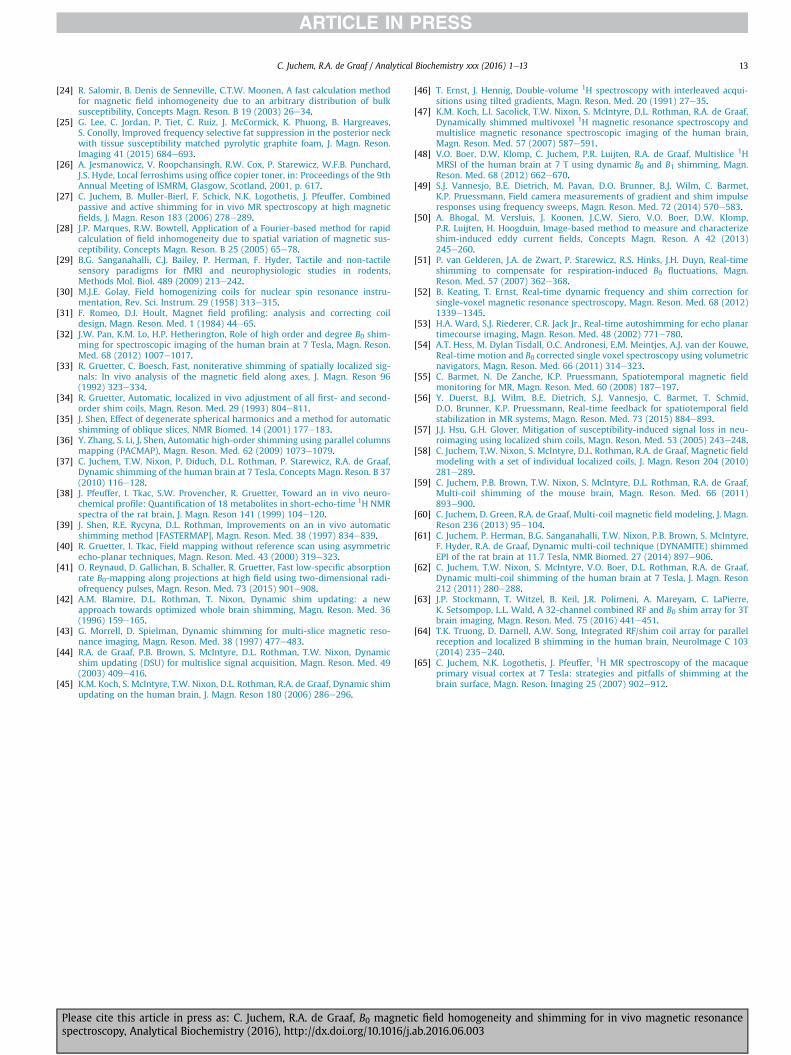
The B0 field focus in the PFC is strongest at its ventral end andfalls off toward the central/dorsal brain (cf. Fig. 9). Therefore,adequate B0 homogeneity can be more easily achieved in the cen-tral PFC. The presentation of those slices, thereby excluding theventral PFC, might suggest an overall B0 shim performance that infact is possible only in the less critical parts of the brain. A morecomplete impression can be achieved, for instance, with a 3Drepresentation focusing on the brain regions critical for B0 shim-ming (Fig. 12, Above: static third-order SH; below: DYNAMITE B0shimming of the human brain at 7 T). In this example, bothmethods remove the largest part of the widespread shallow B0components throughout the brain. However, following SH shim-ming, significant B0 field imperfections remain in the PFC and thetemporal lobes where B0 distortions are localized and complex. Thecombination of improved B0 field modeling capability and dynamicshimming with DYNAMITE B0 shimming accomplishes largely ho-mogeneous B0 conditions throughout the brain, including chal-lenging areas such as the PFC and the temporal lobes.
Please cite this article in press as: C. Juchem, R.A. de Graaf, B0 magnetispectroscopy, Analytical Biochemistry (2016), http://dx.doi.org/10.1016/j.
Summary and outlook: which shimming method is best forme?
The existence of limited B0 homogeneity and the consequentdemand to cope with it secondary to an intended MR investigationcan be vexing. Given the experimental burden of B0 shimming andthe concomitant time requirement, there is a constant temptationto compromise on this step. Magnetic field inhomogeneity, how-ever, can lead to image distortion and signal loss in MRI and todegraded spectral resolution, reduced sensitivity, and additionalvaried artifacts in MRS. All such effects can severely limit or obviatethe validity of the corresponding investigations. Therefore,adequate B0 homogeneity is a necessity, not a choice.
Optimal B0 shimming relies on the accurate knowledge of theemployed basis shapes and amplitudes and the precise synthesis ofthe optimal B0 shim field determined theoretically. Therefore, thecomprehensive, high-quality calibration of any B0 shimmingmethod is of paramount importance because incomplete knowl-edge or erroneous characterization of the shim system results inregular suboptimal B0 shim performance. The B0 shim outcome issimilarly impaired if the strength of the required shim field exceedsthe capacity of the available coil system and amplifier electronics.In such a case, the shapes can be reproduced with the available B0shim system, but the dynamic range appears to be insufficient fortheir compensation. The benefits of combined passive and activeSH B0 shimming have been presented for special cases in which thedynamic range of active shimming alone has not been sufficient[27,65]. In general, however, insufficient dynamic range of the B0shim system inevitably reduces the attainable B0 homogeneity.
A variety of shimming methods has been developed for thepractical realization of B0 correction fields. The B0 distribution canbe altered passively through placement of magnetic materials oractively by driving electrical coils. Active shimming is achieved bycombining a set of basis functions, either orthogonal (e.g., SH) ornon-orthogonal (e.g., MC). Dynamic B0 shimming is inherentlymore powerful than static shimming irrespective of the employed
c field homogeneity and shimming for in vivo magnetic resonanceab.2016.06.003
Fig.12. Comparison of DYNAMITE and static third-order SH modeling for B0 shimmingof the human brain at 7 T. Both methods remove the largest part of shallow B0 com-ponents throughout the brain. However, significant B0 field imperfections remain withSH shimming in the PFC and the temporal lobes, where B0 imperfections are localizedand complex. The combination of improved B0 field modeling capability and dynamicshimming with DYNAMITE B0 shimming allows largely homogeneous B0 conditionsthroughout the brain, including challenging areas in the PFC and the temporal lobes.
C. Juchem, R.A. de Graaf / Analytical Biochemistry xxx (2016) 1e1312
basis set. To date, however, no trivial and effortless B0 shimmingmethod exists that provides perfect B0 homogeneity for all condi-tions. Shimming techniques vary significantly in their performanceas well as the required effort, methodological complexity, andfinancial burden of their implementation. Therefore, MR labora-tories need to decidewhat shimming capabilities to make available,and individuals need to select among the resultant range ofmethods to employ. The best B0 shimming method depends on theMR application for which it is used, and choosing is not a trivial taskgiven that multiple factors, including the species and region de-pendency of apparent magnetic field distortions and the necessarylevel of field homogeneity, are to be considered. For instance,adequate magnetic field homogeneity throughout the entire hu-man brain is required for true 3D whole-brain applications,necessitating robust global static shimming. By contrast, excellentB0 homogeneity can typically be achieved in small volumes forsingle-voxel MRS with second-order SH functions [3]. In addition,magnetic field homogenization of axial slices in dorsal brain re-gions does not face the challenging B0 distortions characteristic ofventral slices; thus, significantly simpler approaches can be suffi-cient. Finally, someMR applications are less susceptible tomagneticfield imperfections than others, and additional insignificant gainsin field homogeneity might not warrant the required additionalinvestment of resources, especially in clinical MRS studies that are
Please cite this article in press as: C. Juchem, R.A. de Graaf, B0 magnetispectroscopy, Analytical Biochemistry (2016), http://dx.doi.org/10.1016/j.
inherently time-constrained. As a result, the successful MRresearcher requires not only broad familiarity with the execution ofvarious B0 shimming methods but also a deep comprehension ofthe underlying theories and characteristics that render them thebest tools for a particular job.
Acknowledgments
The contribution of Hetty Prinsen in the acquisition of the 1HMRspectrum of Fig. 7 and the careful proofreading of the manuscriptby Kelley Swanberg are acknowledged. This workwas supported bythe National Multiple Sclerosis Society (NMSS, RG 4319), the NancyDavis Foundation, and National Institutes of Health (NIH) grantsUL1 TR000142, R01NS062885, and P30-NS052519.
References
[1] V. Govindaraju, K. Young, A.A. Maudsley, Proton NMR chemical shifts andcoupling constants for brain metabolites, NMR Biomed. 13 (2000) 129e153.
[2] J.F. Schenck, The role of magnetic susceptibility in magnetic resonance im-aging: MRI magnetic compatibility of the first and second kinds, Med. Phys. 23(1996) 815e850.
[3] I. Tkac, P. Andersen, G. Adriany, H. Merkle, K. Ugurbil, R. Gruetter, In vivo 1HNMR spectroscopy of the human brain at 7 T, Magn. Reson. Med. 46 (2001)451e456.
[4] S. Michaeli, M. Garwood, X.H. Zhu, L. DelaBarre, P. Andersen, G. Adriany,H. Merkle, K. Ugurbil, W. Chen, Proton T2 relaxation study of water, N-ace-tylaspartate, and creatine in human brain using Hahn and CarrePurcell spinechoes at 4T and 7T, Magn. Reson. Med. 47 (2002) 629e633.
[5] U.E. Emir, E.J. Auerbach, P.F. Van De Moortele, M. Marjanska, K. Ugurbil,M. Terpstra, I. Tkac, G. Oz, Regional neurochemical profiles in the human brainmeasured by 1H MRS at 7 T using local B1 shimming, NMR Biomed. 25 (2012)152e160.
[6] R.A. de Graaf, In Vivo NMR Spectroscopy: Principles and Techniques, JohnWiley, London, 2008.
[7] K.L. Behar, D.L. Rothman, D.D. Spencer, O.A. Petroff, Analysis of macromoleculeresonances in 1H NMR spectra of human brain, Magn. Reson. Med. 32 (1994)294e302.
[8] C. Cudalbu, V. Mlynarik, R. Gruetter, Handling macromolecule signals in thequantification of the neurochemical profile, J. Alzheimers Dis. 31 (Suppl 3)(2012) S101eS115.
[9] A. Haase, J. Frahm, W. Hanicke, D. Matthaei, 1H NMR chemical shift selective(CHESS) imaging, Phys. Med. Biol. 30 (1985) 341e344.
[10] D.L. Rothman, O.A. Petroff, K.L. Behar, R.H. Mattson, Localized 1H NMR mea-surements of g-aminobutyric acid in human brain in vivo, Proc. Natl. Acad. Sci.U. S. A. 90 (1993) 5662e5666.
[11] P.G. Henry, C. Dautry, P. Hantraye, G. Bloch, Brain GABA editing withoutmacromolecule contamination, Magn. Reson. Med. 45 (2001) 517e520.
[12] P. Jezzard, Correction of geometric distortion in fMRI data, NeuroImage 62(2012) 648e651.
[13] M.A. Fernandez-Seara, F.W. Wehrli, Postprocessing technique to correct forbackground gradients in image-based R*2 measurements, Magn. Reson. Med.44 (2000) 358e366.
[14] R. Cusack, M. Brett, K. Osswald, An evaluation of the use of magnetic fieldmaps to undistort echo-planar images, NeuroImage 18 (2003) 127e142.
[15] P. Jezzard, R.S. Balaban, Correction for geometric distortion in echo planarimages from B0 field variations, Magn. Reson. Med. 34 (1995) 65e73.
[16] N. Weiskopf, U. Klose, N. Birbaumer, K. Mathiak, Single-shot compensation ofimage distortions and BOLD contrast optimization using multi-echo EPI forreal-time fMRI, NeuroImage 24 (2005) 1068e1079.
[17] C. Juchem, S. Umesh Rudrapatna, T.W. Nixon, R.A. de Graaf, Dynamic multi-coil technique (DYNAMITE) shimming for echo-planar imaging of the hu-man brain at 7 Tesla, NeuroImage 105 (2015) 462e472.
[18] C. Juchem, T.W. Nixon, S. McIntyre, D.L. Rothman, R.A. de Graaf, Magnetic fieldhomogenization of the human prefrontal cortex with a set of localized elec-trical coils, Magn. Reson. Med. 63 (2010) 171e180.
[19] R. Cusack, N. Papadakis, New robust 3-D phase unwrapping algorithms:application to magnetic field mapping and undistorting echoplanar images,NeuroImage 16 (2002) 754e764.
[20] J.L. Wilson, M. Jenkinson, P. Jezzard, Optimization of static field homogeneityin human brain using diamagnetic passive shims, Magn. Reson. Med. 48(2002) 906e914.
[21] J.L. Wilson, P. Jezzard, Utilization of an intra-oral diamagnetic passive shim infunctional MRI of the inferior frontal cortex, Magn. Reson. Med. 50 (2003)1089e1094.
[22] R. Cusack, B. Russell, S.M. Cox, C. De Panfilis, C. Schwarzbauer, R. Ansorge, Anevaluation of the use of passive shimming to improve frontal sensitivity infMRI, NeuroImage 24 (2005) 82e91.
[23] K.M. Koch, P.B. Brown, D.L. Rothman, R.A. de Graaf, Sample-specific diamag-netic and paramagnetic passive shimming, J. Magn. Reson 182 (2006) 66e74.
c field homogeneity and shimming for in vivo magnetic resonanceab.2016.06.003
C. Juchem, R.A. de Graaf / Analytical Biochemistry xxx (2016) 1e13 13
[24] R. Salomir, B. Denis de Senneville, C.T.W. Moonen, A fast calculation methodfor magnetic field inhomogeneity due to an arbitrary distribution of bulksusceptibility, Concepts Magn. Reson. B 19 (2003) 26e34.
[25] G. Lee, C. Jordan, P. Tiet, C. Ruiz, J. McCormick, K. Phuong, B. Hargreaves,S. Conolly, Improved frequency selective fat suppression in the posterior neckwith tissue susceptibility matched pyrolytic graphite foam, J. Magn. Reson.Imaging 41 (2015) 684e693.
[26] A. Jesmanowicz, V. Roopchansingh, R.W. Cox, P. Starewicz, W.F.B. Punchard,J.S. Hyde, Local ferroshims using office copier toner, in: Proceedings of the 9thAnnual Meeting of ISMRM, Glasgow, Scotland, 2001, p. 617.
[27] C. Juchem, B. Muller-Bierl, F. Schick, N.K. Logothetis, J. Pfeuffer, Combinedpassive and active shimming for in vivo MR spectroscopy at high magneticfields, J. Magn. Reson 183 (2006) 278e289.
[28] J.P. Marques, R.W. Bowtell, Application of a Fourier-based method for rapidcalculation of field inhomogeneity due to spatial variation of magnetic sus-ceptibility, Concepts Magn. Reson. B 25 (2005) 65e78.
[29] B.G. Sanganahalli, C.J. Bailey, P. Herman, F. Hyder, Tactile and non-tactilesensory paradigms for fMRI and neurophysiologic studies in rodents,Methods Mol. Biol. 489 (2009) 213e242.
[30] M.J.E. Golay, Field homogenizing coils for nuclear spin resonance instru-mentation, Rev. Sci. Instrum. 29 (1958) 313e315.
[31] F. Romeo, D.I. Hoult, Magnet field profiling: analysis and correcting coildesign, Magn. Reson. Med. 1 (1984) 44e65.
[32] J.W. Pan, K.M. Lo, H.P. Hetherington, Role of high order and degree B0 shim-ming for spectroscopic imaging of the human brain at 7 Tesla, Magn. Reson.Med. 68 (2012) 1007e1017.
[33] R. Gruetter, C. Boesch, Fast, noniterative shimming of spatially localized sig-nals: In vivo analysis of the magnetic field along axes, J. Magn. Reson 96(1992) 323e334.
[34] R. Gruetter, Automatic, localized in vivo adjustment of all first- and second-order shim coils, Magn. Reson. Med. 29 (1993) 804e811.
[35] J. Shen, Effect of degenerate spherical harmonics and a method for automaticshimming of oblique slices, NMR Biomed. 14 (2001) 177e183.
[36] Y. Zhang, S. Li, J. Shen, Automatic high-order shimming using parallel columnsmapping (PACMAP), Magn. Reson. Med. 62 (2009) 1073e1079.
[37] C. Juchem, T.W. Nixon, P. Diduch, D.L. Rothman, P. Starewicz, R.A. de Graaf,Dynamic shimming of the human brain at 7 Tesla, Concepts Magn. Reson. B 37(2010) 116e128.
[38] J. Pfeuffer, I. Tkac, S.W. Provencher, R. Gruetter, Toward an in vivo neuro-chemical profile: Quantification of 18 metabolites in short-echo-time 1H NMRspectra of the rat brain, J. Magn. Reson 141 (1999) 104e120.
[39] J. Shen, R.E. Rycyna, D.L. Rothman, Improvements on an in vivo automaticshimming method [FASTERMAP], Magn. Reson. Med. 38 (1997) 834e839.
[40] R. Gruetter, I. Tkac, Field mapping without reference scan using asymmetricecho-planar techniques, Magn. Reson. Med. 43 (2000) 319e323.
[41] O. Reynaud, D. Gallichan, B. Schaller, R. Gruetter, Fast low-specific absorptionrate B0-mapping along projections at high field using two-dimensional radi-ofrequency pulses, Magn. Reson. Med. 73 (2015) 901e908.
[42] A.M. Blamire, D.L. Rothman, T. Nixon, Dynamic shim updating: a newapproach towards optimized whole brain shimming, Magn. Reson. Med. 36(1996) 159e165.
[43] G. Morrell, D. Spielman, Dynamic shimming for multi-slice magnetic reso-nance imaging, Magn. Reson. Med. 38 (1997) 477e483.
[44] R.A. de Graaf, P.B. Brown, S. McIntyre, D.L. Rothman, T.W. Nixon, Dynamicshim updating (DSU) for multislice signal acquisition, Magn. Reson. Med. 49(2003) 409e416.
[45] K.M. Koch, S. McIntyre, T.W. Nixon, D.L. Rothman, R.A. de Graaf, Dynamic shimupdating on the human brain, J. Magn. Reson 180 (2006) 286e296.
Please cite this article in press as: C. Juchem, R.A. de Graaf, B0 magnetispectroscopy, Analytical Biochemistry (2016), http://dx.doi.org/10.1016/j.
[46] T. Ernst, J. Hennig, Double-volume 1H spectroscopy with interleaved acqui-sitions using tilted gradients, Magn. Reson. Med. 20 (1991) 27e35.
[47] K.M. Koch, L.I. Sacolick, T.W. Nixon, S. McIntyre, D.L. Rothman, R.A. de Graaf,Dynamically shimmed multivoxel 1H magnetic resonance spectroscopy andmultislice magnetic resonance spectroscopic imaging of the human brain,Magn. Reson. Med. 57 (2007) 587e591.
[48] V.O. Boer, D.W. Klomp, C. Juchem, P.R. Luijten, R.A. de Graaf, Multislice 1HMRSI of the human brain at 7 T using dynamic B0 and B1 shimming, Magn.Reson. Med. 68 (2012) 662e670.
[49] S.J. Vannesjo, B.E. Dietrich, M. Pavan, D.O. Brunner, B.J. Wilm, C. Barmet,K.P. Pruessmann, Field camera measurements of gradient and shim impulseresponses using frequency sweeps, Magn. Reson. Med. 72 (2014) 570e583.
[50] A. Bhogal, M. Versluis, J. Koonen, J.C.W. Siero, V.O. Boer, D.W. Klomp,P.R. Luijten, H. Hoogduin, Image-based method to measure and characterizeshim-induced eddy current fields, Concepts Magn. Reson. A 42 (2013)245e260.
[51] P. van Gelderen, J.A. de Zwart, P. Starewicz, R.S. Hinks, J.H. Duyn, Real-timeshimming to compensate for respiration-induced B0 fluctuations, Magn.Reson. Med. 57 (2007) 362e368.
[52] B. Keating, T. Ernst, Real-time dynamic frequency and shim correction forsingle-voxel magnetic resonance spectroscopy, Magn. Reson. Med. 68 (2012)1339e1345.
[53] H.A. Ward, S.J. Riederer, C.R. Jack Jr., Real-time autoshimming for echo planartimecourse imaging, Magn. Reson. Med. 48 (2002) 771e780.
[54] A.T. Hess, M. Dylan Tisdall, O.C. Andronesi, E.M. Meintjes, A.J. van der Kouwe,Real-time motion and B0 corrected single voxel spectroscopy using volumetricnavigators, Magn. Reson. Med. 66 (2011) 314e323.
[55] C. Barmet, N. De Zanche, K.P. Pruessmann, Spatiotemporal magnetic fieldmonitoring for MR, Magn. Reson. Med. 60 (2008) 187e197.
[56] Y. Duerst, B.J. Wilm, B.E. Dietrich, S.J. Vannesjo, C. Barmet, T. Schmid,D.O. Brunner, K.P. Pruessmann, Real-time feedback for spatiotemporal fieldstabilization in MR systems, Magn. Reson. Med. 73 (2015) 884e893.
[57] J.J. Hsu, G.H. Glover, Mitigation of susceptibility-induced signal loss in neu-roimaging using localized shim coils, Magn. Reson. Med. 53 (2005) 243e248.
[58] C. Juchem, T.W. Nixon, S. McIntyre, D.L. Rothman, R.A. de Graaf, Magnetic fieldmodeling with a set of individual localized coils, J. Magn. Reson 204 (2010)281e289.
[59] C. Juchem, P.B. Brown, T.W. Nixon, S. McIntyre, D.L. Rothman, R.A. de Graaf,Multi-coil shimming of the mouse brain, Magn. Reson. Med. 66 (2011)893e900.
[60] C. Juchem, D. Green, R.A. de Graaf, Multi-coil magnetic field modeling, J. Magn.Reson 236 (2013) 95e104.
[61] C. Juchem, P. Herman, B.G. Sanganahalli, T.W. Nixon, P.B. Brown, S. McIntyre,F. Hyder, R.A. de Graaf, Dynamic multi-coil technique (DYNAMITE) shimmedEPI of the rat brain at 11.7 Tesla, NMR Biomed. 27 (2014) 897e906.
[62] C. Juchem, T.W. Nixon, S. McIntyre, V.O. Boer, D.L. Rothman, R.A. de Graaf,Dynamic multi-coil shimming of the human brain at 7 Tesla, J. Magn. Reson212 (2011) 280e288.
[63] J.P. Stockmann, T. Witzel, B. Keil, J.R. Polimeni, A. Mareyam, C. LaPierre,K. Setsompop, L.L. Wald, A 32-channel combined RF and B0 shim array for 3Tbrain imaging, Magn. Reson. Med. 75 (2016) 441e451.
[64] T.K. Truong, D. Darnell, A.W. Song, Integrated RF/shim coil array for parallelreception and localized B shimming in the human brain, NeuroImage C 103(2014) 235e240.
[65] C. Juchem, N.K. Logothetis, J. Pfeuffer, 1H MR spectroscopy of the macaqueprimary visual cortex at 7 Tesla: strategies and pitfalls of shimming at thebrain surface, Magn. Reson. Imaging 25 (2007) 902e912.
c field homogeneity and shimming for in vivo magnetic resonanceab.2016.06.003