bacillus subtilis chromosome organization oscillates between two … · 2019-07-05 · bacillus...
TRANSCRIPT

Bacillus subtilis chromosome organization oscillatesbetween two distinct patternsXindan Wang, Paula Montero Llopis, and David Z. Rudner1
Department of Microbiology and Immunobiology, Harvard Medical School, Boston, MA 02115
Edited by Sharon R. Long, Stanford University, Stanford, CA, and approved July 2, 2014 (received for review April 23, 2014)
Bacterial chromosomes have been found to possess one of twodistinct patterns of spatial organization. In the first, called “ori-ter”and exemplified by Caulobacter crescentus, the chromosome armslie side-by-side, with the replication origin and terminus at oppositecell poles. In the second, observed in slow-growing Escherichia coli(“left-ori-right”), the two chromosome arms reside in separate cellhalves, on either side of a centrally located origin. These two pat-terns, rotated 90° relative to each other, appear to result from dif-ferent segregation mechanisms. Here, we show that the Bacillussubtilis chromosome alternates between them. For most of the cellcycle, newly replicated origins are maintained at opposite poleswith chromosome arms adjacent to each other, in an ori-ter config-uration. Shortly after replication initiation, the duplicated originsmove as a unit to midcell and the two unreplicated arms resolveinto opposite cell halves, generating a left-ori-right pattern. Theorigins are then actively segregated toward opposite poles, reset-ting the cycle. Our data suggest that the condensin complex and theparABS partitioning system are the principal driving forces underly-ing this oscillatory cycle. We propose that the distinct organizationpatterns observed for bacterial chromosomes reflect a commonorganization–segregation mechanism, and that simple modificationsto it underlie the unique patterns observed in different species.
DNA replication | ParA | chromosome segregation | SMC condensin
Central to reproduction is the faithful segregation of replicatedchromosomes to daughter cells. In eukaryotes, DNA repli-
cation, chromosome condensation, and sister chromatid segre-gation are separated into distinct steps in the cell cycle that aresafeguarded by checkpoint pathways. In bacteria, these processesoccur concurrently, posing unique challenges to genome integrityand inheritance (1, 2). In the absence of temporal control, bac-teria take advantage of spatial organization to promote faithfuland efficient chromosome segregation. The organization of thechromosome dictates where the chromosome is replicated, andthe factors that organize and compact the newly replicated DNAplay a central role in its segregation (1, 2).Studies in different bacteria have revealed strikingly distinct
patterns of chromosome organization that appear to arise fromdifferent segregation mechanisms. In Caulobacter crescentus andVibrio cholerae chromosome I, the origin and terminus are locatedat opposite cell poles, with the two replication arms between them,in a pattern referred to as “ori-ter” (3–5). After replication initi-ation, one of the sister origins is held in place and the other isactively translocated to the opposite cell pole, regenerating the ori-ter organization in both daughter cells (5–11). By contrast, in slow-growing Escherichia coli, the origin is located in the middle of thenucleoid and the two replication arms reside in opposite cellhalves, in a “left-ori-right” pattern (12, 13). Replication initiates atmidcell, and the duplicated origins segregate to the quarter posi-tions followed by the left and right arms on either side, regener-ating the left-ori-right pattern in the two daughter cells.Although chromosome organization was first analyzed in the
Gram-positive bacterium Bacillus subtilis, our understanding ofthe replication–segregation cycle in this bacterium has remainedelusive. In pioneering studies, it was shown that during sporeformation, the replicated origins reside at opposite cell poles and
the termini at midcell in an ori-ter ter-ori organization (14–18). Asimilar ori-ter pattern was observed during vegetative growth (15,19). However, in separate studies, DNA replication was found toinitiate at a midcell-localized origin (20, 21). How these dispa-rate patterns fit into a coherent replication–segregation cycle hasnever been addressed and motivated this study. Our analysishas revealed that the B. subtilis chromosome follows an un-expected and previously unidentified choreography during veg-etative growth in which the organization alternates betweenori-ter and left-ori-right patterns. Our data further suggest thatthe highly conserved partitioning system (parABS) and thestructural maintenance of chromosomes (SMC) condensin com-plex, in conjunction with replication initiation, function as the corecomponents for this oscillating cycle. We propose that this cycleenhances the efficiency of DNA replication and sister chromo-some segregation and provides a unifying model for the diversepatterns of chromosome organization observed in bacteria.
ResultsSporulating B. subtilis Chromosomes Have an ori-ter Organization.Asa first step toward our analysis of chromosome organization inB. subtilis, we modified the cytological tools to visualize chro-mosomal loci in live cells. Previous fluorescently tagged repressorproteins bound to operator arrays caused partial or completeblocks to replisome progression in B. subtilis (15, 22, 23). Wereduced the number of binding sites in the arrays and loweredexpression of the repressor protein fusions using a weak consti-tutive promoter (Materials and Methods). Under these conditions,virtually all cells contained fluorescent foci with no detectableimpact on growth rate, nucleoid morphology, or nucleoid sizedistribution (Fig. S1).
Significance
In bacteria, faithful and efficient DNA segregation is intimatelylinked to the spatial organization of the chromosome. Two dis-tinct organization patterns have been described for bacterialchromosomes (ori-ter and left-ori-right) that appear to arise fromdistinct segregation mechanisms. Here, we show that the Bacillussubtilis chromosome oscillates between them during a replica-tion–segregation cycle. Our data further suggest that the highlyconserved condensin complex and the parABS partitioning systemfunction as the core components that underlie these alternatingpatterns. We propose that this oscillatory cycle enhances the ef-ficiency of DNA replication and sister chromosome segregationand provides a unifying model for the diverse patterns of chro-mosome organization observed in bacteria.
Author contributions: X.W. and D.Z.R. designed research; X.W. performed research; X.W.and P.M.L. contributed new reagents/analytic tools; X.W. and D.Z.R. analyzed data; andX.W. and D.Z.R. wrote the paper.
The authors declare no conflict of interest.
This article is a PNAS Direct Submission.
See Commentary on page 12580.1To whom correspondence should be addressed. Email: [email protected].
This article contains supporting information online at www.pnas.org/lookup/suppl/doi:10.1073/pnas.1407461111/-/DCSupplemental.
www.pnas.org/cgi/doi/10.1073/pnas.1407461111 PNAS | September 2, 2014 | vol. 111 | no. 35 | 12877–12882
MICRO
BIOLO
GY
SEECO
MMEN
TARY

To validate these tools, we analyzed the organization of thereplicated chromosomes during spore formation. At early stagesin this developmental process, the replicated chromosomes adopta structure called the axial filament, in which the origins areanchored at the cell poles and the termini are present nearmidcell (16, 24–26). We visualized six chromosomal loci [−7° (ori)and ±87°, ±120°, and +174° (ter)] during sporulation and de-termined their population-average, normalized position relativeto cell length using automated image analysis software (27) (Fig.1 A–E). The origins were present close to the cell poles at 0.17and 0.83, and the termini were located at midcell (0.5). The ±87°and ±120° loci localized linearly between them. These resultsconfirm and extend previous observations (24, 25, 28) that thesporulating chromosomes are organized linearly along their lengthfrom the poles to midcell in a C. crescentus-like ori-ter ter-ori or-ganization (Fig. 1B).
B. subtilis Maintains a Partially Diploid State. The analysis of chro-mosome organization in C. crescentus and slow-growing E. coli wasfacilitated by their simple cell cycles with no overlapping rounds ofreplication (3, 12, 13). When grown in rich media, B. subtilis, likeE. coli, undergoes multifork replication and the cells are bornwith partially replicated chromosomes. In defined rich medium[doubling time (τ) = 35 min], the cells were born with two origins
and, on average, the number of origins per cell was 3.1 (n = 1,825)(Fig. 1F). To obtain a simpler DNA replication cycle, we grewB. subtilis in minimal medium supplemented with different carbonsources, as was done previously in E. coli, to reduce growth rateand DNA content. Although we could slow growth in glucose (τ =48 min), succinate (τ = 77 min), and sorbitol (τ = 98 min), underall conditions tested, the cells were born with two origins, and theaverage number of origins per cell was 2.8, 2.3, and 2.1 (n > 1,700),respectively (Fig. 1G and Fig. S2A). Even under the slowestgrowth condition, cells were born with greater than 50% of theirchromosome replicated (Fig. S2B). Thus, we were unable to iden-tify conditions in which cells are born with a single unreplicatedchromosome (1-N content). These results suggest that there is alink between DNA replication and cell division that ensures cellsare born with two almost completely replicated chromosomes un-der normal growth conditions, a requirement for successful sporu-lation. We therefore initiated our analysis using conditions in whichcells were artificially restricted to a single copy of the chromosomeby inhibiting replication initiation.
Left-ori-Right Chromosome Organization in Cells with a SingleChromosome. To analyze vegetatively growing cells with 1-NDNA content, we inhibited replication initiation using a tem-perature-sensitive mutant of the helicase loader (DnaB) (29,30). Cells were grown in rich medium at the permissive tem-perature (30 °C, τ = 57 min) to early exponential phase andthen shifted to the restrictive temperature (42 °C, τ = 33 min)(Fig. 2A). After 1 h, 84% of the cells (n = 1,224) had a single-lobed nucleoid containing one replication origin, indicatingthat the chromosome content was reduced to 1-N (Fig. 2A).After 1.5 h, >90% of the cells (n = 2,399) had a single origin.Furthermore, as observed previously by McGinness and Wake(31), the nucleoids adopted a bilobed structure (Fig. 2A).Strikingly, in most cases (92%, n = 2,196), the origin was lo-cated at or close to the center of the two lobes (Fig. 2 A, C, D,and F). This localization pattern is dramatically different fromsporulating cells, in which origins are at the extreme poles (Fig.1C), and vegetatively growing cells with unperturbed replica-tion, in which origins are frequently localized at the nucleoidperiphery (14, 16) (Fig. 1 F and G and Fig. S2A). To determinewhether each nucleoid lobe represents a chromosome arm, wevisualized pairs of loci on the left and right arms. Eighty percentof the cells (n = 1,633) with fluorescently tagged loci at ±87°and 88% of the cells (n = 1,634) with tagged loci at ±120° hadthese markers in separate nucleoid lobes (Fig. 2 B and E).We also monitored loci at −87° and −120° on the same chro-mosome arm. In 86% of the cells (n = 1,804), the two loci werepresent in the same nucleoid lobe (Fig. 2 B and E). Further-more, in the majority of these cells, the −87° locus was closerto the center of the two lobes and the −120° locus was closer tothe nucleoid edge (Fig. 2 B and E).Analysis of a locus close to the replication terminus (+174°)
revealed that most cells had a ter locus directly between the twolobes (64%, n = 2,196) or in close proximity to this position(17%) (Fig. 2 C and F). However, in 18% of the cells, the ter-minus was present near the edge of the nucleoid. To visualize alarger region of the terminus, we used YFP fusion to the repli-cation termination protein (RTP) (32). RTP binds to nine ter-mination sites spanning ∼430 kb of the terminus region (33).RTP-YFP localized as a single focus, clusters of foci, or multiplefoci spanning the bilobed nucleoid (Fig. 2D), consistent with theterminus region connecting the ends of the two lobes. Similarresults were obtained using different strain backgrounds anddifferent growth media, as well as when initiation was blockedusing dnaA(ts) or induction of SirA, an inhibitor of DnaA (34,35). Collectively, these data indicate that under conditions inwhich B. subtilis is restricted to 1-N content, the nucleoid is or-ganized in a pattern like the E. coli chromosome during slow
ori ter
-87˚ +87˚
-87˚ +87˚
-120˚ +120˚
ori
ter
A B
C
D
E -120˚+120˚
0 0.5 1
% ori
0.17 0.83
5
10
0 0.5 1
ter0.50
10
20
30
S750 sorbitol, τ=98min F CH, τ=35min G
0 0.5 1
-87˚0.35 0.65
5
10
0 0.5 1
-120˚0.39 0.61
5
10
0 0.5 1
+120˚0.39 0.62
5
10
0 0.5 1
+87˚0.34 0.65
5
10
Fig. 1. Chromosome organization during sporulation. (A–E) Exponentiallygrowing cells were induced to sporulate and imaged after 2 h. All strainscontained a mutation in spoIIIE (spoIIIE36) (55) to prevent DNA transport. (A)Chromosomemap showing positions of operator arrays. (B) Schematic summaryof cellular localization of loci. Representative micrographs of cells labeled withpairs of loci ori-ter (C, Left), ±87° (D, Left), and ±120° (E, Left) are shown. (C–E,Right) Loci position relative to cell length analyzed (Materials and Methods).The centroid of the Gaussian distribution is shown on the top of each histo-gram. (F and G) Origin number and localization in rich and poor growth media.Origins (green, tetO48/TetR-CFP), nucleoid (blue, HBsu-mYpet), and membrane[red, N-(3-triethylammoniumpropyl)-4-(6-(4-(diethylamino) phenyl) hexatrienyl),pyridinium dibromide (FM4-64)] are shown. (Scale bar: 4 μm.)
12878 | www.pnas.org/cgi/doi/10.1073/pnas.1407461111 Wang et al.

growth, with the two arms present on either side of the origin andgenetic loci arranged linearly from the middle toward the poles.Finally, the terminus region connects the two ends.
Origin-Localized SMC Complexes Are Required for Left-ori-RightOrganization. In E. coli, the left-ori-right organization is shifted toan ori-ter pattern in cells lacking the condensin complex MukBEF(36, 37). We used degradable variants of B. subtilis SMC or itspartner protein ScpB to investigate whether the left-ori-right or-ganization similarly depends on the condensin complex. Degrada-tion of SMC (and ScpB separately) was induced when dnaB(ts)cells were shifted to the restrictive temperature to block replicationinitiation. After 1.5 h, 91% of SMC-degraded cells (n = 1,005) hada more extended chromosome structure, with the replication originlocated at the extreme edge of the nucleoid (Fig. 2G and Fig. S3).Similar results were obtained after ScpB degradation (Fig. 2H).
The Spo0J (ParB) protein in B. subtilis has been shown to recruitSMC complexes to the origin region (28, 38). Accordingly, we in-vestigated the organization of the nucleoid in its absence (Fig. S3).In the majority of the Spo0J mutant cells (70%, n = 999), the originof replication localized to the edge of the nucleoid. This phenotypewas principally due to the absence of Spo0J-mediated recruitmentof SMC rather than the loss of the parABS partitioning activity,because cells lacking Soj (ParA) that are not impaired in SMCrecruitment (28) retained the left-ori-right pattern (Fig. S3). Weconclude that origin-localized condensin complexes play a criticalrole in generating the E. coli-like pattern.
ori-ter and Left-ori-Right Patterns Alternate During the Replication–Segregation Cycle. The two distinct patterns of chromosome or-ganization described above were observed under extreme con-ditions. In one, the origins were physically tethered to the cellpoles during bacterial differentiation (24, 26); in the other,replication initiation was blocked and the cells were restrictedto 1-N content. To investigate whether exponentially growingcells with unperturbed DNA replication possess either pattern,we used time-lapse fluorescence microscopy to visualize chro-mosomal loci during replication–segregation cycles. To ensurethe best time resolution, we used minimal medium supplementedwith sorbitol (τ = 98 min). First, we visualized the origin (Fig.3A). The cells were born with two origins at opposite edges of thenucleoid and remained at the nucleoid periphery for the rest ofthe replication cycle. Previous studies suggested that replicationinitiates in the middle of the nucleoid (15, 20). However, in ourmovies, the origin foci became brighter at or close to the nucleoidperiphery, indicative of their replication at this site (Fig. 3A andFig. S4A, red carets). Similarly, in snapshot images, replisomescolocalized with the brighter origin foci at the nucleoid edge (Fig.S4B). Shortly after their replication, the two new origins migratedas a unit to the middle of the nucleoid (Fig. 3A and Fig. S4A, yellowcarets). This event was followed by their bidirectional segregationto opposite edges of the nucleoid. This cycle is consistent withsnapshot images in which most cells have two origins at the outeredges of the nucleoid (16, 19) (Fig. 1 F and G and Fig. S2A).Next, we tracked markers on the left and right chromosome
arms. For most of the cell cycle, the ±87° loci localized adjacentto each other. However, for a brief period (20–30 min), the twoloci separated, localizing on either side of the nucleoid (Fig. 3B,black arrow, and Fig. S5). After this period of resolution, the twoloci were replicated, segregated, and colocalized again. Similarresults were obtained for the pair of markers at ±120°. To relatethe dynamic movement of the markers on opposite arms to thereplication cycle, we used mCherry–Spo0J (ParB) fusion to lo-calize the origin (14) while simultaneously following the markersat ±87°. When the ±87° markers colocalized, the origin was almostalways present at the edge of the nucleoid (Fig. 3C). However,when the ±87° markers were spatially resolved, the replicatedorigins were either in the middle of the nucleoid between the ±87°markers or had already segregated to the edges of the nucleoid(Fig. 3C). Thus, movement of the duplicated origins to midcellis accompanied by resolution of the left and right arms. This un-precedented choreography can explain some of the unusual local-ization patterns observed previously (15, 19) and is fully supportedby snapshot images in which we visualized fluorescently labeledreplisomes and an origin locus (Fig. S6).Combining the time-lapse microscopy, the three-color images,
and the replisome localization relative to the origin, a pictureemerges of the replication–segregation cycle in B. subtilis (Fig.4). At birth, the two origins are located at opposite edges of thenucleoid, with the partially replicated arms lying side by side. Atthe time of the next round of initiation, replisomes assemble onthe polarly localized origins. The newly replicated origins thenmigrate as a unit to the middle of the nucleoid. At this time, theunreplicated chromosome arms become spatially resolved and
ori RTP
A
-87˚ +87˚ -120˚+120˚ -87˚ -120˚B
E F
G
C D
42˚C 1.5h42˚C 1h30˚C
4±1
65±221±0
2±0
3±1
4±0
80±1%2±0
2±1
6±1
5±1
6±1
88±11±1
1±1
3±1
3±1
3±1
-87˚+87˚ -120˚+120˚ -87˚-120˚
76±3%
16±4
8±1
ori
64±3
17±3
18±0
ter
ScpB degron oridnaBts 42˚C 1.5h
H
ori
ori ter
SMC degron oridnaBts 42˚C 1.5h
Fig. 2. Left-ori-right organization in cells with a single chromosome. Cellswith a dnaB(ts) mutation were shifted from 30 °C to 42 °C for 1.5 h to blocknew rounds of initiation. (A) Origin localization before and after tempera-ture shift. Origins (green), nucleoid (red), and membrane (white) are shown.(B) Representative micrographs of cells labeled at pairs of loci: ±87° (Left),±120° (Center), and −87° and −120° (Right). (C) Cells labeled at ori (red) andter (green). (D) Cells labeled at ori (red) and expressing RTP-YFP (green). (E andF) Quantitative analysis of loci positions. Origin localization in dnaB(ts) 1.5 hafter temperature shift and degradation of SMC (G) or ScpB (H) is shown.(Scale bars: 4 μm.)
Wang et al. PNAS | September 2, 2014 | vol. 111 | no. 35 | 12879
MICRO
BIOLO
GY
SEECO
MMEN
TARY

the replisomes track independently of each other along them(Fig. S7). The left-ori-right pattern of the template DNA persistseven after the origins are segregated to the nucleoid periphery andonly returns to the ori-ter–like organization after the arms them-selves have been replicated.
Partitioning Locus Helps Establish and Maintain the Origins at theNucleoid Edge. We investigated whether the B. subtilis partition-ing locus is responsible for maintaining the origins at the nucleoidperiphery until the next round of replication. Chromosomallyencoded par loci are composed of an ATPase (ParA), a DNAbinding protein (ParB), and a centromere-like sequence (parS)(39). All three are critical for origin segregation in C. crescentus(40, 41), Pseudomonas aeruginosa (42), and V. cholerae chromo-some II (43). In B. subtilis, cells lacking ParA, called Soj, havea very mild defect in chromosome segregation (23). ParB mutantshave a more pronounced phenotype, but this is thought to bea consequence of a failure to enrich SMC complexes at the origin(23, 28, 38). Accordingly, for these experiments, we used a Soj
(ParA) mutant that has no apparent impact on origin-localizedSMC (28). Analysis of origin dynamics in cells lacking Soj revealedthat the directional movement of the newly replicated origins wasabolished (Fig. 5 A and C and Fig. S8). Instead of segregating tothe edge of the nucleoid (Figs. 3A and 5B and Fig. S8), the originsmoved more slowly and in an irregular manner (Fig. 5 A and Cand Fig. S8). Although they reached the nucleoid edge, they fre-quently moved back toward midcell.Next, we investigated whether Soj helps maintain the two
chromosomal arms adjacent to each other in an ori-ter pattern.Time-lapse imaging of markers at ±87° in the Δsoj mutantrevealed that these loci remain resolved for the majority (62 ±8%) of the replication cycle (n = 100) (Fig. 5D and Figs. S9 andS10B). By contrast, in WT cells, these loci were resolved for only37 ± 7% (n = 100) of the replication cycle (Figs. 3B and 5D andFigs. S5 and S10A). Furthermore, instead of alternating betweenarm colocalization and arm resolution as observed in WT cells(Figs. 3B and 5D and Figs. S5 and S10A), the Δsoj mutant hadmore dynamic arm movements, with irregular patterns of armcolocalization and resolution (Fig. 5D and Figs. S9 and S10B).These data indicate that Soj, and presumably the partitioninglocus, facilitates bidirectional origin segregation and plays a cen-tral role in both establishing and maintaining the ori-ter pattern.
DiscussionOur data support a model in which origin-localized condensincomplexes and the partitioning system are the principal drivingforces that underlie the two patterns described here, whereasreplication initiation serves as the switch that triggers the oscil-lation between them. In this model, recruitment of SMC com-plexes to the origin by ParB (28, 38) sets up the left-ori-rightpattern. SMC resolves the replicated origins (44, 45), and byanalogy to the stiffening of eukaryotic centromeres by condensin(46), we hypothesize that SMC rigidifies the origin region con-straining the left and right arms on either side of the originthrough lengthwise condensation (47). Because SMC complexesare present at the origin throughout the cell cycle, force must beapplied to the chromosome to generate the ori-ter pattern. Wehave shown that Soj (ParA) actively segregates origins to thenucleoid periphery and plays an important role in both estab-lishing and maintaining this ori-ter organization. By analogy tochromosome and plasmid segregation in other bacteria (48, 49),we hypothesize that Soj acts by exerting pulling forces on Spo0J
I
IIIII
0 min 10 50
90 130 170100 160150110 140120
70 8060403020
I. ori at pole,arms side by side
II. initiation
III. ori in the middle, arms resolved
ori +87˚-87˚ +87˚-87˚
A
B
0 min 5 25
40 45 50 553530
201510
C
ori
+87˚-87˚
ori
replicated arms+87˚
-87˚
ori
Fig. 3. Alternating patterns of chromosome organization during the cellcycle. (A) Time-lapse progression (5-min intervals) of cells labeled at the or-igin (green) and nucleoid (red) grown in minimal medium supplementedwith sorbitol. Red carets highlight replication initiation at the edge of thenucleoid. Yellow carets show duplicated origins at the center of the nucleoidbefore their segregation. (B) Time-lapse progression (10-min intervals) ofcells labeled at −87° (green) and +87° (red). Black arrows highlight resolu-tion of the left and right loci. (C) Representative micrograph of cells labeledat three chromosomal loci: origin (blue, mCherry-Spo0J), −87° (green), and+87° (red). Stages in the replication–segregation cycle are indicated (I–III)and are interpreted in the schematic model (Right). Origin, −87°, and +87°loci are labeled as blue, green, and red balls, respectively. Chromosomearms are shown as gray lines. (Scale bar: 4 μm.)
Birthori-ter
Initiation at poleori release
Arm resolutionleft-ori-right
ori segregation
New DNA: ori-terTemplate: left-right
ori
ter
left right
B. subtilis C. crescentus
E. coli
Fig. 4. Schematic model of the organization–segregation cycle in B. subtilis,and its comparison with C. crescentus and E. coli. Origins are represented asblack balls, and termini are represented as brown lines in B. subtilis andE. coli and as a brown oval in C. crescentus. The compacted left and rightchromosome arms are shown as thick blue and purple lines (or blobs). Newlyreplicated DNA is shown with a lighter hue, whereas uncompacted DNA isshown as thin lines. In the B. subtilis, newborn cell, unreplicated DNA is shownas a black cloud. The replication–segregation cycle of C. crescentus and E. colicorresponds to two halves of the cycle in B. subtilis.
12880 | www.pnas.org/cgi/doi/10.1073/pnas.1407461111 Wang et al.

(ParB) bound to parS adjacent to the origin. Finally, we proposethat replication initiation triggers the change in organizationpattern by transiently inactivating the forces exerted on the ori-gins. One possibility is that Soj’s role in replication initiation (50)could temporarily relax its segregation function. Alternatively orin addition, replication of the origin region could inactivate thepartitioning system by transiently disrupting the Spo0J–parScomplexes. When force on the origins is lost, the chromosomeresolves into its SMC-mediated left-ori-right pattern. Par-medi-ated segregation of the newly replicated origins initiates a newround of this oscillatory cycle. Intriguingly, in cells that areblocked for replication initiation, the partitioning system appearsto become inactivated, leading to a stable left-ori-right configu-ration (Fig. 2). Chronic inhibition of replication initiation couldmaintain Soj in its replication-active state (50), resulting ina sustained loss of its segregation function. Alternatively, the lossof partitioning activity could be due to a change in the dynamicproperties of the partitioning system (8, 11, 39, 48, 49) resultingfrom a reduced chromosome content in an enlarged cellularcompartment. Systematic analysis of chromosome organizationin P. aeruginosa (42) suggests that its replication–segregation
cycle could follow an alternating pattern similar to the one de-scribed here. Interestingly, this bacterium contains both con-densin complexes and the Par system, and it does not anchor itsorigins at the cell poles. Furthermore, recent quantitative anal-ysis of snapshot images suggests an ori-ter–like chromosome or-ganization in fast-growing E. coli (51). The localization of thereplisome to midnucleoid zones in these fast-growing cells (51)raises the possibility that a transient left-ori-right intermediatemight exist during these overlapping replication cycles. Time-lapse imaging of loci on both replication arms or simultaneousimaging of three loci will be required to establish whether or notthis is the case.In the cycle we report here, the template DNA adopts a left-
ori-right organization, whereas the newly replicated DNA issegregated and organized in an ori-ter pattern. We hypothesizethat this oscillating pattern enhances the efficiency of replicationand segregation of the bacterial chromosome. The left-ori-rightpattern ensures that the two replisomes track on the templateDNA in opposite cell halves (Figs. S6 and S7), providing spatialresolution to the complex topologies that inevitably arise at thereplication forks. The ori-ter pattern of the newly replicatedDNA ensures that the sister chromosomes are segregated as faras possible from each other. Finally, in the specific case ofB. subtilis, the partitioning system also ensures that the originsreside close to the poles, where they can be readily anchored ifthe cell enters the sporulation pathway (24, 26). Initiation ofa new round of replication transiently blocks entry into sporu-lation (52), and our data suggest that it also releases the originsfrom their polar position, allowing the replication–segregationcycle to begin again.Finally, our data raise the possibility that the disparate patterns
of chromosome organization observed in bacteria arise frommodifications to a common organization–segregation mechanism.In the case of E. coli, which lacks a partitioning locus, thecondensin-mediated left-ori-right pattern is established shortlyafter origin segregation and this ground state is maintainedthroughout the replication–segregation cycle (Fig. 4). In thecase of C. crescentus, V. cholerae chromosome I, and sporulatingB. subtilis, distinct polar anchoring mechanisms “lock” chro-mosome organization in the ori-ter pattern, half of the full cycle(Fig. 4). Although the removal of the polar anchoring mecha-nism or addition of a par locus is unlikely to be sufficient torecover the second half of the respective cycles, we propose thatthese factors are the principal drivers in evolving distinctorganization–segregation cycles.
Materials and MethodsGeneral Methods. B. subtilis strains were derived from the prototrophicstrain PY79 . Cells were grown in defined rich casein hydrolysate medium(53) or minimal medium (S750) (54) supplemented with 1% glucose, sor-bitol, or succinate as specified. To monitor chromosome organization duringsporulation, we used a mutant of the SpoIIIE DNA translocase (spoIIIE36)that engages the forespore chromosome after polar division but is blockedin DNA transport (28, 55). Sporulation was induced by resuspension at 37 °Caccording to the method of Sterlini–Mandelstam (53). Images were taken2 h after the initiation of sporulation. E. coli SspB protein was induced with0.5% xylose for degradation of SsrA-tagged proteins (56). Strains, plasmids,and oligonucleotides used in this study are listed in Tables S1–S3. Strain andplasmid constructions are described in SI Materials and Methods.
Fluorescence Microscopy. Fluorescence microscopy was performed with anOlympus BX61 microscope equipped with an UPLFLN 100×/1.3-N.A. phase-contrast oil objective and a CoolSnapHQ cooled CCD camera (Photometrics)or a Nikon Ti microscope equipped with Plan Apo 100×/1.4-N.A. phase-con-trast oil objective and a CoolSnapHQ2 camera. Membranes were stained withtrimethylammonium diphenylhexatriene (Molecular Probes) at 0.01 mMor with FM4-64 [N-(3-triethylammoniumpropyl)-4-(6-(4-(diethylamino)phenyl) hexatrienyl), pyridinium dibromide; Molecular Probes] at 3 μg/mL.DNA was stained with DAPI (Molecular Probes) at 2 μg/mL. Images were
50 100 150 200 min0
1/2L
Lorigin traces (WT)
0 min 5 25
40 45 50 55
3530201510Δsojori
50 100 150 200 min0
1/2L
Lorigin traces (Δsoj)
60 65 70 75
A
B C
Distance of ±87˚ loci relative to cell length 40
20
0
40
20
01 2 3 4 5 6 7 8 9 10 1112 1 2 3 4 5 6 7 8 9 10 frame1 2 3 4 5 6 7 8 9 10 11121 2 3 4 5 6 7 8 9 10 1112
ΔsojWTD % of L
Fig. 5. Impaired origin segregation and chromosome arm dynamics in theabsence of Soj (ParA). (A) Time-lapse progression (5-min intervals) of Δsojcells. The origin (green) and nucleoid (red) are shown. Representative origintraces in WT (B) and Δsoj (C) are shown. The position of the origin wasplotted relative to the total cell length (Fig. S8). (D) Interfocal distances of±87° loci are analyzed in representative WT (Upper) and Δsoj (Lower) cellsin time-lapse progressions (10-min intervals). Each graph plots the distancebetween a pair of ±87° loci relative to the cell length (L) at indicated timesduring their replication cycle. For the purpose of this analysis, the repli-cation cycle of these loci begins when both ±87° loci are replicated (definedas frame 1) and finishes when either of the two loci is replicated again.Because each cell has two pairs of ±87° loci in its replication cycle, themaximum distance of a pair of ±87° loci is 50% of the cell length (Fig. S10).
Wang et al. PNAS | September 2, 2014 | vol. 111 | no. 35 | 12881
MICRO
BIOLO
GY
SEECO
MMEN
TARY

cropped and adjusted using MetaMorph software (Molecular Devices). Finalfigure preparation was performed in Adobe Illustrator (Adobe Systems).
For snapshot imaging, cells were immobilized using 2% (wt/vol) agarosepads containing growth media. For time-lapse imaging, a glass-bottomeddish (Willco dish HBSt-5040; Willco Wells) was used as a coverslip. Expo-nentially growing cells were concentrated at 3,300 × g for 30 s. After re-moval of 90% of the supernatant, 2 μL of the culture was spotted onto theglass-bottomed dish. A 2% (wt/vol) agarose pad containing growth mediawas then laid on top of the bacteria. These cells were imaged on a Well PlateHolder stage (TI-SH-W; Nikon) equipped with a humid, temperature-con-trolled incubator (TC-MIS; Bioscience Tools). The upper face of the pad wasfully exposed, allowing adequate oxygen for growth. The objective was
heated using a Bioptechs objective heater system. Images were acquiredevery 5 or 10 min as specified. Image analyses were performed using theMathWorks MATLAB-based program MicrobeTracker (27). Details of imageanalysis can be found in SI Materials and Methods.
ACKNOWLEDGMENTS. We thank members of the Bernhardt laboratory andthe laboratory of D.Z.R. as well as Viknesh Sivanathan for stimulating discussionsand support, and we thank Alan Grossman, Kevin Griffith, Dirk Landgraf, DavidSherratt, and Rodrigo Reyes-Lamothe for plasmids. Support for this work comesfrom National Institutes of Health Grants GM086466 and GM073831 (to D.Z.R.).X.W. was a long-term fellow of the Human Frontier Science Program. P.M.L. isa Helen Hay Whitney postdoctoral fellow.
1. Reyes-Lamothe R, Nicolas E, Sherratt DJ (2012) Chromosome replication and segre-gation in bacteria. Annu Rev Genet 46:121–143.
2. Wang X, Montero Llopis P, Rudner DZ (2013) Organization and segregation of bac-terial chromosomes. Nat Rev Genet 14(3):191–203.
3. Viollier PH, et al. (2004) Rapid and sequential movement of individual chromosomalloci to specific subcellular locations during bacterial DNA replication. Proc Natl AcadSci USA 101(25):9257–9262.
4. Fogel MA, Waldor MK (2005) Distinct segregation dynamics of the two Vibrio chol-erae chromosomes. Mol Microbiol 55(1):125–136.
5. Fogel MA, Waldor MK (2006) A dynamic, mitotic-like mechanism for bacterial chro-mosome segregation. Genes Dev 20(23):3269–3282.
6. Bowman GR, et al. (2008) A polymeric protein anchors the chromosomal origin/ParBcomplex at a bacterial cell pole. Cell 134(6):945–955.
7. Ebersbach G, Briegel A, Jensen GJ, Jacobs-Wagner C (2008) A self-associating proteincritical for chromosome attachment, division, and polar organization in caulobacter.Cell 134(6):956–968.
8. Schofield WB, Lim HC, Jacobs-Wagner C (2010) Cell cycle coordination and regulationof bacterial chromosome segregation dynamics by polarly localized proteins. EMBO J29(18):3068–3081.
9. Shebelut CW, Guberman JM, van Teeffelen S, Yakhnina AA, Gitai Z (2010) Caulo-bacter chromosome segregation is an ordered multistep process. Proc Natl Acad SciUSA 107(32):14194–14198.
10. Yamaichi Y, et al. (2012) A multidomain hub anchors the chromosome segregationand chemotactic machinery to the bacterial pole. Genes Dev 26(20):2348–2360.
11. Ptacin JL, et al. (2010) A spindle-like apparatus guides bacterial chromosome segre-gation. Nat Cell Biol 12(8):791–798.
12. Nielsen HJ, Ottesen JR, Youngren B, Austin SJ, Hansen FG (2006) The Escherichia colichromosome is organized with the left and right chromosome arms in separate cellhalves. Mol Microbiol 62(2):331–338.
13. Wang X, Liu X, Possoz C, Sherratt DJ (2006) The two Escherichia coli chromosome armslocate to separate cell halves. Genes Dev 20(13):1727–1731.
14. Lin DC, Levin PA, Grossman AD (1997) Bipolar localization of a chromosome partitionprotein in Bacillus subtilis. Proc Natl Acad Sci USA 94(9):4721–4726.
15. Webb CD, et al. (1998) Use of time-lapse microscopy to visualize rapid movement ofthe replication origin region of the chromosome during the cell cycle in Bacillussubtilis. Mol Microbiol 28(5):883–892.
16. Webb CD, et al. (1997) Bipolar localization of the replication origin regions of chro-mosomes in vegetative and sporulating cells of B. subtilis. Cell 88(5):667–674.
17. Glaser P, et al. (1997) Dynamic, mitotic-like behavior of a bacterial protein requiredfor accurate chromosome partitioning. Genes Dev 11(9):1160–1168.
18. Sharpe ME, Errington J (1998) A fixed distance for separation of newly replicatedcopies of oriC in Bacillus subtilis: Implications for co-ordination of chromosome seg-regation and cell division. Mol Microbiol 28(5):981–990.
19. Teleman AA, Graumann PL, Lin DC, Grossman AD, Losick R (1998) Chromosome ar-rangement within a bacterium. Curr Biol 8(20):1102–1109.
20. Lemon KP, Grossman AD (1998) Localization of bacterial DNA polymerase: Evidencefor a factory model of replication. Science 282(5393):1516–1519.
21. Berkmen MB, Grossman AD (2006) Spatial and temporal organization of the Bacillussubtilis replication cycle. Mol Microbiol 62(1):57–71.
22. Bernard R, Marquis KA, Rudner DZ (2010) Nucleoid occlusion prevents cell divisionduring replication fork arrest in Bacillus subtilis. Mol Microbiol 78(4):866–882.
23. Lee PS, Grossman AD (2006) The chromosome partitioning proteins Soj (ParA) andSpo0J (ParB) contribute to accurate chromosome partitioning, separation of repli-cated sister origins, and regulation of replication initiation in Bacillus subtilis. MolMicrobiol 60(4):853–869.
24. Wu LJ, Errington J (2003) RacA and the Soj-Spo0J system combine to effect polarchromosome segregation in sporulating Bacillus subtilis. Mol Microbiol 49(6):1463–1475.
25. Bogush M, Xenopoulos P, Piggot PJ (2007) Separation of chromosome termini duringsporulation of Bacillus subtilis depends on SpoIIIE. J Bacteriol 189(9):3564–3572.
26. Ben-Yehuda S, Rudner DZ, Losick R (2003) RacA, a bacterial protein that anchorschromosomes to the cell poles. Science 299(5606):532–536.
27. Sliusarenko O, Heinritz J, Emonet T, Jacobs-Wagner C (2011) High-throughput, sub-pixel precision analysis of bacterial morphogenesis and intracellular spatio-temporaldynamics. Mol Microbiol 80(3):612–627.
28. Sullivan NL, Marquis KA, Rudner DZ (2009) Recruitment of SMC by ParB-parS or-ganizes the origin region and promotes efficient chromosome segregation. Cell137(4):697–707.
29. Velten M, et al. (2003) A two-protein strategy for the functional loading of a cellularreplicative DNA helicase. Mol Cell 11(4):1009–1020.
30. Rokop ME, Auchtung JM, Grossman AD (2004) Control of DNA replication initiationby recruitment of an essential initiation protein to the membrane of Bacillus subtilis.Mol Microbiol 52(6):1757–1767.
31. McGinness T, Wake RG (1979) Completed Bacillus subtilis nucleoid as a doubletstructure. J Bacteriol 140(2):730–733.
32. Lemon KP, Kurtser I, Grossman AD (2001) Effects of replication termination mutantson chromosome partitioning in Bacillus subtilis. Proc Natl Acad Sci USA 98(1):212–217.
33. Griffiths AA, Andersen PA, Wake RG (1998) Replication terminator protein-basedreplication fork-arrest systems in various Bacillus species. J Bacteriol 180(13):3360–3367.
34. Wagner JK, Marquis KA, Rudner DZ (2009) SirA enforces diploidy by inhibiting thereplication initiator DnaA during spore formation in Bacillus subtilis. Mol Microbiol73(5):963–974.
35. Rahn-Lee L, Gorbatyuk B, Skovgaard O, Losick R (2009) The conserved sporulationprotein YneE inhibits DNA replication in Bacillus subtilis. J Bacteriol 191(11):3736–3739.
36. Badrinarayanan A, Lesterlin C, Reyes-Lamothe R, Sherratt D (2012) The Escherichia coliSMC complex, MukBEF, shapes nucleoid organization independently of DNA repli-cation. J Bacteriol 194(17):4669–4676.
37. Danilova O, Reyes-Lamothe R, Pinskaya M, Sherratt D, Possoz C (2007) MukB coloc-alizes with the oriC region and is required for organization of the two Escherichia colichromosome arms into separate cell halves. Mol Microbiol 65(6):1485–1492.
38. Gruber S, Errington J (2009) Recruitment of condensin to replication origin regions byParB/SpoOJ promotes chromosome segregation in B. subtilis. Cell 137(4):685–696.
39. Gerdes K, Howard M, Szardenings F (2010) Pushing and pulling in prokaryotic DNAsegregation. Cell 141(6):927–942.
40. Mohl DA, Gober JW (1997) Cell cycle-dependent polar localization of chromosomepartitioning proteins in Caulobacter crescentus. Cell 88(5):675–684.
41. Toro E, Hong SH, McAdams HH, Shapiro L (2008) Caulobacter requires a dedicatedmechanism to initiate chromosome segregation. Proc Natl Acad Sci USA 105(40):15435–15440.
42. Vallet-Gely I, Boccard F (2013) Chromosomal organization and segregation in Pseu-domonas aeruginosa. PLoS Genet 9(5):e1003492.
43. Yamaichi Y, Fogel MA, Waldor MK (2007) par genes and the pathology of chromo-some loss in Vibrio cholerae. Proc Natl Acad Sci USA 104(2):630–635.
44. Wang X, Tang OW, Riley EP, Rudner DZ (2014) The SMC condensin complex is re-quired for origin segregation in Bacillus subtilis. Curr Biol 24(3):287–292.
45. Gruber S, et al. (2014) Interlinked sister chromosomes arise in the absence of con-densin during fast replication in B. subtilis. Curr Biol 24(3):293–298.
46. Ribeiro SA, et al. (2009) Condensin regulates the stiffness of vertebrate centromeres.Mol Biol Cell 20(9):2371–2380.
47. Marko JF (2009) Linking topology of tethered polymer rings with applications tochromosome segregation and estimation of the knotting length. Phys Rev E StatNonlin Soft Matter Phys 79(5 Pt 1):051905.
48. Vecchiarelli AG, Mizuuchi K, Funnell BE (2012) Surfing biological surfaces: Exploitingthe nucleoid for partition and transport in bacteria. Mol Microbiol 86(3):513–523.
49. Vecchiarelli AG, Hwang LC, Mizuuchi K (2013) Cell-free study of F plasmid partitionprovides evidence for cargo transport by a diffusion-ratchet mechanism. Proc NatlAcad Sci USA 110(15):E1390–E1397.
50. Murray H, Errington J (2008) Dynamic control of the DNA replication initiation pro-tein DnaA by Soj/ParA. Cell 135(1):74–84.
51. Youngren B, Nielsen HJ, Jun S, Austin S (2014) The multifork Escherichia coli chro-mosome is a self-duplicating and self-segregating thermodynamic ring polymer.Genes Dev 28(1):71–84.
52. Veening JW, Murray H, Errington J (2009) A mechanism for cell cycle regulation ofsporulation initiation in Bacillus subtilis. Genes Dev 23(16):1959–1970.
53. Harwood CR, Cutting SM (1990) Molecular Biological Methods for Bacillus (Wiley,New York).
54. Grossman AD, Losick R (1988) Extracellular control of spore formation in Bacillussubtilis. Proc Natl Acad Sci USA 85(12):4369–4373.
55. Wu LJ, Errington J (1994) Bacillus subtilis SpoIIIE protein required for DNA segrega-tion during asymmetric cell division. Science 264(5158):572–575.
56. Griffith KL, Grossman AD (2008) Inducible protein degradation in Bacillus subtilisusing heterologous peptide tags and adaptor proteins to target substrates to theprotease ClpXP. Mol Microbiol 70(4):1012–1025.
12882 | www.pnas.org/cgi/doi/10.1073/pnas.1407461111 Wang et al.

Supporting InformationWang et al. 10.1073/pnas.1407461111SI Materials and MethodsImage Analysis. Image analyses were performed using theMathWorks MATLAB-based program MicrobeTracker (1).The outline for sporulating cells (Fig. 1 C–E) and cells in time-lapse movies (Fig. 5 B and C and Fig. S8) was determined fromphase-contrast images using built-in algorithms in Microbe-Tracker. For vegetatively growing cells that grow in chains (Fig.S2), the outline of individual cells was determined using cyto-plasmic mCherry, expressed under the control of a constitutivepromoter, Pveg (2). Nucleoid outlines (Figs. S1 and S7) weredetermined using the DNA fluorescent dye DAPI or fluorescentfusions to the nucleoid-associated protein HBsu (HBsu-mYpet orHBsu-mGFP). In conditions where these fluorescence signalswere used for segmentation, the background fluorescence in-tensity was determined by averaging the fluorescence intensityin cell-free regions of the image and subtracted from the imagein MetaMorph. After background subtraction, the images wereinverted in MicrobeTracker and analyzed using the same methodas the phase-contrast images. After segmentation, Microbe-Tracker generated a coordinate system for each cell (or nucleoid,where DAPI or HBsu fusions were used) called a mesh, in whicheach point was described by two coordinates: the distance toa cell pole that was randomly selected and the distance to themidline along the cell length. The mesh was used to calculate cell(or nucleoid) parameters, such as the length, width, and area.The number and cellular position of the replisomes or chro-
mosomal loci were detected using SpotFinder in MicrobeTrackerand recorded into the cell/nucleoid mesh (Fig. 1 and Figs. S1, S2,and S7). For Fig. 1 C–E, the normalized focus position relative tothe cell length was calculated and binned into 21 slices along thecell length (x axis). The height of histograms (y axis) shows thefraction of total foci that falls into each slice of the cell length.A two-peak Gaussian was fit to determine the centroid of thedistribution of all foci. In Fig. 1 C–E, 570, 525, and 558 cells, re-spectively, were analyzed. In Fig. 1 F and G, 1,825 and 1,874 cells,respectively, from two independent experiments were analyzed.In Fig. 2 E and F, nucleoids that contain a single unreplicated
chromosome (1-N) content (a single focus for each locus) weredeemed informative and were analyzed in two independent ex-periments for each strain. A total of 1,633, 1,634, 1,804, and2,196 informative nucleoids were analyzed for strains labeling±87°, ±120°, −87° and −120°, and ori-ter, respectively. Themean and SD between the two experiments are shown.
Plasmid Construction. pWX179 [yycR (−7°)::tetO48 (erm)] wasgenerated by inserting tetO48 [liberated with NheI and HindIIIfrom pLAU29; a gift from I. Lau and D. J. Sherratt (OxfordUniversity, Oxford)] into pNS043 between NheI and HindIII.pNS043 (yycR::erm) is an ectopic integration vector for double-crossover insertions into the yycR locus [a gift from N. Sullivan(Harvard Medical School, Boston) and D.Z.R.].pWX193 [ycgO::PftsW-tetR-cfp (spec)] was generated by
inserting tetR-cfp (amplified from pNS112 using oWX238and oWX239 and digested with HindIII and BamHI) intopWX170 between HindIII and BamHI, replacing lacI-eyfp.pNS112 contains amyE::PspoIIE-tetR-cfp (spec) (a gift fromN. Sullivan and D.Z.R.). pWX170 contains ycgO::PftsW-lacI-eyfp
(spec). The ftsW promoter, PftsW, in pWX170 was amplified fromgenomic DNA of WT Bacillus subtilis using primers oWX245and oWX246.pWX208 [pelB (+174°)::lacO48 (kan)] was generated by in-
serting lacO48 (liberated with EcoRI and HindIII from pLAU23;a gift from I. Lau and D. J. Sherratt) into pKM069 betweenEcoRI and HindIII. pKM069 (pelB::kan) is an ectopic integrationvector for double-crossover insertions into the pelB locus [a giftfrom K. Marquis (Harvard Medical School, Boston) and D.Z.R.].pWX340 [dnaXCter-mypet (cat)] was generated by inserting the
C-terminal region of dnaX (amplified from WT genomic DNAusing oWX217 and oWX329 and digested with EagI and XhoI)into pWX318 between EagI and XhoI. pWX318 contains mypetgene and a chloramphenicol resistance gene. The mypet geneoriginated from the plasmid RRL50 [a gift from R. Reyes-Lamothe(McGill University, Montreal)].pWX564 [pelB::Psoj-mcherry-spo0J (parS*) (tet)] was generated
by three-way ligation to insert mcherry (amplified from pDR201using primers oWX774 and oWX775 and digested with HindIIIand XhoI) and spo0J(parS*) (liberated from pKM256 usingXhoI and BamHI) into pWX516 between HindIII and BamHI.pDR201 contains an mcherry gene that is codon-optimized forB. subtilis. pKM256 contains pelB::Psoj-gfp-spo0J (parS*) (cat), wherethe mutated parS* has seven synonymous changes in the 16-baseparS site in the spo0J gene (3). pWX516 contains pelB::Psoj (tet).
Strain Construction. sacA::hbs-mypet (kan) in BWX721 wasconstructed by direct transformation of a two-way ligation intoB. subtilis, which inserts the hbs gene with its native promoter(amplified using primers odr198 and odr214 and digested withEcoRI and BamHI) into pWX348 between EcoRI and BamHI.pWX348 contains sacA:: mypet (kan). The mypet gene originatedfrom the plasmid RRL50 (a gift from R. Reyes-Lamothe).The in-frame deletion of parA, called Δsoj132 (4), which is
linked to loxP-spec-loxP in BWX2554, was generated using meth-ods described before (5). Specifically, BWX2538 (5) (Δsoj132 loxP-spec-loxP in the JH642 background) was obtained by direct trans-formation of an isothermal assembly product (6) into SV132 (4) tolink the unmarked in-frame deletion Δsoj132 allele to a spectino-mycin resistance gene inserted between noc and yyaB (0.7 kb up-stream of Δsoj132). The isothermal assembly reaction containedthree PCR fragments: (i) noc and its upstream region (amplifiedfrom WT genomic DNA using primers oWX894 and oWX895),(ii) loxP-spec-loxP cassette (amplified from pWX466 using primersoWX438 and oWX439), and (iii) a region downstream of noc andupstream of Δsoj132 containing the yyaB gene (amplified from WTgenomic DNA using primers oWX896 and oWX897). pWX466contains a loxP-spec-loxP cassette. The Δsoj132 loxP-spec-loxP wasthen backcrossed to PY79 twice. The resulting construct was se-quenced across the soj-spo0J region using primers oWX507 andoWX508.lacA::PxylA (Ec) sspB without an antibiotic marker (no a.b.)
in BWX1377 was obtained by transforming pWX480 (5)[lacA::PxylA (Ec) sspB loxP-erm-loxP] to B. subtilis and subse-quently looping out the loxP-erm-loxP cassette using a cre-expressingplasmid pDR244, which contains a spectinomycin resistance geneand a temperature-sensitive replication origin.
1. Sliusarenko O, Heinritz J, Emonet T, Jacobs-Wagner C (2011) High-throughput, subpixelprecision analysis of bacterial morphogenesis and intracellular spatio-temporal dynamics.Mol Microbiol 80(3):612–627.
2. Fukushima T, Ishikawa S, Yamamoto H, Ogasawara N, Sekiguchi J (2003) Transcriptional,functional and cytochemical analyses of the veg gene in Bacillus subtilis. J Biochem 133(4):475–483.
Wang et al. www.pnas.org/cgi/content/short/1407461111 1 of 12

3. Sullivan NL, Marquis KA, Rudner DZ (2009) Recruitment of SMC by ParB-parS organizesthe origin region and promotes efficient chromosome segregation. Cell 137(4):697–707.
4. Lee PS, Grossman AD (2006) The chromosome partitioning proteins Soj (ParA) andSpo0J (ParB) contribute to accurate chromosome partitioning, separation of replicatedsister origins, and regulation of replication initiation in Bacillus subtilis. Mol Microbiol60(4):853–869.
5. Wang X, Tang OW, Riley EP, Rudner DZ (2014) The SMC condensin complex is requiredfor origin segregation in Bacillus subtilis. Curr Biol 24(3):287–292.
6. Gibson DG, et al. (2009) Enzymatic assembly of DNA molecules up to several hundredkilobases. Nat Methods 6(5):343–345.
No arraysA
0 1 2 3 μm
10
20
%
0
Nucleoid length
1.60±0.44μm
lacO48, LacI-mCherryBNo. of ori per nuceloid
50 1 2 3 4
20
40
%
0
60
Nucleoid length
0 1 2 3 μm
10
20
%
0
1.58±0.47μm
P=0.89
tetO120, TetR-mCherryD
5
20
40
%
0
60
0 1 2 3 4
No. of ori per nuceloid
0 1 2 3 μm
10
20
%
0
Nucleoid length
1.63±0.50μm
P=0.59
tetO48, TetR-mCherryC
0 1 2 3 4 5
20
40
%
0
60
No. of ori per nuceloid
0 1 2 3 μm
10
20
%
0
Nucleoid length
1.61±0.48μm
P=0.79
ori
ori
ori
Fig. S1. lacO and tetO labeling systems do not perturb cell growth. Nucleoid length distribution and number of foci per nucleoid were compared in cellsharboring no chromosome locus labeling system (A), lacO48/LacI-mCherry (B), tetO48/TetR-mCherry (C), and tetO120/TetR-mCherry (D). The operator arrayswere inserted at the replication origin (−7°). HBsu-mGFP was used to label the nucleoid (red), and the outline and length of the nucleoid were determinedusing MicrobeTracker. The number of origin foci per nucleoid was determined using SpotFinder. The mean and SD of nucleoid length are displayed in thehistograms. A two-sample Kolmogorov–Smirnov test was performed for nucleoid distribution in B–D against A in MATLAB, and the P values are displayed inthe histograms. A total of 1,716, 1,478, 1,238, and 830 cells were analyzed for A–D, respectively. (Scale bar: 4 μm.)
Wang et al. www.pnas.org/cgi/content/short/1407461111 2 of 12

CH, τ=35min 3.1 ori/cell
a glucose, τ=48min 2.8 ori/cell
b sorbitol, τ=98min 2.1 ori/cell
dsuccinate, τ=77min 2.3 ori/cell
c
-87˚, 2.4 foci /cell
0 1 2 3 4 5 foci/cell
20
40
60
80
0
%
-120˚, 2.1 foci /cell
0 1 2 3 4 5 foci/cell
20
40
60
80
0
%
-87˚ +87˚ Pveg-mCherry
-120˚ +120˚ Pveg-mCherry
a
c
b
d
A
B
+120˚, 2.1 foci/cell
0 1 2 3 4 5 foci/cell
20
40
60
80
0
%
0 1 2 3 4 5 foci/cell
20
40
60
80
0
%+87˚, 2.4 foci /cell
Fig. S2. B. subtilis maintains a partial diploid state. (A) Cells were born with two origins when grown in (a) defined rich casein hydrolysate (CH) medium orminimal medium (S750) supplemented with (b) glucose, (c) succinate, or (d) sorbitol. Nucleoids (blue) were labeled using HBsu-mYpet, origins (green) werelabeled using tetO48/TetR-CFP, and membranes (red) were stained with FM4-64. τ, doubling time. The number of origins per cell was analyzed using a strain inwhich the origins were labeled with tetO48/TetR-CFP, and cytoplasmic mCherry was expressed under the control of a constitutive promoter (Pveg) to determinethe outline of individual cells in MicrobeTracker. A description of the analysis can be found in SI Materials and Methods. A total of 1,825 (a), 1,847 (b), 1,736(c), and 1,874 (d) cells were analyzed in two independent experiments. (B) Cells are born with greater than 50% of their chromosome replicated. Cells weregrown in minimal medium (S750) supplemented with sorbitol (τ = 98 min). mCherry was expressed under Pveg as a cytoplasmic marker (SI Materials andMethods). (B, a and b) Chromosome loci at −87° (lacO48/LacI-YFP) and +87° (tetO48/TetR-CFP) were colored green and red, respectively. The number of foci percell for each locus was determined using MicrobeTracker and is presented in bar graphs. The majority of cells contained two −87° loci and two +87° loci.(B, c and d) Chromosome loci at −120°(lacO48/LacI-YFP) and +120° (tetO48/TetR-CFP) were colored green and red. The number of foci per cell for each locuswas analyzed and is presented in bar graphs. The majority of cells contained two −120° loci and two +120° loci. Examples of cells that are about to undergocytokinesis (yellow carets) or have just completed division (white carets) are highlighted. A total of 993 (a and b) and 1,002 (c and d) cells were analyzed. (Scalebars: 4 μm.)
Wang et al. www.pnas.org/cgi/content/short/1407461111 3 of 12

80±2%
13±3
7±1
82±2
12±2
8±1
15±2
14±4
70±1
4±1
5±2
91±3
WT Δsoj Δspo0J smc degron
A WT B Δsoj
C Δspo0J D smc degron
E
ori, dnaB(ts) 2h at 42˚C
Fig. S3. Origin-localized structural maintenance of chromosome (SMC) complexes are required for left-ori-right organization. Origin localization was analyzedafter inhibition of replication initiation in WT (A), Δsoj (B), Δspo0J (C), and a strain harboring the smc degron (D). Cells were grown in rich medium (CH) at 30 °Cand shifted to 42 °C for 2 h. Cultures were back-diluted to prewarmed media when necessary so that the OD600 was below 0.6 at all times. (D) Degradation ofSMC-SsrA was induced with 0.5% xylose at the time of temperature shift. (E) Localization of the origin was quantified for nucleoids that contained a singleorigin. A total of 940 (A), 980 (B), 999 (C), and 1,005 (D) nucleoids from two independent experiments were analyzed, and the average and SD are shown.Yellow carets highlight the localization of the origin at or close to the center of the nucleoid. White carets show origin localization at the pole of the nucleoid.Replication origin (green) was labeled using tetO48/TetR-CFP. Nucleoids (red) were stained with DAPI. Membranes (blue) were stainedwith FM4-64. (Scale bar: 4 μm.)
Wang et al. www.pnas.org/cgi/content/short/1407461111 4 of 12

nucleoid origin replisome overlayB
a
b
c
d
ori
0 5 25 3530201510
0 5 25 3530201510
0 5 25 3530201510
0 min 5 25 3530201510A
Fig. S4. Replication initiates at or near the nucleoid periphery. (A, a–d) Four representative time-lapse progressions (5-min intervals) of cells labeled at theorigin (green, tetO120/TetR-mCherry) and the nucleoid (red, HBsu-mGFP) grown in minimal medium supplemented with sorbitol. Red carets highlight repli-cation initiation at or close to the edge of the nucleoid, and yellow carets highlight duplicated origins at the center of the nucleoid before segregation. (B)Representative micrographs of cells with labeled origins (red, tetO48/TetR-CFP), replisomes (green, DnaX-mYpet), and nucleoids (blue, HBsu-mCherry) grown inminimal medium supplemented with sorbitol. Yellow carets highlight replication initiation at or close to the edge of the nucleoid. Cells without replisome focihave not yet initiated a new round of replication. (Scale bars: 4 μm.)
Wang et al. www.pnas.org/cgi/content/short/1407461111 5 of 12

A
90 130100 110 12070 80
0 min 10 50 60403020+87˚-87˚
B
90 130100 110 12070 80
0 min 10 50 60403020
C
90 130100 110 12070 80
0 min 10 50 60403020
Fig. S5. Loci on separate chromosome arms resolve before their replication. (A–C) Three representative time-lapse progressions (10-min intervals) of cellslabeled at −87° (green, lacO48/LacI-YFP) and +87° (red, tetO48/TetR-CFP). White arrows highlight the resolution of green and red foci before their replication.(Scale bar: 4 μm.)
Wang et al. www.pnas.org/cgi/content/short/1407461111 6 of 12

A
B
template DNA
i. new born cell; before
replication initiation
ii. initiation of replication
at polar origins
iii. origins released
from polar position;
replisomes track on DNA
iv. origins repositioned;
replisomes split and
track on left right arms
v. origins segregate;
replisomes track on
template DNA
vi. termination
orireplisome
replicated arms
chromosome
i i
vi
ii
iiii
iv
ivviii
S750 sorbitol, τ=98 min
ori, replisome
Fig. S6. Localization of the origins and replisomes in slow-growing B. subtilis. (A) Representative micrograph of origins (red, tetO48/TetR-CFP) and replisomes(green, DnaX-mYpet) in cells grown in minimal medium supplemented with sorbitol (τ = 98 min). Numbers i–vi correspond to the deduced stages of the cellcycle as described in B. (Scale bar: 4 μm.) (B) Stages of the cell cycle. From our analysis of fields of cells from multiple snapshot images, we deduce six differentstages of the cell cycle: (i) B. subtilis cells are born with a partially replicated chromosome, and the two origins localize close to the cell poles; (ii) replisomesform on polarly localized origins at initiation; (iii) replicated origins migrate as a unit from the poles to the middle of the daughter nucleoid, and replisomestrack on ori-proximal regions, which follow the duplicated origins to the midnucleoid region; (iv) replisomes resolve into two foci on either side of the un-segregated origins; (v) origins are segregated to the edges of the nucleoid, and replisomes remain in the midnucleoid region tracking independent of eachother; and (vi) at termination, the two replisome foci fuse and eventually disappear after replication is complete. (Left) Stages and the main events at eachstage are described. (Center) Schematics of the cellular localization of the origins (red dots), replisomes (green dots), and chromosome arms (gray and beigelines) are shown. (Right) Structure of the circular chromosomes (black circles) at each stage is shown.
Wang et al. www.pnas.org/cgi/content/short/1407461111 7 of 12

orireplisomenucleoid
A Slow growth (S750 sorbitol, τ=98 min)
nucleoid
B Fast growth (CH, τ=35 min )
1 2 3 400
40
80
%
No. of oriper nucleoid
C
sorbitol
No. of oriper nucleoid
1 2 3 400
40
80
%
E
CH
1 2 3 40 5 60
15
30%
No. of replisomes in
nucleoids with 2 oriF
CH
No. of replisomes in
nucleoids with 2 ori
1 2 3 40 5 60
20
40
%
D
sorbitol
1 21 2
3 3 4 4 5 6
0replisome number
G Chromosome replication stages in nucleoids with 2 ori orireplisomecohesed oris
nucleoidchromosome
Fig. S7. Evidence that replisomes track independent of each other on separate chromosome arms. Representative micrographs of origins (red, tetO48/TetR-CFP), replisomes (green, DnaX-mYpet), and nucleoids (blue or white, HBsu-mCherry) in cells grown in minimal medium supplemented with sorbitol (A, τ =98 min) or defined rich medium CH (B, τ = 35 min). (Scale bar: 4 μm.) (C and E) Nucleoids were segmented using MicrobeTracker, and the number of origin fociper nucleoid (red bars) was determined using SpotFinder. Approximately 70% of the nucleoids contained two origin foci in both conditions. (D and F) Numberof replisomes in nucleoids that contained two origin foci was determined and plotted in the bar graphs (green bars). (G) Schematic diagrams of chromosomesat different stages of replication. The origin is shown as a small red dot, and replicated but cohesed origins are depicted as a big red dot. The replisome isillustrated as a green dot. The number of resolvable replisomes is indicated above each diagram. (D) Under slow-growth conditions, ∼45% of the nucleoids thatcontain two origins have two resolved replisomes. (F) Under fast-growth conditions, >30% of the nucleoids that contain two origins have two resolved re-plisomes and ∼30% have four or more replisomes. These data support a model in which replisomes track independent of each other on the two chromosomearms and are not present together in a factory. A total of 800 (A) and 916 (B) cells were analyzed.
Wang et al. www.pnas.org/cgi/content/short/1407461111 8 of 12

1/2L
L
origin traces (WT)
50 100 150 2000 250 50 100 150 2000 250 50 100 150 2000 250 50 100 150 2000 min
1/2L
Lorigin traces (Δsoj)
50 100 150 2000 250 50 100 150 2000 250 50 100 150 2000 250 50 100 150 2000 min
Fig. S8. Impaired origin segregation in the absence of Soj (ParA). Representative origin traces from time-lapse progressions (5-min intervals) of WT (Upper)and Δsoj (Lower) cells. The position of the origin was plotted relative to the total cell length (L).
A
B
C
+87˚-87˚
90 130100 110 12070 80
0 min 10 50 60403020
90 130100 110 12070 80
0 min 10 50 60403020
90 130100 110 12070 80
0 min 10 50 60403020
Fig. S9. Two chromosome arms remain resolved for most of the cell cycle in cells lacking Soj. (A–C) Three representative time-lapse progressions (10-minintervals) of cells labeled at −87° (green, lacO/LacI-GFP) and +87° (red, tetO/TetR-mCherry). White arrows highlight the resolution of green and red foci. (Scalebar: 4 μm.)
Wang et al. www.pnas.org/cgi/content/short/1407461111 9 of 12

40
20
0
% of L
40
20
0
1 2 3 4 5 6 7 8 9 10 1112
40
20
0
40
20
0
% of L
40
20
0
40
20
0
1 2 3 4 5 6 7 8 9 10 frame1 2 3 4 5 6 7 8 9 10 11121 2 3 4 5 6 7 8 9 10 1112
1 2 3 4 5 6 7 8 9 10 1112 1 2 3 4 5 6 7 8 9 10 frame1 2 3 4 5 6 7 8 9 10 11121 2 3 4 5 6 7 8 9 10 1112
Distance of ±87˚ loci relative to cell length (WT)
Distance of ±87˚ loci relative to cell length (Δsoj)
A
B
Fig. S10. Chromosome arms are disorganized in the Δsoj mutant. Interfocal distances of ±87° loci are analyzed in representative WT (A) and Δsoj (B) cells intime-lapse progressions (10-min intervals). Each graph plots the distance between a pair of ±87° loci relative to the cell length at indicated times during theirreplication cycle. For the purpose of this analysis, the replication cycle of these loci begins when both ±87° loci are replicated (defined as frame 1) and finisheswhen either of the two loci is replicated again. Because each cell has two pairs of ±87° loci in its replication cycle, the maximum distance of a pair of ±87° loci is50% of the cell length.
Wang et al. www.pnas.org/cgi/content/short/1407461111 10 of 12
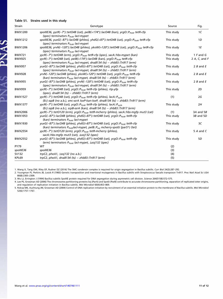
Table S1. Strains used in this study
Strain Genotype Source Fig.
BWX1200 spoIIIE36, yycR(−7°)::tetO48 (cat), pelB(+174°)::lacO48 (kan), ycgO::PftsW tetR-cfp(spec) terminators PftsW lacI-mypet
This study 1C
BWX1212 spoIIIE36, yuxG(−87°)::lacO48 (phleo), yhdG(+87°)::tetO48 (cat), ycgO::PftsW tetR-cfp(spec) terminators PftsW lacI-mypet
This study 1D
BWX1206 spoIIIE36, yrvN(−120°)::lacO48 (phleo), ykoW(+120°)::tetO48 (cat), ycgO::PftsW tetR-cfp(spec) terminators PftsW lacI-mypet
This study 1E
BWX721 yycR(−7°)::tetO48 (erm), ycgO::PftsW tetR-cfp (spec), sacA::hbs-mypet (kan) This study 1 F and GBWX925 yycR(−7°)::tetO48 (cat), pelB(+174°)::lacO48 (kan), ycgO::PftsW tetR-cfp
(spec) terminators PftsW lacI-mypet, dnaB134 (ts) − zhb83::Tn917 (erm)This study 2 A, C, and F
BWX957 yuxG(−87°)::lacO48 (phleo), yhdG(+87°)::tetO48 (cat), ycgO::PftsW tetR-cfp(spec) terminators PftsW lacI-mypet, dnaB134 (ts) − zhb83::Tn917 (erm)
This study 2 B and E
BWX928 yrvN(−120°)::lacO48 (phleo), ykoW(+120°)::tetO48 (cat), ycgO::PftsW tetR-cfp(kan) terminators PftsW lacI-mypet, dnaB134 (ts) − zhb83::Tn917 (erm)
This study 2 B and E
BWX955 yuxG(−87°)::lacO48 (phleo), yrvN(−120°)::tetO48 (cat), ycgO::PftsW tetR-cfp(spec) terminators PftsW lacI-mypet, dnaB134 (ts) − zhb83::Tn917 (erm)
This study 2 B and E
BWX959 yycR(−7°)::tetO48 (cat), ycgO::PftsW tetR-cfp (phleo), rtp-yfp(spec), dnaB134 (ts) − zhb83::Tn917 (erm)
This study 2D
BWX1527 yycR(−7°)::tetO48 (cat), ycgO::PftsW tetR-cfp (phleo), lacA::PxylA(Ec) sspB (no a.b.), smc-ssrA loxP-kan-loxP, dnaB134 (ts) − zhb83::Tn917 (erm)
(1) 2G
BWX1377 yycR(−7°)::tetO48 (cat), ycgO::PftsW tetR-cfp (phleo), lacA::PxylA(Ec) sspB (no a.b.), scpB-ssrA (kan), dnaB134 (ts) − zhb83::Tn917 (erm)
This study 2H
BWX2006 yycR(−7°)::tetO120 (erm), ycgO::PftsW tetR-mcherry (phleo), sacA::hbs-mgfp mut3 (cat) (1) 3A and 5BBWX1053 yuxG(−87°)::lacO48 (phleo), yhdG(+87°)::tetO48 (cat), ycgO::PftsW tetR-cfp
(kan) terminators PftsW lacI-mypetThis study 3B and 5D
BWX1930 yuxG(−87°)::lacO48 (phleo), yhdG(+87°)::tetO48 (cat), ycgO::PftsW tetR-cfp(kan) terminators PftsWlacI-mypet, pelB::Psoj mcherry-spo0J (parS*) (tet)
This study 3C
BWX2554 yycR(−7°)::tetO120 (erm), ycgO::PftsW tetR-mcherry (phleo),sacA::hbs-mgfp mut3 (cat), Δsoj132 (spec)
This study 5 A and C
BWX2552 yuxG(−87°)::lacO48 (phleo), yhdG(+87°)::tetO48 (cat), ycgO::PftsW tetR-cfp(erm) terminators PftsW lacI-mypet, Δsoj132 (spec)
This study 5D
PY79 WT (2)spoIIIE36 spoIIIE36 (3)SV132 trpC2, pheA1, Δsoj132 (no a.b.) (4)KPL69 trpC2, pheA1, dnaB134 (ts) − zhb83::Tn917 (erm) (5)
1. Wang X, Tang OW, Riley EP, Rudner DZ (2014) The SMC condensin complex is required for origin segregation in Bacillus subtilis. Curr Biol 24(3):287–292.2. Youngman PJ, Perkins JB, Losick R (1983) Genetic transposition and insertional mutagenesis in Bacillus subtilis with Streptococcus faecalis transposon Tn917. Proc Natl Acad Sci USA
80(8):2305–2309.3. Wu LJ, Errington J (1994) Bacillus subtilis SpoIIIE protein required for DNA segregation during asymmetric cell division. Science 264(5158):572–575.4. Lee PS, Grossman AD (2006) The chromosome partitioning proteins Soj (ParA) and Spo0J (ParB) contribute to accurate chromosome partitioning, separation of replicated sister origins,
and regulation of replication initiation in Bacillus subtilis. Mol Microbiol 60(4):853–869.5. Rokop ME, Auchtung JM, Grossman AD (2004) Control of DNA replication initiation by recruitment of an essential initiation protein to the membrane of Bacillus subtilis.Mol Microbiol
52(6):1757–1767.
Wang et al. www.pnas.org/cgi/content/short/1407461111 11 of 12

Table S2. Plasmids used in this study
Plasmid Description Refs.
pGK97 rtp-yfp (spec) (1)pWX116S ykoW(+120°)::tetO48 (cat) (2)pWX151 yrvN(−120°)::lacO48 (phleo) (2)pWX154 yhdG(+87°)::tetO48 (cat) (2)pWX159 yuxG(−87°)::lacO48 (phleo) (2)pWX178 yycR(−7°)::tetO48 (cat) (2)pWX179 yycR(−7°)::tetO48 (erm) This studypWX193 ycgO::PftsW tetR-cfp (spec) This studypWX208 pelB(+174°)::lacO48 (kan) This studypWX340 dnaXCter-mypet (cat) This studypWX361 ycgO::PftsW tetR-cfp (spec) terminators PftsW lacI-mypet (2)pWX419 ycgO::PftsW tetR-cfp (phleo) (2)pWX425 ycgO::PftsW tetR-cfp (kan) terminators PftsW lacI-mypet (2)pWX477 scpBCter-ssrA (kan) (2)pWX480 lacA::PxylA (Ec) sspB loxP-erm-loxP (2)pWX510 ycgO::PftsW tetR-mcherry (phleo) (2)pWX564 pelB::Psoj mcherry-spo0J (parS*) (tet) This studypWX570 yycR(−7°)::tetO120 (erm) (2)
1. Lemon KP, Kurtser I, Grossman AD (2001) Effects of replication termination mutants on chromosome partitioning in Bacillus subtilis. Proc Natl Acad Sci USA 98(1):212–217.2. Wang X, Tang OW, Riley EP, Rudner DZ (2014) The SMC condensin complex is required for origin segregation in Bacillus subtilis. Curr Biol 24(3):287–292.
Table S3. Oligonucleotides used in this study
Oligos Sequence Use
odr198 gccCTCGAGttttccggcaactgcgtctttaagcgc BWX721odr214 gccGAATTCaaatcaccttaaatccttgacgagc BWX721oWX217 aaaCGGCCGgtagagcggctcaaaacaacgg pWX340oWX238 gcgAAGCTTacataaggaggaactactatgtctagattagataaaagtaaag pWX193oWX239 gcgGGATCCttacttataaagttcgtccatgcc pWX193oWX245 gcgGAATTCtaaatgataaaactcaaacttattaacag pWX193oWX246 gcgAAGCTTctgcttctaacttccattatctatg pWX193oWX329 cgcCTCGAGtcttttatttcaattaaatccgc pWX340oWX438 gaccagggagcactggtcaac BWX2554oWX439 tccttctgctccctcgctcag BWX2554oWX507 cgtgcttgaattttcaattatttccc BWX2554oWX508 acccgttgcaaaggctcactgggcgc BWX2554oWX774 cgcAAGCTTacataaggaggaactactatggtc pWX564oWX775 tttCTCGAGtccggaacctttgtataattcgtccattccacc pWX564oWX894 cgcggcacagacttgatgaaacgtcc BWX2554oWX895 ctgagcgagggagcagaaggatccttaaaaatataaaaagctctcctgcttttc BWX2554oWX896 gttgaccagtgctccctggtccaaaaggtaatcacttacttttagtgaatat BWX2554oWX897 gatttttcccacgatgtcacctactttc BWX2554
Restriction endonuclease sites are capitalized.
Wang et al. www.pnas.org/cgi/content/short/1407461111 12 of 12